Noninvasive prenatal testing
of aneuploidies: where are we now?
Testes não invasivos para aneuploidias
no pré-natal: onde estamos agora?
AleksAndrA JezelA-stAnek1Małgorzata KrajewsKa-walaseK1
Introduction
Prenatal diagnosis of chromosomal aneuploidies is the most frequent prenatal test offered to pregnant women. In most cases, they are recommended in the following cir-cumstances: maternal age of 35 years or above; positive irst- or second-trimester screen-ing test results, and increased risk of fetal aneuploidies due to family history. Durscreen-ing the irst trimester, screening tests include: nuchal translucency (NT) combined with maternal age; levels of maternal serum pregnancy associated plasma protein-A (PAPP-A) and free
beta-human chorionic gonadotropin (β-hCG) combined with maternal age; combination
of NT measurement, the irst trimester maternal serum analytes (PAPP-A, and free β-hCG
or total hCG) and maternal age, referred to as combined irst trimester screening. The NT measurement is valid when crown-rump length (CRL) is 45–84 mm, corresponding to
11–13+6 week of gestation, while PAPP-A and free β-hCG may be measured between
9–13+6 week of gestation1.
More recently, another option, which is the detection of an increased amount of chro-mosomal material in maternal blood, became available to screen for chromosome aneuploidy. This is called Non-invasive Prenatal Testing (NIPT). Recently, different tests are available, depending on the employed methodologies and algorithms for data analysis. These may involve massively parallel sequencing (MPS), targeted sequencing of speciic chromosomal
segments, or directed sequence analysis of single nucleotide polymorphisms (SNPs)2.
While all these testing methods have limitations, healthcare providers need to be aware of them in order to give their patients reliable information and genetic counseling. In this paper, we focused on NIPT because it is the most promising screening option.
Among the above-mentioned tests, combined irst trimester screening has been demonstrated to have higher detection rates for Down Syndrome (78–91%) and trisomy
18 (91–96%) compared to NT only or serum analytes methods3-5. Since pregnancies
af-fected with trisomy 13 have PAPP-A, free β-hCG, and NT patterns similar to trisomy 18,
this screening is also used to screen for trisomy 136.
1Department of Medical Genetics, The Children’s Memorial Health Institute, Warsaw, Poland.
Conlict of interests: none.
Correspondence
Aleksandra Jezela-Stanek Department of Medical Genetics, The Children’s Memorial Health Institute Aleja Dzieci Polskich 20, 04-736 Warsaw, Poland
Received
07/27/2014
Accepted with modiications
08/22/2014
384 Rev Bras Ginecol Obstet. 2014; 36(9):383-6
Jezela-Stanek A, Krajewska-Walasek M
Obviously, the great advantage of screening options for chromosome aneuploidy is that they are non-invasive. Hence, they are recommended for all pregnancies and usu-ally precede a decision about whether or not to undergo invasive diagnostic testing. On the other hand, screenings have some limitations. The main one is that they do not provide a deinitive diagnosis. Furthermore, they have lower detection rates in multiple pregnancies; variability in the detections rates of trisomy 21, 18, 13, while no information on fetal monosomy X, as well as false-positive
results that, in most laboratories, are higher than 5%7.
Results
The newest and recently introduced prenatal screen-ing method is NIPT, which uses circulatscreen-ing cell-free fetal DNA (cffDNA) in maternal plasma to estimate risk for Down (trisomy 21), Edwards (trisomy 18), and Patau Syndrome (trisomy 13). cffDNA in the plasma of pregnant
women was discovered by Lo et al.8. Later, in 2008, MPS
of the maternal plasma was used to detect material from
fetus with trisomy 219,10. During the following years,
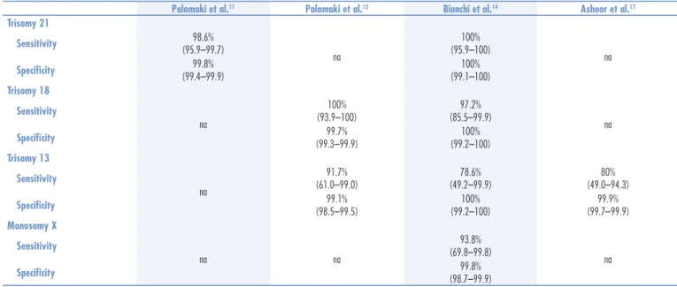
the same technique also detected fetal trisomy 18 and 1311-13, as well as monosomy X14 in high-risk pregnan-cies. The results obtained in the mentioned studies can be seen in Table 1.
In 2012, preliminary results were presented and concluded that cffDNA-based tests may have similar
sensitivity and speciicity in an average risk population15.
The study of Nicolaides et al.1 was conducted in 2,049
pregnant women undergoing routine screening for aneu-ploidies at 11w0d – 13w6d weeks’ gestation. Trisomy risk scores were given for 95.1% (1,949 of 2,049) of the cases,
Palomaki et al.11 Palomaki et al.12 Bianchi et al.14 Ashoor et al.17 Trisomy 21
Sensitivity 98.6%
(95.9–99.7)
na
100% (95.9–100)
na
Speciicity (99.4–99.9)99.8% (99.1–100)100%
Trisomy 18
Sensitivity
na
100% (93.9–100)
97.2% (85.5–99.9)
na
Speciicity (99.3–99.9)99.7% (99.2–100)100%
Trisomy 13
Sensitivity
na
91.7% (61.0–99.0)
78.6% (49.2–99.9)
80% (49.0–94.3)
Speciicity 99.1%
(98.5–99.5)
100% (99.2–100)
99.9% (99.7–99.9)
Monosomy X
Sensitivity
na na
93.8% (69.8–99.8)
na
Speciicity (98.7–99.9)99.8%
na: not analyzed.
Table 1. Results from the last published clinical trials that measured the sensitivity and speciicity of Noninvasive Prenatal Testing in the diagnostics of common aneuploidies
including all eight with trisomy 21 and two among the three with trisomy 18. The trisomy risk score was >99% in the eight cases of trisomy 21 and two of trisomy 18 and <1% in 99.9% (1,937 of 1,939) of the euploid cases.
Results of the study presented by Norton et al.15 showed:
for trisomy 21, a sensitivity of 100% (95.5–100%) and a false-positive rate of 0.03% (95%CI 0.002–0.20); for trisomy 18, a sensitivity of 97.4% (86.5–99.9%) and a false-positive rate of 0.07% (95%CI 0.02–0.25).
Later, Sparks et al.16 evaluated a novel biochemical
assay and algorithm for the prenatal evaluation of risk for fetal trisomy 21 and 18 in a blinded analysis with 167 pregnant women. They performed a digital analysis of the selected regions (DANSR), in combination with a novel algorithm, fetal-fraction optimized risk of trisomy evaluation (FORTE). It allows correctly identifying all
aneuploid cases (36 trisomies 21 and 8 trisomies 18)16.
Moreover, the investigators assayed cell-free DNA from a training set and a blinded validation set of preg-nant women: 250 euploidies, 72 trisomies 21, and 16 trisomies 18. All 167 cases in the blinded validation and 163/171 in the training set passed through the quality control criteria. FORTE produced an individual-ized trisomy risk score for each subject, which correctly discriminated all T21 and T18 cases from the disomic ones. The authors concluded that DANSR and FORTE enable accurate non-invasive fetal aneuploidy detection in a high-risk population, and stated that larger studies including low- and average-risk pregnancies are needed.
Recently, in 2013, studies evaluating the performance of the Harmony Prenatal Test and Panorama Prenatal Test
were reported17. Ashoor et al.17 assessed the performance
385
Rev Bras Ginecol Obstet. 2014; 36(9):383-6
Noninvasive prenatal testing of aneuploidies: where are we now?
13 in a two-phase, blinded, case-control study. In the second phase, after modiication of trisomy 13 algorithm based on data from the irst phase, the test was used to detect trisomy 13 risk scores for 10 cases of trisomy 13 and 1,939 euploid cases. The trisomy 13 risk scores were >99% in eight (80.0%) cases of trisomy 13. In the 1,939 euploid cases, the risk score for trisomy 13 was <0.01% in 1,937 (99.9%), 0.79% in one and >99.0% in one.
The Panorama Prenatal Test was validated by
Nicolaides et al.18 in a population of 242 women with
singleton pregnancies, who had been submitted to cho-rionic villus sampling (CVS) from 11 to 13 weeks. They were referred because irst-trimester screening indicated
an increased risk for trisomy 21, 18 or 1318. Results were
provided for 94.6% (229 cases): 32 cases were correctly identiied as aneuploid, including trisomy 21 (n=25; sensitivity=100%, speciicity=100%), trisomy 18 (n=3), trisomy 13 (n=1), Turner Syndrome (n=2) and triploidy (n=1), with no false-positive or -negative results. In all
these studies (apart of Fairbrother et al.19), NIPT was
in-tegrated as a primary screening test for pregnant women at high-risk of aneuploidy.
Discussion and conclusions
Testing can be done after the tenth week and typically it is performed between 10 to 22 weeks. Interestingly, as
concluded by Norton et al.20 from a population
perspec-tive, a better option for NIPT may be a second-tier test for those patients who screen positive by conventional
aneuploidy screening20. However, before routine
MPS-based population screening for fetal trisomy 21 are widely introduced, additional trials are needed. According to the International Society for Prenatal Diagnosis (ISPD), they should include i. a.: eficacy in low-risk populations or suitable for the diverse sub-populations, such as twins
and IVF donor pregnancies21,22.
Unfortunately, data regarding the clinical validity of NIPT are still limited. These are available only for studies examining the early clinical experience of the Harmony and Verii Prenatal Tests. The limitations are associated with the lack of follow-up information for the majority of studies on pregnancies. For example, in the investigation
by Fairbrother et al.19 with 284 obstetrical patients who
were evaluated by both the Harmony Prenatal Test and traditional irst-trimester screening, only one woman, who had a irst-trimester screening result of one in ive for trisomy 21, elected to have invasive prenatal
diagno-sis, which revealed a normal fetal karyotype19. Another
study, presented by Futch et al.23, involved 6,123 patients
tested with the Verii Prenatal Test. Of 280 fetuses with aneuploidy detected by the NIPT, 94 (33.6%) were con-irmed or the pregnancies resulted in miscarriage, and 14
(0.2%) yielded discordant (likely false-positive) results. Unclassiiable results were obtained in up to 1% of cases for each of the analyzed chromosomes. As a result, it was assumed that the pregnancies that had not yet delivered did not have an undiagnosed aneuploidy (missed by both NIPT and irst-trimester screening).
At this time, NIPT is only recommended for patients from high-risk populations, including advanced maternal age, positive screening test, abnormal ultrasound sugges-tive of aneuploidy, or prior pregnancy with chromosome
aneuploidy21,22,24. It is also recommended that a positive
NIPT conclusion, due to occasional false-positive results, be followed by conirmatory diagnostic testing (chorionic villus sampling, CVS or amniocentesis) prior to making
pregnancy decisions21,22,24. The invasive tests, apart from
giving an accurate diagnosis, also provide important information about the cause and type of trisomy. When Down Syndrome is due to a 21 chromosome transloca-tion, this has important recurrence risk implications for the parents and other family members.
Other risks concerning the NIPT are “unreportable”. From 0.5 to 7% of women who undergo NIPT will not
get a result25 (fact sheet published in 2012 by the National
Coalition for Health Professional Education in Genetics and NSGC). This often happens due to the low amount of fetal DNA in the sample (high maternal weight or early gestational age). Moreover, some laboratories may decline to report results that are near the cutoff.
As one can see, there are still some questions regarding the introduction of NIPT into routine practice. In 2014, in the United Kingdom, this resulted in initiating a na-tional project called “Evaluation of NIPT for aneuploidy in an NHS setting: a reliable accurate prenatal non-invasive
diagnosis (RAPID) protocol”26. The collaborators expect
that this study may be a signiicant contribution for policing decisions around the implementation of NIPT for aneuploidies and allow developing the laboratory standards for testing and reporting, education materials, and counselling strategies.
386 Rev Bras Ginecol Obstet. 2014; 36(9):383-6
Jezela-Stanek A, Krajewska-Walasek M
References
1. Nicolaides KH. Screening for fetal aneuploidies at 11 to 13 weeks. Prenat Diagn. 2011;31(1):7-15.
2. Hayes, Inc. GTE report. Noninvasive prenatal testing (NIPT) for fetal aneuploidy. Lansdale: Hayes, Inc; 2013.
3. Malone FD, Canick JA, Ball RH, Nyberg DA, Comstock CH, Bukowski R, et al. First- trimester or second-trimester screening, or both, for Down’s syndrome. N Engl J Med. 2005;353(19):2001-11. 4. Wald NJ, Rodeck C, Hackshaw AK, Walters J, Chitty L, Mackinson
AM. First and second trimester antenatal screening for Down’s syndrome: the results of the Serum, Urine and Ultrasound Screening Study (SURUSS). J Med Screen. 2003;10(2):56-104.
5. Wapner R, Thom E, Simpson JL, Pergament E, Silver R, Filkins K, et al. First-trimester screening for trisomies 21 and 18. N Engl J Med. 2003;349(15):1405-13.
6. Spencer K, Ong C, Skentou H, Liao AW, Nicolaides KH. Screening for trisomy 13 by fetal nuchal translucency and maternal serum free beta-hCG and PAPP-A at 10–14 weeks of gestation. Prenat Diagn. 2000;20(5):411-6.
7. Wilson KL, Czerwinski JL, Hoskovec JM, Noblin SJ, Sullivan CM, Harbison A, et al. NSGC practice guideline: prenatal screening and diagnostic testing options for chromosome aneuploidy. J Genet Couns. 2013;22(1):4-15.
8. Lo YM, Corbetta N, Chamberlain PF, Rai V, Sargent IL, Redman CW, et al. Presence of fetal DNA in maternal plasma and serum. Lancet. 1997;350(9076):485-7.
9. Chiu RW, Chan KC, Gao Y, Lau VY, Zheng W, Leung TY, et al. Noninvasive prenatal diagnosis of fetal chromosomal aneuploidy by massively parallel genomic sequencing of DNA in maternal plasma. Proc Natl Acad Sci USA. 2008;105(51):20458-63. 10. Fan HC, Blumenfeld YJ, Chitkara U, Hudgins L, Quake
SR. Noninvasive diagnosis of fetal aneuploidy by shotgun sequencing DNA from maternal blood. Proc Natl Acad Sci USA. 2008;105(42):16266-71.
11. Palomaki GE, Kloza EM, Lambert-Messerlian GM, Haddow JE, Neveux LM, Ehrich M, et al. DNA sequencing of maternal plasma to detect Down syndrome: an international clinical validation study. Genet Med. 2011;13(11):913-20.
12. Palomaki GE, Deciu C, Kloza EM, Lambert-Messerlian GM, Haddow JE, Neveux LM, et al. DNA sequencing of maternal plasma reliably identiies trisomy 18 and trisomy 13 as well as Down syndrome: an international collaborative study. Genet Med. 2012;14(3):296-305.
13. Ashoor G, Syngelaki A, Wagner M, Birdir C, Nicolaides KH. Chromosome-selective sequencing of maternal plasma cell-free DNA for irst-trimester detection of trisomy 21 and trisomy 18. Am J Obstet Gynecol. 2012;206(4):322.e1-5.
14. Bianchi DW, Platt LD, Goldberg JD, Abuhamad AZ, Sehnert AJ, Rava RP. Genome-wide fetal aneuploidy detection by maternal plasma DNA sequencing. Obstet Gynecol. 2012;119(5):890-901.
15. Norton ME, Brar H, Weiss J, Karimi A, Laurent LC, Caughey AB, et al. Non-Invasive Chromosomal Evaluation (NICE) study: results of a multicenter, prospective, Cohort study for detection of fetal trisomy 21 and trisomy 18. Am J Obstet Gynecol. 2012;207(2):137. e1-8.
16. Sparks AB, Struble CA, Wang ET, Song K, Oliphant A. Noninvasive prenatal detection and selective analysis of cell-free DNA obtained from maternal blood: evaluation for trisomy 21 and trisomy 18. Am J Obstet Gynecol. 2012;206(4):319.e1-9.
17. Ashoor G, Syngelaki A, Wang E, Struble C, Oliphant A, Song K, et al. Trisomy 13 detection in the irst trimester of pregnancy using a chromosome-selective cell-free DNA analysis method. Ultrasound Obstet Gynecol. 2013;41(1):21-5.
18. Nicolaides KH, Syngelaki A, Gil M, Atanasova V, Markova D. Validation of targeted sequencing of single-nucleotide polymorphisms for non-invasive prenatal detection of aneuploidy of chromosomes 13, 18, 21, X, and Y. Prenat Diagn. 2013;33(6):575-9. 19. Fairbrother G, Johnson S, Musci TJ, Song K. Clinical experience of
noninvasive prenatal testing with cell-free DNA for fetal trisomies 21, 18, and 13, in a general screening population. Prenat Diagn. 2013;33(6):580-3.
20. Norton ME, Rose NC, Benn P. Noninvasive prenatal testing for fetal aneuploidy: clinical assessment and a plea for restraint. Obstet Gynecol. 2013;121(4):847-50.
21. Benn P, Borrell A, Crossley J, Cuckle H, Dugoff L, Gross S, et al. Aneuploidy screening: a position statement from a committee on behalf of the Board of the International Society for Prenatal Diagnosis, January 2011. Prenat Diagn. 2011;31(6):519-22. 22. Benn P, Borrell A, Cuckle H, Dugoff L, Gross S, Johnson JA, et al. Prenatal
detection of Down syndrome using Massively Parallel Sequencing (MPS): a rapid response statement from a committee on behalf of the Board of the International Society for Prenatal Diagnosis, 24 October 2011. Prenat Diagn. 2012;32(1):1-2.
23. Futch T, Spinosa J, Bhatt S, de Feo E, Rava RP, Sehnert AJ. Initial clinical laboratory experience in noninvasive prenatal testing for fetal aneuploidy from maternal plasma DNA samples. Prenat Diagn. 2013;33(6):569-74.
24. Devers PL, Cronister A, Ormond KE, Facio F, Brasington CK, Flodman P. Noninvasive prenatal testing/noninvasive prenatal diagnosis: the position of the National Society of Genetic Counselors. J Genet Couns. 2013;22(3):291-5.
25. National Coalition for Health Professional Education in Genetics [Internet]. Non-Invasive Prenatal Testing (NIPT) FactSheet. 2012 [cited 2014 Apr 10]. Available from: <http://www.nchpeg. org/index.php?option=com_content&view=article&id=384&Ite mid=255>