Functional Behavior of Patients with Conventional Pacemakers
Undergoing Cardiac Resynchronization
Rodrigo Tavares Silva, Martino Martinelli Filho, Carlos Eduardo Batista de Lima, Daniela Garcia Moreno Cabral Martins, Silvana Angelina Dorio Nishióka, Anísio Alexandre Pedrosa, Elizabeth Sartori Crevelari, Roberto Costa, Eduardo Argentino Sosa, José Antonio Franchini Ramires
Instituto do Coração (Heart Institute) do Hospital das Clínicas da Faculdade de Medicina da Universidade de São Paulo, São Paulo, SP - Brazil
Summary
Background: Cardiac resynchronization therapy (CRT) is an efficient treatment for patients with heart failure (HF), severe ventricular dysfunction and intraventricular block. Conventional pacemakers (CPM) implanted in the right ventricular apical area cause alterations in the normal sequence of cardiac activation similar to those induced by LBBB (left bundle-branch block). Therefore, patients with CPM and advanced HF could be candidates to undergo CRT, but as only small numbers of patients have been evaluated so far, definitive conclusions are lacking.
Objective: To assess the clinical and functional outcome of cardiac resynchronization therapy (CRT) in patients with conventional pacemakers.
Methods: Patients with CPM, who were in NYHA HF functional class III/IV class refractory to drug therapy, and
left ventricular ejection fraction (LVEF)<35% underwent CRT. Patients’ clinical-functional behavior was assessed prospectively six months after the procedure. The improvement of one HF-functional class was set as an effective response to the procedure. The following was assessed: QRS duration (ECG), diastolic diameter (LVDd), left ventricular systolic diameter (LVSd) and LVEF seen on the echocardiogram. For the statistical analysis, Student’s paired t test and Spearman’s correlation were used.
Results: Twenty-nine patients (mean age 61.5) were evaluated. Of these, six were females, and chagasic cardiomyopathy was predominant. During the clinical follow-up of 22.7±13 months, 86.2% of the patients benefited from CRT. Within this group, the mean LVEF increased by 18% (p=0.013), QRS duration dropped by 11.8% (p=0.002) and no significant reduction in left ventricular intracavitary diameters was observed.
Conclusion: CRT is effective for patients with CPM and advanced HF as it yields a high rate of response (86.2%),
significantly improves LVEF and reduces QRS duration. (Arq Bras Cardiol 2008; 90(2):138-143)
Key words: Pacemaker, artificial; bundle-branch block; heart failure, congestive; cardiac pacing, artificial.
Mailing address: Rodrigo Tavares Silva •
Rua João Moura, 870 - 05412002 - Pinheiros, São Paulo, SP - Brazil E-mail: [email protected]
Article received on March 25, 2007; revised manuscript received on June 5, 2007; accepted on October 3, 2007.
Introduction
Intraventricular conduction abnormalities are frequent manifestations in heart failure (HF) patients1. In general, they are associated with the severity of impairment of cardiac function, and, in an independent manner, with higher rates of morbidity and mortality in this population2,3. Approximately 25% to 50% of advanced HF patients have increased QRS duration and most have a left bundle-branch block (LBBB) morphology1,3. LBBB is known to cause alterations in the left ventricular activation sequence, thus causing a delay between the interventricular septum and lateral wall contraction3.
Artificial right ventricular apical (RVA) pacing by conventional
pacemakers (CPM) triggers a ventricular activation sequence similar to that occurring in spontaneous LBBB4-6. Depending on the degree of myocardial impairment, this induced electrical dyssynchrony can cause mechanical ventricular dyssynchrony and aggravate progression of the underlying heart disease. This is due to the fact that, in the presence of a significant systolic dysfunction, the following occur: acceleration of myocardial cell injury; changes in ventricular geometry; atrial enlargement and worsening of mitral regurgitation with consequent hemodynamical deterioration6. The recent publication of the DAVID study7, MADIT-II8, Mode Selection Trial9 and PAVE Trial10 has shown clinical evidence of this. Subanalyses of these studies have linked artificial cardiac pacing with higher rates of hospital admissions due to HF, a larger number of cases of atrial fibrillation and mitral failure.
Implantation of the cardiac resynchronizer
Before surgery, all patients underwent digital phlebography of the upper limbs for evaluation of vein patency and planning of the surgical strategy. Previous leads were maintained whenever possible provided they were working properly. The additional lead for stimulation of the LV was implanted preferably by intravenous access through the coronary sinus and positioned in veins that drain into the LV lateral or posterolateral walls. Subclavian/innominate vein stenosis associated with the previously implanted device was not a criterion for exclusion. When this was the case, a new biventricular stimulation system was implanted on the contralateral side; when the pacemaker generator was already implanted in the left thoracic area, patients underwent a minimally invasive left lateral thoracotomy to have the additional lead implanted in the left ventricle.
All pulse generators were replaced with resynchronization devices available on the market at the time of the study. For the first procedures performed, a Y-adaptor was used to connect the RV and LV leads. Later on, independent channel systems were used.
Biventricular pacing programming was done at the discretion of the clinical team in charge of artificial cardiac pacing. Atrioventricular and interventricular pacing intervals were programmed for each patient and guided by echocardiography.
Transthoracic echocardiography
Echocardiographic analysis was performed using M-mode to measure LVDd and LVSd in millimeters (mm), and bidimensional mode to assess LVEF percentage (%). The echocardiography unit of the Heart Institute was responsible for performing all exams in accordance with the guidelines set by the Brazilian Society of Echocardiography, and at the request of the team responsible for the outpatient follow-up. Post-CRT echocardiography was performed with a minimum six-month interval after the surgical procedure for the implantation of the biventricular system.
Twelve-lead electrocardiogram
The duration of the paced QRS complex (milliseconds) was assessed by a surface 12-lead electrocardiogram, calibrated at 0,1 mV/mm and chart speed of 25 mm/sec. The electronic measurement performed by the device, or done manually, was the smallest time interval from one pacemaker spike to the end of the QRS complex in any of the leads.
Statistical analysis
The rate of effective response to CRT was assessed by the calculation of the percentage of patients who had an improvement of one HF NYHA functional class. Spearman’s rank correlation test was used to analyze the classificatory variables, and the paired Student’s t test to analyze the variation in QRS complex duration and the behavior of echocardiographic parameters pre- and post-CRT. The P value was considered statistically significant when smaller than 0.05.
intraventricular block (QRS >120 m). However, this scientific evidence is based on large-scale studies which, invariably, did not include patients with CPM11-14. In a study conducted with 10 patients with HF and CPM, Höijer et al15 have shown that the implantation of a biventricular system yields beneficial effects in clinical, functional and neuroendrocrine parameters. Other studies conducted with limited groups of patients have also shown similar results1,4.
The aim of this study is to assess the clinical and functional outcome of CRT in patients with HF and apical right ventricular CPM.
Method
Patients
Subjects for this study were selected from the 484-patient cohort using cardiac resynchronizer(CR) and being prospectively followed at the Unidade de Estimulação Cardíaca Artificial do Instituto do Coração – HCFMUSP, (Unit of Artificial Cardiac Stimulation of the Heart Institute of the Medical School of University of São Paulo) up to December 2004.
The inclusion criteria were based on the patients’ clinical-functional characteristics before implantation of the CR: 1) LBBB induced by CPM; 2) dilated cardiomyopathy (NYHA functional class III/IV); 3) left ventricular ejection fraction
/9() DQG UHIUDFWRULQHVV WR RSWLPL]HG FOLQLFDO
treatment consisting of at least an angiotensin conversion enzyme inhibitor or an angiotensin receptor blocker, a beta-blocker and spironolactone, all administered at maximum doses, provided there was no contraindication. Besides the above mentioned drugs, diuretics, vessel dilators, digitalis, calcium-channel blockers and antiarrhythmic, and other drugs were also administered, according to the needs and tolerance of each patient.
Exclusion criteria were use of implantable cardioverter defibrillators, dysfunctional pacemakers, post-cardiac transplant, end-stage disease with survival expected to be less than one year, recent cardiovascular events (<3 months), severe obstructive pulmonary disease and patients who were being followed at other institutions.
The endpoint used to assess the clinical response to CRT, after a minimum period of six months of follow-up, was the improvement of at least one NYHA HF functional class. The following were also analyzed: 1) variations in QRS complex duration; 2) variations in LV diastolic and systolic diameters (LVDd and LVSd); and 3) LVEF behavior.
of cardiomyopathy were chagasic (52%) and idiopathic (21%). As to heart failure functional class (HF-FC), 86% of the patients in the group were classified as being FC III. Cardiovascular drug therapy was optimized and over 75% of the patients were concomitantly on ACEI/ARA, betablockers and spironolactone. The mean values of ecocardiographic parameters were: LVEF: 27.7%; LVDd: 69.7 mm and LVSd 63.8 mm. Eighteen patients were in synchronized pacing mode (DDD) and the mean stimulated QRS duration was 185 ms. Time elapsed between implant of the first pacemaker and optimization for CRT was 114.3±80.3 months. Clinical follow-up after the surgical procedure was carried out over 22.7±13 months.
Coronary sinus catheterization for LV lead implantation was performed in 26 patients (89.6%); in 25 patients (86.2%), the device was successfully implanted through a transvenous access. In one patient, despite coronary sinus catheterization, the small number of tributary veins precluded lead fixation. This patient already had a previously implanted device in the left thoracic region, which required conversion to minimally invasive thoracotomy. Failure in coronary sinus endovenous catheterization occurred in three patients; in these patients, the LV lead was also implanted through a left lateral minithoracotomy.
The rate of subclavian/innominate vein stenosis that precluded the passage of the additional lead was 6.8% (two patients); decision was taken to implant a new pacing system on the contralateral side and discontinue the old device. There were no complications from the surgical procedure. Over the clinical follow-up (22.7 months), 86% of the patients had an effective clinical response to CRT. Moreover, most of them (82.7%) remained in NYHA functional class I/II (Figure 1). No significant changes were made in the drug therapy during the follow-up period.
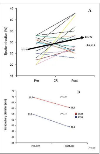
The analysis of the echocardiographic parameters showed an 18% improvement in the patients’ mean LVEF after CRT (P=0.013). No significant reduction of LV intracavitary diameters was observed; with CR, there was a variation of 3.5 mm in the LVDd (P=0.29) and 4.3 mm in the LVSd (P=0.15) (Figure 2). The mean time elapsed from CTR and the echocardiography performed after the procedure was 10.8 months.
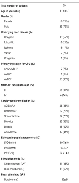
Table 1 - Patient characteristics
Total number of patients 29
Age in years (SD) 61.5±17
Gender (%)
Female 6 (21%)
Male 23 (79%)
Underlying heart disease (%)
Chagasic 15 (52%)
Idiopathic 6 (21%)
Ischemic 5 (17%)
Valvar 2 (7%)
Congenital 1 (3%)
Primary indication for CPM (%)
SND+AVB 1º 2 (7%)
AVB 2º 1 (3%)
AVB 3º 26 (90%)
NYHA HF functional class (%)
III 25 (86%)
IV 4 (14%)
Cardiovascular medication (%)
ACEI/ARA 25 (86%)
Betablocker 22 (76%)
Spironolactone 22 (76%)
Diuretics 25 (86%)
Digitalis 18 (62%)
Amiodarone 12 (41%)
Echocardiographic parameters (SD)
LVDd (mm) 69.7±10
LVSd (mm) 63.8±7
LVEF (%) 27.7±4.8
Stimulation mode (%)
Single-chamber (VVI) 11 (38%)
Dual-chamber (DC) 18 (62%)
Basal stimulated QRS
Duration (ms) 185±34
6'6WDQGDUGGHYLDWLRQ61'VLQXVQRGHGLVHDVH$9%DWULRYHQWULFXODUEORFN $&(,$5$ DQJLRWHQVLQ FRQYHUVLRQ HQ]\PH LQKLELWRUDQJLRWHQVLQ UHFHSWRU
antagonists; LVDd - left ventricule diastolic diameter; LVSd - left ventricule systolic diameter; LVEF - left ventricular ejection fraction; HF - heart failure; ms - milliseconds.
Fig. 1 -Behavior of patients’s NYHA-HF functional class pre-and
post-&5$IWHU&5RIWKHSDWLHQWVUHPDLQHGLQ+))&,,,&5FDUGLDF
resynchronization; HF - heart failure; FC - functional class; NYHA - New
<RUN+HDUW$VVRFLDWLRQ
Results
However, ventricular dyssynchrony induced by CPM in the RV apical area still lacks scientific backing.
LBBB induced by CPM results in interventricular dyssynchrony and causes 30-180 ms delays in LV activation, as well as intraventricular dyssynchrony following variations in the mechanical activation sequence (apex-to-base rather than base-to-apex). Moreover, RV apical pacing leads to heterogeneous distribution of myocardial strain, thinning of the early-activated regions as opposed to the late-activated ones that become thicker. This regional heterogeneity results in LV remodeling, which ultimately leads to contractile and hemodynamic alterations, reducing its functional efficiency. Additional mechanisms, such as neuroendocrine alterations, alterations in L-type calcium channels, in expression of proteins, myofibrillar disarrangement, fibrosis, deposition in fat tissue, tissue perfusion defects and a varied degree of mitral regurgitation all directly contribute to impairment of cardiac function16.
A few drugs were initially used to correct these disturbances, especially beta-blockers; however, they did not show sustained effects17.
CRT began to be employed in 1996 as an invasive therapeutic alternative performed through lateral left thorocotomy and, therefore, did not stir much enthusiasm at first. The advent of the transvenous technique through catheterization of the coronary sinus and the development of LV leads made the procedure less invasive, and its indication has gradually increased worldwide. In this group of patients, the rate of severe stenosis of the venous system associated with the previously implanted device was low (6.8%), resulting in a high rate of success of the transvenous technique through catheterization of the coronary sinus (86.2%). Currently, approximately 95 cardiac ressynchronizers are implanted per year at the Instituto do Coração – FMUSP, with and without cardiac defibrillators, and 85% are performed by transvenous coronary sinus approach.
The classical trials that corroborated CRT as a supporting therapy for patients with severe HF and intraventricular conduction disturbances invariably report clinical and functional improvement11-14. In our study, the rate of clinical response to CRT was 86.2%, a value higher than those reported by Reuter et al (82.4%)18 and Molhoek et al (68%)19. All these studies used similar criteria. In our study, there was an improvement of 1 (one) NYHA HF functional class in 55.1% of our patients, and after CRT, 82.7% remained in functional classes I/II. Despite the apparent subjectivity of the method used for the evaluation of the clinical response, its validation is shown in the profile of the patients with limiting symptoms20-22. Other measurements of post-CRT clinical assessment, such as quality of life questionnaire, the six-minute walk or the ergoespirometric tests have not been used, and this constitutes a limitation of the study.
Most studies conducted to evaluate CRT effects considered the presence of intraventricular conduction disturbance seen on the surface ECG as a specific criterion to document dyssynchrony11-14. In these studies, the rate of non-response to cardiac resynchronization ranges from 20% to 30%23. After biventricular pacing , a significant reduction
(11.8%) in the duration of QRS complex was observed (P=0.002). However, there was no correlation between the reduction in QRS duration and the clinical improvement or echocardiographic parameters.
Discussion
The findings in this study show that CRT is effective for patients with dilated cardiomyopathy refractory to clinical treatment and LBBB induced by CPM. This was one of the largest groups of patients with this type of profile described in literature, characterized by the high prevalence of chagasic cardiomyopathy (52%) and the long period of clinical follow-up after implantation of CR (22.7 months). These findings differ from similar studies in which idiopathic dilated cardiomyopathy was predominant and clinical follow-up was much shorter (6-12 months)11-13.
Over the past few years, CRT has been intensively evaluated in patients with spontaneous LBBB and advanced HF11-14.
Fig. 2 -Analysis of echocardiographic parameters pre- and post-CR. Chart A
VKRZVLQGLYLGXDO/9()YDULDWLRQVEHIRUHDQGDIWHU&5$QLPSURYHPHQW
in the group’s mean LVEF was observed after CR (P=0.013). Chart B shows
WKHYDULDWLRQQRQVLJQLILFDQWLQLQWUDFDYLWDU\GLDPHWHUV/9'G/96HDIWHU
CR. LVEF - left ventricular ejection fraction; CR - cardiac resynchronization; LVDd - left ventricule diastolic diameter; LVSd - left ventricule systolic diameter; HF - heart failure; mm - millimeters.
.
.
1. Horwich T, Foster E, De Marco T, Tseng Z, Saxon L. Effects of resynchronization therapy on cardiac function in pacemaker patients “upgraded” to biventricular devices. J Cardiovasc Electrophysiol. 2004;15 (11): 1284-9.
2. Baldasseroni S, Opasich C, Gorini M, Lucci D, Marchionni N, Marini M, et al, Italian Network on Congestive Heart Failure Investigators: Left bundle-branch block is associated with incresead 1-year sudden and total mortality rate in 5517 outpatients with congestive heart failure: a report from the Italian Network on Congestive Heart Failure. Am Heart J. 2002; 143: 398-405.
3. Baker CM, Christopher TJ, Smith P, Langberg JJ, Delurgio DB, Leon AR. Addition of a left ventricular lead to conventional pacing systems in patients with congestive heart failure: feasibility, safety, and early results in 60 consecutive patients. Pacing Clin Electrophysiol. 2002; 25: 1166-71.
4. Leon AR, Greenberg JM, Kanuru N, Baker CM, Mera FV, Smith AL, et al. Cardiac resynchronization in patients with congestive heart failure and chronic atrial fibrillation. J Am Coll Cardiol. 2002; 39 (8): 1258-63.
5. Vassalo AJ, Cassidy DM, Miller JM, Buxton AE, Marchlinski EF, Josephson EM. Left ventricular endocardial activation during right ventricular pacing: effect of underlying heart disease. J Am Coll Cardiol. 1986; 7 (6): 1228-33.
6. Vernooy K, Verbeek XAAM, Peschar M, Prinzen FW. Relation between abnormal impulse conduction and heart failure. J Interv Cardiol. 2003; 16 (6): 557-62.
7. Wilkoff BL, Cook JR, Epstein AE, Greeme HL, Hallstrom AP, Hsia H, et al. Dual-chamber pacing or ventricular back-up pacing in pacients with as implantable defibrillator: the Dual Chamber and VVI Implantable Defibrillator (DAVID)
References
In our study, the rate of non-response to CRT was much lower (13.8%), and this may be explained by the homogeneity of the cardiac depolarization pattern. Indeed, right apical CPM induces, in a uniform manner, the apex-to-base/RV-LV activation. This modification undoubtedly accounts for the maximum degree of ventricular dyssynchrony, thus ensuring a significant favorable impact through CRT. In this sense, we observed that the mean QRS duration on the surface ECG (pre-CRT) was 179 ms, a mean value higher than that of all classical trials: 174 ms in MUSTIC11; 167 ms in MIRACLE12; 159 ms in COMPANION13 and 160 ms in CARE-HF14. It is worthy mentioning that although this is not the reference pattern to detect ventricular dyssyncrony, there is a direct correlation between the latter and QRS duration1.
In our study, the post-CRT reduction in stimulated QRS complex (21.4 ms) did not correlate with the clinical response rate or with the improvement in echocardiographic parameters. This data is similar to that of several studies that evaluated reduction of stimulated QRS after CRT24. However, a different result is shown in an isolated trial by Horwich et al.1, in which a correlation was found between reduction in QRS duration after CRT (23.08 ms) and echocardiographic parameters.
In our study, the occurrence of reverse remodeling, as observed on echocardiographic modifications, was similar to that in other studies conducted with patients with both spontaneous and pacemaker-induced LBBB1,4,12,14,15. The most relevant finding was the 18% improvement in LVEF (P=0.013), which corresponds to a mean absolute increase of 5% in this parameter. In studies MIRACLE11 and CARE-HF14, the mean absolute increase in LVEF was 4.6% and 6.9%, respectively, whereas Baker et al.3 and Horwich et al.1 reported a mean increase of 6%-7% in their evaluation of CPM patients who underwent CRT.
The reduction in intracavitary diameters observed in our study (3.5 mm in the LVDd and 4.3 mm in the LVSd), despite not being statistically significant, corroborates the effects of CRT on reverse remodeling. In this sense, taking into consideration a larger group of patients, in the subanalysis of echocardiographic parameters in the MIRACLE25study, a significant mean reduction of 3 and 6 mm in LVDd and LVSd was observed six months after CRT.
Clinical trials with patients with HF, ventricular dysfunction and post myocardial infarction have shown that the size and volume of the LV has prognostic value for the occurrence of adverse cardiovascular events, including worsening of HF-FC and sudden cardiac death. In these trials, attenuation of LV dilation or reverse remodeling seems to reduce the rates of these adverse events during clinical follow-up23. Therefore, reverse remodeling promoted by CRT can be one of the mechanisms associated with the benefits of this therapy.
However, new prospective larger-scale studies and with control groups are needed to further add to the results of this study. Moreover, with the continuous evolution of technology, relevant diagnostic tools such as tissue echocardiography and cardiac nuclear magnetic resonance should make criteria used for the selection of patients even more rigorous, and therefore, improve the cost-efficacy of CRT.
Conclusion
Patients with dilated cardiomyopathy, severe HF refractory to clinical therapy and pacemaker-induced LBBB (RV apical stimulation) benefit from cardiac resynchronization therapy. The rate of clinical responsiveness is high (86.2%), and a significant improvement of LVEF is observed on echocardiography (18%). Therefore, despite the need for larger-scale studies, our results show that, in this specific population, cardiac resynchronization is a feasible and highly effective therapy, and is not associated with surgical complications.
Potential Conflict of Interest
The author reports having received financial aid to attend conferences; financial support for other research projects from all pacemaker manufacturers (Biotronik, St. Jude, Medtronic); consulting and lecture fees from St. Jude.
Sources of Funding
There were no external funding sources for this study.
Study Association
Trial. JAMA. 2002; 288: 3115-23.
8. Moss AZ, Zareba W, Hall WJ, Klein H, Wilber DJ, Cannom DS, et al. Prophylatic implatation of a defibrillator in a patients with myocardial infarction and reduced ejection fraction. N Engl J Med. 2002; 346: 877-83.
9. Lamas GA, Lee KL, Sweney MO, Silverman R, Leon A, Yee R, et al. Ventricular pacing or dual-chamber pacing for sinus-node dysfunction. N Engl J Med. 2002; 346 (24): 1854-62.
10. Doshi R, Daoud E, Fellows C, Turk K, Duran A, Hamdan M, et al. Left ventricular-based cardiac stimulation Post AV Nodal Ablation Evaluation (The PAVE Study). J Cardiovasc Electrophysiol. 2005; 16: 1160-5.
11. Cazeau S, Leclercq C, Lavergne T, Walker S, Varma C, Linde C, et al. Effects of multisite biventricular pacing in patients with heart failure and intraventricular conduction delay. N Engl J Med. 2001; 344: 873-80.
12. Abraham WT, Fisher WG, Smith AL, Delurgio DB, Leon AR, Loh E, et al. Cardiac resynchronization in chronic heart failure. N Engl J Med. 2002; 346: 1845-53.
13. Bristow M, Saxon LA, Boehmer J, Krueger S, Kass DA, De Marco T, et al. for the Comparison of Medical Therapy, Pacing, and Defibrillation Heart Failure (COMPANION) Investigators: cardiac resynchronization therapy with or without an implatable defibrillator in advanced chronic heart failure. N Eng J Med. 2004; 350: 2140-50.
14. Cleland JGF, Daubert JC, Erdmann E, Freemantle N, Gras D, Kappenberger L, et al. for the Cardiac Resynchronization - Heart Failure (CARE-HF) Study Investigators. The effect of cardiac resynchronization on morbidity and mortality in heart failure. N Engl J Med. 2005; 352 (15): 1539-49.
15. Höijer CJ, Meurling C, Brandt J. Upgrade to biventricular pacing in patients with conventional pacemakers and heart failure: a double-blind, randomized crossover study. Europace. 2006; 8: 51-5.
16. Dilaveris P, Pantazis A, Giannopoulos G, Synetos A, Gialafos J, Stefanadis C.
Upgrade to biventricular pacing in patients with pacing-induced heart failure: can resyncronization do the trick? Europace. 2006; 8: 352-7.
17. Castro PF, Mc-Nab P, Quintana JC, Bittner A, Greig D, Vergara J, et al. Effects of carvedilol upon intra and interventricular synchrony in patients with chronic heart failure. Am J Cardiol. 2005; 96: 267-9.
18. Reuter S, Garrigue S, Barold SS, Jais P, Hocini M, Haissaguerre M, et al. Comparison of characteristics in responders versus nonresponders with biventricular pacing for drug-resistant congestive heart failure. Am J Cardiol. 2002; 89: 346-50.
19. Molhoek SG, Bax JJ, Erven L, Bootsma M, Boersma E, Steendijk P, et al. Comparison of benefits from cardiac resynchronization therapy in patients with ischemic cardiomyiopathy versus idiopathic dilated cardiomyiopathy. Am J Cardiol. 2004; 93: 860-3.
20. Cleland JGF, Marang R. Trial design for heart failure studies. Eur Heart J. 1999; 20: 1600-1.
21. Rostagno C, Galantini G, Comeglio M, Boddi V, Olivo G, Gastoni Neri Serneri G. Comparison of different methods of functional evaluation in patients with chronic heart failure. Eur J Heart Fail. 2000; 2: 273-80.
22. Bennett JA, Riegel B, Bittner V, Nichols J. Validity and reability of the NYHA classes for measuring research outcomes in patients with cardiac disease. Heart Lung. 2002; 31: 262-70.
23. Kass DA. Ventricular resychronization: pathophysiology and identification of responders. Rev Cardiovasc Med. 2003; 4 (Suppl 2): S3-S13.
24. Kashani A, Barold SS. Significance of QRS complex duration in patients with heart failure. J Am Coll Cardiol. 2005; 46: 2183-92.