Zinc Protoporphyrin Suppresses
β
-Catenin
Protein Expression in Human Cancer Cells:
The Potential Involvement of
Lysosome-Mediated Degradation
Shuai Wang1, Bethany N. Hannafon1, Stuart E. Lind2, Wei-Qun Ding1*
1Department of Pathology, University of Oklahoma Health Sciences Center, Oklahoma City, Oklahoma, United States of America,2Departments of Pathology and Medicine, University of Colorado School of Medicine, Aurora, Colorado, United States of America
Abstract
Zinc protoporphyrin (ZnPP) has been found to have anticancer activity bothin vitroandin vivo. We have recently demonstrated that ZnPP diminishesβ-catenin protein expression in cancer cells. The present study examined the cellular mechanisms that mediate ZnPP’s suppression ofβ-catenin expression. We demonstrate that ZnPP induces a rapid degrada-tion of theβ-catenin protein in cancer cells, which is accompanied by a significant inhibition of proteasome activity, suggesting that proteasome degradation does not directly account for the suppression. The possibility that ZnPP inducesβ-catenin exportation was rejected by the observation that there was no detectableβ-catenin protein in the conditioned medium after ZnPP treatment of cancer cells. Further experimentation demonstrated that ZnPP in-duces lysosome membrane permeabilization, which was reversed by pretreatment with a protein transportation inhibitor cocktail containing Brefeldin A (BFA) and Monensin. More significantly, pretreatment of cancer cells with BFA and Monensin attenuated the ZnPP-induced suppression ofβ-catenin expression in a concentration- and time-dependent manner, indicating that the lysosome protein degradation pathway is likely involved in the ZnPP-induced suppression ofβ-catenin expression. Whether there is cross-talk between the ubiquitin-proteasome system and the lysosome pathway that may account for ZnPP-in-ducedβ-catenin protein degradation is currently unknown. These findings provide a novel mechanism of ZnPP’s anticancer action and reveal a potential new strategy for targeting theβ-catenin Wnt signaling pathway for cancer therapy.
Introduction
Zinc protoporphyrin (ZnPP) belongs to a group of chemical compounds in which the central ion of free heme is replaced by a heavy metal ion, such as zinc, tin (SnPP) or copper (CuPP). Due to ZnPP’s structural similarity to that of free heme, an established substrate of the OPEN ACCESS
Citation:Wang S, Hannafon BN, Lind SE, Ding W-Q (2015) Zinc Protoporphyrin Suppressesβ-Catenin Protein Expression in Human Cancer Cells: The Potential Involvement of Lysosome-Mediated Degradation. PLoS ONE 10(5): e0127413. doi:10.1371/journal.pone.0127413
Academic Editor:Ming Tan, University of South Alabama, UNITED STATES
Received:December 17, 2014
Accepted:April 15, 2015
Published:May 22, 2015
Copyright:© 2015 Wang et al. This is an open access article distributed under the terms of the Creative Commons Attribution License, which permits unrestricted use, distribution, and reproduction in any medium, provided the original author and source are credited.
Data Availability Statement:All relevant data are within the paper.
antioxidant enzyme heme oxygenase-1 (HO-1), ZnPP acts as a competitive inhibitor for HO-1 enzymatic activity [1]. ZnPP has been found to have anticancer activity bothin vitroandin vivo[2–5] and it is generally believed that ZnPP’s anticancer activity is attributed to HO-1
inhi-bition. However, experimental evidence has not been provided to support this assumption. On the contrary, a few studies have suggested that ZnPP’s anticancer action might be independent of HO-1 [5,6]. In our recent report, we demonstrated that neither over-expression nor knock-down of 1 in cancer model systems affects ZnPP’s cytotoxicity, strongly indicating an HO-1-independent action of ZnPP against cancer cells. Our mechanistic studies further revealed that ZnPP is able to rapidly and dramatically suppressβ-catenin protein expression and activi-ty in cancer cells [7].
Becauseβ-catenin is a key player in the canonical Wnt signaling pathway, which is a well ap-preciated target pathway for cancer therapy [8], the significant suppression ofβ-catenin ex-pression and activity reveals an important mechanism of ZnPP’s anticancer activity. A further understanding of how ZnPP suppressesβ-catenin expression in cancer cells may not only help elucidate the cellular mechanisms of ZnPP’s anticancer action, but also provide new cancer therapeutic strategies for targeting theβ-catenin Wnt signaling pathway.
In the present study, we have explored the cellular mechanisms of ZnPP-induced suppression ofβ-catenin expression in human cancer cells. The rapid and dramatic nature of the ZnPP-in-duced suppression ofβ-catenin protein expression strongly suggests that this suppression is pri-marily due toβ-catenin protein degradation.β-catenin protein levels are well controlled by the β-catenin destruction complex that is tightly coupled to the ubiquitin-proteasome system [9]. It is therefore likely that the ubiquitin-proteasome system mediates ZnPP-inducedβ-catenin pro-tein degradation. However, other propro-tein degradation pathways, such as the lysosome-mediated protein degradation pathway [10], may also be involved in this process. In addition, the possibili-ty that ZnPP induces rapid exportation ofβ-catenin from cancer cells cannot be excluded. The present study examined these three potential mechanisms of ZnPP-induced suppression ofβ -catenin expression. To our surprise, ZnPP-induced suppression ofβ-catenin expression is not due to enhanced proteasome activity nor is it mediated by exportation ofβ-catenin. Our results support the involvement of the lysosome-mediated degradation pathway in the ZnPP-induced suppression ofβ-catenin expression.
Material and Methods
Materials
Theβ-catenin, phospho-β-catenin (Ser33/37/Thr41) and K48 (lysine 48)-linkage specific polyubiquitin antibodies were from Cell Signaling Technology, Inc. (Danvers, MA). MG132 and Brefeldin A/Monensin cocktail were from Cayman Chemical (Ann Arbor, MI). Suc-LLVY-AMC was from Anaspec (Fremont, CA). Z-ARR-AMC and Z-LLE-AMC were from Millipore (Billerica, MA). Other fluorescent probes were from Life Technologies (Grand Island, NY). The Corning Spin-X concentrators (6 mL) and monensin sodium salt was from VWR In-ternational LLC (Radnor, PA). Theβ-actin antibody and other chemical reagents were analytic grade and obtained from Sigma-Aldrich (St. Louis, MO).
Cell culture
The A2780 cell line (human ovarian cancer) was a kind gift from Dr. Stephen Howell (University of California, San Diego). The DU145 cell line (human prostate cancer) and MDA-MB-231 cell line (human breast cancer) were purchased from American Type Culture Collection (ATCC, Manassas, VA). A2780 cells were cultivated in RPMI 1640 medium, and DU145 and MDA-MB-231 cells were cultivated in DMEM medium. Both RPMI 1640 and
DMEM mediums were supplemented with 10% fetal bovine serum, 100 IU/ml penicillin, and 100μg/ml streptomycin. Cells were routinely grown in a 75-mm flask at 37°C in a humidified environment containing 5% CO2. All cells were sub-cultivated twice a week and applied to the various experiments as described in the results section.
Preparation and application of ZnPP and SnPP
ZnPP and SnPP were purchased from Frontier Scientific, Inc. (Logan, UT). The manufacturer’s advice and a previous report [11] were followed for proper handling of these compounds. A working stock of ZnPP and SnPP was freshly prepared for each individual experiment. All tubes used to prepare the stock solution were covered by aluminum foil to avoid light reaction with the compounds. The compounds were initially dissolved in complete DMSO, and further diluted with 50% DMSO in 1X PBS buffer prior to addition to the cell culture medium. The final DMSO concentration in the cell culture medium was below 0.5% in all experiments con-ducted. Vehicles were included as controls. Cells were treated with the compounds in indirect low-light conditions and incubated in the dark for various lengths of time prior to individual assays, similar to previous reports [5,11].
Western blot analysis
Protein expression was analyzed by Western blot as we previously described [12,13]. Cells were seeded into 100-mm culture dishes and reached 80% confluence prior to the treatment with ZnPP or SnPP at indicated concentrations and durations. For whole cell lysate, cells were lysed and sonicated on ice for 3 strokes (10 seconds each with 10 seconds interval in between). Insol-uble materials were removed by centrifugation at 15,000×g for 15 min. The supernatants were collected for protein concentration determination. For extracellular protein isolation, cells were treated in Hanks’balanced salt solution (HBSS) for 1.5 hours. After treatment, HBSS was collected and concentrated with Corning Spin-X concentrators. 25 to 40μg of protein were loaded onto each well of a 10% SDS PAGE gel, transferred to a PVDF membrane, and blotted with specific antibodies againstβ-catenin, phosphorylatedβ-catenin, polyubiquitinated pro-teins andβ-actin.
Co-immunoprecipitation
Fluorescence microscopic detection of intracellular ZnPP and lysosome
permeability
Lysosome permeability was analyzed by fluorescence microscopy using the Operetta High Content Imaging System from PerkinElmer (Waltham, MA). A2780 cells were plated in Cell Carrier-96 plate from PerkinElmer (Waltham, MA) at a density of 10,000 cells per well. Forty-eight hours after plating, the cells were treated with ZnPP or SnPP or pre-treated with Brefeldin A/Monensin (21.1μM / 4μM) cocktail for 4 hours followed by treatment with ZnPP. The me-dium was then replaced with fresh meme-dium containing 2.5μM Acridine Orange (AO, Invitro-gen, Carlsbad, CA). After 30 minutes incubation the cells were washed three times with HBSS and viewed under the Operetta. Lysosome permeability was measured using the AO staining [14]. AO was detected by excitation at 500 nm, emission at 526 nm for red, and excitation at 460 nm, emission at 650 nm for green.
Proteasome activity assay
Proteasome activities were measured as previously reported [15]. In brief, A2780 cells were treated with ZnPP, SnPP or MG132 at different concentrations and durations. After treatment, cells were washed with PBS and collected in PBS. Cell pellets were lysed with 250μl lysis buffer (50 mM HEPES, pH 7.5, 5 mM EDTA, 150 mM NaCl and 1% Triton X-100) per 5×10⁶cells by incubating at room temperature for 30 minutes and vortexing every 10 minutes. The lysates were then centrifuged and supernatant collected. A total of 10μg of protein for each sample was incubated with 20 µM fluorogenic substrate (Suc-LLVY-AMC, Z-ARR-AMC or
Z-LLE-AMC) in 100μl assay buffer (20 mM Tris-HCl, pH 7.5) at 37°C for 2 hours. After incu-bation, the fluorescence was read at 380 nm excitation and at 460 nm emission using Molecular Devices Fmax fluorescent microplate reader (Sunnyvale, CA)
Results
K48 specific poly-ubiquitinated proteins, indicating that rather than activating proteasome ac-tivity, ZnPP actually suppresses proteasome activity in our model system. Note that zinc bind-ing compounds have previously been described to inhibit proteasome activity [17,18].
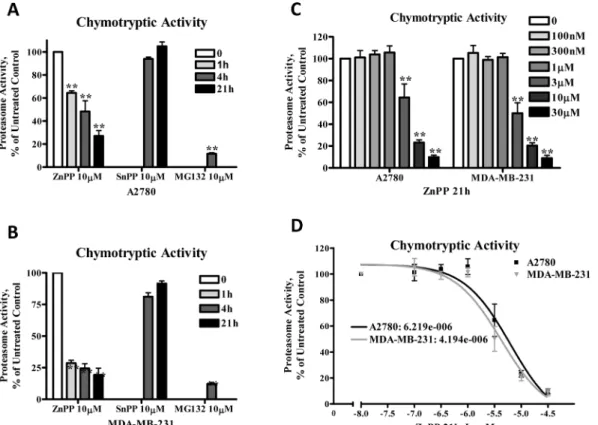
ZnPP’s inhibition of proteasome activity was further confirmed by direct measurement of the 20S proteasome chymotryptic activity, which was analyzed using the fluorophore linked peptide Suc-LLVY-AMC [19]. The eukaryotic 20S proteasome is known to have activities at-tributed to its various protein subunits that are referred to as caspase-like activity (cleaves after Glutamine and Aspartic acid residues), trypsin-like activity (cleaves after the basic amino acids Lysine and Arginine) and chymotrypsin-like activity (cleaves after hydrophobic amino acids) [20]. As shown inFig 3, treatment of A2780 (Fig 3A and 3C) and MDA-MB-231 (Fig 3B and 3C) cells with ZnPP or MG132, but not SnPP, suppressed chymotryptic activity in a time- and concentration-dependent manner. The IC50for ZnPP’s inhibition of proteasome activity was
Fig 1. ZnPP suppressesβ-catenin protein expression in A2780 cells.A2780 cells were treated with 5μM ZnPP for 0.5 or 1 hour. Cell lysates were prepared and western blot was performed using antibodies against
β-catenin andβ-actin.
doi:10.1371/journal.pone.0127413.g001
Fig 2. ZnPP induces accumulation of poly-ubiquitinated proteins.A.A2780 cells were treated with 5μM ZnPP for 15, 30 or 60 minutes. Cell lysates were prepared and western blot was performed using antibodies against phosphorylatedβ-catenin (Ser 33, Ser 37 and Thr 41) andβ-actin.B.A2780 cells were treated with 5μM ZnPP for 0.5 and 4 hours. Cell lysates were prepared and co-IP was performed usingβ-catenin antibody followed by western blot analysis of k48 linkage specific proteins,β-catenin (IPs) orβ-actin (inputs). Shown are representative images of three individual experiments.C.A2780 cells were treated with 10μM ZnPP for 4 and 21 hours, or 10μM MG132 for 21 hours. Cell lysates were prepared and western blot was performed using antibodies against K48-linkage specific polyubiquitinated proteins andβ-actin.
determined to be 6.2μM in A2780 cells and 4.2μM in MDA-MB-231 cells (Fig 3D). ZnPP, but not SnPP, also suppressed the tryptic and caspase-like proteasome activity in A2780 cells as an-alyzed using the fluorophore linked peptides Z-ARR-AMC or Z-LLE-AMC, respectively, [19] (Fig 4). Note that MG132 was more effective in suppressing the chymotryptic activity, rather
Fig 3. ZnPP inhibits chymotryptic proteasome activity in A2780 and MDA-MB-231 cells.A2780(A)or MDA-MB-231(B)cells were treated with 10μM ZnPP, SnPP or MG132 for 4 or 21 hours. Whole cell lysates were prepared and incubated with Suc-LLVY-AMC for 2 hours at 37°C and fluorescence was recorded at 380 nm excitation and 460 nm emission.C,D.A2780 and MDA-MB-231 cells were treated with various concentrations of ZnPP as indicated for 21 hours. Whole cell lysates were prepared and incubated with Suc-LLVY-AMC for 2 hours at 37°C and fluorescence was recorded at 380 nm excitation and 460 nm emission. The IC50was calculated with a nonlinear regression curve (Sigmoidal dose-response equation). Data (mean±SE, n = 3) are expressed as
percentages of untreated control.**,P<0.01, compared to untreated control cells using one-way ANOVA followed by Bonferroni analysis. doi:10.1371/journal.pone.0127413.g003
Fig 4. ZnPP inhibits tryptic and caspase-like proteasome activity.A2780 cells were treated with 10μM ZnPP, SnPP or MG132 for 4 or 21 hours. Whole cell lysates were prepared and incubated with Z-ARR-AMC (tryptic activity)(A)or Z-LLE-AMC (chymotryptic activity)(B)for 2 hours at 37°C and fluorescence was recorded at 380 nm excitation and 460 nm emission. Data (mean±SE, n = 3) are expressed as percentages of untreated control.*,P<0.05,**,P<0.01,
compared to untreated controls using one-way ANOVA followed by Bonferroni analysis.
than the caspase-like activity and did not affect the tryptic activity, results which are consistent with previous reports [21,22].
These results indicate that ZnPP-inducedβ-catenin protein degradation is accompanied by a significant suppression of ubiquitin-proteasome activity, suggesting that proteasome degra-dation does not directly account for ZnPP-induced suppression ofβ-catenin expression.
ZnPP does not promoteβ-catenin protein exportation. A recent report demonstrated that theβ-catenin protein is secreted in exosomes from HEK293 cells, leading to a significant sup-pression of the canonical Wnt signaling pathway [23]. To determine whether ZnPP-induces β-catenin protein secretion, A2780 cells were cultured in HBSS and treated with 5μM or 10μM of ZnPP for 1.5 hours. Whole cell lysates and concentrated extracellular proteins were pre-pared. Approximately 20–30μg of total cell lysate and extracellular proteins were loaded onto a SDS-polyacrylamide gel. Western blot analysis (Fig 5upper) shows thatβ-catenin protein ex-pression was suppressed by ZnPP in the cell lysate and undetectable in the extracellular protein fraction, indicating that ZnPP does not induceβ-catenin protein secretion. Some low molecu-lar bands were only detected in extracellumolecu-lar proteins, not in whole cell lysates, suggesting that these are non-specific. This conclusion was further supported by Coomassie blue gel-staining (Fig 5lower) showing that ZnPP treatment did not alter the extracellular protein profiles. We also treated A2780 cells with 5μM ZnPP for 72 hours and isolated exosomes from the medium. β-catenin was undetectable in the exosome proteins extracts (data not shown), further exclud-ing the possibility thatβ-catenin protein is secreted in exosomes upon ZnPP treatment.
The lysosome pathway is likely involved in ZnPP-inducedβ-catenin protein degradation. We have previously shown that lysosomal enzymes can be released upon enhanced lysosome membrane permeability, leading to cleavage of cellular proteins [14]. To determine whether
Fig 5. ZnPP does not induce secretion ofβ-catenin protein.A2780 cells were cultured in HBSS and treated with ZnPP at the indicated concentrations for 1.5 hours. The conditioned HBSS was collected and the proteins were concentrated. Western blot was performed using antibodies againstβ-catenin(Top).Cell lysates and concentrated protein samples were also separated by SDS-PAGE and stained with Coomassie blue staining(Bottom). Shown are representative images of three individual experiments.
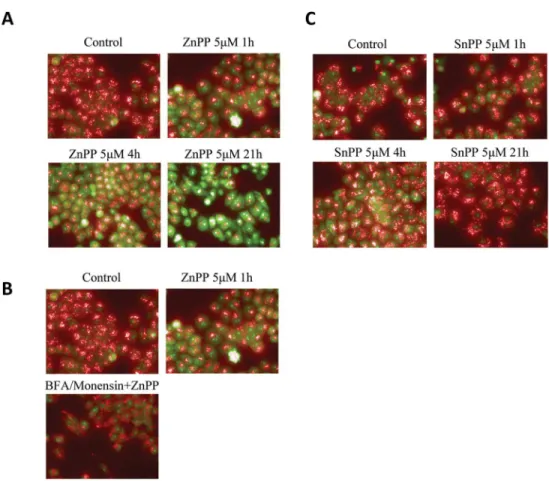
the lysosome protein degradation pathway is involved in ZnPP-induced suppression of β-cate-nin protein expression, we examined lysosome membrane permeability after ZnPP treatment. AO was used to study the lysosome membrane permeability [14,24]. AO preferentially accu-mulates in the lysosomes and will emit red fluorescence when excited under acidic conditions. When the lysosome is permeabilized, AO will relocate to the cytosol where it emits a green fluorescence upon excitation. The shift from red to green fluorescence indicates an increase in lysosome membrane permeability [25]. As shown inFig 6A, treatment with 5μM ZnPP for 1, 4, or 21 hours induced a time-dependent shift in AO staining from red to green, indicating that ZnPP enhances lysosome membrane permeability in A2780 cells. In contrast, treatment with SnPP did not result in significant changes in membrane permeability (Fig 6C), consistent with our previous observation that SnPP doesn’t induce suppression ofβ-catenin protein expression.
BFA is known to block intracellular transportation of lysosomal enzymes [26] and Monen-sin can block acidification of lysosomes [27,28]. Therefore we tested whether a BFA/Monensin cocktail could reverse the lysosome membrane permeability induced by ZnPP. After
Fig 6. ZnPP enhances lysosome membrane permeability.A2780 cells were treated with 5μM ZnPP (A) or 5μM SnPP (B) for 1, 4 or 21 hours. Cells were then incubated with 2.5μM AO for 30 minutes at 37°C. After incubation, cells were washed twice with HBSS. Images were captured using a fluorescent microscope (60X) with excitation at 500 nm, emission at 526 nm for AO green, excitation at 460 nm, emission at 650 nm for AO red.C.A2780 cells were pretreated with 21.2μM Brefeldin A and 4μM Monensin for 4 hours. The cells were then treated with 5μM ZnPP for 1 hour followed by incubation with 2.5μM AO for 30 minutes at 37°C. After incubation, cells were washed twice with HBSS. Images were captured using a fluorescent microscope (60X) with excitation at 500 nm, emission at 526 nm for AO green, and excitation at 460 nm, emission at 650 nm for AO red. Shown are representative images of three individual experiments.
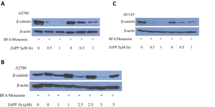
pretreatment of the cells with a BFA/Monensin cocktail (final concentration of 21.2μM BFA and 4μM Monensin) overnight (Fig 6B), a reversal in the ZnPP-induced lysomal membrane permeability was observed. These results support the involvement of the lysosome degradation pathway in the ZnPP-induced suppression ofβ-catenin protein expression. To confirm this as-sumption, A2780 and DU145 cells were treated with the BFA/Monensin cocktail overnight and treated with ZnPP at the indicated concentrations and durations (Fig 7). ZnPP-induced suppression ofβ-catenin protein expression was significantly attenuated by pretreatment with the BFA/Monensin cocktail. The effect of ZnPP onβ-catenin was both dramatic and significant and the reversal by BFA/Monensin was only observed after 0.5 and 1 hour ZnPP treatment (data not shown). However, the attenuation correlated with the concentration and duration of ZnPP treatment. These observations further demonstrate that the lysosome degradation path-way is likely involved in ZnPP-induced suppression ofβ-catenin protein expression.
Discussion
The present study was designed to explore the potential cellular mechanisms that mediate ZnPP-induced suppression ofβ-catenin expression using cancer cell model systems. The most interesting finding from this study is that ZnPP-inducedβ-catenin protein degradation is ac-companied by a significant inhibition of the ubiquitin-proteasome degradation pathway; and lysosome-mediated protein degradation seems to mediate this event. These results further elu-cidate the cellular mechanisms of ZnPP’s anticancer activity and indicate a potential new strat-egy in targeting theβ-catenin Wnt signaling pathway for cancer therapy.
β-catenin protein levels are well controlled by phosphorylation and ubiquitin-proteasome degradation [9]. Therefore, we initially believed that ZnPP would activate the proteasome deg-radation pathway, thereby leading to rapidβ-catenin protein degradation. However, several lines of experimental evidence indicated that proteasome degradation does not directly mediate
Fig 7. The BFA/Monensin cocktail attenuates ZnPP induced suppression ofβ-catenin expression.A2780(A)or DU145(B)cells were pretreated with or without 21.2μM BFA and 4μM Monensin overnight followed by treatment with 5μM of ZnPP for 0.5 or 1 hour.C.A2780 cells were pretreated with 21.2μM BFA and 4μM Monensin overnight followed by treatment with ZnPP for 1 hour at indicated concentrations. Cells were harvested and lysed. Western blot analysis was performed using antibodies againstβ-catenin andβ-actin. Shown are representative images of three individual experiments.
ZnPP-inducedβ-catenin protein degradation. First,β-catenin protein phosphorylation, an event leading to the ubiquitination and subsequent proteasome degradation ofβ-catenin, was not induced by ZnPP in cancer cells. On the contrary, ZnPP treatment rapidly reduced phos-phorylatedβ-catenin protein levels, likely due to the rapid degradation of the total cellularβ -catenin protein. Second, western blot analysis of poly-ubiquitinated proteins showed that ZnPP induces accumulation of poly-ubiquitinated proteins, suggesting that ZnPP acts as a pro-teasome inhibitor rather than an activator. Third, co-IP with aβ-catenin antibody and western blot analysis of K48-linkage specific poly-ubiquitinated proteins demonstrated that poly-ubi-quitinatedβ-catenin accumulated after ZnPP treatment, consistent with its inhibition of pro-teasome activity. Lastly, a direct measurement of the propro-teasome chymotryptic, tryptic and caspase-like activities confirmed that ZnPP significantly suppresses proteasome activities in a time- and concentration-dependent manner in cancer cells. To our knowledge, this is the first demonstration that ZnPP is a proteasome inhibitor. Note that ZnPP’s proteasome inhibitory activity is different from the previously established proteasome inhibitors, such as MG132 [21,22], in that ZnPP seems to have a broader spectrum of proteasome inhibitory activity (Figs
3and4).
The possibility that ZnPP might induceβ-catenin protein exportation through exosomes [23] thereby diminishing cellularβ-catenin protein expression was also not supported by our experimental results.β-catenin protein was undetectable by western blot analysis of the extra-cellular proteins collected from the conditioned media of ZnPP-treated cells. Furthermore, the exosomes isolated from the media did not containβ-catenin proteins. These observations indi-cate that ZnPP does not induceβ-catenin protein secretion from cancer cells.
We have recently reported that zinc ionophores enhance lysosome membrane permeability leading to the release of lysosomal enzymes and cleavage of cellular proteins [14]. In the pres-ent study, the use of AO allowed us to demonstrate that ZnPP enhances lysosome membrane permeability in our cancer cell model system, suggesting that ZnPP’s suppressionβ-catenin protein expression is a result of lysosomal enzyme digestion of cellular proteins. Importantly, ZnPP-induced lysosome membrane permeability could be effectively attenuated by pretreat-ment of the cells with the protein transportation inhibitory cocktail BFA/Monensin [29], which is known to block cellular transportation of lysosome enzymes (BFA, [26]) and inhibits lysosome acidification (Monensin, [27]). While this inhibitory cocktail is not specific to the ly-sosomes, the use of Monensin and BFA to alter lysosome structure and activity has been well documented [30–32]. These observations support the concept that the lysosome-mediated pro-tein degradation pathway is involved in the ZnPP-induced suppression ofβ-catenin expres-sion. Pretreatment of cancer cells with the BFA/Monensin cocktail significantly attenuated the suppression ofβ-catenin protein expression by ZnPP further confirming the involvement of the lysosomal protein degradation pathway in this process. It remains to be determined wheth-er specific lysosomal enzymes are responsible for ZnPP-inducedβ-catenin protein degradation or whether a select group of proteins are degraded through this process in cancer cells. The po-tential interaction of the ubiquitin-proteasome system with the lysosome degradation pathway [33,34] that may account for ZnPP-inducedβ-catenin protein degradation is under active in-vestigation. Given that there are no previous reports on lysosome-mediated suppression ofβ -catenin expression, the findings from the present study provide new insight into ZnPP’s anti-cancer activity and reveal potential new strategies in suppressing the canonical Wnt signaling pathway.
cellular mechanism of ZnPP’s anticancer activity and implicate a new strategy for targeting the canonical Wnt signaling pathway.
Author Contributions
Conceived and designed the experiments: SW BNH SEL WQD. Performed the experiments: SW BNH. Analyzed the data: SW BNH SEL WQD. Contributed reagents/materials/analysis tools: WQD. Wrote the paper: SW BNH SEL WQD.
References
1. Rattan S, Chakder S (2000) Influence of heme oxygenase inhibitors on the basal tissue enzymatic ac-tivity and smooth muscle relaxation of internal anal sphincter. J Pharmacol Exp Ther 294: 1009–1016. PMID:10945853
2. Hirai K, Sasahira T, Ohmori H, Fujii K, Kuniyasu H (2007) Inhibition of heme oxygenase-1 by zinc proto-porphyrin IX reduces tumor growth of LL/2 lung cancer in C57BL mice. Int J Cancer 120: 500–505. PMID:17066448
3. Nowis D, Bugajski M, Winiarska M, Bil J, Szokalska A, Salwa P, et al. (2008) Zinc protoporphyrin IX, a heme oxygenase-1 inhibitor, demonstrates potent antitumor effects but is unable to potentiate antitu-mor effects of chemotherapeutics in mice. BMC Cancer 8: 197. doi:10.1186/1471-2407-8-197PMID:
18620555
4. Fang J, Greish K, Qin H, Liao L, Nakamura H, Takeya M, et al. (2012) HSP32 (HO-1) inhibitor, copoly (styrene-maleic acid)-zinc protoporphyrin IX, a water-soluble micelle as anticancer agent: In vitro and in vivo anticancer effect. Eur J Pharm Biopharm 81: 540–547. doi:10.1016/j.ejpb.2012.04.016PMID:
22576132
5. La P, Fernando AP, Wang Z, Salahudeen A, Yang G, Lin Q, et al. (2009) Zinc protoporphyrin regulates cyclin D1 expression independent of heme oxygenase inhibition. J Biol Chem 284: 36302–36311. doi:
10.1074/jbc.M109.031641PMID:19850937
6. Tanaka S, Akaike T, Fang J, Beppu T, Ogawa M, Tamura F, et al. (2003) Antiapoptotic effect of haem oxygenase-1 induced by nitric oxide in experimental solid tumour. Br J Cancer 88: 902–909. PMID:
12644828
7. Wang S, Avery JE, Hannafon BN, Lind SE, Ding WQ (2013) Zinc protoporphyrin suppresses cancer cell viability through a heme oxygenase-1-independent mechanism: The involvement of the Wnt/beta-catenin signaling pathway. Biochem Pharmacol 85: 1611–1618. doi:10.1016/j.bcp.2013.03.011
PMID:23523860
8. Polakis P (2012) Drugging Wnt signalling in cancer. Embo J 31: 2737–2746. doi:10.1038/emboj.2012. 126PMID:22617421
9. Stamos JL, Weis WI (2013) The beta-catenin destruction complex. Cold Spring Harb Perspect Biol 5: a007898. doi:10.1101/cshperspect.a007898PMID:23169527
10. Ciechanover A (2013) Intracellular protein degradation: from a vague idea through the lysosome and the ubiquitin-proteasome system and onto human diseases and drug targeting. Bioorg Med Chem 21: 3400–3410. doi:10.1016/j.bmc.2013.01.056PMID:23485445
11. Yang G, Nguyen X, Ou J, Rekulapelli P, Stevenson DK, Dennery PA (2001) Unique effects of zinc pro-toporphyrin on HO-1 induction and apoptosis. Blood 97: 1306–1313. PMID:11222374
12. Ding WQ, Liu B, Vaught JL, Yamauchi H, Lind SE (2005) Anticancer activity of the antibiotic clioquinol. Cancer Res 65: 3389–3395. PMID:15833873
13. Zhang X, Yu H, Lou JR, Zheng J, Zhu H, Popescu NI, et al. (2011) MicroRNA-19 (miR-19) regulates tis-sue factor expression in breast cancer cells. J Biol Chem 286: 1429–1435. doi:10.1074/jbc.M110. 146530PMID:21059650
14. Yu H, Zhou Y, Lind SE, Ding WQ (2009) Clioquinol targets zinc to lysosomes in human cancer cells. Biochem J 417: 133–139. doi:10.1042/BJ20081421PMID:18764784
15. Zhang Z, Bi C, Schmitt SM, Fan Y, Dong L, Zuo J, et al. (2012) 1,10-Phenanthroline promotes copper complexes into tumor cells and induces apoptosis by inhibiting the proteasome activity. J Biol Inorg Chem 17: 1257–1267. doi:10.1007/s00775-012-0940-xPMID:23053530
16. Komander D (2009) The emerging complexity of protein ubiquitination. Biochem Soc Trans 37: 937– 953. doi:10.1042/BST0370937PMID:19754430
18. Kim I, Kim CH, Kim JH, Lee J, Choi JJ, Chen ZA, et al. (2004) Pyrrolidine dithiocarbamate and zinc in-hibit proteasome-dependent proteolysis. Exp Cell Res 298: 229–238. PMID:15242777
19. Pan J, Zhang Q, Wang Y, You M (2010) 26S proteasome activity is down-regulated in lung cancer stem-like cells propagated in vitro. PLoS One 5: e13298. doi:10.1371/journal.pone.0013298PMID:
20949018
20. Murata S, Yashiroda H, Tanaka K (2009) Molecular mechanisms of proteasome assembly. Nat Rev Mol Cell Biol 10: 104–115. doi:10.1038/nrm2630PMID:19165213
21. Lee DH, Goldberg AL (1998) Proteasome inhibitors: valuable new tools for cell biologists. Trends Cell Biol 8: 397–403. PMID:9789328
22. Alexandrova A, Petrov L, Georgieva A, Kirkova M, Kukan M (2008) Effects of proteasome inhibitor, MG132, on proteasome activity and oxidative status of rat liver. Cell Biochem Funct 26: 392–398. doi:
10.1002/cbf.1459PMID:18236383
23. Chairoungdua A, Smith DL, Pochard P, Hull M, Caplan MJ (2010) Exosome release of beta-catenin: a novel mechanism that antagonizes Wnt signaling. J Cell Biol 190: 1079–1091. doi:10.1083/jcb. 201002049PMID:20837771
24. Boya P, Kroemer G (2008) Lysosomal membrane permeabilization in cell death. Oncogene 27: 6434– 6451. doi:10.1038/onc.2008.310PMID:18955971
25. Erdal H, Berndtsson M, Castro J, Brunk U, Shoshan MC, Linder S (2005) Induction of lysosomal mem-brane permeabilization by compounds that activate p53-independent apoptosis. Proc Natl Acad Sci U S A 102: 192–197. PMID:15618392
26. Oda K, Nishimura Y (1989) Brefeldin A inhibits the targeting of cathepsin D and cathepsin H to lyso-somes in rat hepatocytes. Biochem Biophys Res Commun 163: 220–225. PMID:2775262
27. Misinzo G, Delputte PL, Nauwynck HJ (2008) Inhibition of endosome-lysosome system acidification en-hances porcine circovirus 2 infection of porcine epithelial cells. J Virol 82: 1128–1135. PMID:
18032516
28. Pohlmann R, Kruger S, Hasilik A, von Figura K (1984) Effect of monensin on intracellular transport and receptor-mediated endocytosis of lysosomal enzymes. Biochem J 217: 649–658. PMID:6231917
29. O'Neil-Andersen NJ, Lawrence DA (2002) Differential modulation of surface and intracellular protein expression by T cells after stimulation in the presence of monensin or brefeldin A. Clin Diagn Lab Immu-nol 9: 243–250. PMID:11874859
30. Lippincott-Schwartz J, Yuan L, Tipper C, Amherdt M, Orci L, Klausner RD, et al. (1991) Brefeldin A's ef-fects on endosomes, lysosomes, and the TGN suggest a general mechanism for regulating organelle structure and membrane traffic. Cell 67: 601–616. PMID:1682055
31. Chi C, Zhu H, Han M, Zhuang Y, Wu X, Xu T (2010) Disruption of lysosome function promotes tumor growth and metastasis in Drosophila. J Biol Chem 285: 21817–21823. doi:10.1074/jbc.M110.131714
PMID:20418542
32. Janda E, Nevolo M, Lehmann K, Downward J, Beug H, Grieco M (2006) Raf plus TGFbeta-dependent EMT is initiated by endocytosis and lysosomal degradation of E-cadherin. Oncogene 25: 7117–7130. PMID:16751808
33. Raiborg C, Stenmark H (2009) The ESCRT machinery in endosomal sorting of ubiquitylated membrane proteins. Nature 458: 445–452. doi:10.1038/nature07961PMID:19325624