Pseudomonas aeruginosa
to Predict Clinical Disease in
Hospitalized Patients
Michel Ledizet1, Thomas S. Murray2¤a, Sailaja Puttagunta3¤b, Martin D. Slade4, Vincent J. Quagliarello3, Barbara I. Kazmierczak3,5*
1L2 Diagnostics, New Haven, Connecticut, United States of America,2Department of Pediatrics (Infectious Diseases), Yale University School of Medicine, New Haven, Connecticut, United States of America,3Department of Medicine, Sections of Infectious Diseases, Yale University School of Medicine, New Haven, Connecticut, United States of America,4Department of Occupational & Environmental Medicine, Yale University School of Medicine, New Haven, Connecticut, United States of America, 5Department of Microbial Pathogenesis, Yale University School of Medicine, New Haven, Connecticut, United States of America
Abstract
Background:Pseudomonas aeruginosais an opportunistic pathogen that frequently causes hospital acquired colonization and infection. Accurate identification of host and bacterial factors associated with infection could aid treatment decisions for patients withP. aeruginosacultured from clinical sites.
Methods:We identified a prospective cohort of 248 hospitalized patients with positiveP. aeruginosacultures. Clinical data were analyzed to determine whether an individual met predefined criteria for infection versus colonization.P. aeruginosa
isolates were tested for the expression of multiple phenotypes previously associated with virulence in animal models and humans. Logistic regression models were constructed to determine the degree of association between host and bacterial factors withP. aeruginosainfection of the bloodstream, lung, soft tissue and urinary tract.
Results:One host factor (i.e. diabetes mellitus), and one bacterial factor, a Type 3 secretion system positive phenotype, were significantly associated withP. aeruginosainfection in our cohort. Subgroup analysis of patients withP. aeruginosaisolated from the urinary tract revealed that the presence of a urinary tract catheter or stent was an additional factor for
P. aeruginosainfection.
Conclusions:Among hospitalized patients with culture-documentedP. aeruginosa,infection is more likely to be present in those with diabetes mellitus and those harboring a Type 3 secretion positive bacterial strain.
Citation:Ledizet M, Murray TS, Puttagunta S, Slade MD, Quagliarello VJ, et al. (2012) The Ability of Virulence Factor Expression byPseudomonas aeruginosato Predict Clinical Disease in Hospitalized Patients. PLoS ONE 7(11): e49578. doi:10.1371/journal.pone.0049578
Editor:Gunnar F Kaufmann, The Scripps Research Institute and Sorrento Therapeutics, Inc., United States of America
ReceivedSeptember 19, 2012;AcceptedOctober 10, 2012;PublishedNovember 12, 2012
Copyright:ß2012 Ledizet et al. This is an open-access article distributed under the terms of the Creative Commons Attribution License, which permits unrestricted use, distribution, and reproduction in any medium, provided the original author and source are credited.
Funding:This work was supported by National Institutes of Health (NIH) grants T32 AI007517 (to SP), T32 AI07210 (to TSM), R42 AI058659 (to BIK) and the Patrick and Catherine Weldon Donaghue Medical Research Foundation (to BIK). The funders had no role in study design, data collection and analysis, decision to publish, or preparation of the manuscript.
Competing Interests:ML is employed by L2 Diagnostics, LLC. This does not alter the authors’ adherence to all the PLOS ONE policies on sharing data and materials.
* E-mail: barbara.kazmierczak@yale.edu
¤a Current address: Department of Basic Medical Sciences, Frank H. Netter MD School of Medicine, Quinnipiac University, Hamden, Connecticut, United States of America
¤b Current address: Durata Therapeutics, Branford, Connecticut, United States of America
Introduction
Hospital acquired infections (HAI) are estimated to complicate 5–10% of hospitalizations in the United States annually, leading to increased health care costs and prolonged hospitalizations [1].
Pseudomonas aeruginosais a frequent cause of HAI, isolated in 16% of urinary tract infections and 18% of pneumonias, particularly ventilator-associated pneumonia [2,3]. A combination of several factors–intrinsic antibiotic and microbicide resistance, prevalence and persistence in the hospital environment, and a propensity to form biofilms on medical devices–lead to relatively high coloni-zation rates by this organism. Prospective studies have shown that a subgroup of colonized patients develop clinically significant
disease, such as ventilator-associated pneumonia. However, many patients do not progress to invasive disease, suggesting that differences in bacterial virulence and/or host susceptibility influence clinical course [4–7].
Many P. aeruginosavirulence factors have been identified over the past decades. The most robust virulence factor in animal models and human studies is the Type 3 secretion system (T3SS), a specialized protein secretion apparatus that allows Gram-negative bacteria such as P. aeruginosa to translocate a specific subset of bacterial effector proteins into the host cell cytosol [8]. Only four
transferase activity; and ExoU, a phospolipase A2 [9–15].
Carefully controlled studies examining virulence of isogenic T3SS mutants in murine models of acute pneumonia have demonstrated that two secreted effectors, ExoU and ExoS, are independently associated with increased virulence in this model. Other virulence factors identified in animal models include those promoting bacterial motility and adhesion (i.e., flagella and Type IV pili) [16–21], degradative enzymes [22–25], and genomic pathogenicity islands [26–31].
Bacterial expression of the T3SS is co-regulated with expression of many bacterial traits associated with acute infection (flagella, type IV pili, secreted proteases) or chronic colonization (biofilm formation). As inverse regulation of acute vs. chronic virulence factors is a feature of many of these regulatory networks, some authors have postulated that bacteria may switch their behavior to favor acute infection or chronic colonization [32]. The natural history of P. aeruginosa in Cystic Fibrosis patients is thought to illustrate such a switch, as younger CF patients are usually acutely infected by T3SS-positive strains, but progress to long-term colonization by T3SS-negative strains that usually display mucoid or hyper-biofilm phenotypes [33,34]. No study has asked, however, whether bacterial expression of particular virulence factors is associated with infection vs colonization in non-CF patients. Identifying such associations might have significant utility for clinical decision-making for hospitalized patients with positive
P. aeruginosacultures. Discriminating between patients at high vs. low risk of acute infection could improve delivery of effective anti-pseudomonal therapy to infected patients, but decrease inappro-priate antibiotic use in colonized patients.
In this study, we prospectively enrolled 248 unique hospitalized patients withP. aeruginosacultured from blood, airway secretions, urine, or deep wound specimens, excluding individuals with Cystic Fibrosis. Subjects were followed clinically for five days after their positive culture to determine whether they met pre-determined clinical criteria for infection vs. colonization with P. aeruginosa. Selected bacterial phenotypes associated in the literature with virulence or colonization were assayed for each bacterial isolate. We employed the statistical tool of factor analysis to determine whether any of the measured bacterial variables grouped together in orthogonal ‘‘factors’’ [35]. The primary endpoint of our study was to determine which, if any, readily measured bacterial traits were associated withP. aeruginosainfection versus colonization of the respiratory or urinary tract.
Methods
Cohort Assembly
This study was approved by the Institutional Review Board of Yale University School of Medicine (HIC #0411027243). All pediatric and adult patients hospitalized at Yale New Haven Hospital (New Haven, CT) with a positive culture forP. aeruginosa
from the respiratory tract, urinary tract, blood, or deep tissue were eligible for study enrollment if they, or designated proxies, provided written consent. Clinical isolates were identified as P. aeruginosa if they grew as non-lactose fermenting colonies on MacConkey agar, were oxidase positive, could utilize acetamide as a sole carbon source, and gave a characteristic zone of inhibition to colistin on Kirby-Bauer plates. The presence of characteristic ‘‘grape-like’’ odor, pigment production and ability to grow at 42uC were also noted. Isolates from enrolled patients were subcultured from either primary specimen plates (i.e. blood or MacConkey agar) or from Kirby-Bauer plates generated by the Clinical Microbiology laboratory to Luria Broth (LB) agar, then coded and banked as 15% glycerol stocks at –80uC. Patient demographics,
risk factors for infection, co-morbid medical conditions, prior and concurrent antibiotic exposure, history of prior P. aeruginosa
cultures, clinical and laboratory features were collected at baseline and for 5 days after the initial culture date. Patients were excluded from the study if they had Cystic Fibrosis (n = 5), hadP. aeruginosa
isolates that would not grow in the defined media required for the Type 3 phenotypic assay (n = 2), or had isolates cultured from a non-eligible site (i.e. a biliary catheter drainage bag) (n = 1).
Clinical Outcomes
Anonymously coded data collection forms were reviewed independently by two Infectious Diseases physicians to determine whether patients met criteria for infection of the respiratory tract (pneumonia), urinary tract (cystitis or pyelonephritis) or soft tissue/ bone (abscess or osteomyelitis), based on definitions established by the National Nosocomial Infection Surveillance (NNIS) system (Table 1) [36]; reviewing physicians were blinded to virulence testing results. Patients who met clinical criteria were categorized as infected; those who did not were categorized as colonized. Bloodstream isolates were considered indicative of infection. If the patient met the criteria for infection, but other potential pathogens were co-cultured withP. aeruginosa, the patient was categorized as ‘‘infection, possibly due toPseudomonas’’. Potential pathogens were considered to be: (1) any other organism isolated from blood or an intraoperative tissue culture, (2) any organism present in .105 cfu/ml in urine, or (3)Staphylococcus aureus, Streptococcus pneumoniae, Hemophilus influenzae, Moraxella catarhalis, any Gram-negative rod not normally isolated from the oropharynx,Mycobacterium tubercu-losis, and dimorphic fungi or molds in respiratory tract specimens. The report of ‘‘normal respiratory flora’’ without the specific identification of one of these microorganisms, or the isolation of
Candida albicansin sputum, was not interpreted as indicating the presence of another potential pathogen. When the two reviewers disagreed, a third independent review was performed to determine clinical assignment of infection versus colonization.
Testing of Type III Protein Secretion by ELISA
Components of the T3SS were detected quantitatively by an ELISA assay as described previously [37]. Monoclonal antibodies against ExoS (SD1, SE1) were used to detect ExoS simultaneously with the other antigens.
Isolation of Monoclonal Antibodies Against ExoS
Motility Assays
Swimming, twitching and swarming motility were measured as previously described [38]. Each isolate was independently assayed twice.
Static Biofilm Formation
Static biofilm formation on polystyrene was measured in triplicate using the method of O’Toole [39].
Secreted Protease Activity
Protease production was measured by a casein proteolysis microplate assay as previously described [38]. The LasB elastase, alkaline protease, and protease IV are all capable of degrading casein [40].
Pathogenicity Island Loci
Genomic DNA was amplified by PCR using primers specific for PAPI-I pathogenicity island loci RL033 (RL033F: 59 AAAGTT-TAGCGCCGACTTCA; RL033R: 59 GAAGTGAGTTGGG-CACCATT) or RL112 (RL112F: 59 GACTGGAATAGACGCC-CAAG; RL112R: 59AAACCTTCGACGGTGATCTG). This pathogenicity island was interrogated based on its association with virulence in several distinct animal models of virulence; the queried loci encode two experimentally identified ‘‘pathogenicity genes’’ [26,31].
Statistical Analysis
Values obtained for all measured bacterial virulence phenotypes were subjected to an exploratory factor analysis [35]. The principal factor method was used to extract factors, followed by promax (oblique) rotation. A ‘‘scree’’ test was used to determine the number of meaningful factors retained for rotation. An item was considered to load on a given factor if the factor loading was 0.50 or greater for that factor and was less than 0.50 for all other factors.
Unadjusted and adjusted logistic regression models were conducted. Parsimonious models were obtained through the use of a forward selection strategy with a significance-to-enter defined as 0.06. The outcome was defined as infection or sepsis caused by
P. aeruginosa vs. colonization. Patients in whom infection was present but could not be attributed to P. aeruginosa (i.e. another
potential pathogen was co-cultured) were coded as unknown and excluded from our analysis. All statistical analyses were performed using SAS 9.1.
Results
Patient Characteristics
A total of 248 patients withP. aeruginosa cultured from blood, urine, respiratory secretions or tissue provided written informed consent and were enrolled in this study at Yale New Haven Hospital (YNHH) from July 2005 through March 2007. Eight patients were found to be ineligible based on defined exclusion criteria. One eligible P. aeruginosa isolate was discarded by the Clinical Microbiology laboratory. Two additional individuals were enrolled twice, during independent hospitalizations under different medical record numbers; the second enrollment event was not analyzed in this study, resulting in a total of 237 unique participants.
Patient age ranged between ,1 to 96 years at the time of enrollment (mean, 58.6 years). One hundred sixty (68%) patients were admitted to YNHH from home, while 48 (20%) were admitted from rehabilitation or extended care facilities. Twenty-six (11%) patients were transferred from other hospitals, while three patients remained at YNHH since their birth. The median elapsed time between hospital admission and the positive
P. aeruginosaculture that triggered enrollment was 4 days (range, 0 to 1749); for 79 patients (33%), a prior positive culture for
P. aeruginosawas documented during either the index or a previous hospitalization.
Host factors historically associated with increased risk of
P. aeruginosainfection were assessed in our cohort [41]. Sixty-five (27%) patients carried a diagnosis of Type I or Type II diabetes mellitus. Fifty-five (23%) patients were deemed immunocompro-mised as a result of chronic steroid use ($10 mg prednisone per day for 30 days or longer), post-transplant immunosuppression, marrow suppressive chemotherapy or HIV/AIDS. Seventy-two patients (30%) carried a diagnosis of asthma, chronic obstructive pulmonary disease or emphysema, or had radiologic evidence of bronchiectasis; these individuals were considered to have ‘‘chronic lung disease’’ in our analysis. The majority of patients (183/237; 77%) had at least one medical device that might be expected to
Table 1.Clinical criteria for infection based on site of culture.
Respiratory tract: Infection (pneumonia)
P. aeruginosacultured from respiratory specimen containing fewer than 25 epithelial cells/HPF.
New and persistent infiltrates are noted on chest x-ray or computed tomography (CT).
Two of the following three criteria are met: (1) fever.38.3uC; (2) WBC.12,000 or bands/metamyelocytes$6% or 1500/ml; (3) altered mental status (patient.70 y).
Symptoms referable to the respiratory tract, evidenced by at least one of the following: (1) new purulent sputum OR increased sputum, secretions or suctioning; (2) new/worsening cough OR dyspnea OR tachypnea; (3) worsening gas exchange (increased requirement for supplemental oxygen, increased ventilator support, PaO2/FiO2#240).
Urinary tract: Infection (cystitis or
pyelonephritis & urosepsis)
Greater than 105cfuP. aeruginosa/ml urine
Pyuria (.10 WBC/HPF)
Soft tissue/bone: Infection (abscess or osteomyelitis)
P. aeruginosacultured from deep wound or intraoperative tissue specimen
Clinical evidence of infection (purulent drainage, pain/erythema/dehiscence) or histopathological confirmation of infection
predispose to infection of the respiratory tract (i.e., endotracheal tube or tracheostomy), urinary tract (i.e., bladder catheter or nephrostomy tube) or blood (i.e., central intravascular catheter). In 144 patients (61%) such a device was present at the anatomical site from which P. aeruginosa was cultured, and was considered a ‘‘relevant foreign device’’ in our analysis.
Clinical Characteristics of Patients
Seventy-one patients (30%) met the criteria for infection of bloodstream, respiratory tract, urinary tract or soft tissue/bone, and had no other potential pathogens cultured from these sites except for P. aeruginosa. An additional 52 patients (22%) met criteria for infection, but at least one other potential pathogen was cultured from the site of infection in addition toP. aeruginosa. One hundred fourteen patients (48%) did not meet the criteria for infection despite a positive culture for P. aeruginosa and were classified as ‘‘colonized’’. This group included 65 individuals with pureP. aeruginosacultures (urine or wound) or normal respiratory flora plusP. aeruginosa, and 49 patients whose cultures yielded a potential pathogen in addition toP. aeruginosa. Table 2 shows the distribution of patient age, gender, ethnicity and selected comorbidities among the groups.
Bacterial Isolates
We collected sputum isolates from 136 patients, urine isolates from 65 patients, and deep wound/tissue isolates from 22 patients. Ten patients had blood isolates; in addition, one patient had positive cultures from both sputum and blood, two from both urine and blood, and one from wound and blood. Table 3 shows the distribution of clinical outcomes by site of culture. Each bacterial isolate was assayed for each virulence phenotype as described in Methods. For the first 75 individuals enrolled in the study, we collected 10 independentP. aeruginosacolonies from the primary culture plates and assayed each individually. Although heterogeneity of T3SS phenotype expression has been reported among isolates cultured from patients with CF [34], we found that only 4 (5.3%) of our tested samples harbored mixtures of T3SS-positive and –negative strains. Given this low rate of T3SS phenotype heterogeneity, only one isolate per unique anatomic site was collected and phenotyped in subsequently enrolled patients.
A number of the bacterial variables that we measured in our study are likely to be biologically interrelated. We applied the statistical tool of factor analysis to identify which variables were most highly interdependent, and found that most of the bacterial phenotypes loaded into three mathematically defined factors [35]. Factor 1 (‘‘Type 3 secretion’’) reflected secretion of the two T3SS structural proteins, PopD and PcrV, and of the T3SS substrates ExoT, ExoS and ExoU. Factor 2 (‘‘Flagellar motility & proteases’’)
consisted of two flagellar based forms of motility (swimming and swarming) and protease production. Factor 3 (‘‘Fimbriae’’) consisted of twitching motility and the presence of two loci, RL033 and RL112, within the PAPI-1 pathogenicity island. Equations for factors 1, 2 and 3 are listed in Figure S1. Biofilm formation did not load strongly onto any of the three factors and was thus evaluated as an independent variable in our models.
Bacterial and Host Variables Associated with Increased Risk of Infection
We tested the measured bacterial phenotypes, as well as host variables potentially associated with altered risk of infection, in both an unadjusted (bivariate) logistic regression model and a parsimonious model resulting from multivariate logistic regression models using a forward selection strategy. All tested model variables are listed in Table 4. Patients with infections that could not be definitely attributed to P. aeruginosa because another potential pathogen was co-cultured were considered to have an ‘‘unknown’’ clinical categorization, and their records were excluded from analysis, lowering the sample size from 237 to 185. Results of the parsimonious (forward selection) model are presented in Table 4. Only two variables, diabetes mellitus and ‘‘Type 3 secretion’’ (Factor 1) showed a significant association with infection vs. colonization (p#0.05). We also ran our models on the subsets of patients with positive cultures from the respiratory tract (n = 105) or urine (n = 59). In patients with positive respiratory specimens, no host or bacterial variable was significantly associated with patient outcome (infection vs. colonization) in the multivariate analysis, and no variables entered the parsimo-nious model. In patients with positive urine cultures, two independent parsimonious models could be constructed. Both contained diabetes mellitus and the presence of a urinary catheter or stent as host variables significantly associated with increased risk of infection, plus a bacterial variable which was either ‘‘Flagellar motility & proteases’’ (odds ratio 2.3; p = 0.02) or ‘‘Type 3 secretion’’ (odds ratio 1.9; p = 0.05).
Discussion
In this study, we hypothesized that distinct virulence traits of the opportunistic pathogen P. aeruginosa would be associated with increased risk of infection vs. colonization in hospitalized patients with positiveP. aeruginosacultures. The presence ofP. aeruginosain conjunction with evidence of host tissue impairment and a local or systemic immune response was indicative of infection (Table 1), while the presence of bacteria with no or minimal tissue damage or inflammation was taken to represent colonization. We assayed a set of bacterial phenotypes, primarily chosen on the basis of their prior association with virulence in animal models of acute infection
Table 2.Patient characteristics as a function of colonization vs. infection status.
Colonization (n = 114)
P. aeruginosaInfection/ Sepsis (n = 71)
Infection, possibly due to
P. aeruginosa(n = 52) pvalue (chi square)
Gender 51 (45%) female 24 (34%) female 24 (46%) female 0.26
Mean age, yrs (95% CI) 58.0 (53.3–62.7) 61.4 (56.4–66.4) 58.3 (51.4–65.2) –
Ethnicity (Hispanic) 21 Hispanic (18%) 5 Hispanic (7.0%) 5 Hispanic (9.6%) 0.06
Diabetes 26 (23%) 27 (38%) 12 (23%) 0.06
Immunocompromised 31 (27%) 17 (24%) 17 (33%) 0.56
Relevant foreign device 67 (59%) 46 (65%) 31 (60%) 0.70
or their co-regulation with such virulence traits. Bacterial phenotypes predominantly associated with persistence in chronic infection models (e.g. production of quorum sensing molecules or pyocyanin) were not considered. Factor analysis of the measured bacterial variables revealed interrelationships between several of these phenotypes. Some groupings were expected, such as that of the five measured T3SS proteins that loaded onto Factor 1. Others were not anticipated, such as the association of twitching motility with the PAPI-1 loci RL033 and RL112 within Factor 3, but are biologically plausible given the recent finding that the PAPI-1 pathogenicity island is transferred by conjugation via a Type IV pilus encoded therein [42]. The association of two flagellar-based forms of motility with protease secretion in Factor 2 may reflect the co-regulation of both flagellar genes and the Type II secretion system by the cyclic AMP dependent transcriptional activator Vfr [43].
Most published studies report bacterial virulence phenotypes as a binary variable (i.e., present/absent). Our study differed in providing quantitative measures for most variables, and this information was retained in the statistically defined factors used in our analysis. Although studies commonly stratify risk factors when considering association with risk of disease (e.g., CD4 count and risk of opportunistic infection, LDL cholesterol and risk of coronary artery disease), this approach is not widespread in
studies of microbial pathogenesis, where two-way comparisons are often made between bacteria that do or do not express a putative virulence trait.
Production and secretion of T3SS components and effectors as measured by Factor 1 was the only bacterial variable that was associated with increased risk of infection in our total cohort (odds ratio 1.4), albeit with apvalue = 0.5. The host risk factor diabetes mellitus was also associated with an increased risk of infection (odd ratio 2.1;p,0.05) in this parsimonious model. The association of T3SS with clinical infection was weaker than we expected based on data from animal models of acute pulmonary infection. Indeed, in the subset of patients with respiratory tractP. aeruginosaisolates, no association between pneumonia and T3SS was observed. Reanalysis of the data applying a less stringent definition of infection, which allowed patients with tracheobronchitis to be classified as infected, did not result in the association of T3SS or any other bacterial variable with increased risk of infection in the multivariate or parsimonious models (data not shown). In the subset of patients with urinary tract infections, two independent parsimonious models could be constructed. The host factors of diabetes mellitus and the presence of a urinary tract catheter or stent were associated with increased risk of infection in both models, along with either bacterial T3SS protein secretion (Factor 1) or the expression of flagellar based motility and protease
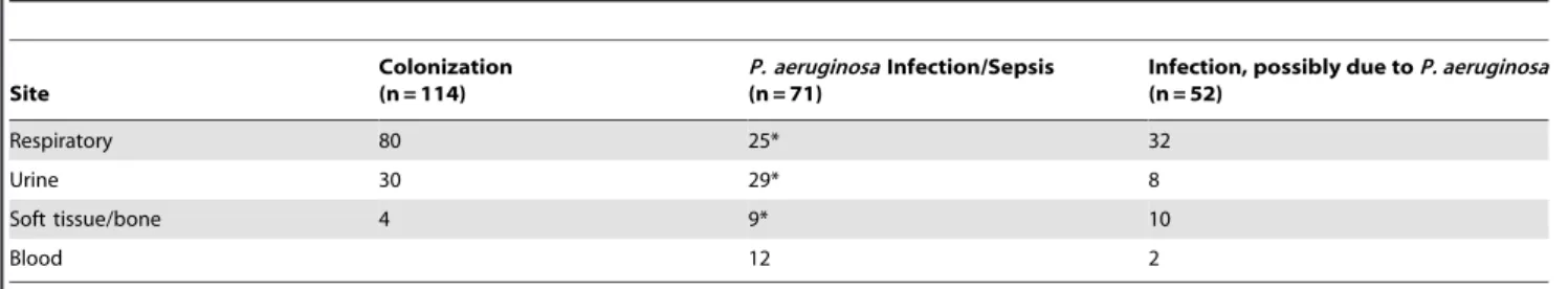
Table 3.Sources of bacterial isolates as a function of clinical categorization.
Site
Colonization (n = 114)
P. aeruginosaInfection/Sepsis (n = 71)
Infection, possibly due toP. aeruginosa (n = 52)
Respiratory 80 25* 32
Urine 30 29* 8
Soft tissue/bone 4 9* 10
Blood 12 2
*Patients withP. aeruginosaisolated concurrently from blood and another site (n = 4) are analyzed with both blood and non-blood sites. doi:10.1371/journal.pone.0049578.t003
Table 4.Parsimonious multivariate analysis of factors associated with risk ofP. aeruginosainfection.
Parameter Level
Parsimonious (forward selection), All culture sites
Parsimonious (forward selection), Urinary tract cultures
p-value Odds Ratio p-value Odds Ratio
estimate L95% U95% estimate L95% U95%
Gender female v male – – – – – – – –
Ethnicity hispanic v non-hispanic – – – – – – – –
Race nonwhite v white – – – – – – – –
Relevant foreign device yes v no – – – – 0.03 6.3 1.2 34
Any chronic lung disease yes v no – – – – – – – –
Immunocompromised yes v no – – – – – – – –
Solid tumor yes v no – – – – – – – –
Diabetes mellitus yes v no 0.03 2.1 1.1 4.3 0.006 12 2.1 70
Age per year – – – – – – – –
Biofilm per unit – – – – – – – –
Factor 1 per unit 0.05 1.4 1.0 1.8 0.05 1.9 0.99 3.8
Factor 2 per unit – – – – – – – –
Factor 3 per unit – – – – – – – –
secretion (Factor 2). A greater propensity for protease secretion by human urinary tract isolates, as opposed to isolates from other body sites, has been reported in several studies (recently reviewed in [44]), suggesting that this group of virulence factors may play a specific role in causing disease at this anatomic site. It is not known whether decreased protease secretion by quorum-sensing mutants underlies the observed attenuation of quorum-sensing deficientP. aeruginosa strains in murine models of acute ascending pyelone-phritis [45].
Several series have examined whether T3SS production byP. aeruginosais associated with poor outcomes for human pulmonary infection. Roy-Burman et al. reported an association between increased mortality and bacterial secretion of one or more T3SS proteins in a study of 108 patients with lower respiratory tract or blood isolates ofP. aeruginosa[11]. The interpretation of this study is confounded by the inclusion of patients with Cystic Fibrosis, whose chronic pulmonary colonization by T3SS-negative strains rarely results in severe, systemic disease. Hauser and colleagues studied a more homogeneous retrospective cohort of 35 patients with an established diagnosis ofP. aeruginosa ventilator-associated pneumonia, and reported that infection with a T3SS-positive isolate was significantly associated with severe disease, defined as death or relapse despite appropriate therapy [10]. These findings were corroborated by a recent study demonstrating increased risk of persistence and relapse associated with T3SS-positive P. aeruginosastrains in patients with ventilator-associated pneumonia [9]. These latter two studies supported an association between T3SS and more severe disease in patients with ventilator-associated pneumonia, but their design did not allow them to assess the influence of this bacterial variable on the risk of infection vs. colonization in the respiratory tract (as was considered in the current study). These studies also did not consider a role for other potential virulence factors in human respiratory infection.
Limitations and Strengths of the Current Study
Our study has limitations. First, all assays required culture and isolation ofP. aeruginosafrom primary clinical specimens, and all bacterial virulence phenotypes were measured in vitro. Although bacterial phenotypes of banked isolates were stable and reproduc-ible upon repeat assay, we only determined whether an isolate could express a particular virulence factor under defined in vitro conditions. Thus our assays measured the potential for an isolate to express a virulence factor in the human host, but did not determine whether those virulence factors were actually expressed in the host. This limitation also applies to other studies that measure virulence factor expression of clinical isolates. Second, the definitions for infection that we applied were based on NNIS system criteria and are congruent with clinical practice. However, our definition of respiratory tract infection was rigorous in requiring radiographic findings consistent with pneumonia, and was not met by patients with tracheobronchitis; in contrast, our definition of urinary tract infection was satisfied by evidence of a local inflammatory response even in patients with a urinary catheter. Nonetheless, our findings did not change when we broadened our criteria for respiratory tract infection to include tracheobronchitis. Third, patients were followed clinically for only 5 days after theP. aeruginosaculture that triggered enrollment was obtained. Any individual who met criteria for infection after this time would nonetheless be considered ‘‘colonized’’ in our analysis.
P. aeruginosa causes nosocomial infections at sites beyond the respiratory tract. A strength of our study cohort is inclusion of patients with positive cultures from lung, urine and deep wound or tissue, making it more representative of patients with positive cultures forP. aeruginosaseen in tertiary care hospitals. Despite the heterogeneity of this patient population, we observed an increased risk ofP. aeruginosainfection associated with T3SS-positive isolates. This association persisted in the subgroup of patients with a urinary tract source ofP. aeruginosa, arguing that T3SS-associated virulence is not specific to the respiratory tract.
Concluding Observations
The T3SS ofP. aeruginosais a dominant virulence factor in acute infections of model organisms that range from insects [46] to mice [8]. In our study, the T3SS was associated with increased risk of infection, but the magnitude of the effect was smaller than might be predicted from studies with inbred mice. Our result illustrates that in human patients, host-related variability has an equal or greater influence on the outcome of host-pathogen encounters than production of virulence factors by bacterial isolates.
Although the culture of certain organisms from clinical specimens, e.g. M. tuberculosis, is diagnostic of infection, the prognostic implications of culturing organisms such asP. aeruginosa
are less clear. The identification of microbial virulence factors that influence the development of infection in humans may aid clinician decision-making in such a setting, and prompted us to undertake this study. Consideration of patient comorbidities that increase the risk and severity of infection, such as diabetes mellitus, is an integral part of treatment algorithms used by clinicians. Our findings suggest that an intrinsic bacterial factor, namely production of T3SS proteins, is also associated with an increased risk of P. aeruginosa infection in a heterogeneous cohort of hospitalized patients. By considering the T3SS phenotype of a clinical P. aeruginosa isolate, clinicians may be able to direct appropriate antimicrobial therapy to patients at higher risk of infection while still limiting the indiscriminate use of antimicrobials associated with increased antibiotic resistance [47]. Our findings also support further investigation into therapeutic strategies that target the T3SS and its effectors [48–52].
Supporting Information
Text S1 Equations determined by factor analysis.
(DOCX)
Acknowledgments
We would like to thank all of the patients who participated in this study. We gratefully acknowledge Roberta Willenkin for assistance with patient consent and enrollment, Maria Lebron and Xiao Bai for technical assistance with bacterial phenotyping assays, and the staff of the Clinical Microbiology Laboratory of Yale-New Haven Hospital.
Author Contributions
Conceived and designed the experiments: ML BIK. Performed the experiments: ML TSM SP BIK. Analyzed the data: ML TSM MDS VJQ BIK. Contributed reagents/materials/analysis tools: MDS. Wrote the paper: ML BIK.
References
1. Wenzel RP, Edmond MB (2001) The impact of hospital-acquired bloodstream infections. Emerging Infect Dis 7: 174–177.
3. Chastre J, Fagon JY (2002) Ventilator-associated pneumonia. Am J Respir Crit Care Med 165: 867–903.
4. Lambert PA (2002) Mechanisms of antibiotic resistance ofPseudomonas aeruginosa. J Royal Soc Med 95: 22–26.
5. Cortes P, Mariscal D, Valles J, Rello J, Coll P (2001) Presence of polyclonal
Pseudomonas aeruginosain an intensive care unit: a 27-month prospective study on molecular epidemiology. Infect Control Hosp Epidemiol 22: 720–723. 6. Parsek MR, Singh PK (2003) Bacterial biofilms: an emerging link to disease
pathogenesis. Ann Rev Microbiol 57: 677–701.
7. Talon D, Mulin B, Rouget C, Bailly P, Thouverez M, et al. (1998) Risks and routes for ventilator-associated pneumonia with Pseudomonas aeruginosa. Am J Respir Crit Care Med 157: 978–984.
8. Hauser AR (2009) The type III secretion system of Pseudomonas aeruginosa: infection by injection. Nat Rev Microbiol 7: 654–665.
9. El Solh AA, Akinnusi ME, Wiener-Kronish JP, Lynch SV, Pineda LA, et al. (2008) Persistent infection withPseudomonas aeruginosain ventilator-associated pneumonia. Am J Respir Crit Care Med 178: 513–519.
10. Hauser A, Cobb E, Bodi M, Mariscal D, Valles J, et al. (2002) Type III protein secretion is associated with poor clinical outcomes in patients with ventilator-associated pneumonia caused byPseudomonas aeruginosa. Crit Care Med 30: 521– 528.
11. Roy-Burman A, Savel RH, Racine S, Swanson BL, Revadigar NS, et al. (2001) Type III protein secretion is associated with death in lower respiratory and systemicPseudomonas aeruginosainfections. J Infect Dis 183: 1767–1774. 12. Vance RE, Rietsch A, Mekalanos JJ (2005) Role of the Type III secreted
exoenzymes S, T and Y in systemic spread ofPseudomonas aeruginosaPA01 in vivo. Infect Immun 73: 1705–1713.
13. Lee VT, Smith RS, Tummler B, Lory S (2005) Activities ofPseudomonas aeruginosa
effectors secreted by the Type III secretion system in vitro and during infection. Infect Immun 73: 1695–1705.
14. Shaver CM, Hauser AR (2004) Relative contributions ofPseudomonas aeruginosa
ExoU, ExoS and ExoT to virulence in the lung. Infect Immun 72: 6969–6977. 15. El-Solh AA, Hattemer A, Hauser AR, Alhajhusain A, Vora H (2012) Clinical outcomes of type IIIPseudomonas aeruginosabacteremia. Crit Care Med 40: 1157– 1163.
16. Nguyen D, Emond MJ, Mayer-Hamblett N, Saiman L, Marshall BC, et al. (2007) Clinical response to azithromycin in cystic fibrosis correlates with in vitro effects onPseudomonas aeruginosaphenotypes. Pediatric Pulmonology 42: 533–541. 17. Mahenthiralingam E, Campbell ME, Speert DP (1994) Nonmotility and phagocytic resistance ofPseudomonas aeruginosaisolates from chronically colonized patients with cystic fibrosis. Infect Immun 62: 596–605.
18. Arora SK, Neely AN, Blair B, Lory S, Ramphal R (2005) Role of motility and flagellin glycosylation in the pathogenesis ofPseudomonas aeruginosaburn wound infections. Infect Immun 73: 4395–4398.
19. Balloy V, Verma A, Kuravi SK, Si-Tahar M, Chignard M, et al. (2007) The role of flagellin versus motility in acute lung disease caused byPseudomonas aeruginosa. J Infect Dis 196: 289–296.
20. Feldman M, Bryan R, Rajan S, Scheffler L, Brunnert S, et al. (1998) Role of flagella in pathogenesis ofPseudomonas aeruginosa pulmonary infection. Infect Immun 66: 43–51.
21. Tang H, Kays M, Prince A (1995) Role ofPseudomonas aeruginosapili in acute pulmonary infection. Infect Immun 63: 1278–1285.
22. Hamood AN, Griswold JA, Duhan CM (1996) Production of extracellular virulence factors byPseudomonas aeruginosaisolates obtained from tracheal, urinary tract, and wound infections. J Surg Res 61: 425–432.
23. Mun JJ, Tam C, Kowbel D, Hawgood S, Barnett MJ, et al. (2009) Clearance of
Pseudomonas aeruginosafrom a healthy ocular surface involves surfactant protein D and is compromised by bacterial elastase in a murine null-infection model. Infect Immun 77: 2392–2398.
24. Preston M, Seed P, Toder D, Iglewski B, Ohman D, et al. (1997) Contribution of Proteases and LasR to the Virulence ofPseudomonas aeruginosaduring Corneal Infections. Infection and Immunity 65: 3086–3090.
25. Woods DE, Schaffer MS, Rabin HR, Campbell GD, Sokol PA (1986) Phenotypic comparison ofPseudomonas aeruginosastrains isolated from a variety of clinical sites. J Clin Microbiol 24: 260–264.
26. He J, Baldini RL, Deziel E, Saucier M, Zhang Q, et al. (2004) The broad host range pathogen Pseudomonas aeruginosastrain PA14 carries two pathogenicity islands harboring plant and animal virulence genes. Proc Natl Acad Sci U S A 101: 2530–2535.
27. Battle SE, Meyer F, Rello J, Kung VL, Hauser AR (2008) Hybrid pathogenicity island PAGI-5 contributes to the highly virulent phenotype of aPseudomonas aeruginosaisolate in mammals. J Bacteriol 190: 7130–7140.
28. Kulasekara BR, Kulasekara HD, Wolfgang MC, Stevens L, Frank DW, et al. (2006) Acquisition and Evolution of the exoU Locus in Pseudomonas aeruginosa. J Bacteriol 188: 4037–4050.
29. Lee DG, Urbach JM, Wu G, Liberati NT, Feinbaum RL, et al. (2006) Genomic analysis reveals thatPseudomonas aeruginosavirulence is combinatorial. Genome Biol 7: R90.91–R90.14.
30. Wiehlmann L, Wagner G, Cramer N, Siebert B, Gudowius P, et al. (2007) Population structure of Pseudomonas aeruginosa. Proceedings of the National Academy of Sciences 104: 8101–8106.
31. Harrison EM, Carter ME, Luck S, Ou HY, Deng Z, et al. (2010) Pathogenicity islands PAPI-1 and PAPI-2 contribute individually and synergistically to the virulence ofPseudomonas aeruginosastrain PA14. Infect Immun 78: 1437–1446. 32. Mikkelsen H, Sivaneson M, Filloux A (2011) Key two-component regulatory
systems that control biofilm formation in Pseudomonas aeruginosa. Environ Microbiol 13: 1666–1681.
33. Hu H, Harmer C, Anuj S, Wainwright CE, Manos J, et al. (2012) Type 3 secretion system effector genotype and secretion phenotype of longitudinally collectedPseudomonas aeruginosaisolates from young children diagnosed with cystic fibrosis following newborn screening. Clin Microbiol Infect: no-no.
34. Jain M, Ramirez D, Seshadri R, Cullina JF, Powers CA, et al. (2004) Type III secretion phenotypes ofPseudomonas aeruginosastrains change during infection of individuals with Cystic Fibrosis. J Clin Microbiol 42: 5229–5237.
35. Hatcher L (1994) A Step-by-Step Approach Using the SAS System for Factor Analysis and Structural Equation Modeling. Cary, NC: SAS Institute, Inc. 36. Garner JS, Jarvis WR, Emori TG, Horan TC, Hughes JM (1988) CDC
definitions for nosocomial infections, 1988. Am J Infect Control 16: 128–140. 37. Li L, Ledizet M, Kar K, Koski RA, Kazmierczak BI (2005) An indirect
enzyme-linked immunosorbent assay for rapid and quantitative detection of type III virulence phenotypes of Pseudomonas aeruginosaisolates. Ann Clin Microbiol Antimicrobiol 4: 22–35.
38. Murray TS, Ledizet M, Kazmierczak BI (2010) Swarming motility, secretion of type 3 effectors and biofilm formation phenotypes exhibited within a large cohort ofPseudomonas aeruginosaclinical isolates. J Med Microbiol 59: 511–520. 39. Merritt JH, Kadouri DE, OToole GA (2005) Growing and analyzing static
biofilms. In: Coico R, Kowalik T, Quarles J, Stevenson B, Taylor RK, editors. Current Protocols in Microbiology: John Wiley & Sons, Inc. 1B.1.1–1B.1.17. 40. Caballero AR, Moreau JM, Engel LS, Marquart ME, Hill JM, et al. (2001)
Pseudomonas aeruginosa protease IV enzyme assays and comparison to the Pseudomonas proteases. Anal Biochem 290: 330–337.
41. Pier GB, Ramphal R (2010)Pseudomonas aeruginosa. In: Mandell GL, Bennett JE, Dolin R, editors. Mandell, Douglas, and Bennett’s principles and practice of infectious diseases. 7th ed. Philadelphia, PA: Churchill Livingstone Elsevier. 2835–2860.
42. Carter MQ, Chen J, Lory S (2010) ThePseudomonas aeruginosapathogenicity island PAPI-1 is transferred via a novel type IV pilus. J Bacteriol 192: 3249– 3258.
43. Wolfgang MC, Lee VT, Gilmore ME, Lory S (2003) Coordinate regulation of bacterial genes by a novel adenylate cyclase signaling pathway. Devlop Cell 4: 253–263.
44. Mittal R, Aggarwal S, Sharma S, Chhibber S, Harjai K (2009) Urinary tract infections caused byPseudomonas aeruginosa: a minireview. J Infect Public Health 2: 101–111.
45. Kumar R, Chhibber S, Harjai K (2009) Quorum sensing is necessary for the virulence ofPseudomonas aeruginosaduring urinary tract infection. Kidney Int 76: 286–292.
46. Miyata S, Casey M, Frank DW, Ausubel FM, Drenkard E (2003) Use of the
Galleria mellonellaCaterpillar as a Model Host To Study the Role of the Type III Secretion System inPseudomonas aeruginosaPathogenesis. Infect Immun 71: 2404– 2413.
47. National Nosocomial Infection Surveillance System (2004) National Nosocomial Infection Surveillance (NNIS) System report, data summary from January 1992 through June 2004, issued October 2004. Am J Infect Control 32: 470–485. 48. Arnoldo A, Curak J, Kittanakom S, Chevelev I, Lee VT, et al. (2008)
Identification of small molecule inhibitors ofPseudomonas aeruginosaexoenzyme S using a yeast phenotypic screen. PLoS Genet 4: e1000005.
49. Lee VT, Pukatzki S, Sato H, Kikawada E, Kazimirova AA, et al. (2007) Pseudolipasin A is a specific inhibitor for phospholipase A2 activity of
Pseudomonas aeruginosacytotoxin ExoU. Infect Immun 75: 1089–1098. 50. Sawa T, Yahr TL, Ohara M, Kurahashi K, Gropper MA, et al. (1999) Active
and passive immunization with thePseudomonasV antigen protects against type III intoxication and lung injury. Nat Med 5: 392–398.
51. Holder IA, Neely AN, Frank DW (2001) PcrV immunization enhances survival of burnedPseudomonas aeruginosa-infected mice. Infect Immun 69: 5908–5910. 52. Francois B, Luyt CE, Dugard A, Wolff M, Diehl JL, et al. (2012) Safety and