664
Revista da Sociedade Brasileira de Medicina Tropical 41(6):664-667, nov-dez, 2008
RELATO DE CASO/CASE REPORT
Pyoderma gangrenosum and ulcerative colitis in the tropics
Pioderma gangrenoso e colite ulcerativa nos trópicos
Olatunji B. Alese
1and David O. Irabor
1ABSTRACT
Pyoderma gangrenosum is a rare inflammatory skin condition, characterized by progressive and recurrent skin ulceration. There may be rapidly enlarging, painful ulcers with undermined edges and a necrotic, hemorrhagic base. Disorders classically associated with pyoderma gangrenosum include rheumatoid arthritis, inflammatory bowel disease, paraproteinemia and myeloproliferative disorders. There have been some reports of the occurrence of pyoderma gangrenosum in Africa, and in Nigeria, but only one specifically reported pyoderma gangrenosum in association with ulcerative colitis. We report on a 45-year-old man who presented with pyoderma gangrenosum associated with ulcerative colitis; the second report in Nigeria. The skin lesions were managed with daily honey wound dressings. Oral dapsone and prednisolone were started. The frequency of the bloody diarrhea decreased, and was completely resolved by the second week after admission. The ulcers also showed accelerated healing. The goal of therapy is directed towards the associated systemic disorder, if present.
Key-words: Pyoderma gangrenosum. Rectal bleeding. Bloody diarrhea. Colitis.
RESUMO
Pioderma gangrenoso é uma rara forma de inflamação dermatológica, caracterizada por ulceração progressiva e recorrente da pele. Pode ocorrer rápida formação de úlceras dolorosas expansivas, com bordas solapadas e base necrótica e hemorrágica. Condições classicamente associadas com pioderma gangrenoso incluem artrite reumatóide, doença inflamatória intestinal, paraproteinemia e doenças mieloproliferativas. Existem alguns relatos da ocorrência de pioderma gangrenoso na África e na Nigéria, mas apenas um relato específico da associação de pioderma gangrenoso e colite ulcerativa. Os autores relatam o caso de um homem de 45 anos que apresentou pioderma gangrenoso associado a colite ulcerativa, sendo este o segundo relato na Nigéria. As lesões de pele foram tratadas com curativos diários de mel. Foram utilizadas dapsona e prednisona via oral. A freqüência de diarréia sanguinolenta diminuiu e se resolveu completamente até a segunda semana após a admissão. As úlceras também mostraram rápida cicatrização. A meta do tratamento deve abordar também a doença sistêmica associada, quando presente.
Palavras-chaves: Pioderma gangrenoso. Sangramento retal. Diarréia sanguinolenta. Colite.
1. Division of Gastrointestinal Surgery, Department of Surgery, University College Hospital, Ibadan, Nigeria.
Address to: Dr. Olatunji B. Alese. Department of Surgery, University College Hospital, PMB 5116, Ibadan, Oyo State, Nigeria. Tel: 234 803 405-8250, 02-2410088; ext. 2500
e-mail: [email protected]
Recebido para publicação em 11/06/2008 Aceito em 29/10/2008
Pyoderma gangrenosum is a type of painful cutaneous ulcer associated with an underlying systemic disease in the majority of cases (including inflammatory bowel disease)6. It is a rare
inflammatory skin condition, characterized by progressive and recurrent skin ulceration, often representing a pathergy response20.
CASE REPORT
A 45-year-old man presented with a one-month history of rectal bleeding and a one-week history of lower abdominal pain in the suprapubic region and left iliac fossa. He started passing mucoid, bloody and watery stools two weeks prior to presentation. He also had low-grade fever for a week and vomiting of two days duration; the vomitus contained recently ingested meals, but there
was no hematemesis, nausea or abdominal swelling. He had taken alternative medical therapy with no relief. There was no history of intercurrent chronic medical illness.
Examination showed that this middle-aged man was not febrile or pale, but was dehydrated. The abdomen was full, and soft with mild suprapubic tenderness. There was no rigidity or rebound tenderness, and no palpable abdominal masses or organomegaly. The bowel sounds were normal. Digital rectal examination revealed perianal fecal soilage and second-degree external hemorrhoids at 6 and 9 o’clock. The rectal mucosa was edematous and tender, no definite mass was felt and the rectum was empty. The prostate gland was not enlarged, while the gloved finger was stained with brown stool.
665
Alese OB and Irabor DOneutrophils (64%), lymphocytes (28%) and monocytes (8%); and a platelet count of 261,000/mm3. A peripheral blood film showed
anisocytosis, microcytosis and hypochromia. The packed cell volume, urinalysis, serum electrolytes and urea were normal. Stool microscopy was negative, as was retroviral and hepatitis B surface antigen screening. The results from an abdominal ultrasound scan were normal. He was admitted to hospital and placed on intravenous antibiotics (ciprofloxacin and metronidazole).
He underwent flexible sigmoidoscopy, which revealed two ulcers at 15cm and 10cm from the anal verge. In the descending colon, there was patchy mucosal ulceration, covered with slough and intervening raised plaques, but not occluding the rectal lumen. Biopsies were reported by the pathologists as showing
nonspecific acute inflammation that might be seen in early cases of ulcerative colitis.
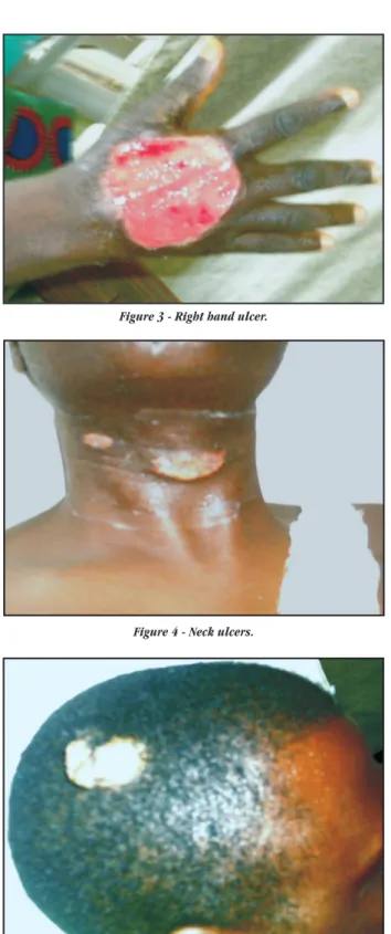
By the seventh day after admission, he had developed superficial ulcerative lesions on the dorsum of the right hand and on the neck and scalp. However, the lesions widened within a few days and new lesions arose on the right gluteus, right inguinal region and left leg. The diagnosis of pyoderma gangrenosum was made, strengthened by the association with ulcerative colitis, with a differential diagnosis of Behcet’s disease. Other investigations included a negative Venereal Disease Research Laboratory (VDRL) test and a reactive Mantoux test (18mm x 20mm). Random blood sugar was normal. Wound biopsy culturing yielded no growth. Wound biopsy histological examination showed chronic inflammation.
The lesions were managed with daily honey wound dressings. Oral dapsone 100mg daily and prednisolone 60mg daily were started. The frequency of the bloody diarrhea decreased, and was completely resolved by the second week after admission. The lesions started healing and the patient was discharged with a prescription of daily honey dressing and oral prednisolone 60mg daily. He has continued to improve in the surgical outpatient clinic, and the prednisolone is being tapered down.
DISCUSSION
Pyoderma gangrenosum is an uncommon type of cutaneous ulceration that continues to be a difficult disorder to diagnose and treat11. It is a neutrophilic dermatosis associated in 70% of
the cases with underlying systemic diseases such as inflammatory bowel disease (IBD), rheumatoid arthritis (RA), monoclonal gammopathy or malignancy16 19. The pathogenesis of pyoderma
gangrenosum is unknown, although a partial defect of cell-mediated immunity may exist5. Other pathophysiological explanations
contemplated include immune dysregulation (including defects in neutrophil chemotaxis), neutrophil hyperreactivity and overexpression of cytokines such as interleukin-86.
There have been some reports of the occurrence of pyoderma gangrenosum in Africa9, and in Nigeria1 2 15, but only one case of
specifically reported pyoderma gangrenosum in association with ulcerative colitis1. The classical presentation is the development
of an erythematous papule or pustule that breaks down to
form an ulcer with purulent discharge and violaceous colored borders spreading peripherally and overhanging the ulcer bed. Alternatively, they may be rapidly enlarging, painful ulcers with purple, undermined edges and a necrotic, hemorrhagic base13.
Although there is no mention of laterality in the literature perused, it was noted that most of our patient’s lesions occurred on the right side of his body (Figures 1, 2, 3, 4 and 5). The significance of this phenomenon is unclear at present. The disorders classically associated with pyoderma gangrenosum are seropositive or seronegative rheumatoid arthritis7, inflammatorybowel
disease21, paraproteinemia22 and myeloproliferative disorders12.
However, it might follow operations such as caesarean delivery12,
abdominal operations (e.g. patients with inflammatory bowel disease following peristomal colostomy)5, breast reduction8,
hysterectomy and salpingo-oophorectomy (on account of uterine fibroids)10, cystectomy (for endometrioma)4 or vulvar pyoderma
gangrenosum18.
Figure 1 - Right inguinal ulcer.
666
Revista da Sociedade Brasileira de Medicina Tropical 41(6):664-667, nov-dez, 2008
Figure 5 - Scalp ulcers, also on the right side. Figure 3 - Right hand ulcer.
Figure 4 - Neck ulcers.
The histological and laboratory findings are nonspecific, but neutrophilic leukocytosis and elevated erythrocyte sedimentation rate are often found3. Biopsy of an early lesion of pyoderma
gangrenosum often demonstratesa dermal neutrophilic abscess. Later-stage lesions show epidermalnecrosis and ulceration, superficial dermal edema and a dense,mixed dermal infiltrate that may extend to the panniculus. Histologicalexamination of the advancing, inflamed border reveals denseperivascular lymphocytic inflammation, which may at times beassociated
with vascular destruction. None of these histological features is however pathognomonic17.
Misdiagnosis of pyoderma gangrenosum is notuncommon. It may be mistaken for vascular occlusive or venous disease, vasculitis, cancer, primaryinfection, drug-induced or exogenous tissue injury and otherinflammatory disorders17.
The treatment includes bed rest, local care, sulfonamides, sulfones and corticosteroids. The goal of therapy is directed towards the associated systemic disorder, if present. High dosages of oral glucocorticoids, sulfasalazine and systemic antibiotics, together with daily wound care, are usually instituted once the diagnosis is suspected13. When these fail, high-dose intravenous
immunoglobulins represent a therapeutic alternative14. There
are reports of successful treatment with etanercept, and a randomized controlled trial using infliximab has been conducted6.
There has been a patient who failed numerous trials of various other immunosuppressive and immunomodulatory regimens, but responded to treatment with adalimumab, which is a fully humanized monoclonal antibody specific for TNF-α6.
Nonetheless, the management of pyoderma gangrenosum continues to be a therapeutic challenge, especially in a tropical population of low socioeconomic level where second-line drugs may not be available.
REFERENCES
1. Afolabi AO. Recurrent leg ulcers in a 16-year old Nigerian girl. African Journal of Medicine and Medical Sciences 32: 93-94, 2003.
2. Ahamefule NC, Olumide YM, Akinkugbe AO, Ayanlowo S, Mohammed TT. Pyoderma gangrenosum: a case report. The Nigerian Medical Practitioner 49: 85-87, 2006.
3. Banga F, Schuitemaker N, Meijer P. Pyoderma gangrenosum after caesarean section: a case report. Published on-line 2006, August 22. Reprod Health 3: 9, 2006.
4. Budak E, Er H. Pyoderma gangrenosum at the incision site following gynecologic surgery. European Journal of Obstetrics, Gynecology, and Reproductive Biology 116: 117-119, 2004.
5. Callen JP, Taylor WB. Pyoderma gangrenosum-a literature review. Cutis; Cutaneous Medicine for the Practitioner 21: 61-64, 1978.
6. Fonder MA, Cummins DL, Ehst BD, Anhalt GJ, Meyerle JH. Adalimumab therapy for recalcitrant pyoderma gangrenosum. Journal of Burns and Wounds 5: e8, 2006.
7. Holt PJ, Davies MG, Saunders KC, Nuki G. Pyoderma gangrenosum: clinical and laboratory findings in 15 patients with special reference to polyarthritis. Medicine (Baltimore) 59: 114-133, 1980.
8. Horner B, El-Mutterdi N. Pyoderma gangrenosum complicating bilateral breast reduction. British Journal of Plastic Surgery 57: 679-681, 2004. 9. Jacyk WK. Pustular ulcerative dermatosis of the scalp. British Journal of
Dermatolology 118: 441-444, 1988.
10. Keohane SG, Graham-Brown RA. Pyoderma gangrenosum complicating hysterectomy for fibroids. Clinical Experimental Dermatology 20: 490-491, 1995.
11. Keskin M, Tosun Z, Ucar C, Savaci N. Pyoderma gangrenosum in a battered child. Annals of Plastic Surgery 57: 228-230, 2006.
667
13. Ljubojevic S, Milavec-Puretic V, Sredoja-Tisma V, Rados J, Kalauz M, Hrstic I. Pyoderma gangrenosum associated with ulcerative colitis. Acta Dermatovenerologica Croatica 14: 35-39, 2006.
14. Meyer N, Ferraro V, Mignard MH, Adamski H, Chevrant-Breton J. Pyoderma gangrenosum treated with high-dose intravenous immunoglobulins: Two cases and review of the literature. Clinical Drug Investigation 26: 541-546, 2006. 15. Obasi OE. Pyoderma gangrenosum and malignant pyoderma in Nigeria. Clinical
and Experimental Dermatology 16: 34-37, 1991.
16. Powell FC, O’Kane M. Management of pyoderma gangrenosum. Dermatologic Clinics 20: 347-355, 2002.
17. Rouphael NG, Ayoub NM, Tomb RR, van der Werf TS, Stienstra Y, van der Graaf WT, Weenig RH, Davis MDP. Skin ulcers misdiagnosed as pyoderma gangrenosum. The New England Journal of Medicine 348: 1064-1066, 2003.
18. Sau M, Hill NC. Pyoderma gangrenosum of the vulva. British Journal of Obstetrics and Gynaecology 108: 1197-1198, 2001.
19. Su WP, Davis MD, Weenig RH, Powell FC, Perry HO. Pyoderma gangrenosum: clinico-pathologic correlation and proposed diagnostic criteria. International Journal of Dermatology 43: 790-800, 2004.
20. Tallon B, Rademaker M, Parkinson G, Whitley B, Swarbrick MJ. Cavitary pyoderma gangrenosum treated with local infusion of corticosteroid. Journal of the American Academy of Dermatology 56: 696-699, 2007. 21. Thornton JR, Teague RH, Low-Beer TS, Read AE. Pyoderma gangrenosum and
ulcerative colitis. Gut 21: 247-248, 1980.
22. Vignon-Pennamen MD, Zelinsky-Gurung A, Janssen F, Frija J, Wallach D. Pyoderma gangrenosum with pulmonary involvement. Archives of Dermatology 125: 1239-1242, 1989.