Journal of Neuroradiology (2008)35, 173—176
D i s p o n i b l e e n l i g n e s u r w w w . s c i e n c e d i r e c t . c o m
CASE REPORT
Simultaneous supratentorial and brainstem abscesses
due to
Listeria monocytogenes
夽
Abc`
es `
a listeria monocytog`
enes : atteinte simultan´
ee
supratentorielle et du tronc c´
er´
ebral
J.P. Soares-Fernandes
a,∗, P. Beleza
b, J.J. Cerqueira
b, M. Ribeiro
a, R. Mar´
e
b,
E. Lourenc
¸o
b, J.F. Rocha
aaDepartment of Neuroradiology, Hospital de S.-Marcos, Braga, Portugal
bDepartment of Neurology (PB, J-JC, RM, EL), Hospital de S.-Marcos, Braga, Portugal
Available online 29 August 2007
KEYWORDS Brain abscess;
Listeria
monocytogenes; Magnetic resonance imaging
Abstract Multiple supratentorial abscesses caused by Listeria monocytogenesare rare. We report the simultaneous occurrence of multiple supratentorial and brainstem abscesses due to
Listeria, in a patient under corticotherapy for an exacerbation of ulcerative colitis. MR imaging features before and after successful conservative treatments are depicted. In immunocompro-mised patients with supratentorial listerial abscesses, the coexistence of brainstem abscedation is exceptional. Despite high mortality associated with listerial abscesses, this case illustrates the possibility of a good clinical outcome, if the appropriate antibiotic regimen is instituted and the immunosuppressant agent is discontinued.
© 2007 Elsevier Masson SAS. All rights reserved.
MOTS CL´ES Listeria
monocytogenes ; Tronc c´er´ebral ; Abc`es ;
Imagerie par r´esonance magn´etique
R´esum´e Les abc`es multiples supratentoriels `aListeria monocytogenessont rares. Nous rap-portons l’observation de la survenue simultan´ee d’abc`es multiples supratentoriels et du tronc c´er´ebral chez un patient sous corticoth´erapie pour colite. Les aspects en imagerie par r´esonance magn´etique (IRM), avant et apr`es un traitement sont d´ecrits. Chez les patients pr´esentant un d´eficit immunitaire, une atteinte list´erienne supratentorielle coexistant avec des abc`es du tronc c´er´ebral est exceptionnelle. Cette observation illustre la possibilit´e d’une ´evolution clinique favorable, si le traitement antibiotique est institu´e et le traitement immunosupresseur arrˆet´e. © 2007 Elsevier Masson SAS. All rights reserved.
夽 Disclosure: the authors have reported no conflicts of interest.
∗Corresponding author.
E-mail address:[email protected](J.P. Soares-Fernandes).
174 J.P. Soares-Fernandes et al.
Introduction
Listeria monocytogenesis an intracellular Gram-positive rod that is known to cause systemic and central nervous system (CNS) infection, afflicting mainly the immunossuppressed and perinatal—neonatal populations[12].
CNS involvement by L. monocytogenes includes menin-gitis and other less common manifestations, such as meningoencephalitis, rhomboencephaplitis, cerebritis and abscesses [2,9,14]. Brain abscesses account for approxi-mately 10% of CNS listerial infections and are seen in 1% of all listerial infections[10].
In 2003, Cone et al. [6] reviewed published cases of brain abscesses due toL. monocytogenes. Thirty of the 40 observed were solitary ones and only four presented with more than three abscesses.
We present a case of multiple Listeriabrain abscesses, describe its imaging findings and discuss the potential phys-iopathologic mechanism of this infection.
Case report
A 46-year-old woman presented to our emergency depart-ment with a five-day history of general malaise, fever, headaches and progressive lethargy. She took prednisolone (40 mg per day) for an exacerbation of her ulcerative coli-tis 6 weeks before and had recently been diagnosed to have diabetes mellitus.
On examination, the patient was feverish (40.4◦C),
comatose, with conjugate eye deviation to the right. She also exhibited left hemiparesis and evident neck stiffness.
Laboratory analysis showed a mild leukocytosis (13.1×10⁹/l) and elevated C-reactive protein (75 mg/l). A
lumbar puncture revealed an opening pressure of 490 mm of water, a leukocyte count of 50 cells/mm3(consisting of
Figure 2 A, DWI, and B, ADC images demonstrate restricted
diffusion in the lenticular nucleus.
Figure 2L’image pond´er´ee en diffusion (A) et la cartographie ADC (B) montrent une restriction de la diffusion au niveau du noyau lenticulaire droit.
67% neutrophils), a glucose level of 42 mg/dl (serum level of 168 mg/dl) and a protein level of 1.14 g/l.
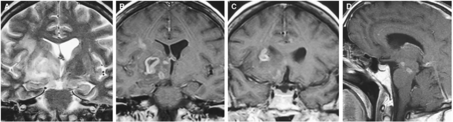
MR imaging displayed multiple rounded lesions, with vari-able sizes, involving the right thalamus, basal ganglia and frontal white matter. The midbrain structures were bilater-ally involved. The lesions were characterized by prolonged T2 (Fig. 1A), restricted water diffusion (Fig. 2) and ring-like or nodular enhancement after contrast administration (Fig. 1B—D).
Cultures of blood and CSF samples yielded L. mono-cytogenes. An ultrasound study of the abdomen failed to reveal hepatic lesions. The patient was treated with intravenous ampicillin (8 g per day) for eight weeks and gentamicin (300 mg per day) for 4 weeks. She recovered her consciousness within five weeks and was discharged with slight bilateral restriction of ocular abduction and
Figure 1 A, coronal T2-weighted image demonstrates pathological high signal in the right thalamus, lenticular nucleus and frontal subcortical white matter. The right frontal horn and the third ventricle are deformed due to lesional mass effect. No hypo-intense rim is seen. B, C and D, post-gadolinium injection, T1-weighted MR imaging; B and C (coronal images) show supratentorial ring-enhancing lesions in the right thalamus, lenticular nucleus, head of caudate and frontal subcortical white matter. Note smaller lesions exhibiting nodular enhancement, in the same locations; D (sagital image) reveals abscess-like lesions in cerebral peduncle and transition between pons and midbrain.
Simultaneous supratentorial and brainstem abscesses due toListeria monocytogenes 175
moderate left-sided hemiparesis (grade 4/5). She could walk with unilateral support. Outpatient physiotherapy was arranged.
MR images obtained before discharge showed nearly com-plete resolution of lesions, with residual enhancement in the right basal ganglia, after contrast administration. At that time, CSF white blood cell count was of 6 cells/mm3;
glucose and protein levels were normal. No bacteria were cultured.
Discussion
L. monocytogenes is believed to reach the CNS by haematogenous spread from the gastrointestinal tract. Ingestion of Listeria-contaminated food is considered to be the source of virtually all human infections [6]. Once ingested, it penetrates into Peyer’s patches of the small intestine gaining access to the mesenteric lymph nodes and then to the blood. In our patient, intestinal lesions of ulcera-tive colitis may have facilitated invasion. Previously, listerial disease has also been associated to perforating duodenal ulcer, caecal gangrene, Crohn’s disease, carcinoma of the rectum and shigellosis[15].
Once in the bloodstream,Listeriatravels mainly in non-listericidal macrophages spreading from cell to cell without contact with the extracellular space[7].
In the CNS, meningitis may follow as the organism attaches to the epithelial cells on the choroid plexuses. Cerebritis and subsequent abscess formation result from penetration in the brain parenchyma, through the cerebral capillary endothelium, via middle cerebral artery. There-fore, bacteraemia is a laboratory finding in about 86% of
Listeria abscesses. Bacteraemia is an unusual laboratory finding in brain abscesses caused by other bacteria, occur-ring in 11% of cases[8]. As in our case, listerial brain abscess have been associated with meningitis in up to 38% of the patients[8].
On the other hand, listerial rhombencephalitis may be explained by axonal transport of food borneListeriato the brainstem, after entering cranial nerve endings [1]. The proposition of two different mechanisms for two different lesions (supratentorial abscesses and rhombencephalitis) strengthens the presumed blood dissemination through the perforating arteries of the basal ganglia and brainstem in our patient.
The limited number of reported cases of multiple brain abscesses caused byL. monocytogenesprovided few reports of MR imaging correlations. Involvement of the subcortical grey matter, such as the thalamus and the basal ganglia, are more common than in abscesses due to other agents, occurring in up to 21%[8,10]. The frontoparietal subcortical white matter is another elective location.
Seventy-five percent of patients presenting more than one listerial supratentorial brain abscess are immuno-suppressed versus 58% of those with solitary abscesses, suggesting that suppressed immunity is a risk factor for lis-terial brain abscesses and even more so with multiple ones
[6]. Differently, rhombencephalitis often occurs in otherwise healthy adults, with only 8% of cases found in immunosup-pressed patients[2].
Since listerial abscesses are rare entities, alternative diagnosis of more common conditions occurring in the immunocompromised patient must be considered. Toxoplas-mosis also presents with multiple lesions, involving the basal ganglia. However, normal ADC values are usually seen[5]. Pyogenic abscesses are infrequently multiple and typically show restricted diffusion[4]. Additionally, a rim of hyperintensity on T1-weighted images and low signal on long-TR sequences due to the presence of free radi-cals is characteristically found. In our case, basal ganglia abscesses showed restricted diffusion, without a rim of free radicals.
The combination of ampicillin (for a minimum of 4 weeks) and gentamicin (for 2—4 weeks) is the regimen of choice for the treatment of listerial brain abscess
[11,13]. Trimethoprim—sulphametoxazole may be a reason-able alternative for the treatment of patients with allergy to penicillin.
Mortality is high and not significantly different in patients with more than one abscess (44%) from those with a single abscess (40%)[6]. However, it is significantly higher than in non-listerial supratentorial brain abscess.
Finally, we excluded the concomitant presence of hep-atic abscesses, which seems to predict a dismal outcome. In Braun’s review of liver abscesses due toL. monocytogenes [3], all the patients with multiple liver abscesses expired, while those with solitary ones survived.
Despite high mortality associated with listerial abscesses, this case illustrates the possibility of a good clinical out-come, if the appropriate antibiotic regimen is instituted and the immunosuppressant agent is discontinued.
References
[1] Antal EA, Loberg EM, Dietrichs E, Maehlen J. Neuropatholog-ical findings in 9 cases ofListeria monocytogenesbrain stem encephalitis. Brain Pathol 2005;15(3):187—91.
[2] Armstrong RW, Fung PC. Brainstem encephalitis (romboen-cephalitis) due to Listeria monocytogenes: case report and review. Clin Infect Dis 1993;16:689—702.
[3] Braun TI, Travis D, Dee RR, Nieman RE. Liver abscess due to Listeria monocytogenes: case report and review. Clin Infect Dis 1993;17:267—9.
[4] Chang SC, Lai PH, Chen WL, Weng HH, Ho JT, Wang JS, et al. Diffusion-weighted MRI features of brain abscess and cystic or necrotic brain tumors: comparison with conventional MRI. Clin Imaging 2002;26(4):227—36.
[5] Chong-Han CH, Cortez SC, Tung GA. Diffusion-weighted MR imaging of cerebral toxoplasma abscess. Am J Roentgenol 2003;181:1711—4.
[6] Cone LA, Leung MM, Byrd RG, Annunziata GM, Lamm RY, Herman BK. Multiple cerebral abscesses because of Lis-teria monocytogenes: three case reports and a literature review of supratentorial listerial brain abscess(es). Surg Neurol 2003;59:320—8.
[7] Drevets DA, Canono BP, Campbell PA. Listericidal and non-listericidal mouse macrophages differ in complement receptor type 3-mediated phagocytosis ofL. monocytogenesand in pre-venting escape of the bacteria into the cytoplasm. J Leukoc Biol 1992;52:70—9.
[8] Eckburg PB, Montoya JG, Vosti KL. Brain abscess due to Liste-ria monocytogenes: five cases and a review of the literature. Medicine (Baltimore) 2001;80:223—35.
176 J.P. Soares-Fernandes et al.
[10] Lorber B. Listeriosis. Clin Infect Dis 1997;25:763—81. [11] Michelet C, Leib SL, Bentue-Ferrer D, Tauber MG. Comparative
efficacies of antibiotics in a rat model of meningoencephalitis due toListeria monocytogenes. Antimicrob Agents Chemother 1999;43:1651—6.
[12] Milonakis E, Hohmann EL, Calderwood SB. Central nervous system infection withListeria monocytogenes: 33 years’ expe-rience at a general hospital and review of 776 episodes from the literature. Medicine 1998;77:313—24.
[13] Moellering Jr RC, Medoff G, Leech I, Wennersten C, Kunz LJ. Antibiotic synergism againstListeria monocytogenes. Antimi-crob Agents Chemother 1972;1(1):30—4.
[14] Nieman RE, Lorber B. Listeriosis in adults: a changing pattern: report of eight cases and review of the literature, 1968—1978. Rev Infect Dis 1980;2:207—27.