online | memorias.ioc.fiocruz.br
The mc
2-CMX vaccine induces an enhanced immune response
against Mycobacterium tuberculosis compared to Bacillus
Calmette-Guérin but with similar lung inflammatory effects
Fábio Muniz de Oliveira1, Monalisa Martins Trentini2, Ana Paula Junqueira-Kipnis2, André Kipnis1/+
1Universidade Federal de Goiás, Instituto de Patologia Tropical e Saúde Pública, Laboratório de Bacteriologia Molecular, Goiânia, GO, Brasil 2Universidade Federal de Goiás, Instituto de Patologia Tropical e Saúde Pública,
Laboratório de Imunopatologia das Doenças Infecciosas, Goiânia, GO, Brasil
Although the attenuated Mycobacterium bovis Bacillus Calmette-Guérin (BCG) vaccine has been used since 1921, tuberculosis (TB) control still proceeds at a slow pace. The main reason is the variable efficacy of BCG protec-tion against TB among adults, which ranges from 0-80%. Subsequently, the mc2-CMX vaccine was developed with promising results. Nonetheless, this recombinant vaccine needs to be compared to the standard BCG vaccine. The objective of this study was to evaluate the immune response induced by mc2-CMX and compare it to the response generated by BCG. BALB/c mice were immunised with both vaccines and challenged with Mycobacterium tubercu-losis (Mtb). The immune and inflammatory responses were evaluated by ELISA, flow cytometry, and histopathology. Mice vaccinated with mc2-CMX and challenged withMtbinduced an increase in the IgG1 and IgG2 levels against CMX as well as recalled specific CD4+ T-cells that produced T-helper 1 cytokines in the lungs and spleen compared with BCG vaccinated and challenged mice. Both vaccines reduced the lung inflammatory pathology induced by the Mtb infection. Themc2-CMX vaccine induces a humoral and cellular response that is superior to BCG and is effi-ciently recalled after challenge with Mtb, although both vaccines induced similar inflammatory reductions.
Key words: recombinant vaccine - tuberculosis - inflammation - mouse
doi: 10.1590/0074-02760150411
Financial support: CNPq, CAPES, FAPEG + Corresponding author: akipnis@ufg.br Received 25 October 2015
Accepted 24 February 2016
Tuberculosis (TB) has been studied since the 460 years B.C. (Benedek 2004); however, during the current post-genomic era, TB remains one of the most important public health problems worldwide. Nine million new TB cases were reported in 2013 and, despite the significant advances in treating the disease over the last few decades, 1.5 million deaths due to Mycobacterium tuberculosis (Mtb), the causative agent of TB, occurred (WHO 2014). Additionally, the global scenario was aggravated by the increasingly high numbers of reported multidrug-resist-ant (MDR)-TB strains. Of all of the registered TB cases in 2013, 3.5% were due to MDR-TB, representing 480,000 cases that resulted in the deaths of 210,000 individuals (WHO 2014). Therefore, several research groups are seek-ing alternatives to fight TB, includseek-ing developseek-ing new vaccines, because the best way to overcome a disease is to prevent infection and/or disease development.
Bacillus Calmette-Guérin (BCG) vaccine is very efficient in protecting children against severe forms of TB; however, its efficacy wanes with time and is highly variable among adult individuals (0-80%), the age group with the highest incidence of the disease (Fine 1995,
These studies indicated the beneficial use of the recombi-nant fusion CMX protein in the context of a new TB vac-cine. In this regard and considering the limitations of BCG, a recombinant vaccine composed of the avirulent strain of Mycobacterium smegmatis mc2 155 expressing the
recom-binant fusion protein CMX (mc2-CMX) was constructed.
When tested in a murine model, this vaccine induced a specific immune response with protective efficacy against Mtb infection (Junqueira-Kipnis et al. 2013). However, the differences in the immune responses induced by the mc2
-CMX and BCG vaccines have not yet been studied. The aim of this study was to compare the immune responses induced by the mc2-CMX and BCG vaccines.
MATERIALS AND METHODS
Animals - The study was conducted in six-eight-week-old BALB/c mice from the Institute of Tropical Pathology and Public Health at the Federal University of Goiás (UFG), city of Goiânia, state of Goiás, Brazil, an-imal facilities that were housed in HEPA-filtered racks and fed with water and a standard diet ad libitum. The temperature was maintained between 20-24ºC, with a relative humidity between 40-70%, and light/dark cycles of 12 h. The mice were maintained and handled in ac-cordance with the rules of the Brazilian Society of Sci-ence in Laboratory Animals. This study was approved by the Ethical Committee of UFG under protocol 229/11.
Vaccine preparation - Aliquots of the mc2
-CMX-vac-cine, which were previously produced as described by Jun-queira-Kipnis et al. (2013), were removed from the -80ºC freezer and the concentration was adjusted to 1 x 10⁸
colo-ny-forming unit (CFU)/mL with phosphate-buffered saline (PBS) containing 0.05% Tween 80. The same procedure was used to prepare the vaccine inoculum of BCG Moreau; however, the concentration was adjusted to 107 CFU/mL.
The control groups received PBS with 0.05% Tween 80. The vaccine diluted for each experiment was plated onto 7H11 media to confirm the inoculum concentration.
Immunisation - Sixteen BALB/c mice were divided into four groups of four mice each: PBS, PBS + Mtb (infection), BCG + Mtb, and mc2-CMX + Mtb. The PBS,
infection, and mc2-CMX + Mtb groups received two
im-munisations (100 mL/immunisation, subcutaneous injec-tions) with an interval of 15 days between injections. The BCG + Mtb group (100 mL/immunisation, subcutaneous injection) received a single immunisation. In all vaccine immunisations, the vaccine inoculum was plated onto 7H11 agar to confirm the concentration.
Intravenous infection with Mtb H37Rv - Thirty days after the last immunisation, the animals were challenged with Mtb H37Rv prepared as described by Junqueira-Kipnis et al. (2013). On the day of infection, the inoc-ulum was diluted to a concentration of 108 CFU/mL in
PBS with 0.05% Tween 80, and 100 mL (107 CFU) was
administered intravenously (via the retroorbital plexus). Seventy days after infection, the mice were sacrificed to analyse their cellular immune responses and the patho-logical changes in their lungs.
ELISA - Blood samples were collected from the mice in each group 15 days before and 30 days after challenge. The collected blood was incubated for 1 h at 37ºC, cen-trifuged at 1,200 g at 4ºC for 15 min to separate the se-rum and subsequently stored at -20ºC. To determine the levels of the anti-CMX antibodies of IgG1 and IgG2a classes in the serum, an ELISA was performed and op-timised as described by Junqueira-Kipnis et al. (2013).
Lung and spleen cell preparation - Seventy days after infection, all mice were euthanised by cervical disloca-tion and their lungs and spleens were collected. The lung digestion was performed in a solution of type IV DNase (30 µg/mL) (Sigma-Aldrich, USA) and Collagenase III (0.7 mg/mL) (Sigma-Aldrich) for 30 min at 37ºC. The lung cell suspension was obtained by passing the digest-ed tissue through a 70 µm cell strainer. The erythrocytes were lysed with lysis solution (0.15 M NH4Cl and 10 mM KHCO3) and the cells were then washed and resuspend-ed in complete RPMI (cRPMI) mresuspend-edium (Junqueira-Kip-nis et al. 2003). Finally, the viable cells were counted and adjusted to a density of 1 x 10⁶ cells/mL. The splenocytes
were obtained after passing the organ through a 70 µm cell strainer (BD Biosciences, USA) and immediately resuspended in RPMI medium (RPMI-1640) (GIBCO, USA). The erythrocytes were lysed with lysis solution and the cells were then washed and resuspended in cRP-MI medium. Finally, the viable cells were counted and adjusted to a density of 1 x 10⁶ cells/mL.
Intracellular cytokine profile in the lung and spleen - To identify the cytokines that were produced by the CD4+ T-cells in the lung and spleen, the cells were
cul-tured without stimulation (cRPMI medium) during 4 h at 37ºC in a 5% CO2 incubator. Next, 3 µM monensin was added (BD Biosciences) for an additional 6 h. Then, the cells were stained with an anti-CD4-PerCP antibody (BD Pharmingen®, USA) and fixed and permeabilised
with Perm Fix/Perm Wash (BD Pharmingen®). The cells
were then stained with the following antibodies for 30
min: IL-2-PE, TNF-α-FITC, and IFN-γ-APC or IgG2a/
IgG1 isotypes control (all antibodies used were from eBioscience®, USA). All analyses were performed on
50,000 events acquired in a BD Biosciences FACSCali-bur flow cytometer (Araújo Jorge Hospital, Goiânia) and the data were analysed with the FlowJo 8.7 software. The lymphocytes were selected based on their size (forward scatter) and granularity (side scatter).
Histopathological analysis - For the histopatholog-ical analysis of the lungs, the right caudal lobes of the lungs from each mouse were collected 70 days after in-fection and fixed with 10% buffered formalin. The fol-lowing parameters were evaluated under the microscope at 5X, 10X, 20X, 40X, and 100X magnifications: the in-tensity of the inflammatory infiltrate and the presence or absence of foamy macrophages and necrotic areas.
inflam-matory foci and foamy mononuclear macrophages, with 1 being the minimal number of events in the fields and 4 when one or two events were observed in several fields. The presence of lesions, inflammatory foci, diffuse mon-onuclear infiltrates, and foamy macrophages were giv-en a score betwegiv-en 5-7, where a score of 5 was received when three-five events per field was observed and a score of 7 represented samples with six-eight events per field. Histological samples that presented lesions, inflamma-tory foci, a moderate, diffuse mononuclear infiltrate, foamy macrophages, and necrosis received scores from 8-10 with a score of 8 attributed to samples presenting eight-10 inflammatory foci per field with little necrosis and a score of 10 attributed to samples that exhibited ac-cumulated lesions and necrosis associated with the foci and the loss of the lung architecture.
Statistical analysis - The results were tabulated with Excel (v.14.3.4, 2011 for Mac) and the Prism software (v.6.0a, GraphPad). The differences between groups were assessed with a two-tailed Student’s t test after a nonparametric (Mann-Whitney U) test. The results were considered significantly different when p < 0.05.
RESULTS
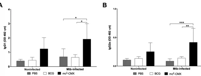
The humoral immune response against CMX induced by the mc2-CMX vaccine is two times higher than that in-duced by BCG - Because the proteins chosen to comprise the recombinant vaccine are produced by most of the my-cobacteria species, it is necessary to know if there is a difference in the immunogenicity of recombinant fused protein in two different live vectors: M. smegmatis and M.bovis BCG. Fig. 1 depicts the timeline of experimental procedures as well as vaccination and Mtbchallenge (Fig. 1). Fifteen days after the last immunisation, blood sam-ples were collected from all mice to perform an ELISA. As shown in Fig. 2A, B, mice immunised with the mc2
-CMX vaccine had similar serum levels of the anti--CMX antibodies of both the IgG1 and IgG2a classes compared to the group immunised with BCG or PBS (Fig. 2).
To assess whether Mtb infection could recall the im-mune response induced by previous vaccination, blood samples were collected from all animals at 30 days after the infection and the levels of the anti-CMX antibodies were determined. As shown in Fig. 2A, B, the levels of the CMX-specific antibodies of both the IgG1 and IgG2a
Fig. 1: timeline scheme of the experimental design. Mice were immunised once with Bacillus Calmette-Guérin (BCG) [106 colony-forming unit (CFU)/mouse] or twice with mc2-CMX (107 CFU/mouse) with 15 days interval. Thirty days after first immunisation, blood was collected from mice and assayed in an ELISA for IgG1 and IgG2a antibodies against CMX. Forty-five days after initial immunisation, mice were intravenously challenged with Mycobacterium tuberculosis (Mtb) H37Rv strain (107 CFU/mouse). Thirty days after challenge, blood was collected for ELISA. Seventy days after challenge, mice were euthanised and organs were removed for flow cytometry and histopathological analyses.
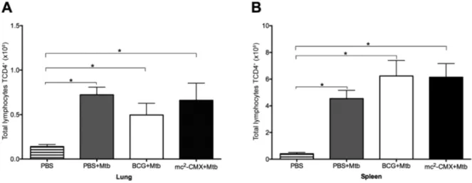
Fig. 3: CD4+ T lymphocytes in the lungs and spleen of the mice. The mice were immunised and challenged with Mycobacterium tuberculosis (Mtb) 30 days after the last immunisation. Seventy days after challenge, the lungs and spleen were collected and analysed by flow cytometry. The cells were cultivated ex vivo without stimuli for 4 h at 37ºC. The total number of CD4+ T lymphocytes in the lungs (A) and spleen (B) were determined. Mice that were not immunised or infected were used as controls [phosphate-buffered saline (PBS)]. BCG: Bacillus Calmette-Guérin; *: p < 0.05 indicates a significant difference between groups.
Fig. 4: percentage of CD4+ T lymphocytes that produce inflammatory cytokines in the lungs of the Mycobacterium tuberculosis (Mtb)-challenged mice. The mice were immunised and (Mtb)-challenged with Mtb 30 days after the last immunisation. Seventy days after challenge, the lungs were collected and analysed by flow cytometry. The cells were cultivated ex vivo without stimuli for 4 h at 37ºC. The number of
classes were significantly higher in the mice immunised with the mc2-CMX vaccine compared to the animals
from the PBS or BCG groups (Fig. 2). These results show that the mc2-CMX-vaccine induced a specific humoral
immune response against CMX only after challenge, while this was not observed after vaccination with BCG.
The mc2-CMX-vaccine induces a greater number of CD4+ T-cells that produce IFN-g and TNF-a in the lungs of the infected mice compared to BCG - The control of an Mtb infection is mainly related to the phenotypic profile of the cells that migrate to the site of infection and their effector activity (Cooper 2009). Therefore, we assessed if prior immunisation with mc2-CMX or BCG could
modu-late the frequency of these cells in the lung and spleen 70 days after Mtb infection. As shown in Fig. 3A, Mtb infec-tion alone was able to induce an increased migrainfec-tion of CD4+ T lymphocytes to the lungs of the mice, regardless
of their immunisation status (Fig. 3A). The same result was observed in the spleen (Fig. 3B), where there was an increase in these populations in all groups challenged with Mtb compared to the nonchallenged group.
Cytokine production by CD4+ effector T
lympho-cytes is an important factor in the control of Mtb infec-tion, particularly when they exhibit characteristics of a T-helper (Th)1-type response, such as IFN-g, TNF-a, and IL-2 production. Thus, we evaluated the profile of the cytokines produced by the CD4+ T-cells in the lungs
of the mice immunised with each vaccine and challenged with Mtb (gating strategy of representative dot plots is shown in Supplementary Figure). As shown in Fig. 4A,
the mice immunised with mc2-CMX vaccine had a
sig-nificantly higher percentage of IFN-g producing cells compared to the group immunised with BCG; however, there was no difference compared to the infected group (PBS + Mtb) (Fig. 4A). The percentage of cells producing TNF-a was also significantly higher in the group immu-nised with the mc2-CMX vaccine compared to the BCG
group (Fig. 4B), but this increase was not significantly different compared to the group that received PBS (PBS + Mtb). In determining the percentages of CD4+ T-cells
that produced IL-2, the mice immunised with mc2-CMX
and BCG had significantly higher levels than the PBS group, but only the mc2-CMX had significantly higher
levels when compared to the infection group (Fig. 4C).
Both mc2-CMX and BCG vaccines reduce the severi-ty of the lung lesions in BALB/c mice infected with Mtb - After 70 days of infection, the lungs were collected from all mice for histological evaluations. In the infection group (PBS + Mtb) (Fig. 5B), the intravenous challenge with Mtb induced severe and diffuse lung inflammation, which led to the consolidation of the lung parenchyma. Large inflammatory agglomerates containing neutro-phils, mononuclear cells, and foamy macrophages were also present (Fig. 5B). In contrast, the mice immunised with the BCG or mc2-CMX vaccines had preserved the
lung parenchyma architecture, with few inflammatory foci compared to the infection group (Fig. 5C, D).
To enhance the comparisons of the immune response induced by the vaccines following Mtb infection, a score was attributed to the lung lesions observed in each
group. Significantly more lesions were observed in the infection group (PBS + Mtb) compared to the groups im-munised with the BCG or mc2-CMX vaccines (Fig. 6),
but there were no differences in the lesion scores of the two vaccinated groups. The responses induced by the BCG and mc2-CMX vaccines were able to preserve the
lung architecture, significantly avoiding the immunopa-thology of Mtb infection.
DISCUSSION
In this study, we compared the immune response and efficacy of the recombinant vaccine with the widely used BCG Moreau vaccine. The mc2-CMX vaccine increased
the production of antibodies specific for the CMX pro-tein in BALB/c mice compared to the BCG vaccine. When assessing the immunological profile induced by the recombinant vaccine against the challenge with Mtb, we observed significant differences in the IFN-g and TNF-a levels in the mc2-CMX-treated mice compared
to the BCG-treated mice. Although the vaccines induced different immunological profiles, they were both effec-tive in reducing lung injury in BALB/c mice.
Although they are crucial in the control of infec-tions, such as Leishmania major infections(Woelbing et al. 2006), the role of antibodies in the immune response against TB is not clear. However, studies have shown the importance of B-cells in the generation of a protective immune response against Mtb infection (Maglione et al. 2007, Maglione & Chan 2009). Torrado et al. (2013) ob-served that mice deficient in antibody maturation are more susceptible to Mtb infection, but they could not determine the role of the antibodies. However, increased production
of IL-10 was observed in the deficient mice, resulting in an increased susceptibility to infection. This shows that the induction of a humoral immune response is important in developing a protective immune response. However, by itself, it may not be effective in infection control, although it is involved in the modulation of the immune response by aiding in T-cell proliferation, differentiation, and sur-vival (Abebe & Bjune 2009, Torrado et al. 2013).
In our studies, the mc2-CMX-vaccine was able to
induce significant levels of CMX-specific antibodies of the IgG1 and IgG2a classes (Fig. 2) compared to the BCG vaccine. Interestingly, BCG contains the protein antigens present in CMX (Ag85C, MPT-51, and HspX); however, it did not induce the production of anti-CMX antibodies. The lack of the humoral immune response in mice immunised with the BCG vaccine may have been due to the reduced expression of the HspX protein in this vaccine, as several studies have shown that BCG does not induce a specific immune response against HspX in humans or mice (Geluk et al. 2007, Shi et al. 2010, Spratt et al. 2010). Furthermore, de Sousa et al. (2012) demon-strated that the major rCMX-induced humoral immune response in mice was towards the HspX protein.
IFN-g and TNF-a present key roles in TB protection, and the Th1 subpopulation of CD4+ T-cells secrete those
cytokines that activate macrophages (Cooper 2009, Bold & Ernst 2012) and contribute to the migration of these cells to the site of infection, particularly the lungs. IFN-g and TNF-a can induce the microbicidal actions of mac-rophages, such as the production of reactive oxygen and nitrogen intermediates and autophagy induction (Flynn et al. 1993, Saunders et al. 2002, Gutierrez et al. 2004). Another protective mechanism of TNF-a is to aid in the development of granulomas (Kindler et al. 1989, Flynn et al. 1995, Ramakrishnan 2012).
In this study, we observed a migration of CD4+ T
lymphocytes to the lungs in all groups challenged with Mtb, demonstrating that infection alone was capable of inducing the migration of these cells (Fig. 3). However, when evaluating the profile of the Th1 cytokines pro-duced by these cells in the lung, we found that IFN-g and TNF-a were increased in the mice immunised with the mc2-CMX vaccine compared to the group immunised
with the BCG vaccine (Fig. 3A, B). Similar results were obtained by Zhang et al. (2010), who demonstrated that IFN-g production by the CD4+ T-cells was increased in
the group of mice that were immunised with M. smeg-matis expressing a CFP10-ESAT6 fusion protein com-pared to the BCG group (Zhang et al. 2010).
Although IFN-g is crucial for Mtb infection pro-tection, some studies have shown that the BCG-in-duced protection is not only IFN-g-dependent, because BCG-vaccinated IFN-g-deficient mice challenged with Mtb exhibited better infection control than the mice de-pleted of CD4+ T lymphocytes (Cowley & Elkins 2003,
Elias et al. 2005, Abebe 2012). Therefore, CD4+ T-cells
can control the Mtb infection through mechanisms that are not exclusively dependent on IFN-g; alternatively, other cells, such as natural killer (NK) cells, can control the infection (Cowley & Elkins 2003, Mittrucker et al. 2007, Abebe 2012). We observed this phenomenon in our
studies, because of the mice immunised with the BCG vaccine showed a significant reduction in lung injury, similar to mice immunised with mc2-CMX (Figs 5, 6).
Additionally, mice vaccinated with mc2-CMX and
chal-lenged with Mtb presented higher levels of IL-2 than in-fected animals; therefore IL-2 could be involved in those protective mechanisms. IL-2 induces the activation and proliferation of Th1 CD4+ T-cells and CD8+ T-cells and
consequently results in the suppression of Mtb replica-tion (Orme 1993, Kim et al. 2000, Williams et al. 2006, Seder et al. 2008). Furthermore, IL-2 can activate NK cells to produce IFN-g and consequently increase Mtb elimination by macrophages (Esin et al. 2013). It seems that BCG vaccinated mice and challenged with Mtb also show the same trend in increase of IL-2, however future work should be done to confirm this hypothesis using a higher number of animals (Fig. 4C).
Polyfunctional cells has been associated with the protection induced by vaccines (Darrah et al. 2007, Lin-denstrom et al. 2009, Derrick et al. 2011), therefore BCG protection may comprise the development of Th cells that express more than one cytokine. Our group showed that Mtb infection significantly reduces the frequency of tri-ple positive CD4+ T-cells in the spleen of nonimmunised
mice that was not observed in mice previously vaccinated with mc2-CMX, suggesting the importance of those cells
in TB protection (Junqueira-Kipnis et al. 2013). This hy-pothesis is corroborated by the algorithm developed by Boyd et al. (2015). Here we hypothesise that polyfunc-tional T-cells could compensate for the lower levels of
IFN-γ positive cells induced by BCG vaccination.
The lungs of the mice from the infection group (PBS + Mtb) showed significantly increased percentages of Th1 cytokines (Fig. 4C) accompanied by excessive tis-sue injury with inflammatory lymphocytic and mac-rophagic clusters, characteristics of the lack of infection control (Figs 5, 6, and data not shown). This phenome-non is likely related to the fact that a protective immune response to TB is not limited to or solely dependent on the production of Th1 cytokines, but on the balance of the immune response as a whole (Walzl et al. 2011). Pre-vious studies using the wild type strain of the M. smeg-matis vaccine showed that, even though it was capable of inducing similar numbers of CD4+ T-cells that produce
Th1-type cytokines, as observed in this study using the mice immunised with the mc2-CMX vaccine, the former
was not able to reduce the bacterial load in the lungs of the BALB/c mice. This could be primarily due to the capacity of the mc2-CMX vaccine in inducing IL-17
pro-duction from the CD4+ T-cells in the lungs of the
BAL-B/c mice, a phenomenon that was also observed for the M. smegmatis Immune Killing Evasion - CMX vaccine (Junqueira-Kipnis et al. 2013).
The recombinant CMX fusion protein has been shown to play an effective role in improving vaccine ef-ficiency. Studies by da Costa et al. (2014) showed that the addition of this protein to the BCG vaccine increased its ability to induce the production of both IL-17 and Th1-type cytokines (IFN-g and TNF-a) by the CD4+
T-cells (da Costa et al. 2014). Thus, we believe that the addition of the CMX protein induces the development
of a balanced immune response that is capable of im-proving the control of Mtb infection. Thus, the immune response induced by a vaccine that aims to replace or improve BCG should not simply increase the immune response, but instead provide a more balanced immune response by optimising the host defense mechanisms and reducing the inflammatory lesions, thus improving infection control and preserving the architecture of the infected organ (Ottenhoff 2012).
This study demonstrated that both mc2-CMX and
BCG vaccines were able to prevent the deleterious effects in the lungs of mice infected with Mtb, likely through different immune mechanisms than BCG. Moreover, this path should be followed to obtain a vaccine that can replace or enhance BCG by providing immunogenic properties that are absent in the BCG vaccine, such as the induction of a humoral immune response.
REFERENCES
Abebe F 2012. Is interferon-g the right marker for bacille Calmette-Guerin-induced immune protection? The missing link in our understanding of tuberculosis immunology. Clin Exp Immunol 169: 213-219.
Abebe F, Bjune G 2009. The protective role of antibody responses during Mycobacterium tuberculosis infection. Clin Exp Immunol 157: 235-243.
Andersen P, Doherty TM 2005. The success and failure of BCG - implications for a novel tuberculosis vaccine. Nat Rev Microbiol 3: 656-662.
Benedek TG 2004. The history of gold therapy for tuberculosis. J Hist Med Allied Sci59: 50-89.
Bold TD, Ernst JD 2012. CD4+ T-cell-dependent IFN-g production by CD8+ effector T-cells in Mycobacterium tuberculosis infection. J
Immunol189: 2530-2536.
Bottai D, Frigui W, Clark S, Rayner E, Zelmer A, Andreu N, de Jonge MI, Bancroft GJ, Williams A, Brodin P, Brosch R 2015. Increased protective efficacy of recombinant BCG strains ex-pressing virulence-neutral proteins of the ESX-1 secretion sys-tem. Vaccine33: 2710-2718.
Boyd A, Almeida JR, Darrah PA, Sauce D, Seder RA, Appay V, Gorochov G, Larsen M 2015. Pathogen-specific T-cell polyfunc-tionality is a correlate of T-cell efficacy and immune protection. PLoS ONE 10: e0128714.
Cole ST, Brosch R, Parkhill J, Garnier T, Churcher C, Harris D, Gordon SV, Eiglmeier K, Gas S, Barry III CE, Tekaia F, Badcock K, Bash-am D, Brown D, Chillingworth T, Connor R, Davies R, Devlin K, Feltwell T, Gentles S, Hamlin N, Holroyd S, Hornsby T, Jagels K, Krogh A, McLean J, Moule S, Murphy L, Oliver K, Osborne J, Quail MA, Rajandream MA, Rogers J, Rutter S, Seeger K, Skelton J, Squares R, Squares S, Sulston JE, Taylor K, Whitehead S, Bar-rell BG 1998. Deciphering the biology of Mycobacterium tubercu-losis from the complete genome sequence. Nature393: 537-544.
Cooper AM 2009. Cell-mediated immune responses in tuberculosis. Annu Rev Immunol27: 393-422.
Cowley SC, Elkins KL 2003. CD4+ T-cells mediate IFN-g -indepen-dent control of Mycobacterium tuberculosis infection both in vi-tro and in vivo. J Immunol171: 4689-4699.
Darrah PA, Bolton DL, Lackner AA, Kaushal D, Aye PP, Mehra S, Blanchard JL, Didier PJ, Roy CJ, Rao SS, Hokey DA, Scanga CA, Sizemore DR, Sadoff JC, Roederer M, Seder RA 2014. Aerosol vac-cination with AERAS-402 elicits robust cellular immune responses in the lungs of rhesus macaques but fails to protect against high-dose Mycobacterium tuberculosis challenge. J Immunol193: 1799-1811.
Darrah PA, Patel DT, De Luca PM, Lindsay RW, Davey DF, Flynn BJ, Hoff ST, Andersen P, Reed SG, Morris SL, Roederer M, Seder RA 2007. Multifunctional TH1 cells define a correlate of vaccine-me-diated protection against Leishmania major. Nat Med 13: 843-850.
de Sousa EM, da Costa AC, Trentini MM, de Araújo Filho JA, Kip-nis A, Junqueira-KipKip-nis AP 2012. Immunogenicity of a fusion protein containing immunodominant epitopes of Ag85C, MPT51, and HspX from Mycobacterium tuberculosis in mice and active TB infection. PLoS ONE7: e47781.
Derrick SC, Yabe IM, Yang A, Morris SL 2011. Vaccine-induced anti-tuberculosis protective immunity in mice correlates with the magnitude and quality of multifunctional CD4 T-cells. Vaccine 29: 2902-2909.
Elias D, Akuffo H, Britton S 2005. PPD induced in vitro interferon g production is not a reliable correlate of protection against Myco-bacterium tuberculosis. Trans R Soc Trop Med Hyg99: 363-368.
Esin S, Counoupas C, Aulicino A, Brancatisano FL, Maisetta G, Bot-tai D, Di Luca M, Florio W, Campa M, Batoni G 2013. Interaction of Mycobacterium tuberculosis cell wall components with the human natural killer cell receptors NKp44 and Toll-like receptor 2. Scand J Immunol77: 460-469.
Fine PE 1995. Variation in protection by BCG: implications of and for heterologous immunity. Lancet346: 1339-1345.
Flynn JL, Chan J, Triebold KJ, Dalton DK, Stewart TA, Bloom BR 1993. An essential role for interferon g in resistance to Mycobac-terium tuberculosis infection. J Exp Med178: 2249-2254.
Flynn JL, Goldstein MM, Chan J, Triebold KJ, Pfeffer K, Lowenstein CJ, Schreiber R, Mak TW, Bloom BR 1995. Tumor necrosis fac-tor-alpha is required in the protective immune response against Mycobacterium tuberculosis in mice. Immunity2: 561-572.
Geluk A, Lin MY, van Meijgaarden KE, Leyten EM, Franken KL, Ottenhoff TH, Klein MR 2007. T-cell recognition of the HspX protein of Mycobacterium tuberculosis correlates with latent M. tuberculosis infection but not with M. bovis BCG vaccination. Infect Immun75: 2914-2921.
Gutierrez MG, Master SS, Singh SB, Taylor GA, Colombo MI, Deret-ic V 2004. Autophagy is a defense mechanism inhibiting BCG and Mycobacterium tuberculosis survival in infected macro-phages. Cell119: 753-766.
Hesseling AC, Marais BJ, Gie RP, Schaaf HS, Fine PE, Godfrey-Faussett P, Beyers N 2007. The risk of disseminated Bacille Calmette-Guerin (BCG) disease in HIV-infected children. Vaccine25: 14-18.
Junqueira-Kipnis AP, de Oliveira FM, Trentini MM, Tiwari S, Chen B, Resende DP, Silva BD, Chen M, Tesfa L, Jacobs Jr WR, Kipnis A 2013. Prime-boost with Mycobacterium smegmatis recombi-nant vaccine improves protection in mice infected with Mycobac-terium tuberculosis. PLoS ONE8: e78639.
Junqueira-Kipnis AP, Kipnis A, Jamieson A, Juarrero MG, Diefen-bach A, Raulet DH, Turner J, Orme IM 2003. NK cells respond to pulmonary infection with Mycobacterium tuberculosis, but play a minimal role in protection. J Immunol171: 6039-6045.
Kalra M, Grover A, Mehta N, Singh J, Kaur J, Sable SB, Behera D, Sharma P, Verma I, Khuller GK 2007. Supplementation with RD antigens enhances the protective efficacy of BCG in tuberculous mice. Clin Immunol125: 173-183.
Kaufmann SH, Hussey G, Lambert PH 2010. New vaccines for tuber-culosis. Lancet375: 2110-2119.
Kim JJ, Yang JS, Montaner L, Lee DJ, Chalian AA, Weiner DB 2000. Coimmunization with IFN-g or IL-2, but not IL-13 or IL-4 cDNA can enhance Th1-type DNA vaccine-induced immune responses in vivo. J Interferon Cytokine Res20: 311-319.
Kindler V, Sappino AP, Grau GE, Piguet PF, Vassalli P 1989. The inducing role of tumor necrosis factor in the development of bac-tericidal granulomas during BCG infection. Cell56: 731-740.
Lindenstrom T, Agger EM, Korsholm KS, Darrah PA, Aagaard C, Sed-er RA, Rosenkrands I, AndSed-ersen P 2009. TubSed-erculosis subunit vac-cination provides long-term protective immunity characterized by multifunctional CD4 memory T-cells. J Immunol 182: 8047-8055.
Maglione PJ, Chan J 2009. How B cells shape the immune response against Mycobacterium tuberculosis. Eur J Immunol39: 676-686.
Maglione PJ, Xu J, Chan J 2007. B-cells moderate inflammatory progres-sion and enhance bacterial containment upon pulmonary challenge with Mycobacterium tuberculosis. J Immunol178: 7222-7234.
Mahairas GG, Sabo PJ, Hickey MJ, Singh DC, Stover CK 1996. Mo-lecular analysis of genetic differences between Mycobacterium bovis BCG and virulent M. bovis. J Bacteriol178: 1274-1282.
Marongiu L, Donini M, Toffali L, Zenaro E, Dusi S 2013. ESAT-6 and HspX improve the effectiveness of BCG to induce human dendritic cells-dependent Th1 and NK cells activation. PLoS ONE8: e75684.
Mittrucker HW, Steinhoff U, Kohler A, Krause M, Lazar D, Mex P, Miekley D, Kaufmann SH 2007. Poor correlation between BCG vaccination-induced T-cell responses and protection against tu-berculosis. Proc Natl Acad Sci USA104: 12434-12439.
Orme IM 1993. Immunity to mycobacteria. Curr Opin Immunol 5: 497-502.
Ottenhoff TH 2012. The knowns and unknowns of the immunopatho-genesis of tuberculosis. Int J Tuberc Lung Dis16: 1424-1432.
Ottenhoff TH, Kaufmann SH 2012. Vaccines against tuberculosis: where are we and where do we need to go? PLoS Pathog8: e1002607.
Ottenhoff TH, Verreck FA, Lichtenauer-Kaligis EG, Hoeve MA, Sanal O, van Dissel JT 2002. Genetics, cytokines, and human infectious disease: lessons from weakly pathogenic mycobacteria and salmonellae. Nat Genet32: 97-105.
Philipp WJ, Nair S, Guglielmi G, Lagranderie M, Gicquel B, Cole ST 1996. Physical mapping of Mycobacterium bovis BCG Pasteur re-veals differences from the genome map of Mycobacterium tubercu-losis H37Rv and from M. bovis. Microbiology142 (Pt 11): 3135-3145.
Rachman H, Kaufmann SH 2007. Exploring functional genomics for the development of novel intervention strategies against tubercu-losis. Int J Med Microbiol297: 559-567.
Ramakrishnan L 2012. Revisiting the role of the granuloma in tuber-culosis. Nat Rev Immunol12: 352-366.
Saunders BM, Frank AA, Orme IM, Cooper AM 2002. CD4 is quired for the development of a protective granulomatous re-sponse to pulmonary tuberculosis. Cell Immunol216: 65-72.
Seder RA, Darrah PA, Roederer M 2008. T-cell quality in memory and protection: implications for vaccine design. Nat Rev Immunol 8: 247-258.
Shaban K, Amoudy HA, Mustafa AS 2013. Cellular immune respons-es to recombinant Mycobacterium bovis BCG constructs express-ing major antigens of region of difference 1 of Mycobacterium tuberculosis. Clin Vaccine Immunol20: 1230-1237.
Spratt JM, Britton WJ, Triccas JA 2010. In vivo persistence and pro-tective efficacy of the bacille Calmette Guerin vaccine overex-pressing the HspX latency antigen. Bioeng Bugs1: 61-65.
Torrado E, Fountain JJ, Robinson RT, Martino CA, Pearl JE, Rangel-Moreno J, Tighe M, Dunn R, Cooper AM 2013. Differential and site specific impact of B-cells in the protective immune response to Mycobacterium tuberculosis in the mouse. PLoS ONE8: e61681.
Trentini MM, de Oliveira FM, Gaeti MP, Batista AC, Lima EM, Kip-nis A, Junqueira-KipKip-nis AP 2014. Microstructured liposome sub-unit vaccines reduce lung inflammation and bacterial load after Mycobacterium tuberculosis infection. Vaccine32: 4324-4332.
Walzl G, Ronacher K, Hanekom W, Scriba TJ, Zumla A 2011. Immuno-logical biomarkers of tuberculosis. Nat Rev Immunol11: 343-354.
Wang C, Fu R, Chen Z, Tan K, Chen L, Teng X, Lu J, Shi C, Fan X 2012. Immunogenicity and protective efficacy of a novel recom-binant BCG strain overexpressing antigens Ag85A and Ag85B. Clin Dev Immunol2012: 563838.
WHO- World Health Organization 2014. Global tuberculo-sis report 2014. Available from: apps.who.int/iris/bitstre am/10665/137094/1/9789241564809_eng.pdf.
Williams MA, Tyznik AJ, Bevan MJ 2006. Interleukin-2 signals dur-ing primdur-ing are required for secondary expansion of CD8+ mem-ory T-cells. Nature441: 890-893.
Woelbing F, Kostka SL, Moelle K, Belkaid Y, Sunderkoetter C, Ver-beek S, Waisman A, Nigg AP, Knop J, Udey MC, von Stebut E 2006. Uptake of Leishmania major by dendritic cells is mediated by Fcg receptors and facilitates acquisition of protective immu-nity. J Exp Med203: 177-188.
Yuan X, Teng X, Jing Y, Ma J, Tian M, Yu Q, Zhou L, Wang R, Wang W, Li L, Fan X 2015. A live attenuated BCG vaccine overexpress-ing multistage antigens Ag85B and HspX provides superior pro-tection against Mycobacterium tuberculosis infection. Appl Mi-crobiol Biotechnol99: 10587-10595.
Zhang H, Peng P, Miao S, Zhao Y, Mao F, Wang L, Bai Y, Xu Z, Wei S, Shi C 2010. Recombinant Mycobacterium smegmatis express-ing an ESAT6-CFP10 fusion protein induces anti-mycobacterial immune responses and protects against Mycobacterium tubercu-losis challenge in mice. Scand J Immunol72: 349-357.
Supplementary data
Representative dot plots of flow cytometry analysis of intracellular cytokines [(interferon (IFN)-γ, tumour necrosis factor (TNF)-α, and in
-terleukin (IL)-2] from naïve noninfected mice [phosphate-buffered saline (PBS)], infected mice [PBS + Mycobacterium tuberculosis (Mtb)],
immunised Mtb challenged mice (BCG + Mtb or mc2-CMX + Mtb), and isotype controls (isotype control). The cells were cultivated ex vivo
without stimuli for 4 h at 37ºC. Lymphocytes where gated according to their size and granularity, and CD4+ T-cells were evaluated for the ex


