Brazilian Journal of Microbiology (2012): 449-455 ISSN 1517-8382
CHARACTERIZATIONS OF A NEW CORDYCEPS CICADAE ISOLATE AND PRODUCTION OF ADENOSINE AND
CORDYCEPIN
Yongjun Wang1*, Yanbin Guo2, Liqin Zhang1, Jia Wu1
1
Key Laboratory of Forest Protection, College of Forestry and Biotechnology, Zhejiang Agricultural and Forestry University,
Lin’an 311300 China; 2College of Resources and Environmental Sciences, China Agricultural University, Beijing 100094 China.
Submitted: November 30, 2010; Approved: January 16, 2012.
ABSTRACT
Cordyceps is a fastidious pathogenic fungus infecting insects, and recent years have witnessed rapid
progress in its medical properties. In this study, a wild isolate, C. cicadae MP12, was characterized through
in vitro cultivation and its nuclear small-subunit (SSU) ribosomal DNA (rDNA) data. In vitro culture of C.
cicadae MP12 was established by growing its fruiting bodies in a solid matrix. C. cicadae MP12 was
inoculated into Cryptotympana atrata cicada pupae for in vivo culture, where the fungi developed its fruiting
body as well. The contents of adenosine and cordycepin in dried fruiting bodies after culture were
1421.45μg/g and 1398.12 μg/g, respectively. Therefore, the established cultures from this study could be
used for the production of various medically important metabolic substances.
Key words: Cordyceps cicadae, cultivation, adenosine, cordycepin.
INTRODUCTION
Cordyceps is one of the entomopathogenic fungi capable
of invading a specific insect and kills the host eventually. After
hibernating the winter inside the host, it forms a fruiting body
on the surface of the insect’s cadaver during the summer. C.
sinensis, is the most popular Cordyceps, which is a parasite of
the Hepialus armoricanus larvae. The fungus has long been
used to promote longevity, relieve exhaustion and treat
numerous diseases in Chinese traditional medicines (19).
Recent studies have demonstrated that various species in this
genus possess multiple pharmacological properties, including
anti-tumor, anti-microbial, anti-inflammatory and
immunomodulatory effects (5). Furthermore, a variety of
effective chemical constituents including cordycepin,
adenosine, ergosterol, and myriocin have been isolated from
various Cordyceps species (9, 16, 26, 27).
Adenosine is widely distributed in Cordyceps spp. It is
also known to have a widespread effect on coronary and
cerebral circulation (4), control of blood flow (1), prevention of
cardiac arrhythmias (18). Cordycepin (3’-deoxyadenosine) is a
nucleoside analogue, which exhibits a broad spectrum of
biological activity (2, 7). Cordycepin can be converted into its
5’-mono, di and triphosphates intracellularly to inhibit the
activity of ribose-phosphate pyrophosphokinase and
5’-phosphoribosyl- 1’- pyrophosphate amidotransferase in the
Wang, Y. et al. C. cicadae isolate and production of adenosine and cordycepin
de novo biosynthesis of purines and/or the synthesis of nucleic
acids, causing the tumor, metastatic and
microbial effects (6, 11, 13, 15, 20, 22). In addition, the
anti-leukemic activity of cordycepin combined with an adenosine
deaminase inhibitor and the inhibitory effect of its analogues of
2’, 5’-oligoadenylate on human immunodeficiency virus
infection have also been reported (8).
A C. militaris-like isolate of C. cicadae MP12 was
obtained from the fruiting body forming on the cicada from
Hubei province, China. In this study, the biological and
morphological characters were described. To assess the
potential of C. cicadae MP12 as a new fungal resource for
medical applications, the in vitro culture methods were
established and the contents of adenosine and cordycepin were
measured.
MATERIALS AND METHODS
Microorganisms and insect
C. cicadae MP12 (its fruiting body looked like C.
militaris) was obtained from the Shennongjia Mountains,
Hubei province, China. Cryptotympana atrata cicada pupae
were used as the host for in vivo culture of the fungus in this
study.
In vitro culture
Cultures of C. cicadae MP12 were grown on Sabouraud
dextrose agar (Difco Laboratories) slants at 25ºC and
maintained on the same medium at 2 to 4°C. One loopful of
spores from 7- to 14-day cultures was used to inoculate a liquid
medium consisting of 4% fructose and 1% Neopeptone (Difco;
pH adjusted to 7.5 after autoclaving by using sterile 1.0 N
NaOH). Each 250-ml flask contained 60 ml of medium, and
cultures were incubated at 250 rpm on a rotary shaker for the
specified duration.
After 3-days of culture in the flask, 2 ml of mycelia
culture was inoculated into a transparent glass bottle containing
RSM medium (rice 40g,glucose 0.4g,tryptone 0.2g,MgSO4
0.8g, K2HPO4 0.4g, vitamin B complex 0.05g, H2O2 50 ml, pH
7.0-7.2; autoclaved for 20 min at 121°C). The bottle was sealed
by a ventilated film after inoculation. The inoculated bottles
were incubated in a culture room (25ºC, 12h) for mycelium
growth and fruiting body formation.
In vivo culture
The spores were suspended in phosphate salt buffer (pH
7.5) after 14-day culture in plates and diluted to ~106
spores/ml. The cicada pupae were collected from nature and
surface sterilized using 75% (V/V) ethanol for 5 min. Each
pupa was then placed into a transparent glass bottle and
sprayed with 1ml spore suspension. The pupae were incubated
in a culture room (25ºC, 12h) for infection to occur.
Conidial measurements
Between twenty to thirty conidia from 10–20 day old
cultures were suspended in 0.01% Tween 40 and mixed with
an equal volume of agarose. Conidia were observed using a
Nikon E600 microscope equipped with Nikon DXM 1200
digital camera and Nikon ACT-1 image capture software.
DNA extraction and PCR
DNA was extracted from the fungal cultures by a
modified CTAB method as previously described (21).
Approximately 1150 base pairs (bp) of the nuclear
small-subunit ribosomal DNA (SSU rDNA) was amplified using
PCR. Amplifications and sequencing reactions were performed
as described in Sung et al (21).
Sequence analysis
Sequences were edited using BioEdit version 7.0.4.1. The
programs DNADIST, NEIGHBOR, DNAPARS, SEQBOOT,
and CONSENSE, present in the PHYLIP package (Version
3.68) were used to perform the maximum parsimony analyses
Wang, Y. et al. C. cicadae isolate and production of adenosine and cordycepin
regions were excluded from the data matrix before analysis.
Relative support of the resulting trees was determined by 1000
bootstrap replications on informative characters only with the
known search options (3). The phylogenetic tree was generated
using TreeView version 1.6.
Extraction and analysis of adenosine and cordycepin
The fruiting bodies and myceliaof C. cicadae MP12 were
kept in a vacuum incubator at 50ºC overnight for drying. The
samples were ground and extracted in deionized water.
Chromatographic separations were performed using a
Waters HPLC system (Millipore, Waters Division, Milford,
MA). The HPLC column was a C8 reversed-phase column for
nucleotides (YMC-Pack FA), which was packed with Si 60
(particle size 5 µm, 250-4.6 mm). Analysis of the
chromatograms was performed using Waters Millennium
software. Extractions of C. cicadae MP12 fruiting body were
routinely monitored at a wavelength of 258nm. Peak area
measurements were used to quantitate retinoid amounts using a
standard curve based on cordycepin (Sigma-Aldrich, St. Louis,
MO) and adenosine standards (Sigma-Aldrich, St. Louis, MO)
and normalized using an internal standard. The solvent system
for elution from the YMC-Pack FA column consisted of
methyl alcohol:Milli-Q water (85:15, by vol). Separations were
made at a flow rate of 0.7 ml/min. The column temperature
was kept constant at 25°C. The retention time (RT, in min) on
the YMC-Pack FA column ranged from 1 to 18 min.
Data analysis
Statistical significance between the groups was
determined by paired t-test and one-way ANOVA for repeated
measures. Results with p<0.05 were considered statistically
significant. Data were assessed using the SPSS software
(version 15.0, SPSS Inc., Chicago, Illinois).
RESULTS
Cultivation of C. cicadae MP12
After storage of the fungus in the Sabouraud dextrose agar
plate for 7 days (Fig. 1), C. cicadae MP12 spores were
collected and suspended in phosphate salt buffer (pH 7.8) and
diluted to 108 spores/ml. The suspensions were inoculated into
the RSM medium to induce sexual phase. The fruiting bodies
formed after 14 days since inoculation and reached at ~ 8 cm in
length and ~0.8 cm in width 29 days later (Fig. 2A). The
fruiting bodies were bright orange on the outside and milky
inside. The average weight of the dried fruiting body was about
8.91g (statistical data not shown).
About two hundred cicada pupae were used for in vivo
cultivation of the fungi. The pupae were soaked in spore
suspension for 30 min. The cicada died 6 days after
inoculation. 95 % of insects were inoculated successfully. The
fruiting bodies started to grow on the insect surface 16 days
since cicada died and reached at ~8 cm in length and ~0.8 cm
in width 29 days later (Fig 2C, D). Bright orange fruiting
bodies were observed. Figure 2D illustrated the fruiting body
of C. cicadae MP12 grown on a cicada after metamorphosis.
Figure 1. The colony of C. cicadae MP12 growing on the Sabouraud dextrose agar plate (A) and line drawings of hyphae
Wang, Y. et al. C. cicadae isolate and production of adenosine and cordycepin
Figure 2. Sexual reproduction of C. cicadae MP12. A: Fruiting bodies growing on the RSM medium; B: The mature stroma
from the fruiting body growing on the RSM medium; C and D:
C. cicadae MP12 on cicadae pupae after inoculation. Scale
bars: A, C-D = 10 mm; B = 100 μm.
Morphological characters and molecular data of C.cicadae
MP12
In the conidial stage of C. cicadae MP12, displayed an
abundance of branched septate mycelium and the conidiospore
started germinating after 7 days at 25℃ on Sabouraud dextrose
agar (Fig 1A). The aerial mycelium is cottony in texture and
whitish yellow-colored in the middle, the reverse side of
cultures is yellowish. The microscopic observation showed that
conidiospores were produced in stringed chains. Stromata was
observed solitary or occasionally with serveral, simple or rarely
branched, on the larva of cicada and RSA medium. Stipe was
fleshly, ochraceous orange to red, clavate, 5-50×0.5-6 mm.
Fertile area on the terminal was reddish orange to reddish,
cylindrical, elliptical to fusiform, 3-10×1-6 mm. Perithecia
were crowded, loosely embedded, ordinal-likely in orientation,
elliptical to fusiform to obclavate, 200-600×150-250 µm. The
typical orange bat-shaped stromata were fruiting from cicadae
and RSM medium 14 days after inoculation using
conidiospores. The oval-shaped perithecia were presented on
the stroma with cream-colored prosenchyma inside (Fig 2 B).
After SSU rDNA amplification, a 1150 bp DNA fragment
was obtained and sequenced (NCBI access number
HM536623). Final alignments for phylogenetic analysis
included characters from various species of Clavicipitaceae
used in Philip alignments. Parsimony analysis showed that C.
cicadae MP12 was closer to another C. cicada (NCBI access
number DQ838788) and C. inegoensis phylogenetically (Fig.
3). The topology of the most-parsimonious tree and the
Cordyceps phylogenetic pattern were globally similar to the
results of a larger phylogenetic analysis of the Clavicipitaceae
with more intensive taxon sampling (21).
Figure 3. The most-parsimonious tree from the maximum
parsimony analysis of SSU rDNA
data. Numbers above nodes are
nonparametric bootstrap values
from 1000 replications. MP12
Wang, Y. et al. C. cicadae isolate and production of adenosine and cordycepin
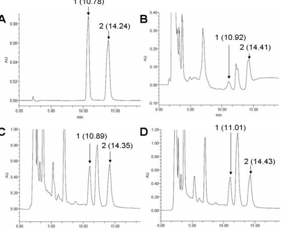
Production of adenosine and cordycepin in C. cicadae
MP12
The peaks of adenosine and cordycepin were identified at
the Retention Time of 10.78 min and 14.24 min, repectively
(Fig. 4A). Two peaks corresponding to adenosine and
cordycepin from the extract of dried mycelium (10.92 min and
14.41 min) and fruiting bodies from in vitro culture (10.89 min
and 14.35 min) and in vivo culture (11.01 min and 14.43 min)
were detected and after a peak correction (p<0.05) (Fig. 4B, C,
D). Standard adenosine concentrations ranging from 50 to 500
μg/ml were used to establish the linear curve between concentration and peak area. The equation that Y =
62.75X-550.73 (Y, concentration, μg/ml; X, peak area, mU×min) was
established after three replicates (p<0.05). With the same
procedure, standard cordycepin concentrations ranging from 50
to 500 μg/ml came to the equation Y = 34.14X-443.50 (Y,
concentration, μg/ml; X, peak area, mU×min) (p<0.05) in the
chromatograms. According to these two standard models, the
content of adenosine in the dried mycelium was 82.39 μg/g,
much lower than the extract of dried fruiting body cultured in
RSM medium (1398.12 μg/g) and dried fruiting body growing
on cicada (1421.45μg/g). Also, the contents of cordycepin were
64.98μg/g in the mycelium, 1548.12 μg/g in the dried fruiting
body cultured in the RSM medium, and 1578.43 μg/g in the
dried fruiting body growing in the cicada larvae. The contents
of adenosine and cordycepin in the fruiting body showed no
significant difference between culturing in RSM medium and
harvest on cicada (p<0.05).
Figure 4. Chromatograms (HPLC) of water solution extracted from C. cicadae MP12. A: Standard adenosine (80 μg/ml) and cordycepin (100 μg/ml); B: water extract of dried mycelium; C: water extract of the harvested fruiting body of C. cicadae MP12
cultured in the RSM medium; D: water extract of harvested fruiting body of C. cicadae MP12 growing on cicadae pupae of after
Wang, Y. et al. C. cicadae isolate and production of adenosine and cordycepin
DISCUSSION
Cordyceps (Fr.) includes pathogens of species from nine
orders of arthropods and parasites of one genus, Elaphomyces.
In the Cordyceps genus, C. militaris is one of the most
frequently collected species. However, it is also one of the
most variable species in the genus with respect to morphology
and host affiliation (21). Sung et al. conducted the most
extensive multigene phylogenetic analyses to provide a basis
for the phylogenetic classification of Cordyceps and the
clavicipitaceous fungi, and determined the clear classification
of this genus (21). Also, based on the evolution of host
specificity, the phylogenetic relationships and the stromata
morphological characters, five monophyletic groups in
Cordyceps genus were identified: the truffle-cicada clade,
cicada clade A, cicada clade B, the scale-moth clade, and the
moth clade (17). Previously, C. cicadae MP12 was also
considered non-specific with C. militaris according to its
macroscopical morphology. In the present study, inoculation of
C. cicadae MP12 on cicadae and morphological characters,
including clavate stroma, oval crowed perithecia, branched
mycelium and conidiospores producing in stringed chains,
grouped this isolate into the cicada clade A, which includes C.
inegoensis and C. paradoxa. The high similarity of SSU rDNA
sequences between C. inegoensis and C. cicadae MP12 is
consistent with this group. The anamorphic and cultural
differences from C. inegoensis suggested that isolate MP12
was more similar to C. cicadae.
Various bioactive constituents from Cordyceps species
have been reported. These include cordycepin and other
anti-bacterial and anti-tumour adenosine derivatives, ophicordin, an
anti-fungal agent, myriocin, L-tryptophan, etc (16, 26). In this
study, the contents of adenosine and cordycepin were measured
by the use of HPLC. In the chromatographic data, peaks
corresponding to standard adenosine and cordycepin were
identified. These results indicated that the contents of both
adenosine and cordycepin in C. cicadae MP12 were about the
same as those in C. militaris (13, 24, 25).
The fruiting body culture on a large scale using in vitro
media has recently been developed to produce cordycepin due
to its limited supply from natural sources. Therefore, several
methods have been established for cultivation of varied
Cordyceps species (10, 12, 14, 23). In this case, an economical
and high efficient cultivation for C. cicadae in vitro and in vivo
was first-time established to produce the fruiting body. This
culturing would help to identify other pharmacologically active
metabolites from C. cicadae in the future.
ACKNOWLEDGEMENTS
This work was supported by a grant from Qianjiang
Talents Grant, No. 2010R10098 funded by the Department of
Science & Technology of Zhejiang Province, China and by a
grant from National Natural Science Foundation of China
(NSFC 30901153). We are thankful to Dr. Anliang Chen for
the assistance on HPLC work.
REFERENCES
1. Berne, R.M. (1980). The role of adenosine in the regulation of coronary blood flow. Circ. Res. 47, 807–813.
2. Cunningham, K.G.; Manson, W.; Spring, F.S.; Hutchinson, S.A. (1950). Cordycepin, a metabolic product isolated from cultures of Cordyceps militaris (Linn.) Link. Nature 166, 949.
3. Felsenstein, J. (1988). Phylogenies from molecular sequences: inference and reliability. Annu. Rev. Genet. 22, 521–565.
4. Hermann, S.C.; Feigl, E.O. (1992). Adrenergic blockade blunts adenosine concentration and coronary vasodilation during hypoxia. Circ. Res. 70, 1203–1216.
5. Holliday, J.; Cleaver, M. (2008). Medicinal Value of the Caterpillar Fungi Species of the Genus Cordyceps (Fr.) Link (Ascomycetes). A Review. Int. J. Med. Mushrooms 10, 219–234.
6. Ioannidis, P.; Courtis, N.; Havredaki, M.; Michailakis, E.; Tsiapalis, C.M.; Trangas, T. (1999). The polyadenylation inhibitor cordycepin (3’dA) causes a decline in MYC mRNA levels without a.ecting c-MYC protein levels. Oncogene 18, 117–125.
Wang, Y. et al. C. cicadae isolate and production of adenosine and cordycepin
as 3'-deoxyadenosine. Biochem. Biophys. Res. Commun. 14, 456–457. 8. Kodama, E.N.; McCaffrey, R.P,; Yusa, K.; Mitsuya, H. (2000).
Antileukemic activity and mechanism of action of cordycepin against terminal deoxynucleotidyl transferase-positive (TdT+) leukemic cells.
Biochem. Pharmacol. 59, 273–281.
9. Kuo, Y.C.; Weng, S.C.; Chou, C.J.; Chang, T.T.; Tsai, W.J. (2003). Activation and proliferation signals in primary human T lymphocytes inhibited by ergosterol peroxide isolated from Cordyceps cicadae. Br. J. Pharmacol. 140, 895–906.
10. Lee, S.Y.; Nakajima, I.; Ihara, F.; Kinoshita, H.; Nihira, T. (2005). Cultivation of entomopathogenic fungi for the search of antibacterial compounds. Mycopathologia 160, 321–325.
11. Legraverend, M.; Robert, I. (1978). Glazer inhibition of the phosphorylation of non-histone chromosomal proteins of rat liver by cordycepin and cordycepin triphosphate. Cancer Res. 38, 1142–1146. 12. Lin, Q.Y.; Song, B.; Huang, H.; Li, T.H. (2010). Optimization of selected
cultivation parameters for Cordyceps guangdongensis. Lett. Appl. Microbiol. 51, 219–225.
13. Ling, J.Y.; Sun, Y.J.; Zhang, H.; Lv, P.; Zhang, C.K. (2002). Measurement of cordycepin and adenosine in stroma of Cordyceps sp. by capillary zone electrophoresis (CZE). J. Biosci. Bioeng. 94, 371–374. 14. Mao, X.B.; Zhong, J.J. (2004). Hyperproduction of cordycepin by
two-stage dissolved oxygen control in submerged cultivation of medicinal mushroom Cordyceps militaris in bioreactors. Biotechnol. Prog. 20, 1408–1413.
15. Müller, W.E.G.; Seibert, G.; Beyer, R.; Breter, H.J.; Maidhof, A.; Zahn, R.K. (1977). Effect of cordycepin on nucleic acid metabolism in L5178Y cells and on nucleic acid-synthesizing enzyme systems. Cancer Res. 37, 3824–3833.
16. Ng, T.B.; Wang, H.X. (2005). Pharmacological actions of Cordyceps, a prized folk medicine. J. Pharm. Pharmacol. 57, 1509–1519.
17. Nikoh, N.; Fukatsu, T. (2000). Interkingdom host jumping underground: Phylogenetic analysis of entomoparasitic fungi of the genus Cordyceps.
Mol. Biol. Evol. 17, 629–638.
18. Pelleg, A.; Porter, R.S. (1990). The pharmacology of adenosine.
Pharmacotherapy 10, 157–174.
19. Russell, R.; Paterson, M. (2008). Cordyceps – A traditional Chinese medicine and another fungal therapeutic biofactory? Phytochemistry 69, 1469–1495.
20. Shin, S.; Lee, S.; Kwon, J.; Moon, S.; Lee, S.; Lee, C-K.; Cho, K.; Ha, N-J.; Kim, K. (2009). Cordycepin suppresses expression of diabetes regulating genes by inhibition of lipopolysaccharide-induced inflammation in macrophages. Immune. Netw. 9, 98–105.
21. Sung, G-H.; Hywel-Jones, N.L.; Sung, J-M.; Luangsa-Ard, J.J.; Shrestha, B.; Spatafora, J.W. (2007). Phylogenetic classification of Cordyceps and the clavicipitaceous fungi. Stud. Mycol. 57, 5–59.
22. Wong, Y.Y.; Moon, A.; Duffin, R.; Barthet-Barateig, A.; Meijer, H.A.; Clemens, M.J.; de Moor, C.H. (2010). Cordycepin inhibits protein systhesis and cell adhesion through effects on signal transduction. J. Biol. Chem. 285, 2610–2621.
23. Wongsa, P.; Tasanatai, K.; Watts, P.; Hywel-Jones, N. (2005). Isolation and in vitro cultivation of the insect pathogenic fungus Cordyceps unilateralis. Mycol. Res. 109, 936–940.
24. Xie, C.Y.; Gu, Z.X.; Fan, G.J.; Gu, F.R.; Han, Y.B.; Chen, Z.G. (2009). Production of cordycepin and mycelia by submerged fermentation of
Cordyceps militaris in mixture natural culture. Appl. Biochem. Biotechnol. 158, 483–492.
25. Yang, F.Q.; Li, D.Q.; Feng, K.; Hu, D.J.; Li, S.P. (2010). Determination of nucleotides, nucleosides and their transformation products in
Cordyceps by ion-pairing reversed-phase liquid chromatography-mass spectrometry. J. Chromatogr. A. 1217, 5501–5510.
26. Yu. J.; Xu, H.; Mo, Z.; Zhu, H.; Mao, X. (2009). Determination of myriocin in natural and cultured Cordyceps cicadae using 9-fluorenylmethyl chloroformate derivatization and high-performance liquid chromatography with UV-detection. Anal. Sci. 25, 855–859. 27. Yu, R.; Yang, W.; Song, L.; Yan, C.; Zhang, Z.; Zhao, Y. (2007).
Structural characterization and antioxidant activity of a polysaccharide from the fruiting bodies of cultured Cordyceps militaris. Carbohydr. Polym. 70, 430–436.