online | memorias.ioc.fiocruz.br
Hepatitis B virus (HBV) (family Hepadnaviridae, genus Orthohepadnavirus) is an etiologic agent of acute and chronic liver disease in humans. It is estimated that more than 2 billion people in the world have been in-fected with HBV. Of these, approximately 360 million are chronically infected and at risk of serious illness and death from cirrhosis and hepatocellular carcinoma (WHO 2004). At least 65 million of all chronically in-fected individuals live in Africa (WHO 2004, Kramvis & Kew 2007). The prevalence of HBV surface antigen (HBsAg) has been reported to vary substantially among African countries from less than 5% to up to 15% (Sut-cliffe et al. 2002, Msuya et al. 2006, Pirillo et al. 2007). Sub-Saharan Africa is a region where the prevalence of anti-HBV antibodies and HBsAg is very high (Pawlotsky et al. 1995, Kurbanov et al. 2005, Makuwa et al. 2006, Bekondi et al. 2007). Antibody to HBV core antibody (anti-HBc) prevalence in western Africa is estimated to be more than 85% (Kramvis & Kew 2007). Regions of low, intermediate and high prevalence of HBsAg can also be found in Africa. Despite this high number, the epidemiological and molecular patterns of HBV are still poorly documented in some countries, including Angola.
Angola is a country with an estimated population of 17 million and a total area of 1.24 million km2. Located on the west coast of Southern Africa, Angola is bordered by the Democratic Republic of Congo (formerly Zaire), Zambia and Namibia. Of the few studies that have been conducted in these countries, it has been suggested that Namibia is a region of high (18%) HBsAg prevalence (Botha et al. 1984, Steele et al. 1995) Zambia and the Democratic Republic of Congo are of intermediary endemicity, with 6.5% and 5.4% HBsAg prevalence, respectively (Oshitani et al. 1995, André 2000, Batina et al. 2007). The results from the few previous studies conducted in Angola showed the presence of HBV in pregnant women with or without jaundice (Strand et al. 2003) and estimated the HBsAg prevalence to be 13% among patients and healthcare workers from a hospital in Mucusso, Angola (Steele & Bos 1996).
HBV is classified into eight genotypes (A-H) that are related to their geographical origins. The genotypes are based on the greater than 8% sequence divergence that ex-ists throughout the entire genome of the virus (Kramvis et al. 2005, Schaefer 2007). Most genotypes have been divided into subgenotypes with distinct virological and epidemiological properties. Recently, two new genotypes, designated I (Olinger et al. 2008) and J (Tatematsu et al. 2009), have also been proposed. In Africa, genotypes A, D and E are the most predominant. It is well established that genotype A is prevalent in Northwest Europe, Af-rica and the AmeAf-ricas. Genotype A, which circulates in Africa, was initially divided into two subgroups: A1 and A2. Subgroup A1 has been previously identified in three countries in sub-Saharan Africa, including South Africa, Zimbabwe and Malawi, which suggests that subgroup A1
Financial support: CNPq, FAPERJ
+ Corresponding author: [email protected] Received 18 May 2010
Accepted 12 August 2010
Epidemiology and molecular characterization
of hepatitis B virus in Luanda, Angola
Fatima Valente1,2, Barbara Vieira do Lago1, Carlos Augusto Velasco de Castro3,
Adilson José de Almeida4, Selma A Gomes1, Caroline Cordeiro Soares1/+
1Laboratório de Virologia Molecular 3Laboratório de AIDS e Imunologia Molecular,
Instituto Oswaldo Cruz-Fiocruz, Rio de Janeiro, RJ, Brasil
2Fundação Eduardo dos Santos, Ministério da Saúde, Angola
4Hospital Universitário Gaffrée e Guinle, Universidade Federal do Estado do Rio de Janeiro, Rio de Janeiro, RJ, Brasil
An estimated 360 million people are infected with hepatitis B virus (HBV) worldwide. Among these, 65 million live in Africa. Despite the high levels of hepatitis B in Africa, HBV epidemiology is still poorly documented in most African countries. In this work, the epidemiological and molecular characteristics of HBV infection were evaluated among the staff, visitors and adult patients (n = 508) of a public hospital in Luanda, Angola. The overall prevalence of hepatitis B core antibody (anti-HBc) and hepatitis B surface antigen was 79.7% and 15.1%, respectively. HBV infection was higher in males and was more prevalent in individuals younger than 50 years old. HBV-DNA was detected in 100% of HBV “e” antigen-positive serum samples and in 49% of anti-hepatitis Be antibody-positive samples. Thirty-five out of the 40 HBV genotypes belonged to genotype E. Circulation of genotypes A (4 samples) and D (1 sample) was also observed. The present study demonstrates that HBV infection is endemic in Luanda, which has a predominance of genotype E. This genotype is only sporadically found outside of Africa and is thought to have emerged in Africa at a time when the trans-Atlantic slave trade had stopped.
evolved within the indigenous populations of some Afri-can countries (Sugauchi et al. 2003). Studies of subgroup A2, classified as an “European” genotype, suggest that it may have been introduced to Europe by Portuguese traders who travelled through southern Africa during the XV century (Kramvis & Kew 2007). Hannoun et al. (2005) have also speculated that the A2 subgenotype evolved from a subgroup of A1 isolates. A third subgeno-type, named A3, was originally isolated in Cameroon (Kurbanov et al. 2005) and was subsequently identified in isolates from Gambia (Hannoun et al. 2005) and Mali (Olinger et al. 2006). The subgenotypes A4 and A5 were identified in Mali and Nigeria, respectively (Hannoun et al. 2005, Olinger et al. 2006). Currently, two new sub-genotypes have been proposed, including A6, which was identified in patients from the Belgian Congo and Rwanda (Pourkarim et al. 2010), and A7, which was isolated in a subset of individuals from Cameroon (Hübschen et al). Genotype D is widespread, with a high prevalence in the Mediterranean and Middle East regions. At least seven subgenotypes within genotype D are known (designated D1-D7) (Meldal et al. 2009). Genotype D is the dominant genotype in northern Africa, with isolates clustering with subgenotype D1. South African isolates of genotype D clustered with subgenotype D3 (Kramvis & Kew 2007). Genotype E is the most prevalent genotype in western and central Africa. Genotype E isolates have low genetic vari-ability with unique features, such as an in-frame deletion of three nucleotides in the 5′-pre-S1 and, as deduced from the amino acid sequence of HBsAg, the rare serotype, ayw4. Another unique feature of genotype E isolates is a second start codon in the pre-S1 region (Kramvis & Kew 2007). Due to its low genetic variability and the fact that this genotype is exclusively found in Africa or African de-scendants, Mulders et al. (2004) suggested that the intro-duction of genotype E into the human population is a more recent event that occurred from the mid to the late XIX century, at a time when the slave trade was already over.
The aim of the present study was to determine the prevalence and risk factors associated with HBV infec-tion among staff, visitors and patients of a public hospi-tal in Luanda, Angola, and to assess the HBV genotypes of these individuals.
PATIENTS, MATERIALS AND METHODS
Study population - Five hundred and eight individuals, including staff, visitors and patients of the Divine Provi-dence Hospital in Luanda, were investigated. The popula-tion ranged from 18-76 years of age. Three hundred and twenty-six individuals were female and 182 were male. Informed consent was obtained from all participants. The individuals were asked about their socio-demographic characteristics (age, ethnicity, place of birth, schooling, occupation, marital status and the number of people liv-ing in the same house), exposure to risk factors linked to blood transmission, such as surgery, circumcision, organ transplant, blood transfusions, tattoos, body piercing/ear-rings or sharing of personal tools, family history or self-infected by HBV. Volunteers were also asked about their sexual behavior, including sexual orientation, number of sexual partners and condom use and their histories of
sexually transmitted diseases. Information about the hu-man immunodeficiency virus (HIV) status, use of drugs for HIV therapies, HBV vaccination and use of injected drugs for each of the individuals was also collected.
Although the study was conducted at a hospital in Lu-anda, only 33.4% of the individuals were born in Luanda and 66.1% were born in other Angolan provinces. Blood samples and sera were collected and stored at -20°C until use. The protocol used in this study was approved by the Ethical Committee of Oswaldo Cruz Foundation and by the Ethical Committee of the Republic of Angola.
Serological tests - Serum samples were screened by commercial enzyme linked immunosorbent assay (ELI-SA) for the presence of HBsAg (Biokit, Barcelona, Spain), anti-HBc, anti-HBc IgM and HBV surface antibody (anti-HBs) (Diasorin, Sallugia, Italy). HBsAg-positive samples were also tested for the presence of HBV “e” antigen (HBeAg) and anti-hepatitis Be antibody (anti-Hbe) (Dia-sorin, Sallugia, Italy). These samples were also investi-gated for the presence of antibodies againstHIV using the Vironostika HIV Uni-Form II Plus O (bioMérieux, Marcy l’Etoile, France). This commercial ELISA is useful for de-tecting antibodies against HIV-I, II and group O.
HBV sequencing - PCR products from pre-S/S regions were loaded onto 2% agarose gels and DNA bands were extracted from the agarose gels. Nucleotide sequences from these regions were determined by direct sequencing using the BigDye Terminator kit (Applied Biosystems, Foster City, CA, USA) with specific, internal HBV prim-ers. Sequencing reactions were analyzed on an ABI373 automated sequencer (Applied Biosystems). Bioinfor-matics analyses of the sequences were performed using the Molecular Evolutionary Genetics Analysis software, version 4 (Tamura et al. 2007). Phylogenetics analyses were performed by comparing the sequences that were obtained in this study with international sequences that are representative of other genotypes, including specific African sequences.
Statistical analyses - A bivariate analysis was per-formed using the Chi-square (χ2) test for independence with Yate’s continuity correction and χ2 for trends when appropriate to compare proportions. A two-tailed p value < 0.05 was considered statistically significant. All calculations, including multiple logistic regression analy-sis, were performed with the Statistical Package for the Social Sciences (SPSS for Windows, release 12.0; SPSS, Inc, Chicago, IL, USA).
RESULTS
Epidemiology of HBV - Among the 508 individuals that were investigated for HBV epidemiological risk fac-tors and serological markers, 182 (35.8%) were male and 326 (64.2%) were female. The median age of the partici-pants was 34 years (range from 18-76 years). The overall anti-HBc prevalence was 79.7% (n = 405), indicating that a high proportion of the population had been previously exposed to HBV. HBsAg/anti-HBc serological markers
were found in 77 (15.1%) subjects [38 male (M), 39 fe-male (F)]. The anti-HBs/anti-HBc-positive serological pattern, corresponding to a resolved past HBV infection, was observed in 233 (45.8%) individuals (77 M, 156 F). As much as 6.3% of the population, which corresponds to 25 out of the 77 (32%) HBsAg-positive individuals, simultaneously showed HBsAg, anti-HBs and anti-HBc markers. All HBsAg-positive samples were negative for anti-HBc IgM. Twenty-two (4.3%) individuals (8 M, 14 F) were positive for anti-HBs only, a serological pattern associated with a previous HBV vaccination, although only eight people declared that they had been vaccinated against HBV. This suggests that most of these individu-als were positive for anti-HBs with undetectable levels of anti-HBc. One hundred and twenty-eight (14.8%) in-dividuals (52 M, 76 F) were positive for anti-HBc alone and the remaining volunteers (21 M, 59 F, 15.7% of the total) had no HBV serological markers.
Several risk factors for HBV infection were evalu-ated (see Patients, Materials and Methods); however, only a few of them were significantly different between the HBV-infected and non-infected populations (Table). The seroprevalence of HBsAg was higher in men when com-pared to women (21.4% vs. 11.6%, p = 0.0046). Previous self-history of hepatitis was significant for anti-HBc posi-tivity (p = 0.0465) and extremely significant (p = 0.0004) for HBsAg positivity. The relationship of circumcision with HBV serological markers was evaluated only in men. Circumcision was significantly associated with anti-HBc positivity (p = 0.004), but not with HBsAg positivity. Considering that only a few men (11/182) declared that they had earrings or piercings, this variable was analyzed only in women. No significant association was found be-tween earrings or piercings and HBV markers.
TABLE
Epidemiological factors related to hepatitis B virus (HBV) core antibody (anti-HBc) and HBV surface antigen (HBsAg) positivity
Total group (n = 508)
Anti-HBc positivity (n = 405)
HBsAg positivity (n = 77)
n (%) n (%) p value n (%) p value
Gender Male Female
182 (35.8) 326 (64.2)
153 (84.1) 252 (77.3)
0.0885
39 (21.4) 38 (11.6)
0.0049
Surgery history No
Yes NR
416 (81.9) 81 (15.9) 11 (2.2)
332 (79.8) 65 (80.2)
8 (72.7)
0.9281
61 (14.7) 15 (18.5) 1 (9.1)
0.4757
Circumcision history in men No
Yes
43 (23.6) 139 (76.4)
29 (67.4) 122 (87.8)
0.004
9 (20.9) 30 (21.6)
0.927
Family history of hepatitis No
Yes NR
366 (72.1) 102 (20.1) 40 (7.8)
292 (79.8) 81 (79.4)
32 (80)
0.9346
50 (13.7) 19 (18.6) 8 (20)
Total group (n = 508)
Anti-HBc positivity (n = 405)
HBsAg positivity (n = 77)
n (%) n (%) p value n (%) p value
Previous self-history of hepatitis No Yes NR 439 (86.4) 54 (10.6) 15 (3) 343 (78.1) 49 (90.7) 13 (86.7) 0.0469 55 (12.5) 17 (31.5) 5 (33.3) 0.0004 Transfusion history No Yes NR 435 (85.6) 66 (13) 7 (1.4) 344 (79.1) 57 (86.4) 4 (57.1) 0.2247 64 (14.7) 13 (19.7) 0 (0) 0.3881 Tattooing No Yes NR 444 (87.4) 56 (11) 8 (1.6) 352 (79.3) 48 (85.7) 5 (62.5) 0.3385 63 (14.2) 14 (25) 0 (0) 0.0554
Earrings/piercings in women No Yes 27 (8.3) 299 (91.7) 18 (66.7) 234 (78.3) 0.168 3 (11.1) 35 (11.7) 0.926
Sharing piercing-cutting objects No Yes NR 382 (75.2) 114 (22.4) 12 (2.4) 299 (78.3) 96 (84.2) 10 (83.3) 0.2116 57 (14.9) 19 (16.7) 1 (8.3) 0.7597 Biohazardous accidents No Yes NR 385 (75.8) 109 (21.4) 14 (2.8) 300 (77.9) 94 (86.2) 11 (78.6) 0.0763 51 (13.2) 23 (21.1) 3 (21.4) 0.0606 Sexual activity No Yes NR 39 (7.7) 466 (91.7) 3 (0.6) 27 (69.2) 378 (81.1) 0 (0) 0.1141 2 (5.1) 75 (16.1) 0 (0) 0.1100
Past multiple sexual partners No Yes NR 330 (65) 61 (12) 117 (23) 272 (82.4) 49 (80.3) 84 (71.8) 0.8332 47 (14.2) 11 (18) 19 (16.2) 0.5693
Present multiple sexual partners No Yes NR 443 (87.2) 36 (7.1) 29 (5.7) 349 (78.8) 32 (88.9) 24 (82.7) 0.2183 65 (14.7) 7 (19.4) 5 (17.2) 0.5975 Condom use No/ sporadically Always NR 422 (83.1) 48 (9.4) 38 (7.5) 338 (80.1) 40 (83.3) 27 (71) 0.7309 65 (15.4) 10 (20.8) 2 (5.3) 0.4440 HIV coinfection No Yes NR 401 (78.9) 105 (20.7) 2 (0.4) 315 (78.5) 88 (83.8) 2 (100) 0.2916 56 (14) 19 (18.1) 2 (100) 0.3649 STD history No Yes NR 271 (53.5) 211 (41.5) 26 (5) 211 (77.8) 173 (82) 21 (80.7) 0.3155 42 (15.5) 32 (15.2) 3 (11.5) 0.9200
Previous HBV vaccination No Yes NR 491 (96.6) 8 (1.6) 9 (1.8) 394 (80.2) 4 (50) 7 (77.8) 0.0952 75 (15.3) 1 (12.5) 1 (11.1) 0.8285
There were no significant differences in HBsAg prev-alence among adults between age groups younger than 50 years (17.6%, 15.1% and 17.2% in age groups of 18-29, 30-39 and 40-49, respectively); however, the HBsAg prevalence decreased to 6.9% in people over 50 years old. HBsAg is more prevalent in men of all age groups (Fig. 1A). Most (71.4%) HBsAg-positive samples were anti-HBe positive. anti-HBeAg was found in 18.2% of men and the remaining 10.4% were either positive (4 samples) or nega-tive (4 samples) for both HBeAg and anti-HBe.
HBV/HIV co-infection - Among the studied popula-tion, 210 (42%) individuals declared that they have had an HIV detection test and 72 of these individuals de-clared that they were HIV positive (all 72 individuals were in treatment for HIV/AIDS at the hospital). The entire study population was also tested in our laboratory for HIV seropositivity, confirming anti-HIV in these 72 people and in 33 more individuals. HIV prevalence was highest in adults aged 30-39 years old (30.3%) and low-est in people older than 50 years of age (11.1%). Fig. 1B shows that the highest prevalence of HIV in women was in the individuals aged 30-39 years old (33.6%) (30-39 years old) while in men it was in the 40-49 years old group (26.9%). Co-infection with HBV was found in 18% (19/105) of the HIV-positive population.
HBV-DNA detection and genotypes - HBV-DNA was detected in 41 out of 77 (53%) of HBsAg-positive samples, without significant difference depending on their anti-HBs status (13/25, 52% among anti-anti-HBs positive people and 28/52, 54% among anti-HBs negative individuals).
All 14 (100%) HBeAg-positive/anti-HBe-negative sam-ples were HBV-DNA positive. Among HBeAg-negative/ anti-HBe positive samples, 27/55 (49%) were HBV-DNA positive. Pre-S/S PCR-RFLP analysis permitted the de-tection of HBV genotypes in 40 out of the 41 HBV-DNA-positive samples. Thirty-five samples belonged to geno-type E, four belonged to genogeno-type A and one belonged to genotype D. Fifteen samples (10 genotype E, 4 genotype A and 1 genotype D) that were genotyped by PCR-RFLP were also submitted to nucleotide sequencing.
Phyloge-Age groups
Percentage
B
15
5
0 20 25 30 35 40
10
18-29 30-39 40-49 ≥ 50
15
5
0 20 25 30 35
10
Percentage
A
18-29 30-39 40-49 ≥ 50
Age groups
Fig. 1A: prevalence of hepatitis B virus surface antigen according to der and age (black columns: female gender; white columns: male gen-der); B: prevalence of anti-human immunodeficiency virus by gender and age (black columns: female gender; white columns: male gender).
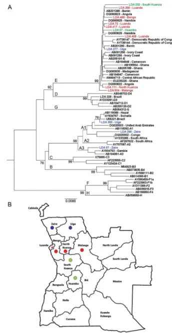
netics analyses (Fig. 2) confirmed the results of all RFLP genotypes and showed that genotype E isolates from An-gola (sequences named by their LDA numbers followed by birth places) did not cluster separately from those within Africa. Among the four genotype A isolates, two clustered within subgenotype A1 (LDA 296-Zaire and LDA300-Uíge) and one clustered within subgenotype A3 (LDA 57-Zaire). The last sample from genotype A, LDA 495-Luanda, clustered within subgenotype A2 (the phy-logeny was obtained by alignment of the S region because the pre-S/S region of this sample was not able to be ampli-fied by PCR; data not shown). Interestingly, subgenotypes A1 and A3 were derived from people who were born to the north of Luanda (Fig. 2).
DISCUSSION
Despite the high endemicity of HBV infection in sub-Saharan Africa, little is known about HBV seroepi-demiology and genotype distribution in many African countries, such as Angola. The only previously pub-lished study of our knowledge about HBV epidemiology, conducted more than 10 years ago in Angola (Steele & Bos 1996), showed a high (79%) prevalence of HBV. The present study confirmed that extremely high HBV rates exist in Angola because 79.7% of the studied population was anti-HBc positive, which indicates previous contact with the virus, and 15.1% of the individuals were carri-ers of HBsAg, which is a marker of active HBV infec-tion. Despite the burden of hepatitis B in Angola, only a few individuals declared that they had been vaccinated against HBV. Our data show that Luanda is a hyperen-demic area for HBV infection and emphasizes the need for HBV prevention programs for adults to reduce the spread of this important human pathogen. It is known that HBeAg levels may vary greatly from one African population to another (Kramvis & Kew 2007). In our study, the levels of HBeAg (18.2%) were similar to those found in adults in Benin (19%) and slightly lower than those found in adults in Ethiopia (23%) and Zimbabwe (25%). Yet, HBeAg levels found in Luanda were much higher than those recorded in other African countries, such as the Central African Republic (4.5%) and Nigeria (2.3%) (Kramvis & Kew 2007).
Risk factors for HBV infection may vary consider-ably depending on epidemiological data, the geographic region and the social, cultural and demographic charac-teristics of different populations. Some predisposing risk factors have been associated with HBV infection in dif-ferent African populations, such as unprotected sexual activities (Abou et al. 2009), multiple sexual partners and blood transfusions (Adoga et al. 2009). In this cohort of Angolans, HBV infection was not associated with the most common predisposing risk factors, which is prob-ably due to the very high rate of HBV infection in this cohort (about 80%). The knowledge of a past hepatitis infection was the only statistically significant factor as-sociated with both anti-HBc and HBsAg markers.
Differences in HBsAg rates between genders were found in some populations, but not in others (Kramvis & Kew 2007). In our study, HBsAg prevalence was higher in men than in women (p = 0.0049).
In sub-Saharan Africa, the predominant HBV trans-mission route is horizontal, mostly due to children being infected during early childhood (under 5 years of age) (Kew 1996, Martinson et al. 1998, Dumpis et al. 2001). Perinatal transmission seems to play a minor role in Af-rica, which is in contrast to what is observed in other high-prevalence endemic areas, such as southeast Asia, where materno-fetal transmission is the dominant route of transmission (Zhang et al. 1998). In low-endemic coun-tries, such as western Europe, sexual transmission and high-risk behavior represent major factors (François et al. 2008). Here, no significant differences in HBsAg preva-lence were found between adults in the range of 18-49 years old. Individuals younger than 18 years old were not analyzed. Consequently, it was not possible to determine which route of transmission is more important: horizon-tal-childhood transmission or sexual transmission. Con-sidering that Angola is a hyperendemic area for HBV infection, it is possible that a higher rate of transmission occurs during childhood than in adulthood.
Studies conducted in Africa showed that rates of HBV co-infection among HIV-positive individuals vary from 6.1% in Kenya (Harania et al. 2008) to 25.9% in Nigeria (Uneke et al. 2005). Among the individuals from Divine Providence Hospital, 18% of HIV patients were co-infected with HBV. Despite HBV and HIV sharing common routes of transmission (parenteral and sexual), our data suggest that the two viruses were differentially transmitted and/or maintained between genders and age groups. Indeed, HBV infection was more prevalent in men than in women in all age groups while HIV showed a different pattern depending on age. In younger people, HIV was more prevalent in women, while in older peo-ple, HIV was more prevalent in men (Fig. 1B).
In this study, HBV DNA was detected in 53% of the HBsAg-positive samples. This rate was lower than the 84% obtained in previous studies with HBsAg-positive samples from Brazilian patients using the same method (Araujo et al. 2004) and the 98% obtained with other meth-ods (real time PCR, transcription-mediated amplification) among HBsAg-positive samples from Ghana, a country where genotype E is circulating (Allain et al. 2003). Dif-ferences in methodologies, HBeAg positivity rates and/or viral loads may account for these differences.
et al. 2003, Kramvis et al. 2005, Vray et al. 2006, Kramvis & Kew 2007, Hübschen et al. 2008). In our study, most of the HBV isolates belonged to genotype E, which supports the idea that this genotype is the most prevalent genotype in west Africa (Bekondi et al. 2007, Kramvis & Kew 2007).
Genotype A is found predominantly in southern, eastern and central Africa (Kramvis & Kew 2007). Pre-vious studies have suggested that isolates of genotype A have a longer natural history in Africa than the two other genotypes, D and E (Andernach et al. 2009a, b). Most of the African-derived genotype A isolates that have been sequenced to date belong to subgenotype A1, which is also the most prevalent subgenotype in Brazil (Araujo et al. 2004, Mello et al. 2007). Subgenotype A1 is thought to have been introduced into Brazil during the slave trade (Motta-Castro et al. 2008). Angola was one of the main suppliers of the slave trade and it is reasonable to think that subgenotype A1 was introduced into Brazil via An-gola. Yet, only about 10% of the samples characterized here belonged to genotype A. Further studies with geno-type A/subgenogeno-type A1 from Brazil and Africa should be conducted to elucidate the origin of subgenotype A1, which is frequently detected in Brazilian individuals and rarely found in Luanda, the main port of the slave trade. In conclusion, the present study showed that HBV is highly endemic in Luanda, with a predominance of genotype E and circulation of genotype A subgenotypes A1, A2 and A3. Further molecular characterization of complete HBV nucleotide sequences from Angola will allow the assessment of their genetic variability, possible molecular signatures and patterns of mutations and de-letions. The possibility that recombination events have taken place between genotypes A and E for all HBV genes should also be considered.
ACKNOWLEDGEMENTS
To Francisco CA Mello, for helping with phylogenetic tree, and to Dr Christian Niel, for critical reading of the manuscript. DNA sequencing was performed by the Platform of Genomics and DNA Sequencing/PDTIS-PDTIS/Fiocruz.
REFERENCES
Abou MA, Eltahir YM, Ali AS 2009. Seroprevalence of hepatitis B virus and hepatitis C virus among blood donors in Nyala, South Dar Fur, Sudan. Virol J 6: 146.
Adoga MP, Banwat EB, Forbi JC, Nimzing L, Pam CR, Gyar SD, Agabi YA, Agwale SM 2009. Human immunodeficiency virus, hepatitis B virus and hepatitis C virus: sero-prevalence, co-in-fection and risk factors among prison inmates in Nasarawa State, Nigeria. J Infect Dev Ctries 3: 539-547.
Allain JP, Candotti D, Soldan K, Sarkodie F, Phelps B, Giachetti C, Shyamala V, Yeboah F, Anokwa M, Owusu-Ofori S, Opare-Sem O 2003. The risk of hepatitis B virus infection by transfusion in Kumasi, Ghana. Blood 101: 2419-2425.
Andernach IE, Hübschen JM, Muller CP 2009a. Hepatitis B virus: the genotype E puzzle. Rev Med Virol 19: 231-240.
Andernach IE, Nolte C, Pape JW, Muller CP 2009b. Slave trade and hepatitis B virus genotypes and subgenotypes in Haiti and Af-rica. Emerg Infect Dis 15: 1222-1228.
André F 2000. Hepatitis B epidemiology in Asia, the Middle East and Africa. Vaccine 18 (Suppl. 1): S20-S22.
Araujo NM, Mello FC, Yoshida CF, Niel C, Gomes SA 2004. High proportion of subgroup A’ (genotype A) among Brazilian isolates of Hepatitis B virus. Arch Virol 149: 1383-1395.
Batina A, Kabemba S, Malengela R 2007. Infectious markers among blood donors in Democratic Republic of Congo (DRC). Rev Med Brux 28: 145-149.
Bekondi C, Olinger CM, Boua N, Talarmin A, Muller CP, Le Faou A, Venard V 2007. Central African Republic is part of the West-Afri-can hepatitis B virus genotype E crescent. J Clin Virol 40: 31-37.
Botha JF, Ritchie MJ, Dusheiko GM, Mouton HW, Kew MC 1984. Hepatitis B virus carrier state in black children in Ovamboland: role of perinatal and horizontal infection. Lancet 1: 1210-1212.
Dumpis U, Holmes EC, Mendy M, Hill A, Thursz M, Hall A, Whittle H, Karayiannis P 2001. Transmission of hepatitis B virus in-fection in Gambian families revealed by phylogenetic analysis. J Hepatol 35: 99-104.
François G, Dochez C, Mphahlele MJ, Burnett R, Van Hal G, Me-heus A 2008. Hepatitis B vaccination in Africa: mission accom-plished? South Afr J Epidemiol Infect 23: 24-28.
Hannoun C, Söderström A, Norkrans G, Lindh M 2005. Phylogeny of African complete genomes reveals a West African genotype A subtype of hepatitis B virus and relatedness between Somali and Asian A1 sequences. J Gen Virol 86: 2163-2167.
Harania RS, Karuru J, Nelson M, Stebbing J 2008. HIV, hepatitis B and hepatitis C coinfection in Kenya. AIDS 22: 1221-1222.
Hübschen JM, Andernach IE, Muller CP 2008. Hepatitis B virus gen-otype E variability in Africa. J Clin Virol 43: 376-380.
Hübschen JM, Mbah PO, Forbi JC, Otegbayo JA, Olinger CM, Char-pentier E, Muller CP 2010. Detection of a new subgenotype of hepatitis B virus genotype A in Cameroon but not in neighbour-ing Nigeria. Clin Microbiol Infect, in press.
Kew MC 1996. Progress towards the comprehensive control of hepa-titis B in Africa: a view from South Africa. Gut 38 (Suppl. 2): S31-S36.
Kramvis A, Kew MC 2007. Epidemiology of hepatitis B virus in Af-rica, its genotypes and clinical associations of genotypes. Hepa-tol Res 37: S9-S19.
Kramvis A, Restorp K, Norder H, Botha JF, Magnius LO, Kew MC 2005. Full genome analysis of hepatitis B virus genotype E strains from South-Western Africa and Madagascar reveals low genetic variability. J Med Virol 77: 47-52.
Kurbanov F, Tanaka Y, Fujiwara K, Sugauchi F, Mbanya D, Zekeng L, Ndembi N, Ngansop C, Kaptue L, Miura T, Ido E, Hayami M, Ichimura H, Mizokami M 2005. A new subtype (subgenotype) Ac (A3) of hepatitis B virus and recombination between genotypes A and E in Cameroon. J Gen Virol 86: 2047-2056.
Makuwa M, Souquière S, Telfer P, Apetrei C, Vray M, Bedjabaga I, Mouinga-Ondeme A, Onanga R, Marx PA, Kazanji M, Roques P, Simon F 2006. Identification of hepatitis B virus subgenotype A3 in rural Gabon. J Med Virol 78: 1175-1184.
Martinson FE, Weigle KA, Royce RA, Weber DJ, Suchindran CM, Lemon SM 1998. Risk factors for horizontal transmission of hepatitis B virus in a rural district in Ghana. Am J Epidemiol 147: 478-487.
Meldal BH, Moula NM, Barnes IH, Boukef K, Allain JP 2009. A novel hepatitis B virus subgenotype D7 in Tunisian blood donors. J Gen Virol 90: 1622-1628.
MM, Martins RM, Gomes SA 2007. Hepatitis B virus genotypes circulating in Brazil: molecular characterization of genotype F isolates. BMC Microbiol 7: 103.
Motta-Castro AR, Martins RM, Araujo NM, Niel C, Facholi GB, Lago BV, Mello FC, Gomes SA 2008. Molecular epidemiology of hepatitis B virus in an isolated Afro-Brazilian community. Arch Virol 153: 2197-2205.
Msuya SE, Mbizvo EM, Hussain A, Sam NE, Stray-Pedersen B 2006. Seroprevalence of hepatitis B and C viruses among women of child-bearing age in Moshi Urban, Tanzania. East Afr Med J 83: 91-94.
Mulders MN, Venard V, Njayou M, Edorh AP, Bola Oyefolu AO, Kehinde MO, Muyembe Tamfum JJ, Nebie YK, Maiga I, Ammer-laan W, Fack F, Omilabu SA, Le Faou A, Muller CP 2004. Low genetic diversity despite hyperendemicity of hepatitis B virus genotype E throughout West Africa. J Infect Dis 190: 400-408.
Niel C, Moraes MT, Gaspar AM, Yoshida CF, Gomes SA 1994. Genetic diversity of hepatitis B virus strains isolated in Rio de Janeiro, Brazil. J Med Virol 44: 180-186.
Olinger CM, Jutavijittum P, Hübschen JM, Yousukh A, Samountry B, Thammavong T, Toriyama K, Muller CP 2008. Possible new hepatitis B virus genotype, southeast Asia. Emerg Infect Dis 14: 1777-1780.
Olinger CM, Venard V, Njayou M, Oyefolu AO, Maïga I, Kemp AJ, Omilabu SA, le Faou A, Muller CP 2006. Phylogenetic analysis of the precore/core gene of hepatitis B virus genotypes E and A in West Africa: new subtypes, mixed infections and recombina-tions. J Gen Virol 87: 1163-1173.
Oshitani H, Kasolo F, Tembo C, Mpabalwani M, Mizuta K, Luo N, Suzuki H, Numazaki Y 1995. Hepatitis B virus infection among pregnant women in Zambia. East Afr Med J 72: 813-815.
Palumbo E, Scotto G, Faleo G, Cibelli DC, Saracino A, Angarano G 2007. Prevalence of HBV-genotypes in immigrants affected by HBV-related chronic active hepatitis. Arq Gastroenterol 44: 54-57.
Pawlotsky JM, Bélec L, Grésenguet G, Deforges L, Bouvier M, Duval J, Dhumeaux D 1995. High prevalence of hepatitis B, C, and E markers in young sexually active adults from the Central African Republic. J Med Virol 46: 269-272.
Pirillo MF, Bassani L, Germinario EA, Mancini MG, Vyankandondera J, Okong P, Vella S, Guiliano M 2007. Seroprevalence of hepatitis B and C viruses among HIV-infected pregnant women in Uganda and Rwanda. J Med Virol 79: 1797-1801.
Pourkarim MR, Lemey P, Amini-Bavil-Olyaee S, Maes P, Van Ranst M 2010. Novel hepatitis B virus subgenotype A6 in African-Bel-gian patients. J Clin Virol 47: 93-96.
Schaefer S 2007. Hepatitis B virus taxonomy and hepatitis B virus genotypes. World J Gastroenterol 7: 14-21.
Sitnik R, Sette H Jr, Santana RA, Menezes LC, Graça CH,
Dasto-li GT, Silbert S, Pinho JR 2007. Hepatitis B virus genotype E detected in Brazil in an African patient who is a frequent traveler. Braz J Med Biol Res 40: 1689-1692.
Steele AD, Bos P 1996. Hepatitis B and C virus infection in adult volunteers in Angola. S Afr Med J 86: 701-702.
Steele AD, Bos P, Joubert JJ, Bafort JM, Lecatsas G, Aspinall S 1995. Serologic markers for hepatitis B virus and hepatitis A virus in Bushmen in West Caprivi, Namibia. East Afr Med J 72: 30-32.
Strand RT, Franque-Ranque M, Bergström S, Weiland O 2003. Infec-tious aetiology of jaundice among pregnant women in Angola. Scand J Infect Dis 35: 401-403.
Sugauchi F, Orito E, Kato H, Suzuki S, Kawakita S, Sakamoto Y, Fukushima K, Akiba T, Yoshihara N, Ueda R, Mizokami M 2003. Genotype, serotype, and phylogenetic characterization of the complete genome sequence of hepatitis B virus isolates from Malawian chronic carriers of the virus. J Med Virol 69: 33-40.
Sutcliffe S, Taha TE, Kumwenda NI, Taylor E, Liomba GN 2002. HIV-1 prevalence and herpes simplex virus 2, hepatitis C virus, and hepatitis B virus infections among male workers at a sugar estate in Malawi. J Acquir Immune Defic Syndr 31: 90-97.
Suzuki S, Sugauchi F, Orito E, Kato H, Usuda S, Siransy L, Arita I, Sakamoto Y, Yoshihara N, El-Gohary A, Ueda R, Mizokami M 2003. Distribution of hepatitis B virus (HBV) genotypes among HBV carriers in the Cote d’Ivoire: complete genome sequence and phylogenetic relatedness of HBV genotype E. J Med Virol 69: 459-465.
Tamura K, Dudley J, Nei M, Kumar S 2007. MEGA4: Molecular Evo-lutionary Genetics Analysis (MEGA) software version 4.0. Mol Biol Evol 24: 1596-1599.
Tatematsu K, Tanaka Y, Kurbanov F, Sugauchi F, Mano S, Mae-shiro T, Nakayoshi T, Wakuta M, Miyakawa Y, Mizokami M 2009. A genetic variant of hepatitis B virus divergent from known human and ape genotypes isolated from a Japanese pa-tient and provisionally assigned to new genotype J. J Virol 83: 10538-10547.
Uneke CJ, Ogbu O, Inyama PU, Anyanwu GI, Njoku MO, Idoko JH 2005. Prevalence of hepatitis-B surface antigen among blood do-nors and human immunodeficiency virus-infected patients in Jos, Nigeria. Mem Inst Oswaldo Cruz 100: 13-16.
Vray M, Debonne JM, Sire JM, Tran N, Chevalier B, Plantier JC, Fall F, Vernet G, Simon F, Mb PS 2006. Molecular epidemiology of hepatitis B in Dakar, Senegal. J Med Virol 78: 329-334.
WHO - World Health Organization 2004. Hepatitis B vaccines. Wkly Epidemiol Rec 79: 255-263.