HEV, TTV and GBV-C/HGV markers
in patients with acute viral hepatitis
1Departamento de Gastroenterologia, Universidade de São Paulo, São Paulo,
SP, Brasil
2Instituto Adolfo Lutz, São Paulo, SP, Brasil
3Serviço de Gastro-Hepatologia, Universidade Federal da Bahia and Hospital São
Rafael, Salvador, BA, Brasil
4Fundação Oswaldo Cruz (FioCruz), Salvador, BA, Brasil 5Laboratório Central da Bahia (LACEN), Salvador, BA, Brasil
A.C. Lyra1,3,J.R.R. Pinho2,
L.K. Silva4, L. Sousa2,
C.P. Saraceni2, E.L. Braga3,
J.E. Pereira3, M.A.S. Zarife5,
M.G. Reis4, L.G.C. Lyra3,
L.C. da Silva1 and
F.J. Carrilho1
Abstract
The aim of the present study was to evaluate the prevalence of HEV, TTV and GBV-C/GBV-C/HGV in patients with acute viral hepatitis A, B and non-A-C. Weevaluated sera of 94 patients from a sentinel program who had acute hepatitis A (N = 40), B (N = 42) and non-A-C (N = 12); 71 blood donors served as controls. IgM and anti-HEV IgG antibodies were detected by enzyme immunoassay using commercial kits. TTV and GBV-C/HGV were detected by nested PCR; genotyping was done by sequencing and phylogenetic analysis. Anti-HEV IgG was present in 38, 10 and 17% of patients with hepatitis A, B and non-A-C. Four patients with hepatitis A and 1 with non-A-C hepatitis also had anti-HEV IgM detected in serum. TTV was detected in 21% of patients with acute hepatitis and in 31% of donors. GBV-C/HGV was detected in 9% of patients with hepatitis, and in 10% of donors. We found TTV isolates of genotypes 1, 2, 3, and 4 and GBV-C/HGV isolates of genotypes 1 and 2. Mean aminotransferase levels were lower in patients who were TTV or GBV-C/HGV positive. In conclu-sion, the detection of anti-HEV IgM in some acute hepatitis A cases suggests co-infection with HEV and hepatitis E could be the etiology of a few cases of sporadic non-A-C hepatitis in Salvador, Brazil. TTV genotype 1, 2, 3 and 4 isolates and GBV-C/HGV genotype 1 and 2 strains are frequent in the studied population. TTV and GBV-C/HGV infection does not appear to have a role in the etiology of acute hepatitis.
Correspondence
A.C. Lyra
R. Sócrates Guanaes Gomes, 84/401 40283-320 Salvador, BA Brasil
Fax: +55-71-452-9589 E-mail: aclyra@atarde.com.br
Research supported in part by Alves de Queiroz Family Fund for Research.
Publication supported by FAPESP.
Received February 19, 2004 Accepted January 3, 2005
Key words
•Acute hepatitis •Hepatitis E •TTV •GBV-C/HGV •Genotype •Brazil
Introduction
Acute viral hepatitis A, B, and non-A-C remains animportant cause of morbidity in cities of Northeastern Brazil. The prevalence of hepatitis A virus (HAV) infection closely correlates with the degree of environmental
sanitation and the prevailing socioeconomic and hygiene conditions. In Brazil, serolence studies have indicated a high preva-lence of anti-HAV antibodies in the adult population (1-3).
infection and its seroprevalence has been reported to be approximately 8% in several regions of Brazil, including the Northeast (4,5). Following acute hepatitis B, chronic infection occurs in 1.7% of cases in some areas of Brazil (6). Sexual transmission ap-peared to play a major role in the spread of HBV in a Brazilian study (7).
A few cases of acute non-A-C hepatitis have been reported in the city of Salvador, Bahia, Brazil, and these patients were less likely to evolve toward chronic infection compared with acute hepatitis C (8).
Hepatitis E virus (HEV) is a single-stranded positive sense RNA virus whose genome was first cloned and sequenced in 1990 and is approximately 7.5 kb in length (9,10). It has been recognized as an etiologic agent responsible for epidemic and endemic cases of acute viral hepatitis worldwide, es-pecially in developing countries (11-13). Previous studies have demonstrated sero-positivity for anti-HEV IgG antibodies in populations from Brazil, including patients with non-A-C hepatitis, suggesting that HEV may be responsible for some cases of acute non-A-C hepatitis in this country (14-16). Nevertheless, detection of anti-HEV IgM in serum of patients with acute hepatitis has not been reported in Brazil.
In 1997, a new virus designated transfu-sion-transmitted virus (TTV) was isolated from the serum of a patient with post-trans-fusion non-A-E hepatitis who was GBV-C/ HGV-RNA negative (17). TTV is a single-stranded, circular, non-enveloped DNA vi-rus with at least 3800 base pairs and at least two open reading frames (18). The results of studies concerning the role of TTV in acute and chronic liver diseases have been contro-versial (19-21).
GBV-C/GBV-C/HGV was initially re-ported as a putative non-A-C hepatitis virus; however, there is increasing evidence that this virus does not cause hepatitis (22,23). Recently, GBV-C viremia has been associ-ated with prolonged survival among
HIV-positive subjects (24,25).
The aim of the present study was to evalu-ate the prevalence of HEV, TTV and GBV-C/GBV-C/HGV infection in patients with acute viral hepatitis A, B and non-A-C from a sentinel program in the city of Salvador, Northeastern Brazil, and to determine if co-infections with these viruses correlated with the severity of the necro-inflammatory activ-ity.
Material and Methods
Selection of patients and donors
We evaluated sera from 94 patients who attended the acute hepatitis outpatient serv-ice of the Federal University of Bahia be-tween 1995 and 1999. These patients were referred by primary care public institutions in the city of Salvador, Bahia, through a sentinel program for hepatitis. All public primary care health units in the city of Salva-dor were instructed to refer patients with jaundice to the central laboratory of the State of Bahia located in the city of Salvador. These primary care units assist subjects who live in neighborhoods of low socioeconomic level. Patients were subjected to laboratory tests, serum samples were stored and pa-tients with alanine aminotransferase (ALT) levels higher than twice the upper limit of normal were referred to the outpatient serv-ice of the Federal University of Bahia and evaluated by a nurse and a hepatologist. If clinical suspicion of acute hepatitis was con-firmed the patient was subjected to a com-plete laboratory work-up that included viral markers.
exclude other liver diseases.
Sera from 71 volunteer blood donors were used as controls. These subjects had donated blood at the main Blood Center in the State of Bahia (HEMOBA), located in the city of Salvador. We selected 71 consecutive sub-jects who had donated blood in 1999 and who fulfilled criteria for blood donation. Thus, all were seronegative for all serologic tests performed at the Blood Center, includ-ing tests for hepatitis B surface antigen (HBsAg) and antibody to hepatitis B core antigen (anti-HBc) and hepatitis C antibody (anti-HCV), and all had normal ALT levels. The institutional review boards approved the study and the protocol conformed to the ethical guidelines of the Helsinki declara-tion and resoludeclara-tion 196/CONEP/MS.
Case definition
All patients had signs and symptoms of acute hepatitis, as well as ALT and aspartate aminotransferases (AST) at least two times the upper limit of normal. Patients were further divided into three categories accord-ing to the followaccord-ing serological features:
Acute hepatitis A. This group consisted of 40 patients seropositive for anti-HAV IgM and seronegative for anti-HBc IgM, HBsAg and anti-HCV by enzyme immu-noassay (EIA).
Acute hepatitis B. This group contained 42 patients positive for HBsAg and anti-HBc IgM and negative for anti-HAV and anti-HCV by EIA.
Acute non-A-Chepatitis. This group in-cluded 12 patients negative for anti-HAV, anti-HBc IgM, HBsAg and anti-HCV. All patients were also HCV-RNA negative by PCR (26). All patients had hepatic auto-antibody titers lower than 1/40; had no his-tory of potentially hepatotoxic drug use in the 15 days that preceded the beginning of symptoms; all had an estimated alcohol con-sumption of less than 40 g/day, and an ab-dominal ultrasonography showing normal
liver texture and no dilated common bile duct. Serum ceruloplasmin was measured in patients who were less than 30 years old and all were found to have normal levels.
Laboratory methods
Serological tests. Anti-HAV IgM and anti-HBc and HBsAg IgM were detected by EIA (Abbott Laboratories, Abbott Park, IL, USA, and Wiesbaden-Delkenheim, Ger-many). Anti-HCV was tested using a sec-ond-generation EIA (Abbott Laboratories). Anti-HEV IgM and anti-HEV IgG were as-sayed by EIA (Genelabs Diagnostics, Sin-gapore, and Abbott Laboratories, respec-tively). All assays were performed in dupli-cate according to manufacturer instructions.
TTV-DNA detection. TTV-DNA was de-tected in serum by a nested PCR assay using sense primers NG059 and NG061, and anti-sense primer NG063 described previously (27); the second anti-sense oligonucleotide (5' GTK GGK TAC CAY TTA GCT CTC ATT C 3') was denoted JR01. Briefly, nucleic acid was extracted as previously reported (28) and amplified using standard PCR con-ditions by a first round of PCR performed with primers NG059 and JR01 for 35 cycles (94ºC, 30 s; 60ºC, 45 s; 72ºC, 60 s), plus an additional cycle of 72ºC for 7 min. Identical conditions and primers NG061 and NG063 were used for the second round of amplifica-tion. After 1.5% agarose gel electrophoresis, samples producing a single band of 271 bp were considered to be positive. Stringent precautions were taken to avoid contamina-tion as previously described (29).
GBV-C/HGV-RNA detection. GBV-C/
sets of primers for the NS3 region (NS3.2-a.2, NS3.2-s1, GBVc-a1, GBVc-s1) (22,31) and 5’NCR region (5’NCR-s1, 5’NCR-a1, 5’NCR-s2, 5’NCR-a2) (32) were used. A PCR “touchdown” thermocycling protocol was used for the NS3 region (22,30). After agarose gel electrophoresis, samples pro-ducing a single band of 171 bp for the NS3 region, and of 203 bp for the 5’NCR region were considered to be positive.
Sequencing and genotyping. Both strands of TTV and GBV-C/HGV PCR products were sequenced using the ABI Prism BigDye Terminator Ready Reaction Kit version 1.0 (Applied Biosystems, Foster City, CA, USA). Reactions were analyzed with an automated DNA sequencer (ABI model 377). Sequences were validated after visual inspection and analysis using the Phred-Phrap-Consed soft-ware, and then aligned using the CLUSTAL X software, version 1.81 (33). TTV and GBV-C/HGV isolates were genotyped by comparison of nucleotide or amino acid mutations with sequences of previously pub-lished genomes, and by phylogenetic
analy-sis using the neighbor-joining method of the MEGA program (version 2.1) (34); the dis-tance matrix was calculated with the Kimura 2-parameter model (35).
Statistical analysis
Data regarding continuous variables are reported as means ± SD. The means for the three groups with acute hepatitis were com-pared by Kruskal-Wallis ANOVA followed by Dunn’s test. The means for TTV (or C/HGV)-positive and TTV (or GBV-C/HGV)-negative patients were compared by the Mann-Whitney U-test. Proportions were compared by the chi-square test or Fisher exact test when appropriate. All analy-ses were performed using the SPSS package (SPSS for Windows release 11.0; SPSS Inc., Chicago, IL, USA) and the SigmaStat for Windows software (version 2.03, SPSS Inc.). A P value ≤0.05 was considered to be statis-tically significant.
Results
The clinical features of the patients are summarized in Table 1. Fifty-nine patients were males and 35 were females. Mean age was higher for patients with hepatitis B and non-A-C compared to patients with hepatitis A. Mean ALT and AST levels were highest in the group of patients with acute hepatitis B.
Anti-HEV IgG was detected more fre-quently in patients with acute hepatitis A (38%) than in patients with acute hepatitis B (10%) (P = 0.003; Figure 1). Anti-HEV IgG was detected in 15 patients with acute hepa-titis A and 4 patients with acute hepahepa-titis B; 2 patients from the non-A-C group were also anti-HEV IgG positive. Four of the 15 pa-tients with acute hepatitis A and 1 of the 2 patients with non-A-C hepatitis who had HEV IgG were also positive for anti-HEV IgM, while none of the 4 patients from the hepatitis B group was positive (Figure 1).
Table 1. Clinical features of the 94 patients studied.
Group Number Sex (male: Age (years) ALT (x ULN)a AST (x ULN)a
of patients female) (mean ± SD)* (mean ± SD)** (mean ± SD)***
Hepatitis A 40 30:10 14.28 ± 8.95 18.55 ± 14.48 12.05 ± 11.64 Hepatitis B 42 25:17 32.19 ± 12.99 30.93 ± 20.77 26.36 ± 21.62 Hepatitis 12 4:8 31.70 ± 6.30 19.00 ± 12.83 8.83 ± 7.06
non-A-C
Total 94 59:35 24.50 ± 15.58 24.14 ± 18.31 18.03 ± 18.07
aAlanine aminotransferase (ALT) and aspartate aminotransferase (AST) levels are
reported as multiple upper limit of normal (x ULN). The normal range for ALT is 12-50 U/l and the normal range for AST is 12-46 U/l.
*Comparison of age: P < 0.001 for comparison of age between the three groups (Kruskal-Wallis test); P < 0.05 for comparison of hepatitis A vs hepatitis B group and for comparison of hepatitis A vs non-A-C (Dunn test); P = NS for comparison of hepatitis B vs non-A-C (Dunn test).
**Comparison of ALT levels: P = 0.007 for comparison of ALT levels between the three groups (Kruskal-Wallis test); P < 0.05 for comparison of hepatitis A vs hepatitis B group; P = NS for comparison of hepatitis A vs non-A-C and for comparison of hepatitis B vs non-A-C (Dunn test).
Among the 4 patients with serological mark-ers for HAV and HEV, mean ALT and AST levels were 9.3 ± 4.6 and 7.5 ± 4.4 times the upper limit of normal, respectively, while among the remaining 36 patients infected only with HAV, ALT and AST levels were 19.6 ± 14.9 and 12.6 ± 12.1 times the upper limit of normal (P = 0.179 and P = 0.417 for comparison of ALT and AST, respectively). Serum TTV-DNA was detected in 20 of 94 patients (21%) with acute hepatitis, and in 22 of 71 blood donors (31%). TTV was detected in 8 of 40 (20%) patients with acute hepatitis A, 11 of 42 (26%) with acute hepa-titis B, and 1 of 12 (8%) patients with non-A-C hepatitis (Table 2). There was no statisti-cally significant difference in mean age or mean aminotransferase levels between TTV-positive and TTV-negative patients (Table 3).
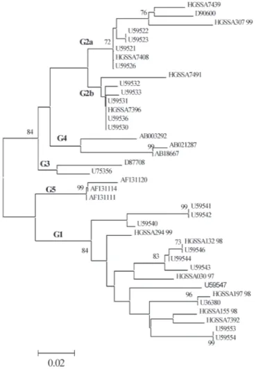
TTV genotyping was carried out for 26 of 42 subjects who were TTV positive (GenBank accession numbers AY137347-AY137372). Six of these subjects had acute hepatitis B, 2 had acute hepatitis A and 18 were blood donors. They were selected ac-cording to the availability of extra amounts of serum. We found TTV isolates of geno-types 1a, 1b, 2, 3, and 4 (Figure 2). The distribution of TTV genotypes showed that genotype 1 was the most prevalent in the study population.
Serum GBV-C/HGV-RNA was detected in 13 of 165 individuals using primers for the NS3 region (8%), and in 15 of 165 using primers of the 5’NCR region (9%). GBV-C/ HGV-RNA was detected in 6 (NS3 region) and 8 (5’NCR region) of 94 patients (6 and 9%, respectively) with acute hepatitis, and in 7 of 71 (10%) blood donors, for both the NS3 and 5’NCR regions (Table 2). Mean ALT levels were lower in C/HGV-RNA-positive patients compared to GBV-C/HGV-RNA-negative patients, although this difference did not reach statistical sig-nificance.
GBV-C/HGV genotyping was carried out
Table 2. Prevalence of serum TTV-DNA and GBV-C/HGV-RNA in patients with acute hepatitis and in blood donors.
Group Number of TTV-DNA- GBV-C/HGV-
GBV-C/HGV-5’NCR-patients positive NS3-positive positive patients (%) patients (%) patients (%)
Blood donors 71 22 (31)* 7 (10) 7 (10)**
Hepatitis A 40 8 (20) 0 (0) 1 (2.5)
Hepatitis B 42 11 (26) 5 (12) 6 (14)
Hepatitis non-A-C 12 1 (8)* 1 (8) 1 (8)**
Total 165 42 (25) 13 (8) 15 (10)
*P = 0.165 for comparison of TTV-DNA prevalence between blood donors and pa-tients with non-A-C hepatitis (Fisher exact test);
**P = 1.000 for comparison of GBV-C/HGV-5’NCR prevalence between blood donors and patients with non-A-C hepatitis (Fisher exact test).
Anti-HEV IgG negative Anti-HEV IgG positive
Hepatitis non-A-C
Hepatitis B
Hepatitis A Hepatitis non-A-C
Hepatitis B
Hepatitis A
2 10
4 38
25 15
0 10 20 30 40 50
Number of patients
0 5 10 15 20
Number of patients
1 1
4 4 0
11
Anti-HEV IgM negative Anti-HEV IgM positive
Figure 1. A, Prevalence of serum anti-HEV IgG antibodies in patients with acute hepatitis. Anti-HEV IgG was significantly more prevalent in patients with hepatitis A compared to the hepatitis B group (P = 0.003, chi-square test). Comparison between hepatitis A vs non-A-C groups showed P = 0.294, and comparison between the hepatitis B group and the non-A-C hepatitis group showed P = 0.605 (Fisher exact test). B, Prevalence of serum anti-HEV IgM antibodies in patients with acute hepatitis who were anti-HEV IgG positive.
A B
Table 3. Comparison of acute hepatitis patients with and without serum TTV-DNA or GBV-C/HGV-RNA.
Clinical data Acute hepatitis A, B and non-A-C (N = 94)
TTV-DNA TTV-DNA GBV-C/HGV-
GBV-C/HGV-positive negative 5’NCR positive 5’NCR negative
Number of patients 20 74 8 86
Sex (male:female) 14:6 45:29 3:5 56:30
Age (years, 23.40 ± 12.50 24.80 ± 16.37 32.25 ± 18.01 23.78 ± 15.25 mean ± SD)
Max ALT (x ULN) 22.95 ± 17.93 24.46 ± 18.52 21.88 ± 14.26 24.35 ± 18.70 Max AST (x ULN) 15.95 ± 13.76 18.59 ± 19.11 18.38 ± 14.54 18.00 ± 18.43
Discussion
Confirmed acute cases of HEV infection have not been documented in Brazil. How-ever, our results suggest that symptomatic hepatitis E is the etiologic agent of some of the cases of acute sporadic hepatitis in the population that seeks primary medical care in the city of Salvador. We found that 2 of 12 patients with acute non-A-C hepatitis were anti-HEV IgG seropositive and one of them also had anti-HEV IgM detected in serum. To our knowledge, this is the first report of detection of anti-HEV IgM in serum of a patient with acute non-A-C hepatitis in Sal-vador, Northeastern Brazil. Interestingly, we also found seropositivity for anti-HEV IgM in 4 patients who had been diagnosed with acute hepatitis A, while none of the patients who had acute hepatitis B were anti-HEV IgM positive. Detection of HEV by PCR would be necessary to confirm the presence of the virus and to determine that co-infec-tion of HAV and HEV indeed occurred. Nevertheless, our findings suggest that there was co-infection with HAV and HEV. Both viruses share a similar mode of transmission by the fecal-oral route (29). Thus, patients who are infected with HAV are probably at a higher risk to be co-infected with HEV. In fact, the prevalence of HEV IgG anti-body was significantly higher in the group of subjects with hepatitis A compared to the group with acute hepatitis B. This is in agree-ment with a previous study from Brazil that reported a higher rate of HEV IgG anti-body seropositivity in patients with acute hepatitis A compared to patients with acute hepatitis B and controls (14).
Among 26 serum samples subjected to TTV genotyping, 12 isolates were genotype 1. It should be noted that the primers we used do not permit the detection of all TTV geno-types, although they allow the detection of the main TTV genotypes reported in other studies. TTV genotypes 1 and 2 have been described in several countries including
Ja-Figure 2. Neighbor-joining tree of 183 nt fragments of TTV. This corre-sponds to nt 1960-2142 of strain TA278 from previously published refer-ence strains and from our study population. For previously published reference strains, the GenBank accession numbers are indicated. The genotype designations used correspond to designations from previous publications. They are indicated on the branches. The bootstrap test of phylogeny was performed with 1000 replicates and values equal to or higher than 70 are indicated.
pan, Thailand, United Kingdom, and Ger-many. Other genotypes have also been re-ported in these regions (19,36,37).
We found that TTV was highly prevalent among all groups studied, although the virus was less frequently detected in the non-A-C group. Moreover, ALT and AST levels were lower in patients infected with TTV com-pared to patients who were TTV negative, but these differences did not reach statistical significance. These findings suggest that TTV does not have an etiologic role in patients with acute hepatitis of unknown etiology and that it does not add to the severity of the necro-inflammatory activity of acute hepati-tis caused by HAV or HBV.
The results of the studies that have ana-lyzed the role of TTV in acute and chronic liver diseases have been controversial (17,19, 20,27,36). Nevertheless, it appears that TTV does not have a role in the etiology of acute and chronic liver disease. In a Japanese study, TTV was detected in 29% of patients with hepatitis A, 24% of patients with acute hepa-titis B, 43% of patients with non-A-E hepati-tis, and in 37% of the controls. There was no significant difference in aminotransferase levels between TTV-positive and TTV-nega-tive patients (14). These findings appear to be similar to ours.
We were able to identify only GBV-C/ HGV isolates of genotypes 1 and 2. These appear to be the most prevalent GBV-C/ HGV genotypes found in Africa, and North America and Europe, respectively. GBV-C/ HGV genotype 3 isolates are found in Asia and among native American Indians from Colombia and Venezuela (38,39). Perhaps this high prevalence of GBV-C/HGV geno-types 1 and 2 in our population may be explained by the fact that the population of Salvador mainly consists of ians and of miscegenation of African-Brazil-ians and Whites (Mulattos),many of whom have a European and African ethnic back-ground.
As previously demonstrated by other
in-vestigators, the data reported here do not support a role for GBV-C/HGV in the etiol-ogy of acute non-A-C hepatitis. The high frequency of GBV-C/HGV in blood donors (10%) is in agreement with rates found in blood donor populations in other Brazilian cities and elsewhere (22,23,30).
Finally, it is interesting to note that the serum samples analyzed in this study were from patients who were referred by primary care public institutions through a sentinel program for hepatitis. Therefore, our results
correspond to findings observed in patients who seek primary care health institutions in the city of Salvador and are not necessarily referred to tertiary institutions. On the other hand, this is a retrospective study and pa-tients were selected on the basis of the avail-ability of stored serum samples. Therefore, further studies are necessary to confirm and extend our findings.
Acknowledgments
We wish to thank the following persons for their assistance and/or support for this study: Dr. Aurelino Reis (Hemocentro da Bahia, Salvador, BA, Brazil), Dr. Nelma Santana, Dr. Marcos C. Lyra, Dr. Maxuel Oliveira (Serviço de Gastro-Hepatologia, Universidade Federal da Bahia and Hospital São Rafael, Salvador, BA, Brazil) and Jussara Botto (Laboratório Central da Bahia (LACEN), Salvador, BA, Brazil).
References
1. Pannuti CS, de Mendonca JS, Pereira ML, Carvalho MJ & Amato Neto V (1985). Sporadic acute viral hepatitis A, B and non-A non-B. A prospective study of 150 consecutive cases in São Paulo, Brazil.
Tropical and Geographical Medicine, 37: 136-138.
2. Abuzwaida AR, Sidoni M, Yoshida CF & Schatzmayr HG (1987). Seroepidemiology of hepatitis A and B in two urban communities of Rio de Janeiro, Brazil. Revista do Instituto de Medicina Tropical de São Paulo, 29: 219-223.
3. Vitral CL, Yoshida CF, Lemos ER, Teixeira CS & Gaspar AM (1998). Age-specific prevalence of antibodies to hepatitis A in children and adolescents from Rio de Janeiro, Brazil, 1978 and 1995. Relation-ship of prevalence to environmental factors. Memórias do Instituto Oswaldo Cruz, 93: 1-5.
4. dos Santos JI, Lopes MA, Deliege-Vasconcelos E, Couto-Fernandez JC, Patel BN, Barreto ML, Ferreira Junior OC & Galvao-Castro B (1995). Seroprevalence of HIV, HTLV-I/II and other perinatally-trans-mitted pathogens in Salvador, Bahia. Revista do Instituto de Medi-cina Tropical de São Paulo, 37: 343-348.
5. Clemens SA, da Fonseca JC, Azevedo T, Cavalcanti A, Silveira TR, Castilho MC & Clemens R (2000). Hepatitis A and hepatitis B seroprevalence in 4 centers in Brazil. Revista da Sociedade Brasi-leira de Medicina Tropical, 33: 1-10.
6. Ferraz ML, Yoradjian A, Barbieri A, Figueiredo V, Lopes Neto E, Cruz CN & Silva AE (1998). Epidemiology of acute hepatitis B in a univer-sity hospital in São Paulo, Brazil: retrospective study of two five-year periods. São Paulo Medical Journal, 116: 1695-1699. 7. Lewis-Ximenez LL, do O KM, Ginuino CF, Silva JC, Schatzmayr HG,
Stuver S & Yoshida CF (2002). Risk factors for hepatitis B virus infection in Rio de Janeiro, Brazil. BMC Public Health, 2: 26. 8. Parana R, Vitvitski L, Andrade Z, Trepo C, Cotrim H, Bertillon P, Silva
F, Silva L, de Oliveira IR & Lyra L (1999). Acute sporadic A, non-B hepatitis in Northeastern non-Brazil: etiology and natural history. Hep-atology, 30: 289-293.
9. Reyes GR, Purdy MA, Kim JP, Luk KC, Young LM, Fry KE & Bradley DW (1990). Isolation of a cDNA from the virus responsible for enterically transmitted non-A, non-B hepatitis. Science, 247: 1335-1339.
10. Bradley DW, Purdy MA & Reyes GR (1991). Hepatitis E virus ge-nome. Molecular features, expression of immunoreactive proteins and sequence divergence. Journal of Hepatology, 4 (Suppl):
S152-S154.
11. Hyams KC, Purdy MA, Kaur M, McCarthy MC, Hussain MA, el-Tigani A, Krawczynski K, Bradley DW & Carl M (1992). Acute spo-radic hepatitis E in Sudanese children: analysis based on a new Western blot assay. Journal of Infectious Diseases, 165: 1001-1005.
12. Bryan JP, Tsarev SA, Iqbal M, Ticehurst J, Emerson S, Ahmed A, Duncan J, Rafiqui AR, Malik IA & Purcell RH (1994). Epidemic hepatitis E in Pakistan: patterns of serologic response and evidence that antibody to hepatitis E virus protects against disease. Journal of Infectious Diseases, 170: 517-521.
13. Paul DA, Knigge MF, Ritter A, Gutierrez R, Pilot-Matias T, Chau KH & Dawson GJ (1994). Determination of hepatitis E virus seropreva-lence by using recombinant fusion proteins and synthetic peptides.
Journal of Infectious Diseases, 169: 801-806.
14. Parana R, Cotrim HP, Cortey-Boennec ML, Trepo C & Lyra L (1997). Prevalence of hepatitis E virus IgG antibodies in patients from a referral unit of liver diseases in Salvador, Bahia, Brazil. American Journal of Tropical Medicine and Hygiene, 57: 60-61.
15. Souto FJ, Fontes CJ, Parana R & Lyra LG (1997). Short report: further evidence for hepatitis E in the Brazilian Amazon. American Journal of Tropical Medicine and Hygiene, 57: 149-150.
16. Goncales NS, Pinho JR, Moreira RC, Saraceni CP, Spina AM, Stucchi RB, Filho AD, Magna LA & Goncales Junior FL (2000). Hepatitis E virus immunoglobulin G antibodies in different populations in Cam-pinas, Brazil. Clinical and Diagnostic Laboratory Immunology, 7: 813-816.
17. Nishizawa T, Okamoto H, Konishi K, Yoshizawa H, Miyakawa Y & Mayumi M (1997). A novel DNA virus (TTV) associated with el-evated transaminase levels in post-transfusion hepatitis of unknown etiology. Biochemical and Biophysical Research Communications, 241: 92-97.
18. Mushahwar IK, Erker JC, Muerhoff AS, Leary TP, Simons JN, Birkenmeyer LG, Chalmers ML, Pilot-Matias TJ & Dexai SM (1999). Molecular and biophysical characterization of TT virus: Evidence for a new virus family infecting humans. Proceedings of the National Academy of Sciences, USA, 96: 3177-3182.
20. Ikeda H, Takasu M, Inoue K, Okamoto H, Miyakawa Y & Mayumi M (1999). Infection with an unenveloped DNA virus (TTV) in patients with acute or chronic liver disease of unknown etiology and in those positive for hepatitis C virus RNA. Journal of Hepatology, 30: 205-212.
21. Tanaka H, Okamoto H, Luengrojanakul P, Chainuvati T, Tsuda F, Tanaka T, Miyakawa Y & Mayumi M (1998). Infection with an unenveloped DNA virus (TTV) associated with posttransfusion non-A to G hepatitis in hepatitis patients and healthy blood donors in Thailand. Journal of Medical Virology, 56: 234-238.
22. Simons JN, Leary TP, Dawson GJ, Pilot-Matias TJ, Muerhoff AS, Schlauder GG, Desai SM & Mushahwar IK (1995). Isolation of novel virus-like sequences associated with human hepatitis. Nature Medi-cine, 1: 564-569.
23. Halasz R, Weiland O & Sallberg M (2001). GB virus C/hepatitis G virus. Scandinavian Journal of Infectious Diseases, 33: 572-580. 24. Williams CF, Klinzman D, Yamashita TE et al. (2004). Persistent GB
virus C infection and survival in HIV-infected men. New England Journal of Medicine, 350: 981-990.
25. Tillmann HL, Heiken H & Knapik-Botor A (2001). Infection with GB virus C and reduced mortality among HIV-infected patients. New England Journal of Medicine, 345: 715-724.
26. Silva LK, Parana R, Souza SP, Berby F, Kay A, Trepo C, Santana N, Cotrim H, Lyra LG & Reis MG (2000). Hepatitis C virus genotypes in a northeastern area of Brazil. American Journal of Tropical Medicine and Hygiene, 62: 257-260.
27. Okamoto H, Nishizawa T, Kato N, Ukita M, Ikeda H, Iizuka H, Miyakawa Y & Mayumi M (1998). Molecular cloning and character-ization of a novel DNA virus (TTV) associated with posttransfusion hepatitis of unknown etiology. Hepatology Research, 10: 1-16. 28. Chomczynski P & Sacchi N (1987). Single-step method of RNA
isolation by acid guanidinium thiocyanate-phenol-chloroform extrac-tion. Analytical Biochemistry, 162: 156-159.
29. Kwok S & Higuchi R (1989). Avoiding false positives with PCR.
Nature, 339: 237-238.
30. Pinho JR, Zanotto PM, Ferreira JL et al. (1999). High prevalence of
GB virus C in Brazil and molecular evidence for intrafamilial trans-mission. Journal of Clinical Microbiology, 37: 1634-1637.
31. Leary TP, Muerhoff AS, Simons JN, Pilot-Matias TJ, Erker JC, Chalmers ML, Schlauder GG, Dawson GJ, Desai SM & Mushahwar IK (1996). Consensus oligonucleotide primer for the detection of GB virus C in human cryptogenic hepatitis. Journal of Virological Meth-ods, 56: 119-121.
32. Erker JC, Desai SM & Mushahwar IK (1998). Rapid detection of GB virus C RNA by reverse transcription-polymerase chain reaction (RT-PCR) using primers derived from the 5’nontranslated region. Jour-nal of Virological Methods, 70: 1-5.
33. Thompson JD, Gibson TJ, Plewniak F, Jeanmougin F & Higgins DG (1997). The CLUSTAL_X windows interface: flexible strategies for multiple sequence alignment aided by quality analysis tools. Nucleic Acids Research, 25: 4876-4882.
34. Kumar S, Tamura K, Jakobsen IB & Nei M (2001). MEGA2: molecu-lar evolutionary genetics analysis software. Bioinformatics, 17: 1244-1245.
35. Kimura M (1980). A simple method for estimating evolutionary rates of base substitutions through comparative studies of nucleotide sequences. Journal of Molecular Evolution, 16: 111-120.
36. Gimenez-Barcons M, Forns X, Ampurdanes S et al. (1999). Infection with a novel human DNA virus (TTV) has no pathogenic significance in patients with liver diseases. Journal of Hepatology, 30: 1028-1034.
37. Kanda T, Yokosuka O, Ikeuchi T, Seta T, Kawai S, Imazeki F & Saisho H (1999). The role of TT virus infection in acute viral hepatitis.
Hepatology, 29: 1905-1908.
38. Tanaka Y, Mizokami M, Orito E et al. (1998). GB virus C/hepatitis G virus infection among Colombian native Indians. American Journal of Tropical Medicine and Hygiene, 59: 462-467.