ISSN 0100-879X
BIOMEDICAL SCIENCES
AND
CLINICAL INVESTIGATION
www.bjournal.com.br
www.bjournal.com.br
Volume 43 (5) 381-496 May 2011
Braz J Med Biol Res, May 2011, Volume 44(5) 445-452
doi:
10.1590/S0100-879X2011007500044
Gadolinium increases the vascular reactivity of rat aortic rings
J.K. Angeli, D.B. Ramos, E.A. Casali, D.O.G. Souza, J.J.F. Sarkis, I. Stefanon, D.V. Vassallo and
C.R. Fürstenau
Faculdade de Medicina de Ribeirão Preto Campus
Ribeirão Preto
Institutional Sponsors
The Brazilian Journal of Medical and Biological Research is partially financed by
analiticaweb.com.br S C I E N T I F I C Hotsite of proteomics metabolomics
Gadolinium increases the vascular reactivity
of rat aortic rings
J.K. Angeli
1, D.B. Ramos
3,
E.A. Casali
3,4, D.O.G. Souza
3, J.J.F. Sarkis
3†,
I. Stefanon
1, D.V. Vassallo
1,2and C.R. Fürstenau
31Departamento de Ciências Fisiológicas, Universidade Federal do Espírito Santo, Vitória, ES, Brasil 2Escola de Ensino Superior da Santa Casa de Misericórdia de Vitória, EMESCAM, Vitória, ES, Brasil 3Departamento de Bioquímica, Instituto de Ciências Básicas da Saúde,
Universidade Federal do Rio Grande do Sul, Porto Alegre, RS, Brasil
4Laboratório de Fisiologia e Nutrição Experimental, Centro Universitário Metodista do IPA, Porto Alegre, RS, Brasil
Abstract
Gadolinium (Gd) blocks intra- and extracellular ATP hydrolysis. We determined whether Gd affects vascular reactivity to con-tractile responses to phenylephrine (PHE) by blocking aortic ectonucleoside triphosphate diphosphohydrolase (E-NTPDase). Wistar rats of both sexes (260-300 g, 23 females, 7 males) were used. Experiments were performed before and after incubation of aortic rings with 3 µM Gd. Concentration-response curves to PHE (0.1 nMto 0.1 mM) were obtainedin the presence and absence of endothelium, after incubation with 100 µM L-NAME, 10 µM losartan, or10 µM enalaprilat. Gd significantlyincreased the maximum response (control: 72.3 ± 3.5; Gd: 101.3 ± 6.4%) and sensitivity (control: 6.6 ± 0.1; Gd: 10.5 ± 2.8%) to PHE. To investigate the blockade of E-NTPDase activity by Gd, we added 1 mM ATP to the bath. ATP reduced smooth muscle tension and Gd increased its relaxing effect (control: -33.5 ± 4.1; Gd: -47.4 ± 4.1%). Endothelial damage abolished the effect of Gd on the contractile responses to PHE (control: 132.6 ± 8.6; Gd: 122.4 ± 7.1%). L-NAME + Gd in the presence of endothelium reduced PHE contractile responses (control/L-NAME: 151.1 ± 28.8; L-NAME + Gd: 67.9 ± 19% AUC). ATP hydrolysis was reduced after Gd administration, which led to ATP accumulation in the nutrient solution and reduced ADP concentration, while adenosine levels remained the same. Incubation with Gd plus losartan and enalaprilat eliminated the pressor effects of Gd. Gd increased vascular reactivity to PHE regardless of the reduction of E-NTPDase activity and adenosine production. Moreover, the increased reactivity to PHE promoted by Gd was endothelium-dependent, reducing NO bioavailability and involving an increased stimulation of angiotensin-converting enzyme and angiotensin II AT1 receptors.
Key words: Gadolinium; E-NTPDase; Adenosine; Angiotensin II; AT1 receptor
Introduction
Correspondence: J.K. Angeli, Programa de Pós-Graduação em Ciências Fisiológicas, Centro de Ciências da Saúde, Universidade Federal do Espírito Santo, Av. Marechal Campos, 1468, 29040-090 Vitória, ES, Brasil. Fax: +55-27-3335-7350. E-mail: [email protected]
†In memoriam.
Received August 23, 2010. Accepted March 18, 2011. Available online April 1, 2011. Published May 16, 2011. Gadolinium (Gd) is a trivalent lanthanide cation able at
10 µMto block stretch-activated calcium channels (1) and to attenuate post-ischemic myocardial stunning (2). It is currently used as a magnetic resonance contrast medium, gadobenate dimeglumine (Gd-BOPTA) (3), but concern for contrast-induced nephropathy has been reported (4). Ad-ditionally, Gd interacts with pathways involved in intra- and extracellular adenosine-5’-triphosphate (ATP) hydrolysis (5). Escalada et al. (6) showed that 3 µM Gd has a potent inhibitory action on ectonucleoside triphosphate diphos-phohydrolase (E-NTPDase) activity in the electric organ of Torpedo marmorata. When used as a chelator, Gd can
also affect the activity of angiotensin-converting enzyme (ACE) via a transmetallation effect with zinc (7).
446 J.K. Angeli et al.
The activity of extracellular nucleotides is terminated by a subset of enzymes called ectonucleotidases. Among them, the NTPDase family seems to be the most important. E-NTPDase1 is the major ectonucleotidase expressed in the vasculature (9) and its activity limits platelet activation by hydrolyzing ADP (10). It has been recently demonstrated that the modulation of the expression level and activity of E-NTPDase1 in vascular smooth muscle cells (VSMCs)
influences the constrictor effect of nucleotides (11). The
same group has also shown that this enzyme controls the relaxation dependent on nucleotide receptor activation (12). E-NTPDase2 hydrolyses nucleoside triphosphates more rapidly than nucleoside diphosphates. E-NTPDase1 is predominantly expressed by endothelial and VSMCs, while E-NTPDase2 is associated with the adventitial surfaces (13). Together with the E-NTPDases, ecto-5’-nucleotidase is also responsible for the end of nucleotide signaling by converting adenosine monophosphate (AMP) to adenos-ine (14). However, the putative effect of Gd on vascular reactivity by inhibiting ectonucleotidase activity has not been studied. Thus, the present study was undertaken to investigate whether Gd might alter vascular reactivity to phenylephrine by blocking E-NTPDase activity as well as by affecting the renin-angiotensin system.
Material and Methods
Chemicals
L-phenylephrine hydrochloride (PHE), acetylcholine
chloride (Ach), L-NAME (Nω-nitro-L-arginine methyl ester
hydrochloride), enalaprilat, and gadolinium chloride were purchased from Sigma (USA). Sodium pentobarbital was obtained from Fontoveter (Brazil). These chemicals were dissolved in distilled water. All other reagents were also of analytical grade.
Animals
Wistar rats of both sexes (260-300 g, 23 females and 7 males) were used in the present study. The care and use of laboratory animals were in accordance with NIH guidelines. All rats had free access to tap water and were fed rat chow ad libitum. All experiments were conducted in compliance with the guidelines for biomedical research as stated by the Brazilian Societies of Experimental Biol-ogy and approved by the Institutional Ethics Committee, Escola Superior de Ciências da Santa Casa de Misericórdia (CEUA-EMESCAM).
Tissue bath studies
Animals were anesthetized with sodium pentobarbital (35 mg/kg, ip) and killed by exsanguination. A section of the thoracic aorta was removed and placed in cold oxygenated Krebs-Henseleit bicarbonate buffer of the following composi-tion: 131 mM NaCl, 4.7 mM KCl, 18 mM NaHCO3, 2.5 mM
CaCl2, 1.2 mM KH2PO4, 1.2 mM MgSO4, 11 mM glucose,
and 0.01 mM EDTA. The buffer was kept at 36.5°C and was gassed with 95% O2 and 5% CO2 to maintain the pH at
7.4. The aorta was cleaned of fat and connective tissue and
cut into 4-5-mm long rings. Four to five rings were obtained
from each aorta. Rings were mounted between parallel wires in tissue baths (5 mL volume). Rings were stretched to an optimal resting tension of 1 g. Resting tension was maintained at this values and no changes were observed after incubations with Gd or drugs. Isometric tension was recorded using an isometric force displacement transducer (GRASS FT03, RI, USA) connected to a data acquisition system (MP100 Biopac Systems, Inc., USA).
Experimental protocols
After 30-45 min of equilibration, each aortic ring was exposed twice to 75 mM KCl to determine its maximum contractility. Each ring was washed three times sequen-tially, re-equilibrated and allowed to relax to baseline. Thirty minutes later, the rings were contracted with 0.1 µM PHE, and 10 µM ACh was added to assess the integrity of the endothelium. A relaxation of 90% or more indicated the functional integrity of the endothelium. Each ring was washed sequentially, re-equilibrated and allowed to relax to baseline. After 30 min, cumulative concentration-response curves were generated for PHE (0.1 nMto 300 µM). In other experiments, the PHE concentration-response curve was constructed in endothelium-denuded rings. The endothelium was removed by gently rubbing the intimal surface with a stainless steel rod. The effectiveness of endothelium
remov-al was confirmed by the absence of the relaxation induced
by 10 µMACh in aortas pre-contracted with PHE. After stabilization and completion of the endothelial in-tegrity test, a concentration-response curve to PHE (ranging from 0.1 nMto 300 µM) was constructed in the presence and absence of 3 µM Gd (30-min incubation). The ability of Gd to reduce ATP/ADP metabolism by E-NTPDase inhibition was checked using precontractions with 0.1 µM PHE followed by the addition of 1 mM ATP. The amount of relaxation in response to ATP was measured before and after incubation with Gd.
To determine if adenosine production and the involve-ment of nitric oxide (NO) are endothelium-dependent, cumulative concentration-response curves were generated for PHE (0.1 nMto 300 µM) under control conditions, after 100 µM L-NAME, and after incubation with 100 µM L-NAME plus Gd administration. Also, to test whether Gd affects the generation of angiotensin II, AT1 receptors were blocked
with 10 µM losartan, and ACE activity was blunted with 10 µM enalaprilat.
Biochemical studies
To determine the ability of Gd in blocking E-NTPDase
activity, we used a modified preparation of thoracic aorta previously reported by Levitsky et al. (15). Briefly, about 50
connec-tive tissue were removed with a microscissor. The aorta was
then manually processed with a motor-driven Teflon-glass
homogenizer in 0.7 mL medium containing 100 mM KCl, 30 mM Tris-HCl, pH 7.4, and 5.0 mM sodium azide. Each homogenate was transferred to an Eppendorf tube (1.5 mL) and centrifuged at 1250 g for 2 min (Centrifuge 5402, Eppendorf). The supernatant (S1) was collected, and the pellet (P1) was rehomogenized in 0.5 mL of the isolation medium. The second homogenate was centrifuged under similar conditions as described above and the extraction procedure was repeated. The pellets (P1 to P3) were dis-carded, and the supernatants (S1 to S3) were pooled and used for further determination of E-NTPDase activity.
E-NTPDase activity in aorta preparations was deter-mined at 37°C using a reaction mixture containing 45 mM Tris-HCl, 0.1 mM EDTA, 2.0 mM CaCl2, 0.5 mM KCl, and 10 mM glucose + 210 mM sucrose, pH 8.0, in a final volume of 200 μL. About 10 µg aorta protein, measured by the method
of Bradford (16), was added to each tube and the enzyme reactions were initiated by the addition of substrates (ATP or
ADP) at a final concentration of 2.0 mM. Gd was added to the reaction mixture from water solution at a final concentration of
75 or 100 µM, and E-NTPDase activity was measured in the presence or absence (control group) of Gd. These concentra-tions, although higher than the one used in vivo, were selected since the sensitivity of enzymes to inhibitors is reduced in in vitro experiments carried out to assess enzymatic activities (17). After 8 min of incubation, the enzyme reactions were
stopped with trichloroacetic acid (TCA, 5% final concentra -tion). Incubation time and protein concentration were chosen in order to ensure linearity of the enzymatic reactions. The amount of inorganic phosphate (Pi) released was determined colorimetrically as described by Chan et al. (18). Controls to correct for non-enzymatic hydrolysis of substrates were obtained by adding aorta preparations after the reactions had been stopped with TCA. All experiments were performed in triplicate, and enzyme activities are reported as nmol Pi released per minute per milligram of protein.
The metabolism of nucleotides was also checked by high performance liquid chromatography (HPLC). We determinedif the concentration of the hydrolysis products of E-NTPDase activity would change in the nutrient solu-tion after the addisolu-tion of 1 mM ATP, either in the absence or in the presence of Gd. After incubation with ATP, the supernatant sample of aortic rings in different conditions (with or without Gd and/or PHE) was collected and cen-trifuged (14,000 g for 10 min). Fifty-microliter aliquots of each supernatant were applied to a reverse-phase HPLC system using a 25-cm C18 Shimadzu column (Shimadzu,
Japan) at 260 nm, with a mobile phase containing 60 mM KH2PO4, 5.0 mM tetrabutylammonium chloride, pH 6.0, in
30% methanol, according to themethod ofVoelter et al. (19). Purine peaks (ATP, ADP, AMP, adenosine, and inosine)
were identified by their retention times and quantified by
comparison with standards. The results are reportedas the
amount of purines in nmol/mL.
Statistical analysis
Contractile responses are reported as a percentage of the maximum response to 75 mM KCl. Relaxation responses to ACh are reported as the percentage of relaxation of the maximum contractile response. For each concentration-re-sponse curve, the maximum effect (Emax) and the
concentra-tion of agonist that produced 50% of the maximal response (log EC50) were calculated using non-linear regression
analysis (GraphPad Prism Software, USA). The sensitivity of the agonists is reported as pD2 (-log EC50). Results were
analyzed by the Student t-test and by analysis of variance (ANOVA) followed by the Fisher post hoc test (GB-STAT, version 4.0, Dynamic Microsystem Inc., USA). Differences
were considered to be statistically significant when P ≤ 0.05.
To compare the effects of L-NAME on contractile responses to PHE, the results were reported as “differences” in the area under the concentration-response curve (dAUC) to PHE in control and in experimental situations. AUC was calculated from the individual curve plots (GraphPad Prism Software), and the difference was reported as percentage of the AUC of the corresponding control situation. These values give information about whether the magnitude of the effect of L-NAME is different in control or after incubation with Gd. Data are reported as means ± SEM with the exception of E-NTPDase activity, which is reported as means ± SD. Differences were analyzed by the Student t-test or
one-way ANOVA followed by the Bonferroni test. P ≤ 0.05 was considered to be significant.
Results
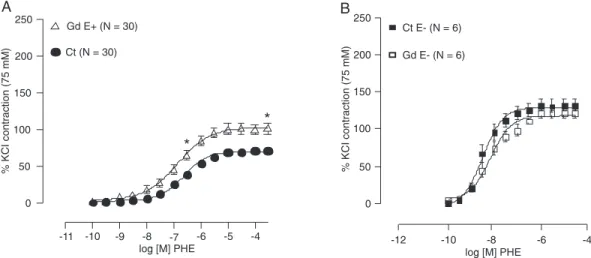
Figure 1 shows that after incubation with Gdfor 30 min, there was an increase in the contractile responses to PHE. The concentration-response curves show that Emax
(control: 72.3 ± 3.52; Gd: 101.3 ± 6.40%; P < 0.05) and the sensitivity (control: 6.6 ± 0.16; Gd: 10.5 ± 2.81%; P < 0.05) to PHE increased after incubation with Gd.
As the E-NTPDase and ecto-5’-nucleotidase cascade metabolizes ATP to adenosine, we tested the inhibitory ac-tion of Gd on these enzymes after the addiac-tion of 1 mM ATP to the tissue bath. Figure 2 illustrates the action of Gd on the ATP hydrolyzing activity of E-NTPDase. It can be seen that after incubation of intact rings (with endothelium) with Gd there was an increase in the vasorelaxation produced by ATP (control: -33.5 ± 4.10; Gd: -47.4 ± 4.12%), suggesting that less ATP hydrolysis occurred.
To investigate the endothelial dependence of the actions of Gd, the same protocol was repeated with endothelium-denuded aortic rings. Figure 1B shows that, after endothelial damage, the leftward displacement of the concentration-response curve to PHE was eliminated. No changes in Emax
(control: 132.6 ± 8.60; Gd: 122.4 ± 7.16%)or pD2 (control:
448 J.K. Angeli et al.
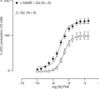
action of Gd is dependent on the presence of a functional endothelial layer, and the increased reactivity might result from the inhibition of vasorelaxation or from the stimulation of the release of a vasoconstrictor, we tested the putative role of NO using 100 µM L-NAME. Figure 3 shows %dAUC changes suggesting that Gd reduced the effects of L-NAME (control/L-NAME: 151.1 ± 28.8; L-NAME + Gd: 67.9 ± 19% AUC; Emax:
L-NAME: 143.6 ± 6.2; L-NAME + Gd: 142.9 ± 7.3%, or pD2:
L-NAME + Gd: 7.13 ± 0.1; L-NAME: 6.99 ± 0.23%). These observations could result from the blockade of
adenosine production, which is a final product of the action
of ectonucleotidases that exhibits vasodilator properties. Figure 4 shows that E-NTPDaseactivity was inhibited by 75
µM Gd (ATP hydrolysis) and by 100 µM Gd (both ATP and ADP hydrolysis) in aorta homogenates. Table 1 shows that, after ATP administration, Gd reduced ATP breakdown and
ADP generation, but the final concentration of adenosine
was similar to that in the control condition. To determine whether PHE could affect these results a similar protocol was used in the presence of PHE. Similar results were obtained (data not shown). According to these results, Gd might modulate extracellular ATP concentration but the increased vascular reactivity to PHE could not be explained by a reduction in adenosine production.
Since we knew that metals such as mercury at low concentration stimulate ACE activity (20) and Gd can take
Figure 1. A, Concentration-response curves to phenylephrine (PHE) in control (Ct) rat aortic rings and in rings incubated with gadolinium (Gd) for 30 min. An increased vascular reactivity was observed in rings with intact en-dothelium (E+) after Gd administration. An increase of the maximal response (Emax) and sensitivity to PHE also
occurred (*P ≤ 0.05). B, Concentration-response curves to PHE in control rings (Ct E-) and in rings with damaged endothelium (E-) incubated with Gd for 30 min (Gd E-). Results are reported as means ± SEM. The maximal response (Emax) and sensitivity to PHE did not change (P ≥ 0.05; Student t-test).
the place of zinc in the ACE molecule by transmetallation (7), we investigated whether Gd could produce a similar effect. Figure 5 shows that the action of Gd was blocked by both losartan, an AT1 receptor blocker (Emax: losartan: 63.6
± 11.3%; losartan + Gd: 67.6 ± 8.10%), and by enalaprilat (Emax: enalaprilat: 69.7 ± 6.24%; enalaprilat + Gd: 70.3 ±
8.15%), an ACE inhibitor. Also, the sensitivity (pD2) to PHE
was not altered suggesting that Gd stimulates ACE.
Figure 3. Concentration-response curves to phenylephrine (PHE) in control rat aortic rings (Ct), and in rings incubated for 30 min with 100 µM L-NAME (L-NAME; A) and with gadolinium + L-NAME (L-NAME + Gd; B). Panel C shows the difference in area under the concentration-response curve (dAUC) before and after L-NAME in control and Gd-incubated rings. *P ≤ 0.05 Ct/L-NAME vs L-NAME + Gd (Student t-test). Data are reported as means ± SEM.
Figure 4. Effect of gadolinium chloride (GdCl3) on ATP (A) and ADP (B) hydrolysis by aortic rings. Data are reported asmeans ± SD
450 J.K. Angeli et al.
Discussion
The results presented here show that Gd, in addition to blocking ATP and ADP hydrolysis, increasedthe vascular reactivity of rat aortic ringsto PHE by a mechanism involving the stimulation of angiotensin II AT1 receptors and reduced
NO bioavailability. Gd had no effect on the concentration-response curves to PHE when the endothelial layer was removed, suggesting dependence on the endothelium for the increase in vascular reactivity induced by Gd.
Gadolinium is a potent inhibitor of E-NTPDases at the concentration of 3 µM in the electric organ of Torpedo
(6). However, the role of E-NTPDases in the regulation of vascular tone is not completely understood. Since the enzymes that generate adenosine are expressed in the vessel wall, we performed experiments to investigate the
efficiency of Gd in blocking NTPDase in the rat aorta to
determine whether this inhibition could affect the vascular tone. Gd proved to be a potent inhibitor of NTPDase in the rat aorta. Sinceadenosine is a vasodilator molecule in several vascular beds (21), the blockade of its generation by inhibiting E-NTPDase activity should increase vascular reactivity.
To confirm if Gd interacts with ATP breakdown, we
used another protocol measuring the aortic ring relaxation by ATP before and after incubation with Gd. Rings were precontracted with PHE and ATP was added to the bath. An enhanced relaxation effect was observed after incubation with Gd. Assuming that ATP degradation was reduced after incubation with Gd, our results suggest that Gd blocked E-NTPDase activity at the endothelial level.
Indeed, our findings showed that, in the presence of
endothelium, incubation with Gd caused an upward dis-placement of the concentration-response curves to PHE.
The finding that Gd increased the contractile responses to
PHE supports the idea that the diminished generation of adenosine could participate in a mechanism to regulate vascular tone.
In order to test whether the increased reactivity to PHE induced by incubation with Gd could have been produced Figure 5. Concentration-response curves to phenylephrine (PHE)
before and after 3 µM gadolinium (Gd) in controls (Ct), after 10 µM enalaprilat (Panel A) or after 10 µM losartan incubation (Panel B) in aortic rings with intact endothelium. Data are reported as means ± SEM. *Emax P ≤ 0.05 - Gd vs Ct, enalaprilat and
losar-tan, enalaprilat + Gd and losartan + Gd (Student t-test).
Table 1. Effect of gadolinium (Gd) on purine metabolism in rat aortic rings.
ATP ADP AMP ADO INO
Control 0 8.50 ± 4.50 3.25 ± 1.00 1.75 ± 0.50 0
Gd 1.75 ± 0.25 4.25 ± 1.00 2.95 ± 0.73 1.50 ± 0.75 0
ATP 291.75 ± 53.75 20.25 ± 2.25 3.00 ± 0.50 1.25 ± 1.00 1.00 ± 0.75
ATP + Gd 666.25 ± 5.25* 4.00 ± 7.25* 3.50 ± 1.75 1.00 ± 0 2.50 ± 1.50
by reducing the release of endothelium-derived relaxing factor, we performed a pharmacological blockade of these pathways. According to our results, incubation with L-NAME in the presence of Gd potentiated the vasoconstrictor re-sponse to PHE, but to a lesser extent than in the control
group. These findings suggest that the effects of Gd were
dependent on the endothelium and NO.
The next protocol was performed to assess E-NTPDase activity in aortic homogenate preparations in the presence or absence of Gd. Several studies have reported the ability of Gd to decrease nucleotide hydrolysis (6,22,23). In the present study, we observed that Gd was capable of inhibit-ing both ATP and ADP hydrolysis. This result supports the idea that the effect of Gd regarding vascular reactivity was dependent on the inhibition of ATP breakdown, which in turn could reduce adenosine production.
To clarify this issue, we performed experiments to mea-sure ATP breakdown to ADP and adenosine. Surprisingly, as shown in Table 1, ADP formation was reduced by Gd, but adenosine concentration was unaffected. As seen in Table 1, ATP concentration increased after Gd administration, suggesting that Gd was an effective inhibitor of ATP hydro-lysis. The reduction of ATP breakdown explains our results showing that the vasodilator effect of ATP increased after incubation with Gd. However, Gd also inhibits permeable ATP channels in rat hepatocytes and HTC hepatoma cells (24), suggesting that ATP transfer through the sarcolemma could be affected, possibly resulting in the maintenance of higher extracellular ATP concentration. As expected, ADP formation was reduced by Gd. However, adenosine
concentration was unaffected. This finding ruled out the
possibility that Gd was increasing vascular reactivity by reducing adenosine generation.
Metals such as mercury, at low concentrations stimulate ACE activity (20) and Gd can take the place of zinc in the ACE molecule by transmetallation (7). We investigated whether Gd could produce a similar effect. ACE is a metal-lopeptidase that converts angiotensin I to angiotensin II, which has different biological actions and is considered to be the major effector of the renin-angiotensin system (25). Angiotensin II acts mainly through the AT1 receptor
that is expressed in many tissues, including adrenals, kidneys, heart, aorta, lungs, liver, testes, pituitary gland, and brain. When binding to the AT1 receptor, angiotensin II
promotes vasoconstriction and pressor effects, also
caus-ing thrombosis, inflammation and vascular and myocardial
hypertrophy (26).
We used losartan to block angiotensin II AT1 receptors
and enalaprilat to block ACE, and both pre-incubations blocked the effects of Gd on vascular reactivity. These results suggest that Gd was able to increase the produc-tion of angiotensin II by stimulating ACE. A previous report showed that Gd might affect ACE activity via a transmetal-lation effect with zinc (7) and we recently demonstrated that another metal, mercury, at low concentration, is capable of stimulating ACE (20). Moreover, studies have shown that at lower concentrations (6 nM and 0.5 to 10 mM), mercury induces vasoconstriction in rat caudal arteries (20,27). Part of these effects are mediated by the increase in the production of reactive oxygen species and of prostanoids
via cyclooxygenase (20,27). This finding clarified one of
the mechanisms of increased vascular reactivity produced by Gd, which depends on an increased production of an-giotensin II and an enhanced stimulation of AT1 receptors,
similar to the effects reported for low concentrations of mercury (20,27).
Potential limitations of the study
In the present study, we used aortic rings knowing that adenosine is more important for the regulation of tone in small arteries. However, other reports have shown that the aorta has adenosine receptors. We then decided that
for a first approach the use of aortic rings would be better
for us to evaluate the actions of Gd. Moreover, the main goal was to show that E-NTPDases could play a role in the control of vascular tone modulating NO release, and since NO plays an important role in the modulation of aortic tone
we chose aortic rings. However, according to our findings,
we believe that the results obtained in the present study are acceptable.
Our results show that Gd promoted an increase of vas-cular reactivity to PHE. They did not support the idea that the action of Gd depends on the reduction of adenosine production by the inhibition of E-NTPDase activity. However, the present results show that the in vitro exposure to 3 µM Gd induces endothelial dysfunction in aortic segments of rats since Gd increased the release of vasoconstrictors from the endothelium through ACE activation and reduced
bioavailability of NO. Based on these findings, we suggest
that Gd might have toxicological consequences and might be considered to be a health risk factor.
Acknowledgments
452 J.K. Angeli et al.
References
1. Caldwell RA, Clemo HF, Baumgarten CM. Using gadolinium to identify stretch-activated channels: technical consider-ations. Am J Physiol 1998; 275: C619-C621.
2. Nicolosi AC, Strande JL, Hsu A, Fu X, Su J, Gross GJ, et al. Gadolinium limits myocardial infarction in the rat: dose-response, temporal relations and mechanisms. J Mol Cell Cardiol 2008; 44: 345-351.
3. Klein C, Gebker R, Kokocinski T, Dreysse S, Schnackenburg B, Fleck E, et al. Combined magnetic resonance coronary artery imaging, myocardial perfusion and late gadolinium enhancement in patients with suspected coronary artery disease. J Cardiovasc Magn Reson 2008; 10: 45.
4. Perazella MA. Gadolinium-contrast toxicity in patients with kidney disease: nephrotoxicity and nephrogenic systemic fibrosis. Curr Drug Saf 2008; 3: 67-75.
5. Burnstock G. Pathophysiology and therapeutic potential of purinergic signaling. Pharmacol Rev 2006; 58: 58-86. 6. Escalada A, Navarro P, Ros E, Aleu J, Solsona C,
Martin-Satue M. Gadolinium inhibition of ecto-nucleoside triphos-phate diphosphohydrolase activity in Torpedo electric organ.
Neurochem Res 2004; 29: 1711-1714.
7. Corot C, Idee JM, Hentsch AM, Santus R, Mallet C, Goulas V, et al. Structure-activity relationship of macrocyclic and linear gadolinium chelates: investigation of transmetallation effect on the zinc-dependent metallopeptidase angiotensin-converting enzyme. J Magn Reson Imaging 1998; 8: 695-702.
8. Ralevic V, Burnstock G. Involvement of purinergic signaling in cardiovascular diseases. Drug News Perspect 2003; 16: 133-140.
9. Enjyoji K, Sevigny J, Lin Y, Frenette PS, Christie PD, Esch JS, et al. Targeted disruption of cd39/ATP diphosphohydro-lase results in disordered hemostasis and thromboregula-tion. Nat Med 1999; 5: 1010-1017.
10. Robson SC, Kaczmarek E, Siegel JB, Candinas D, Koziak K, Millan M, et al. Loss of ATP diphosphohydrolase activity with endothelial cell activation. J Exp Med 1997; 185: 153-163. 11. Kauffenstein G, Drouin A, Thorin-Trescases N, Bachelard
H, Robaye B, Orleans-Juste P, et al. NTPDase1 (CD39) controls nucleotide-dependent vasoconstriction in mouse.
Cardiovasc Res 2010; 85: 204-213.
12. Kauffenstein G, Furstenau CR, Orleans-Juste P, Sevigny J. The ecto-nucleotidase NTPDase1 differentially regulates P2Y1 and P2Y2 receptor-dependent vasorelaxation. Br J Pharmacol 2010; 159: 576-585.
13. Sevigny J, Sundberg C, Braun N, Guckelberger O, Csiz-madia E, Qawi I, et al. Differential catalytic properties and vascular topography of murine nucleoside triphosphate diphosphohydrolase 1 (NTPDase1) and NTPDase2 have implications for thromboregulation. Blood 2002; 99: 2801-2809.
14. Frassetto SS, Schetinger MR, Schierholt R, Webber A, Bo-nan CD, Wyse AT, et al. Brain ischemia alters platelet ATP
diphosphohydrolase and 5’-nucleotidase activities in naive and preconditioned rats. Braz J Med Biol Res 2000; 33: 1369-1377.
15. Levitsky DO, Clergue M, Lambert F, Souponitskaya MV, Le Jemtel TH, Lecarpentier Y, et al. Sarcoplasmic reticulum calcium transport and Ca2+-ATPase gene expression in
thoracic and abdominal aortas of normotensive and spon-taneously hypertensive rats. J Biol Chem 1993; 268: 8325-8331.
16. Bradford MM. A rapid and sensitive method for the quantita-tion of microgram quantities of protein utilizing the principle of protein-dye binding. Anal Biochem 1976; 72: 248-254. 17. Bohmer AE, Pochmann D, Sarkis JJ. In vitro effect of
homo-cysteine on nucleotide hydrolysis by blood serum from adult rats. Chem Biol Interact 2006; 160: 159-164.
18. Chan KM, Delfert D, Junger KD. A direct colorimetric assay for Ca2+-stimulated ATPase activity. Anal Biochem 1986;
157: 375-380.
19. Voelter W, Zech K, Arnold P, Ludwig G. Determination of selected pyrimidines, purines and their metabolites in serum and urine by reversed-phase ion-pair chromatography. J Chromatogr 1980; 199: 345-354.
20. Wiggers GA, Stefanon I, Padilha AS, Pecanha FM, Vassallo DV, Oliveira EM. Low nanomolar concentration of mercury chloride increases vascular reactivity to phenylephrine and local angiotensin production in rats. Comp Biochem Physiol C Toxicol Pharmacol 2008; 147: 252-260.
21. Shryock JC, Belardinelli L. Adenosine and adenosine recep-tors in the cardiovascular system: biochemistry, physiology, and pharmacology. Am J Cardiol 1997; 79: 2-10.
22. Buffon A, Ribeiro VB, Wink MR, Casali EA, Sarkis JJ. Nucleotide metabolizing ecto-enzymes in Walker 256 tumor cells: molecular identification, kinetic characterization and biochemical properties. Life Sci 2007; 80: 950-958. 23. Rucker B, Almeida ME, Libermann TA, Zerbini LF, Wink MR,
Sarkis JJ. E-NTPDases and ecto-5’-nucleotidase expres-sion profile in rat heart left ventricle and the extracellular nucleotide hydrolysis by their nerve terminal endings. Life Sci 2008; 82: 477-486.
24. Roman RM, Feranchak AP, Davison AK, Schwiebert EM, Fitz JG. Evidence for Gd3+ inhibition of membrane ATP
permeability and purinergic signaling. Am J Physiol 1999; 277: G1222-G1230.
25. Kifor I, Dzau VJ. Endothelial renin-angiotensin pathway: evidence for intracellular synthesis and secretion of angio-tensins. Circ Res 1987; 60: 422-428.
26. Touyz RM, Berry C. Recent advances in angiotensin II sig-naling. Braz J Med Biol Res 2002; 35: 1001-1015.