Radiotherapy modulates expression of EGFR,
ERCC1 and p53 in cervical cancer
V.H. de Almeida
1,2, A.C. de Melo
1, D.D. Meira
3, A.C. Pires
4, A. Nogueira-Rodrigues
1,
H.K. Pimenta-Inada
1, F.G. Alves
1, G. Moralez
1, L.S. Thiago
1, C.G. Ferreira
1and C. Sternberg
11Divisão de Pesquisa Clínica e Desenvolvimento Tecnológico, Instituto Nacional de Câncer, Rio de Janeiro, RJ, Brasil 2Instituto de Bioquímica Médica Leopoldo De Meis, Universidade Federal do Rio de Janeiro, Rio de Janeiro, RJ, Brasil 3Departamento de Ciências Biológicas, Universidade Federal do Espírito Santo, Vitória, ES, Brasil 4Fonte Medicina Diagnóstica, Niterói, RJ, Brasil
Abstract
Cervical cancer is a public health problem and the molecular mechanisms underlying radioresistance are still poorly understood. Here, we evaluated the modulation of key molecules involved in cell proliferation, cell cycle and DNA repair in cervical cancer cell lines (CASKI and C33A) and in malignant tissues biopsied from 10 patients before and after radiotherapy. The expres-sion patterns of epidermal growth factor receptor (EGFR), exciexpres-sion repair cross-complementation group 1 (ERCC1) and p53 were evaluated in cancer cell lines by quantitative PCR and western blotting, and in human malignant tissues by immuno-histochemistry. The mutation status ofTP53 gene was evaluated by direct sequencing. Among cell lines, absent or weak modulations of EGFR, ERCC1 and p53 were observed after exposure to 1.8 Gy. Conversely, increased expressions of p53 (5/10 patients; P=0.0239), ERCC1 (5/10 patients; P=0.0294) and EGFR (4/10 patients; P=0.1773) were observed in malignant tissues after radiotherapy with the same radiation dose.TP53mutations were found only in one patient. Here we show that a single dose of radiotherapy induced EGFR, ERCC1 and p53 expression in malignant tissues from cervical cancer patients but not in cancer cell lines, highlighting the gap betweenin vitroandin vivoexperimental models. Studies on larger patient cohorts are needed to allow an interpretation that an upregulation of p53, EGFR and ERCC1 may be part of a radioresistance mechanism.
Key words: Cervical cancer; Radiotherapy; Epidermal growth factor receptor (EGFR); p53; Excision repair cross-complementation group 1 (ERCC1)
Introduction
Cervical cancer is a public health problem represent-ing the fourth most common cancer in women worldwide, with more than 500,000 new cases per year (1). The combined treatment, involving cisplatin-based chemo-therapy and radiochemo-therapy, has been established as the standard therapeutic approach for patients with locally advanced disease. Radiotherapy alone has also been used for patients with early disease (2). However, patients with stage III and IVA tumors have 5-year survival rates lower than 50% (3), and novel strategies to improve the prognosis of these patients are needed. An increased understanding of the molecular mechanisms leading to radioresistance may offer novel radiosensitizing strategies.
Virtually all cervical cancers (99% or more) are caused by high-risk human papillomavirus (HPV) (4). E6 and E7 HPV oncoproteins have been known to play a major role
in malignant transformation of epithelial cells, mainly by inhibiting p53 and Rb tumor suppressor proteins. On the other hand, the role of the viral E5 protein remains poorly understood. Some studies indicate that E5 may increase epidermal growth factor receptor (EGFR) recycling to the cell surface and enhance growth factor signal transduc-tion (5). Indeed, previous reports have shown EGFR to be frequently expressed in cervical cancer (around 80%) and correlated with disease progression (6,7). EGFR belongs to the HER tyrosine kinase receptor family and EGFR activation leads to dimerization, autophosphorylation and activation of downstream signaling pathways which regu-late cell proliferation, survival and transformation (8).
The most critical DNA lesion induced by radiation is DNA double-strand breaks. In mammalian cells, these lesions are predominantly repaired by non-homologous
Correspondence: C. Sternberg:<[email protected]>
Present addresses of C.G. Ferreira: Instituto D’Or de Pesquisa e Ensino, Rio de Janeiro, RJ, Brasil, and C. Sternberg: Sociedade Brasileira de Oncologia Clínica, Belo Horizonte, MG, Brasil.
end joining (NHEJ), which protein machinery is mainly composed by DNA-dependent protein kinase (DNA-PK), Ku-70 and Ku-80 (9). Beyond a critical role in DNA repair, activated DNA-PK also phosphorylates proteins involved in cell cycle checkpoints and cell death pathway, such as p53 (10). However, in most cervical cancers, inactivation of p53 is mainly caused by HPV infection (11). Recently, excision repair cross-complementation group 1 (ERCC1) protein also was described to facilitate DNA repair of double-strand breaks induced by radiotherapy (12).
Genotoxic stress can induce EGFR autophosphoryla-tion, activating signaling pathways that promote prolifera-tion and survival, but EGFR can also modulate DNA damage response by interacting with components of the DNA repair machinery (13). Ionizing radiation induces EGFR internalization to nucleus, association of EGFR with DNA-PK, enhancing kinase activity of the DNA-PK complex and increasing the repair of DNA strand breaks (14). In addition, EGFR can also modulate positively ERCC1 expression in prostate cancer cell lines (15) and physically interact with ERCC1 (16), suggesting a new role for EGFR in DNA repair independent of DNA-PK. Indeed, combination of radiotherapy and EGFR inhibitors can improve local tumor control compared to irradiation alone and this strategy has been introduced into clinical radio-therapy practice (17). Preclinical data from our group showed that cetuximab, a monoclonal antibody anti-EGFR, can sensitize gynecological cancer cell lines to chemoradiation (18).
In the current study, we have evaluated the radiation-induced modulation of EGFR, ERCC1 and p53 in cervical cancer cell lines and in malignant tissues from 10 patients diagnosed with cervical cancer.
Material and Methods
Cell lines and irradiation
CASKI and C33A cells were cultured in RPMI 1640 medium (Thermo Fisher Scientific, USA) supplemented with 10% fetal bovine serum and incubated in a humidified atmosphere at 37°C in 5% CO2. Cell line authentication
via STR profiling was performed to confirm their identity. In addition, cells were tested periodically for mycoplasma contamination by Mycosensor PCR assay (Agilent, USA). For radiotherapy, cells were exposed tog-rays from a60Co source (Theratron 780C; Theratronics International Limited, Canada) at a dose rate of 2.0 Gy/min.
Clonogenic assay
In order to establish the radiosensitivity profile of each cell line, cells were seeded onto 12-well plates at a con-centration of 150 cells per well. After 24 h, cells were irradiated (2, 4, and 6 Gy) and incubated for 72 h. Cells were then washed with PBS and allowed to proliferate in fresh medium for 10 more days. Colonies were stained with 0.1% crystal violet.
Western blot analysis
Cells were incubated in serum-free medium for 4 h followed by irradiation (1.8 Gy). After 24h, cells were lysed and 50 mg of proteins from each sample were run on a 6–12% SDS–PAGE gel and transferred to a PVDF
Hybond-P (GE Healthcare, Brazil) membrane. Membranes were incubated with antibodies against EGFR (1:500; #2232, Cell Signaling Technology, USA), ERCC1 (1:100; 8F1 clone, Thermo Fisher Scientific, USA), p53 (1:500; G59-12 clone, BD Pharmingen, USA) and HSC70 (heat shock cognate 70 protein; 1:5000; Santa Cruz Biotechnology, USA). After incubation with secondary antibodies, immunoblots were detected using the ECL reagent (GE Healthcare, Brazil). Predicted molecular weights for EGFR, ERCC1, p53, and HSC70 are 170, 39, 53, and 70 kDa, respectively.
Gene expression analysis
Cell lines were incubated in serum-free medium for 4 h followed by irradiation (1.8 Gy). After 24 h of treatment, total RNA was extracted using TRIzol Reagent (Thermo Fisher Scientific). From each sample, 1.0mg of total RNA was reverse transcribed to cDNA. Then, quantitative PCR was performed on the aliquots of the cDNA using Taqman Fast Real-Time PCR Master Mix (Thermo Fisher Scientific). The gene expression profile was evaluated using the ABI PRISM 7500 Fast Real-Time PCR System (Thermo Fisher Scientific). The Taqman gene expression assay references are Hs01076078_m1 (EGFR), Hs00157415_
m1 (ERCC1), Hs00153349_m1 (TP53) and 4326317E
(GAPDH). The 2 DDCTmethod was utilized to analyze
the fold increase.
Patient eligibility
Ten patients diagnosed with stage IIB–IVA cervical
squamous cell carcinoma, age 18–70 years, and clinical
indi-cation for radiotherapy were enrolled. All patients provided written informed consent before participation. The study was approved by the Institutional Review Board (CEP-INCA) under registry 143/09 and followed the Helsinki Declaration.
Clinical study design
A prospective trial included locally advanced cervical squamous cell carcinoma patients. Briefly, all 10 patients were treated with teletherapy - 45Gy divided into 25 fractions, which means that each fraction is equivalent to 1.8 Gy. This dose was the same used in cell culture experiments pre-viously described. A tumor biopsy was performed before treatment and a second biopsy was taken 24 h after thefirst radiotherapy fraction. Cisplatin, when indicated, was pre-scribed afterwards. The expression pattern of EGFR, ERCC1, and p53 was compared between malignant tissues before treatment and after thefirst teletherapy with 1.8 Gy dose.
Tissue microarray (TMA) and immunohistochemistry
in 10% formalin and embedded in paraffin. Serial TMA sections were cut, mounted on glass slides and dried at 56° C before dewaxing in xylene and rehydration in alcohol. All sections were subjected to heat-induced epitope retrieval in citrate buffer followed by inhibition of endogenous peroxidase (peroxidase block, RE7101, Novocastra, UK). Incubation of primary antibodies against EGFR (D38B1, 1:25, Cell Signaling Technology), ERCC1 (1:100; 8F1 clone, Thermo Fisher Scientific), or p53 (1:100; G59-12 clone, BD Pharmingen) was performed for 1 h. Slides were incubated with NovoLinktpolymer (RE7112, Novo-castra), further developed with diaminobenzidine chromo-gen (RE7105, Novocastra), andfinally stained with Mayer hematoxylin, dehydrated and mounted with Canadian balsam. EGFR, ERCC1 and p53 expressions were eval-uated semi-quantitatively as positive cells after counting 300–500 tumor cells, being scored as negative (no staining),
1+(o25% of positive cells), 2+(25–75% of positive cells)
or 3+ (475% cells staining positively). Negative controls were obtained by omitting the primary antibody.
TP53 mutation status
Only samples with at least 70% of malignant cells were sent to DNA extraction. Genomic DNA was extracted from paraffin-embedded tissues with NucleoSpin FFPE DNA kit (Macherey-Nagel, Germany) following the manufacturer’s instructions. TP53 exons 5-9 were amplified by PCR using
primers and cycling parameters according to International Agency for Research on Cancer (IARC) protocol (20). The PCR products were then purified with Illustra GFX PCR DNA and gel Band Purification kit (GE Healthcare, Brazil) and directly sequenced in both directions in the ABI 3130XL DNA sequence analyzer (Thermo Fisher Scientific) using the same primers and Big-Dye 3.1 Terminator cycle sequencing chemistry (Thermo Fisher Scientific). The elec-tropherograms were visualized manually on the program Sequencher 4.7 (Gene Codes Corporation, USA).
Statistical analysis
Quantitative experiments comparing two conditions in cell culture were analyzed by unpaired Student’s t-test. Immunohistochemistry comparing score intensity between EGFR, p53 and ERCC1 staining before and after radio-therapy was analyzed by paired Student’st-test. All sta-tistical analyses were performed using GraphPad Prism Software (USA) and the P values were considered significant wheno0.05.
Results
We evaluated the radiosensitivity of two cervical cancer cell lines (C33A and CASKI) by clonogenic assay after increasing doses of radiation (2, 4, and 6 Gy). As shown in Figure 1A, C33A was highly sensitive to
Figure 1. Radiosensitivity profile of cervical cancer cell lines.A, Cells were irradiated (2, 4, and 6 Gy) and incubated for 72 h. Then, cells were washed and allowed to proliferate in fresh medium for 10 more days. The number of colony-forming units in treated cultures is reported as the surviving fraction. *Po0.05 (two-tailed unpaired t-test). Representative images of CASKI cells (B) and C33A
radiotherapy while CASKI exhibited a radioresistance profile. Representative pictures of colonies of CASKI (Figure 1B) and C33A cells (Figure 1C) exposed to increasing doses of radiotherapy show the dose-response effect.
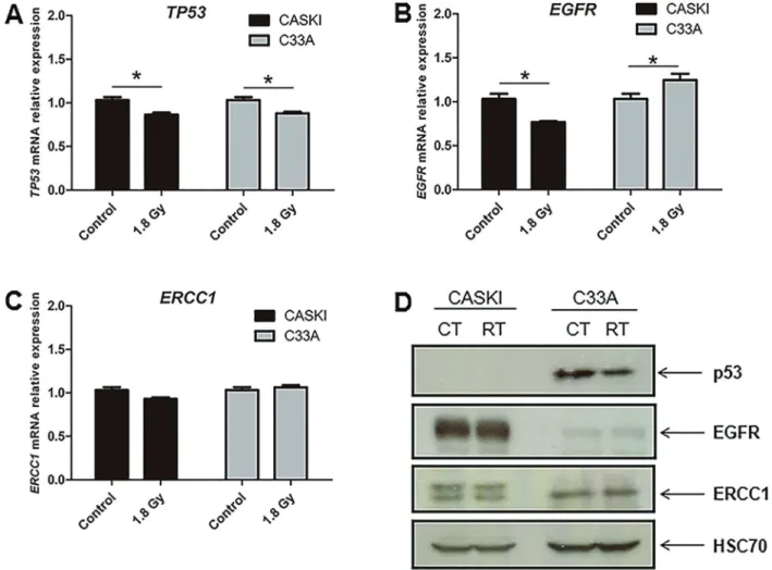
Since the dose of each radiation therapy session was 1.8 Gy for each individual patient enrolled in the clinical study, we treated each cell line with the same radiation dose in order to study the expression pattern and compare with the clinical study findings. Overall, absent or weak modulation of p53 (Figure 2A), EGFR (Figure 2B) and ERCC1 (Figure 2C) were observed in cancer cell lines herein investigated at mRNA level. Since the highest radiation-induced variations observed at mRNA levels do not reach 30% and considering that no difference was observed at protein levels (Figure 2D), these slight
mRNA variations may have no functional impact. There-fore, independently of the resistance/sensitivity status to radiation therapy, none of the studied cell lines showed important variations at EGFR, ERCC1, and p53 expres-sion profile induced by radiation. It is important to note that the HPV-16 positive CASKI cell line (21) does not present p53 expression (Figure 2D). Finally, although C33A cell line does not exhibit HPV infection (21), it presentsTP53
gene mutation (22).
The exploratory clinical study herein conducted was intended to confirm and validate thein vitrofindings. Ten patients, with a median age of 47.5 years (range: 33–66),
diagnosed with cervical squamous cell carcinoma were prospectively evaluated in this study. Immunohistochem-ical analysis of tumor samples before radiation showed
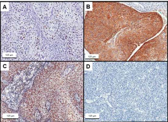
positive EGFR staining in 9/10 cases (90%), positive ERCC1 staining in 5/10 cases (50%) and positive p53 staining in 5/10 cases (50%; Table 1). Representative microphotographs of immunohistochemistry for p53 (A), EGFR (B), ERCC1 (C), and an unstained sample (D) are depicted in Figure 3.
Our immunohistochemical analysis on tissue micro-arrays revealed that malignant tissues from 7 of 10 patients exhibited a radiation-induced phenotypic change (Figure 4A). After treatment with 1.8 Gy, an increased expression of p53 as well as of ERCC1 was found in half of the patients while EGFR was positively modulated in
Table 1.Scoring of p53, EGFR and ERCC1 immunohistochemistry in malignant tissues from cervical cancer patients.
Patients Before radiotherapy After radiotherapy
p53 EGFR ERCC1 p53 EGFR ERCC1
1 – 2+ – 1+ 3+ 2+
2 – – – 2+ 3+ 1+
3 1+ 3+ – 1+ 2+ –
4 1+ 2+ 1+ 1+ 3+ 2+
5 – 2+ 3+ 1+ 3+ 3+
6 – 2+ – – 2+ –
7 – 3+ 1+ 1+ 3+ 3+
8 2+ 3+ 3+ 2+ 3+ 3+
9 1+ 3+ – 2+ 3+ 3+
10 1+ 3+ 3+ 1+ 3+ 3+
The dose of radiotherapy was 1.8 Gy. Negative score indicates absence of expression in cancer cells; 1+:o25% of positive tumor cells; 2+: 25–75% of
positive tumor cells, and 3+:475% of positive tumor cells.
4 of 10 patients (Table 1, Figure 4A). The simultaneous induction of p53 and EGFR protein expression occurred in 3 of 10 patients, while EGFR and ERCC1 was found in 3 of 10 patients; p53 and ERCC1 induction was detected in 4 of 10 patients, while all 3 proteins (EGFR, p53 and ERCC1) were jointly stimulated in 2 out of 10 individuals (Figure 4A). The medium score of p53 staining increased from 0.6 before radiotherapy to 1.2 after radiation (P= 0.0239, Figure 4B). Although radiation therapy upregu-lates EGFR expression in 4 tumors, the median score of immunohistochemistry staining raised from 2.3 to 2.8, with no statistical significance (P=0.1773, Figure 4C). Prob-ably, this occurred because half of the tumors exhibited
EGFR staining with maximum intensity even before radiation therapy. The medium score of ERCC1 staining increased from 1.1 before treatment to 2.0 after radio-therapy (P=0.0294, Figure 4D).
Since mutations may prevent the production of a functional protein, we have investigated possible muta-tions on exons 5-9 ofTP53gene through direct sequenc-ing in malignant tissue samples. No mutations were found in tumor samples except for patient #6 (Table 2) who showed two missense mutations (P142S and H179Y). Patient #6 was also one of the few who failed to exhibit ERCC1 and EGFR protein modulation after radiation therapy in tumor tissues. This is notable due
to the fact that, sinceTP53gene is not mutated in 9 of 10 cervical tumors, it is not unlikely that p53 protein would display a regulatory role at several genes after radiation therapy, including EGFR and/or ERCC1.
Discussion
Locally advanced cervical cancer treatment has been based on cisplatin and radiation therapy since 1999 (2), and after almost 20 years there are no major advances on the treatment of this type of cancer. The failure to respond to radiotherapy is a major concern in cervical cancer patients. To this end, studies aiming to detect the molec-ular mechanisms underlying radioresistance are on high demand. Identification of molecular pathways implicated in the adaptive response of tumor cells to radiation may allow the prediction of treatment outcome and enhance cancer cell killing through employment of selective inhibitors for these pathways.
In the present study, we sought to investigate cell survival and DNA repair proteins potentially implicated in the modulation of radioresistance using both in vitro
and clinical studies. Indeed, it is well known that several
findings from in vitro experiments with immortalized cell lines are not confirmed in well-conductedin vivostudies. Herein, we observed that although classical cervical cancer cell lines (one radioresistant and one radiosensitive) did not shown any radiation-induced modulation of EGFR, p53, or ERCC1, almost all malignant tissues obtained from cervical cancer patients exhibited a radiation-induced phenotypic change of at least one of these proteins.
The p53 protein expression is not generally observed in tumor cells infected by HPV, as E6 viral oncoprotein causes p53 inactivation by promoting its degradation (11). Curiously, p53 expression was detected at pre-radiotherapy
biopsies of cervical cancer. Moreover, there was p53 induction after radiation therapy in 50% of cases. This result suggests that p53 may not be completely inactivated in HPV infected tumor cells. Besides cyto-toxic therapies that cause DNA damage, other condi-tions are capable to induce p53 expression, such as hypoxia (23). It has been demonstrated that hypoxia is a common feature in solid tumors, including cervical cancer (24). Hypoxia is capable to induce p53 expres-sion even in cells infected by HPV through decreasing MDM2 expression (a negative endogenous regulator of p53) and uncoupling the interaction of p53 with the E6 viral protein (25). To date, other groups have already shown p53 expression in cervical tumors by immuno-histochemistry (26–28).
These studies, along with ours, suggest that p53 should not be simply considered not expressed in cervical cancer cells. However, the interpretation of p53 induction and its impact in cellular response is not easy to extrapolate. As revised by Ferreira and colleagues (23), the presence of functional p53 could both sensitize or promote resistance in tumor cells exposed to genotoxic therapy, depending on the model utilized and experimental conditions used.
An important regulation between p53 and EGFR was reported previously. Wild type p53 is capable to induce the expression of heparin-binding EGF-like growth factor (HB-EGF), a known EGFR ligand (29). Moreover, a respon-sive site to wild type p53 was identified at the proximal promoter of the EGFR gene (30). These mechanisms reveal a new aspect of p53 function operating beyond apoptosis induction and tumor suppression, what could explain the positive concomitant modulation of p53 and EGFR in 30% of tumors.
EGFR is commonly expressed in normal cells of the basal layer of epithelium but overexpression in tumor cells
Table 2.TP53gene mutation status.
Patients Exon 5 Exon 6 Exon 7 Exon 8 Exon 9
1 WT WT WT WT WT
2 WT WT WT WT WT
3 WT WT WT WT WT
4 WT WT WT WT WT
5 – WT WT WT WT
6 Mutant WT WT WT WT
7 WT WT WT WT WT
8 WT WT WT WT WT
9 WT WT WT WT –
10 WT WT WT – –
is closely associated with reduced survival in patients with cervical cancer (6). It is estimated that 90% of cervical carcinomas overexpress EGFR (7), the same magnitude observed here. Furthermore, EGFR has been validated by our group as an important therapeutic target in cervical cancer in preclinical model (18) and in a phase II clinical trial (31). In addition to its physical interaction with components of DNA repair machinery (14,16), EGFR also up-regulates theERCC1gene expression through MAPK signaling pathway (15). This mechanism may explain concurrent induction of EGFR and ERCC1 in 30% of tumor tissues in our study.
ERCC1 is a key protein for DNA repair, especially for nucleotide excision repair (NER). This mechanism is associated with DNA damage repair caused by chemical adducts. In lung cancer, high ERCC1 expression is correlated to platinum chemotherapy resistance (32). However, the ERCC1 role in radioresistance and in repair of DNA damage caused by radiation is recent. Kawashima and colleagues (33) described that ERCC1 silencing promoted sensitization to radiotherapy in bladder cancer. Ahmad and colleagues (12) demonstrated a direct role of ERCC1 in repair of DNA double strand break, the major damage caused by radiation therapy, using in vitro and
in vivoERCC1 knockout models. Therefore, the regulatory
axis p53-EGFR-ERCC1 may be activated in tumor cells exposed to radiationin vivo.
Finally, immortalized cell lines have been extensively used to understand the fundamental cancer cell biology mechanisms. Despite its huge contribution to the under-standing of cancer biology, many in vitro findings fail to reproduce the complex cellular and molecular interactions taking place in individual patients. Our prospective investigational clinical study allowed us to identify potential radioresistance biomarkers that were not modu-lated in cervical cancer cell lines, highlighting the gap between in vitro and in vivo experimental models. However, the findings reported in this study need to be confirmed in a larger patient cohort to draw a definitive conclusion on the role of the p53, EGFR, and ERCC1 proteins in radioresistance of cervical carcinoma.
Acknowledgments
This work was supported by grants from Conselho Nacional de Desenvolvimento Científico e Tecnológico (CNPq, MCTI, Brazil), Fundac¸ão do Câncer (Brazil), and
Ministério da Saúde (MS, Brazil).
References
1. Torre LA, Siegel RL, Ward EM, Jemal A. Global cancer incidence and mortality rates and trends - an update.Cancer Epidemiol Biomarkers Prev2016; 25: 16–27, doi: 10.1158/ 1055-9965.EPI-15-0578.
2. Rose PG, Bundy BN, Watkins EB, Thiqpen JT, Deppe G, Maiman MA, et al. Concurrent cisplatin-based radiotherapy and chemotherapy for locally advanced cervical cancer.N Engl J Med 1999; 340: 1144–1153, doi: 10.1056/NEJM199904153401502. 3. Tierney JF, Vale C, Symonds P. Concomitant and
neoadju-vant chemotherapy for cervical cancer.Clin Oncol2008; 20: 401–416, doi: 10.1016/j.clon.2008.04.003.
4. Bosch FX, Lorincz A, Muñoz N, Meijer CJ, Shah KV. The causal relation between human papillomavirus and cervical cancer. J Clin Pathol 2002; 55: 244–265, doi: 10.1136/ jcp.55.4.244.
5. Kim MK, Kim HS, Kim SH, Oh JM, Han JY, Lim JM, et al. Human papillomavirus type 16 E5 oncoprotein as a new target for cervical cancer treatment. Biochem Pharmacol 2010; 80: 1930–1935, doi: 10.1016/j.bcp.2010.07.013. 6. Tian WJ, Huang ML, Qin QF, Chen Q, Fang K, Wang
PL. Prognostic impact of epidermal growth factor receptor overexpression in patients with cervical cancer: a meta-analysis. PLoS One 2016; 11: e0158787, doi: 10.1371/ journal.pone.0158787.
7. Cerciello F, Riesterer O, Sherweif M, Odermatt B, Ciernik IF. Is EGFR a moving target during radiotherapy of carcinoma of the uterine cervix?Gynecol Oncol2007; 106: 394–399, doi: 10.1016/j.ygyno.2007.04.019.
8. Citri A, Yarden Y. EGF-ERBB signalling: towards the systems level. Nat Rev Mol Cell Biol 2006; 7: 505–516, doi: 10.1038/nrm1962.
9. Mahaney BL, Meek K, Lees-Miller SP. Repair of ioniz-ing radiation-induced DNA double strand breaks by non-homologous end-joining. Biochem J2009; 417: 639–650, doi: 10.1042/BJ20080413.
10. Lees-Miller SP, Sakaguchi K, Ullrich SJ, Appella E, Anderson CW. Human DNA-activated protein kinase phos-phorylates serines 15 and 37 in the amino-terminal trans-activation domain of human p53. Mol Cell Biol 1992; 12: 5041–5049, doi: 10.1128/MCB.12.11.5041.
11. Scheffner M, Werness BA, Huibregtse JM, Levine AJ, Howley PM. The E6 oncoprotein encoded by human papillo-mavirus types 16 and 18 promotes the degradation of p53.Cell1990; 63: 1129–1136, doi: 10.1016/0092-8674(90) 90409-8.
12. Ahmad A, Robinson AR, Duensing A, van Drunen E, Beverloo HB, Weisberg DB, et al. ERCC1-XPF endonu-clease facilitates DNA double-strand break repair.Mol Cell Biol2008; 28: 5082–5092, doi: 10.1128/MCB.00293-08. 13. Chen DJ, Nirodi CS. The epidermal growth factor receptor: a
role in repair of radiation-induced DNA damage.Clin Cancer Res 2007; 13: 6555–6560, doi: 10.1158/1078-0432.CCR-07-1610.
14. Dittmann K, Mayer C, Fehrenbacher B, Schaller M, Raju U, Milas L, et al. Radiation-induced epidermal growth factor receptor nuclear import is linked to activation of DNA-dependent protein kinase.J Biol Chem2005; 280: 31182– 31189, doi: 10.1074/jbc.M506591200.
signaling. Radiat Res 2003; 159: 439–452, doi: 10.1667/ 0033-7587(2003)159[0439:EGFAIR]2.0.CO;2.
16. Liccardi G, Hartley JA, Hochhauser D. Importance of EGFR/ ERCC1 interaction following radiation-induced DNA damage. Clin Cancer Res2014; 20: 3496–3506, doi: 10.1158/1078-0432.CCR-13-2695.
17. Sartor CI. Mechanisms of disease: Radiosensitization by epidermal growth factor receptor inhibitors.Nat Clin Pract Oncol2004; 1: 80–87, doi: 10.1038/ncponc0048.
18. Meira DD, de Almeida VH, Mororó JS, Nóbrega I, Bardella L, Silva RL, et al. Combination of cetuximab with chemoradia-tion, trastuzumab or MAPK inhibitors: mechanisms of sensitisation of cervical cancer cells. Br J Cancer 2009; 101: 782–791, doi: 10.1038/sj.bjc.6605216.
19. Pires ARC, Andreiuolo F da M, de Souza SR. TMA for all: a new method for the construction of tissue microarrays without recipient paraffin block using custom-built needles. Diagn Pathol2006; 1: 14, doi: 10.1186/1746-1596-1-14. 20. International Agency for Research on Cancer, Detection of
TP53 mutations by direct sequencing (IARC protocol, 2010 update) http://p53.iarc.fr/Download/TP53_DirectSequencing_ IARC.pdf. Accessed December 16, 2016.
21. Kessis TD, Slebos RJ, Nelson WG, Kastan MB, Plunkett BS, Han SM, et al. Human papillomavirus 16 E6 expres-sion disrupts the p53-mediated cellular response to DNA damage. Proc Natl Acad Sci USA 1993; 90: 3988-3992, doi: 10.1073/pnas.90.9.3988.
22. Muller PA, Vousden KH. Mutant p53 in cancer: new functions and therapeutic opportunities.Cancer Cell2014; 25: 304–317, doi: 10.1016/j.ccr.2014.01.021.
23. Ferreira CG, Tolis C, Giaccone G. p53 and chemosensitivity. Ann Oncol 1999; 10: 1011–1021, doi: 10.1023/A:100836 1818480.
24. Fyles A, Milosevic M, Hedley D, Pintilie M, Levin W, Manchul L, et al. Tumor hypoxia has independent predictor impact only in patients with node-negative cervix cancer. J Clin Oncol2002; 20: 680–687, doi: 10.1200/JCO.2002.20.3.680. 25. Alarcón R, Koumenis C, Geyer RK, Maki CG, Giaccia AJ. Hypoxia Induces p53 Accumulation through MDM2
Down-Regulation and Inhibition of E6-mediated Degrada-tion.Cancer Res1999; 59: 6046–6051.
26. Haensgen G, Krause U, Becker A, Stadler P, Lauten-schlaeger C, Wohlrab W, et al. Tumor hypoxia, p53, and prognosis in cervical cancers.Int J Radiat Oncol Biol Phys 2001; 50: 865–872, doi: 10.1016/S0360-3016(01)01523-1. 27. Portari EA, Russomano FB, de Camargo MJ, Machado Gayer
CR, da Rocha Guillobel HC, Santos-Rebouc¸as CB, et al.
Immunohistochemical expression of Cyclin D1, p16Ink4a, p21WAF1, and Ki-67 correlates with the severity of cervical neoplasia. Int J Gynecol Pathol 2013; 32: 501–508, doi: 10.1097/PGP.0b013e31826f5cf6.
28. Shukla S, Dass J, Pujani M. p53 and bcl2 expression in malignant and premalignant lesions of uterine cervix and their correlation with human papilloma virus 16 and 18. South Asian J Cancer 2014; 3: 48–53, doi: 10.4103/ 2278-330X.126524.
29. Fang L, Li G, Liu G, Lee SW, Aaronson SA. p53 induction of heparin-binding EGF-like growth factor counteracts p53 growth suppression through activation of MAPK and PI3K/ Akt signaling cascades.EMBO J2001; 20: 1931–1939, doi: 10.1093/emboj/20.8.1931.
30. Sheikh MS, Carrier F, Johnson AC, Ogdon SE, Fornace AJ Jr. Identification of an additional p53-responsive site in the human epidermal growth factor receptor gene promotor.Oncogene 1997; 15: 1095–1101, doi: 10.1038/sj.onc.1201264.
31. Nogueira-Rodrigues A, Moralez G, Grazziotin R, Carmo CC, Small IA, Alves FG, et al. Phase 2 trial of erlotinib com-bined with cisplatin and radiotherapy in patients with locally advanced cervical cancer. Cancer2014; 120: 1187–1193, doi: 10.1002/cncr.28471.
32. Rosell R, Mendez P, Isla D, Taron M. Platinum Resistance Related to a Functional NER Pathway.J Thorac Oncol2007; 2: 1063–1066, doi: 10.1097/JTO.0b013e31815ba2a1. 33. Kawashima A, Nakayama M, Kakuta Y, Abe T, Hatano K,