9
SUMMARY
BACKGROUND AND OBJECTIVES: Assessing
patients´ pain complaints is essential for determining adequate diagnosis and therapeutic interventions in oro-facial pain (OFP). Thus, the aim of this study was to ver-ify the frequency of reported pain complaints compared to those marked on patients´ body pain maps.
METHOD: Data were collected from the Orofacial Pain Clinic archives (532 patients) of the Orofacial Pain Clin-ic, Araraquara Dental School. All individuals answered a questionnaire to report their pain complaints and com-pleted a body map indicating their pain areas.The fre-quency of reported pain complaints was compared to the frequency of painful sites identiied on body maps. Nine anatomic regions were considered: head, face, neck, shoulders, arms, chest, abdomen, back, and legs. In addi-tion, sensitivity, speciicity and kappa values were calcu -lated comparing the pain reports to body pain drawings, the latter being considered the golden standard.
RESULTS:Mean age of total sample was 33.5 ± 13.8
Body pain maps improve the report of painful complaints in patients
with orofacial pain*
Mapas de dor corporal aprimoram os relatos das queixas dolorosas em pacientes com
dor orofacial
Ana Lúcia Franco
1, Gabriel Henrique Farto Runho
2, José Tadeu Tesseroli de Siqueira
3, Cinara Maria Camparis
4.
* Received from Department of Dental Materials and Prosthodontics. Araraquara Dental School (UNESP). Ara-raquara, SP.
1. Graduate Student in Oral Rehabilitation - PhD Program Araraquara Dental School of Universidade Estadual Paulista
(UNESP). Araraquara, SP, Brazil.
2. General Dentist. Araraquara, SP, Brazil.
3. Teacher Orofacial Pain Clinic, Dentistry Division, Hospital das Clinicas, Medical School of Universidade de São Paulo
(USP). São Paulo, SP, Brazil.
4. Teacher Department of Dental Materials and Prosthodon-tics of Araraquara Dental School of Universidade Estadual
Paulista (UNESP). Araraquara, SP, Brazil.
Correspondence to: Ana Lúcia Franco, M. D. Rua Humaita, 1680 – Centro 14801-903, Araraquara-SP.
Phone: 55 16 3301-6412. Fax: 55 16 3301-6406. E-mail: [email protected]
years, 33.9 ± 13.9 for females and 31.7 ± 13.1 for males. Higher prevalence of pain was observed among female patients. For both genders, the regions of greater pain re-ports were located in the upper body areas and a signii -cant difference between pain reports and pain drawings was observed for body regions below the neck. Body pain maps demonstrated superiority against pain reports in assessing pain complaints during anamnesis.
CONCLUSION: Major pain reports were not an effec-tive method to identify all pain complaints because body maps showed the presence of additional pains in OFP patients.
Keywords: Drawings, Facial pain, Pain, Self-report, Temporomandibular joint.
RESUMO
JUSTIFICATIVA E OBJETIVOS:Identiicar as que -ixas dolorosas dos pacientes é essencial para determinar diagnósticos e intervenções terapêuticas adequadas em dor orofacial (DOF). Assim, o objetivo deste estudo foi veriicar a frequência das queixas de dor relatadas com -parando-as àquelas marcadas pelos pacientes em mapas de dor.
RESULTADOS: A média etária da amostra foi de 33,5 ± 13,8 anos, 33,9 ± 13,9 anos para as mulheres e 31,7 ± 13,1 anos para os homens. Foi observada uma maior pre-valência de dor entre as mulheres. Em ambos os gêneros, as regiões com mais queixas de dor estavam localizadas na parte superior do corpo e uma diferença signiica-tiva entre os relatos de dor e os desenhos de dor foi ob-servada para as regiões abaixo do pescoço. Os mapas de dor corporal demonstraram superioridade sobre os relatos de dor na identiicação das queixas dolorosas du -rante a anamnese.
CONCLUSÃO: O relato da queixa principal não foi um método eiciente para conhecer todas as queixas dol -orosas, pois os mapas corporais evidenciaram a presença de dores adicionais em pacientes com DOF.
Descritores: Articulação temporomandibular, Autor-relato, Desenhos, Dor, Dor orofacial.
INTRODUCTION
Orofacial pain (OFP) is a common problem with high incidence in the population. The etiology of chronic OFP remains unclear and the treatment of patients with chronic OFP conditions, i.e. temporomandibular disor-ders (TMD), is inluenced by its clinical assessment1.
Thus, identifying patient´s chief complaint is essential for determining adequate diagnosis and therapeutic interventions.Although the screening protocols of OFP patients include questions about the presence of pain in adjacent facial areas such as head, neck, ears and shoul-ders, the other body areas are rarely considered by dental professionals.
Previous studies have reported an association between OFPand general pain conditions2-7. An example of those
relationships is ibromyalgia, which, if not known, may dificult a successful OFP treatment8.
An important information to be identiied in chronic pain patients is pain distribution and how it is felt9. Human
body schemes, in which patient draws the speciic loca -tion and distribu-tion of pain areas, have been useful for diagnosing and treating several chronic pain conditions
2,9,10.It is suggested that by using body pain maps there
is a greater possibility of patients indicating other pain areas – out of face – that could not have been reported by them in the chief complaint.
Thus, the aim of this study was to verify the frequency of reported pain complaints compared to those marked on patient´s body pain maps. We also investigated if body maps constitute an important tool to improve general as-sessment of pain in patients with OFP.
METHOD
This study was carried out after the approval of the Institution’s Research Ethics Committee (CAAE 0019.0.199.000-05) and data were collected from med-ical records of patients treated in the Orofacial Pain Clinic, Araraquara Dental School (UNESP) – from 2000 to 2004.
Trained graduate students applied standardized ques -tionnaires consisting of an interview and a systematic evaluation of cervical, cranial, facial, dental, and other oral structures according a clinical protocol to detail: (a) chief complaint; (b) general pain characteristics when it was the chief complaint (location, intensity, quality, dur-ation, time of pain worsening); (c) presence of headache and other body pain complaints; and (d) the patient’s medical history.
After the interview, patients were asked to mark all pain sites on a human body map. Nine potential pain sites (head, face, neck, shoulders, arms, chest, abdomen, back, and legs) could be distinguished. This assessment and diagnosis criterion has been extensively used for evaluation and classiication of pain patients 2,9,10.
Data were collected from two parts of clinical records. Chief complaint areas were collected based on patients’ reports and body pain areas were marked by patients on body maps.
Descriptive analysis were used to compare the fre-quency of reported complaints to painful sites identiied on body maps, overall and by gender, in all anatomic regions. In addition, sensitivity, speciicity and kappa index tests were performed to compare reports effective-ness to body pain draws, the latter considered the golden standard.
RESULTS
Graph 2 relects patients´ body pain maps. As observed in pain reports, regions with more pain draws were face (85.34%), head (63.72%), neck (56.95%) and shoulders
Table 1 – Sensitivity, speciicity and Kappa index values for pain reports as compared to body pain maps (golden standard), considering
patients who answered to both.
Region Sensitivity Especiicity Kappa Index
Face (n = 495) 52.1 43.2 -0.016
Head (n = 497) 87.3 10.1 -0.032
Neck (n = 496) 39.4 95.4 0.300
Shoulders (n = 495) 19.1 96.8 0.198
Back (n = 497) 8.7 99.7 0.123
Arms (n = 497) 7.9 100.00 0.137
Chest (n = 496) 4.4 100.00 0.080
Abdomen (n = 497) 6.7 99.8 0.111
Legs (n = 494) 3.5 100.00 0.059
(29.70%). Other areas ranged from 2% to 21%, espe-cially back (21.80%), legs (10.90%) and arms (7.14%). Sensitivity and speciicity values for pain reports com -pared to body pain maps, as well as their respective Kappa index, are described in table 1. Most reported areas did not agree or weakly agreed to those marked on maps (kappa values ranged from -0.32 to 0.3). As it can be observed, reported pain complaints did not reach satisfactory values for the effective diagnosis of patients’ pain complaints.
DISCUSSION
Many aspects may interact within human body to deter-mine diseases onset and progression, and also the develop-ment of persistent pain. Environdevelop-mental parameters such as ethnicity, culture, stress and gender are also essential variables3,11. With regard to gender, our study has shown a
higher prevalence of females seeking OFP treatment. In addition, a higher prevalence of pain was also observed in females, suggesting that they are at higher risk for de-veloping pain conditions12. Emerging evidences suggest
that both male and female hormones may contribute to marked gender-related differences in the occurrence of musculoskeletal pain13. Gender differences may be also
expressed by pain processing in nociceptive pathways as well as by environmental and cultural issues11.
Our results conirm previous indings that regions of greater pain reports are located in the upper body areas, both for males and females. However, 69% to 76% of TMD patients also present pain outside of head and fa-cial regions2,14. A correlation between OFP and referred
pain in other body parts is also described in the liter-ature. Referred pain is considered a risk factor for the onset of TMD among females and may also inluence the maintenance of TMD in both genders14.
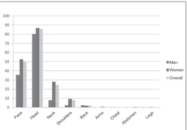
Graph 1 – Pain reports frequency for each identiiable body region,
overall and per gender. Face Hea
d Neck
Shou
lders Back Arms Che st
Abdo men Legs
Men
Women
Overall
Graph 2 – Pain areas frequency drawn for each identiiable body
region, overall and per gender.
Men
Women
Face Hea d
Neck Shou
lders Back Arms Che st
Abdo men Legs
In addition, some OFP predictors were also identiied, being one of them the presence of widespread pain.15
For example, greater prevalence of cervical spine symp-toms has been observed in patients with TMD16. Central
sensitization, neural convergences, neuroplasticity and descending inhibitory pain system dysfunction are some phenomena that may contribute to the maintenance and spread of pain in those patients17. Like other chronic pain
conditions, TMD may lead to states of comorbid depres-sion and anxiety, worsening patients´ painful manifesta-tions1,10,14. Recent indings observed that psychological
distress is common in orofacial pain patients, particu-larly on those with widespread pain18.
A signiicant difference between pain reports and pain draws was observed on body regions below the neck, both for males and females, which may be due to people believing that signs and symptoms outside the mouth should not be reported to dentists10. The only exception
was for head, whose reported complaints were superior to map drawings. This may be due to a misinterpretation of the pain site, because both facial pain and headaches are in the OFP context. While headaches are deined as pain above the orbitomeatal line, facial pain is anterior to the pinna and beneath the orbitomeatal line, above the neck19.
Patients who present to primary care practitioners are often treated for their syndromes, while co-existing complaints are often ignored20. The attention of the
den-tist is generally focused on the orofacial region. Patients, in turn, also tend to limit their pain descriptions to the fa-cial area, because they do not expect to receive treatment for symptoms outside mouth or face. Some possible fail-ures in this professional approach are highlighted, such as the presence of generalized pain conditions that can be involved in the maintenance of patient´s pain, and, if not known, may dificult OFP treatment10.
In line with the literature, our indings suggest that a thorough approach may reveal co-existing pain regions. Self-reports were only good for detecting face and head pain, but not for detecting pain in other body areas. Thus, both information from self reports and body maps seem essential during an OFP interview, since the former seem to complement the latter and vice versa. This approach allows dentists to gather more complete information that will lead to a better diagnosis and, consequently, to global patients’ treatment.
In general, patients have marked more painful areas on the map than those reported as chief complaints. In our study, pain maps were better than pain reports to identify painful areas. So, we suggest that dentists use both meth-ods in the anamnesis of OFP patients.
CONCLUSION
Chief complaint report was not an effective method to identify all pain complaints, because body maps have evi-denced the presence of additional pains in OFP patients
REFERENCES
1. Aggarwal VR, McBeth J, Zakrzewska JM, et al. Are reports of mechanical dysfunction in chronic oro-facial pain related to sensitization? A population based study. Eur J Pain 2008;12(4):501-7.
2. Türp JC, Kowalski CJ, O’Leary N, et al. Pain maps from facial pain patients indicate a broad pain geog-raphy. J Dent Res 1998;77(6):1465-72.
3. Macfarlane TV, Glenny AM, Worthington HV. Sys-tematic review of population-based epidemiological studies of oro-facial pain. J Dent 2001;29(7):451-67. 4. Macfarlane TV, Gray RJM, Kincey J, et al. Factors associated with the temporomandibular disorder, pain dysfunction syndrome (PDS): Manchester case-control study. Oral Dis 2001;7(6):321-30.
5. Macfarlane TV, Blinkhorn AS, Davies RM, et al. Oro -facial pain: just another chronic pain? Results from a population-based survey. Pain 2002;99(3):453-8.
6. Macfarlane TV, Blinkhorn AS, Davies RM, et al. Predictors of outcome for orofacial pain in the gener-al population: a four-year follow-up study. J Dent Res 2002;83(9):712-7.
7. Macfarlane TV, Worthington HV. Association be-tween orofacial pain and other symptoms: a population-based study. Oral Biosci Med 2004;1(1):45-54.
8. Yunus MB. Central Sensitivity Syndromes: a new para -digm and group nosology for ibromyalgia and overlap -ping conditions, and the related issue of disease versus illness. Semin Arthritis Rheum 2008;37(6):339-52. 9. Wenngren A, Stalnacke BM. Computerized assess -ment of pain drawing area: a pilot study. Neuropsychiatr Dis Treat 2009;5:451-6.
10. Türp JC, Kowalski CJ, Stohler CS. Temporoman -dibular disorders-pain outside the head and face is rare-ly acknowledged in chief complaint. J Prosthet Dent 1997;78(6):592-5.
11. Benoliel R, Svensson P, Heir GM, et al. Persistent orofacial muscle pain. Oral Dis 2011;17(1):23-41. 12. Sa KN, Baptista AF, Matos MA, et al. Chronic pain and gender in Salvador population, Brazil. Pain 2008;139(3):498-506.
14. John MT, Miglioretti DL, LeResche L, et al. Wide-spread pain as a risk factor for dysfunctional temporo-mandibular disorder pain. Pain 2003;102(3):257-63. 15. Aggarwal VR, Macfarlane GJ, Farragher TM, et al. Risk factors for onset of chronic oro-facial pain – Re-sults of the North Cheshire oro-facial pain prospective population study. Pain 2010;149(2):354-9.
16. Bevilaqua-Grossi D, Chaves TC, de Oliveira AS. Cervical spine signs and symptoms:perpetuating rather than predisposing factors for temporomandibular disor-ders in women. J App Oral Sci 2007;15(4):259-64. 17. Sarlani E, Greenspan JD. Evidence for generalized hyperalgesia in temporomandibular disorders patients. Pain 2003;102(3):221-6.
18. Mcmillan AS, Wong MCM, Zheng J, et al. Widespread pain symptoms and psychological distress in southern Chi-nese with orofacial pain. J Oral Rehabil 2010;37(1):2-10. 19. Headache Classiication Subcommittee of the Inter -national Headache Society. The Inter-national Clas-siication of Headache Disorders: 2nd ed. Cephalalgia
2004;24(1):9-160.
20. Aggarwal VR, McBeth J, Zakrzewska JM, et al. The epidemiology of chronic syndromes that are frequently unexplained: do they have common associated factors? Int J Epidemiol 2006;35(2):468-76.
Submitted in September 12, 2011.