2010 Nov-Dec;18(6):1138-44 www.eerp.usp.br/rlae
Corresponding Author: Maria Luiza Gonzalez Riesco
Universidade de São Paulo. Escola de Enfermagem. Av. Dr. Enéas de Carvalho Aguiar, 419
Bairro Cerqueira César
CEP: 05403-000 São Paulo, SP, Brasil E-mail: [email protected]
Perineal Muscle Strength During Pregnancy and Postpartum: the
Correlation Between Perineometry and Digital Vaginal Palpation
1Maria Luiza Gonzalez Riesco
2Adriana de Souza Caroci
3Sonia Maria Junqueira Vasconcellos de Oliveira
4Maria Helena Baena de Moraes Lopes
5Digital vaginal palpation performed during clinical practice can help diagnose urinary,
intestinal and sexual disorders, while perineometry is more useful for performing perineal
exercises with biofeedback. This study verifies whether there is a correlation between values
of Pelvic Floor Muscle Strength (PFMS) obtained through perineometry performed with an
electronic perineometer and through digital vaginal palpation using the Oxford scale. This
is a prospective cohort study with 330 measurements carried out in 110 women. Data were
collected from 2007 to 2008 in the health service system in Itapecerica da Serra, São Paulo,
Brazil. Evaluations were carried out at three points in time: up to 12 weeks of pregnancy;
between 36-40 weeks; and between 42-60 days postpartum. The Spearman coefficient
indicated a strong positive correlation between the two evaluation methods for the three
evaluations (p<0.0001). The conclusion is that both methods are valid for measuring PFMS
during pregnancy and after delivery.
Descriptors: Pelvic Floor; Electromyography; Perineum; Lacerations.
1 Supported by Fundação de Apoio à Pesquisa do Estado de São Paulo, process # 07/00357-5.
2 Free Lecture, Associate Professor, Escola de Enfermagem, Universidade de São Paulo, SP, Brazil. E-mail: [email protected]. 3 Ph.D. in Nursing, Professor, Centro Universitário Adventista de São Paulo, SP, Brazil. E-mail: [email protected]. 4 Free Lecture, Associate Professor, Escola de Enfermagem, Universidade de São Paulo, SP, Brazil. E-mail: [email protected]. 5 Free Lecture, Associate Professor, Faculdade de Ciências Médicas, Universidade Estadual de Campinas, SP, Brazil. E-mail:
Avaliação da força muscular perineal durante a gestação e pós-parto: correlação entre perineometria e palpação digital vaginal
Na prática clínica, a palpação vaginal digital auxilia no diagnóstico de disfunções urinárias,
intestinais e sexuais, enquanto a perineometria é mais utilizada para realizar exercícios perineais com biofeedback. O objetivo foi veriicar se existe correlação entre os valores da força muscular perineal (FMP), avaliada pela perineometria, utilizando o perineômetro
eletrônico, e por meio da palpação digital vaginal, utilizando a escala de Oxford. O estudo
deriva de coorte prospectiva, com 330 mensurações, em 110 mulheres. A coleta de
dados ocorreu em 2007 e 2008, em serviços de saúde de Itapecerica da Serra, São
Paulo. A avaliação foi realizada em três momentos: até 12 semanas de gestação, entre 36-40 semanas, entre 42-60 dias pós-parto. O coeiciente de Spearman indicou forte correlação positiva entre os dois métodos de avaliação, nos três momentos (p<0,0001).
Conclui-se que ambos os métodos são válidos para mensurar a FMP durante a gravidez
e após o parto.
Descritores: Soalho Pélvico; Eletromiograia; Períneo; Lacerações.
Evaluación de la fuerza muscular perineal durante la gestación y posparto: correlación entre perineometría y palpación digital vaginal
En la práctica clínica, la palpación vaginal digital auxilia en el diagnóstico de disfunciones
urinarias, intestinales y sexuales, en cuanto la perineometría es más utilizada para realizar ejercicios perineales con biofeedback. El objetivo fue veriicar se existe correlación entre los valores de la Fuerza Muscular Perineal (FMP) evaluada por la perineometría utilizando
el perineómetro electrónico, y por medio de la palpación digital vaginal, utilizando la
escala de Oxford. El estudio deriva de una cohorte prospectiva, con 330 mensuraciones
en 110 mujeres. La recolección de datos ocurrió en 2007 y 2008, en servicios de salud
de Itapecerica de la Serra, en Sao Paulo. La evaluación fue realizada en tres momentos:
hasta 12 semanas de gestación; entre 36 y 40 semanas; y, entre 42 y 60 días posparto. El coeiciente de Spearman indicó una fuerte correlación positiva entre los dos métodos de evaluación, en los tres momentos (p<0,0001). Se concluye que ambos métodos son
válidos para mensurar la FMP durante la gravidez y después del parto.
Descriptores: Suelo Pélvico; Electromiografía; Perineo; Laceraciones.
Introduction
Damage caused to a woman’s pelvic loor (PF) can lead to diminished or the loss of Pelvic Floor Muscle
Strength (PFMS) and consequently to genital prolapse,
fecal and urinary incontinence and constipation. About
one third of adult women have Urinary Incontinence(1-2)
(UI), which can impair a woman’s physical, sexual,
domestic, and professional and leisure activities(2-4).
Pregnancy, vaginal delivery, parity, duration of the second stage of labor, dificulty in fetal extraction during a cesarean section, newborn’s weight, perineal trauma
and other mechanical, endocrinal and neural factors can lead to reduction or loss of the pelvic loor muscle tone causing genitourinary disorders(1-10).
Altered PFMS has been the focus of studies and
research due to the evolution of equipment and exams
that make its evaluation and prognosis more precise(10).
To evaluate the PF muscles and diagnose disorders in the
genitourinary and anal tracts, different exams are used:
magnetic resonance, manometry, anal endosonography,
translabial ultrasound, electromyography, perineometry,
digital vaginal palpation, and neurophysiological and
urodynamic studies of the PF(2,5-8,10-13).
Perineometry and digital vaginal palpation are the
most frequently used methods to measure PFMS in
clinical practice(11-13). Evaluating PFMS can be essential
www.eerp.usp.br/rlae
Studies evaluating PFMS through digital vaginal
palpation and perineometry revealed that even though
these are different methods, they are positively
correlated(11,13). Other authors, however, veriied that
there is no signiicant correlation between digital vaginal palpation and perineometry(15).
The following question was investigated in this
study: Can digital vaginal palpation be used to evaluate
PFMS as an alternative to perineometry?
Therefore, this study veriies whether there is correlation between the two methods, perineometry and
digital vaginal palpation, in PFMS evaluation in pregnant
and postpartum women.
Method
This is a prospective cohort study addressing the
evaluation of PFMS through perineometry and vaginal
digital palpation. The population was composed of pregnant women who attended antenatal care in ive Primary Care Units (PCU) and whose deliveries were
performed in two hospitals in Itapecerica da Serra, SP,
Brazil. The inclusion criteria were: being primigesta with
up to 12 weeks of pregnancy. Exclusion criteria were:
multiple pregnancies; abdominal or previous urogenital
surgery; hormonal therapy; diseases that can interfere
in PFMS; refusal of the women to allow digital vaginal
palpation or the insertion of a perineometer into the vagina; dificulty in understanding the Portuguese language or communication dificulties.
The parameter adopted to calculate the sample
size of women in the cohort study was the difference
between the average PFMS evaluated in postpartum
women who had cesarean section and vaginal delivery
with perineal laceration(12). An alternative formula was
used to determine the sample in order to compare two
averages when the groups have different sizes(16).
A total of 136 pregnant women would be necessary
to compose the cohort, assuming a probability of type
one as being 5% and power of 80%. Because it is a
longitudinal study with the possibility of dropouts, the
number of participants was increased 50%, hence
204 women needed to be recruited. Aiming to ensure
the maximum number in the estimated sample, 226
pregnant women were included in the study.
Data were collected between February 2007 and
August 2008. The data collection form was exclusively
developed for this study. The instrument and equipment
used were previously tested and assistants were trained
in the data collection technique.
Data collection was carried out at three points
in time: at the beginning of the pregnancy (up to 12
weeks), at the end of pregnancy (36 to 40 weeks) and
during puerperium (42 to 60 days postpartum).
Antenatal and puerperal consultations were carried
out during data collection followed by the services’
protocol. In the period of hospitalization for childbirth,
one of the researchers visited the participants in the
hospital and scheduled the return visit to the PCU to
perform a postpartum evaluation. If any of the women
missed the consultations, the researcher would visit
them at home.
The study was approved by the Research Ethics
Committee (604/2006/CEP-EEUSP). The participation
of the women was voluntary and they signed free
and informed consent forms. Researchers have no
agreements with the manufacturers or distributors of
the equipment used in this study.
Methods used to measure PFMS
All the participants underwent two methods of PFMS
measurement: perineometric (electronic perineometer)
and digital vaginal palpation (Oxford scale). A table to
randomly apply the sequence of PFMS measurement
methods was used through a statistical program
aiming to avoid biased data. The sequence cards were
put in opaque envelopes opened only at the moment
of the PFMS measurements. Hence, perineometry or vaginal digital palpation could be either the irst or last measurement performed.
Perineometry
The electronic pressure perineometer model Perina
996-2 Quark was used. It registers the potential action
of PF muscle contractions and translates their intensity
to visual signs through a numerical scale graded from
0 to 46.4 mmHg. This device does not discriminate between pelvic loor and abdominal contractions. The chosen perineometer met all the requirements of the
safety standard for electrometrical equipments and is
registered in the Brazilian National Sanitary Surveillance
Agency, Ministry of Health.
Surface Electromyography
To control abdominal relaxation during PFMS
measurement, a surface electromyography model
Bio-ADS1200 Lynx was used to detect, through external
electrodes, electrical activity of the muscle during
Riesco MLG, Caroci AS, Oliveira SMJV, Lopes MHBM.
www.eerp.usp.br/rlae
to a computer; a graphic showing muscle activity
appears in the screen. Records of PFMS indicated in the
perineometer were only considered when the surface
electromyography indicated abdominal muscle activity
was compatible with rest (EMG scale between 0 and 10
microvolts).
Digital Vaginal Palpation
The Oxford Scale(17) was used to classify PFMS as
follows: Grade 0 – no contraction; Grade 1 – hint of
non-sustained small contraction; Grade 2 – low intensity but
sustained contraction; Grade 3 – moderate contraction
with increased intravaginal pressure, compressing the ingers and presenting small elevation of the vaginal wall; Grade 4 – satisfactory contraction, compressing the ingers of the examiner with elevation of the vaginal wall toward the pubic symphysis.
Procedures for measuring PFMS
1. Place the woman in the gynecological
position with the genital region and inferior limbs
naked and protected by a sheet; 2. Connect the four
electromyography electrodes to the rectus-abdominal
muscles (two electrodes on the right side and two on
the left side, between the top edge of the pubis and
the umbilical region); 3. Put on procedure gloves; 4.
Instruct the woman to make contractions as if “holding”
urine using only the PF muscles and avoiding contracting
abdomen, thigh and buttocks muscles.
Perineometer
1. Zero the scale pressure level; 2. Cover the elastic
tube with a disposable but non-lubricated condom; 3.
Lubricate the condom with lubricating gel; 4. Introduce
the tube three to four centimeters in the vagina; 5.
Instruct the woman to relax the PF muscles; 6. Ask the
woman to contract and keep the voluntary contraction of the pelvic loor muscles around the vaginal tube as long as possible in a sequence of three sessions with an
interval of 15 seconds between each session; 7. Keep
the vaginal tube in all PFMS measurements; 8. Register
the strongest voluntary contraction of the PF muscles; 9.
Rest for one minute before initiating the digital vaginal
palpation (in case it was not previously performed in a
random fashion).
Digital vaginal palpation
1. Introduce the two distal phalanges of the index and middle ingers into the vagina with lubricating gel; 2. Ask the woman to contract and keep the voluntary contraction of the pelvic loor muscles around the examiner’s inger as long as possible in a sequence of three sessions with an interval of 15 seconds between each session; 3. Keep the inger in the vagina during all PFMS measurements; 4. Record the highest classiication of contraction according to the Oxford scale; 5. Rest for one minute before beginning
perineometry (in case it was not previously performed
in a random fashion).
Data treatment and analysis
Data were entered twice in the Epi-info 6. The
database was validated and imported into Excel. Spearman’s ρ coeficient was computed in each of the measurements to verify whether there was
correlation between the PFMS values obtained in the two
measurement methods; probability of a type one error
was considered at 5%.
Results
A total of 116 women dropped out of the study
among the 226 women that met the inclusion criteria.
Due to the high number of women who decided not
to continue the study, a comparative analysis was performed between the women included in the inal sample and those excluded from the study, aiming to
verify whether the losses were random and did not inluence the sample result, especially in relation to PFMS. Hence, the inal sample was composed of 110
women who completed all the cohort measurements.
Considering that the measurements of PFMS were
carried out at three different points of time, a total of
330 measurements were obtained.
The pregnant women’s average age was 21.4±5.1
years; 73.6% of the women reported being non-white;
44.5% had a paid job and 35.5% were housewives.
The PFMS values obtained through perineometry
and digital vaginal palpation grouped according to
www.eerp.usp.br/rlae
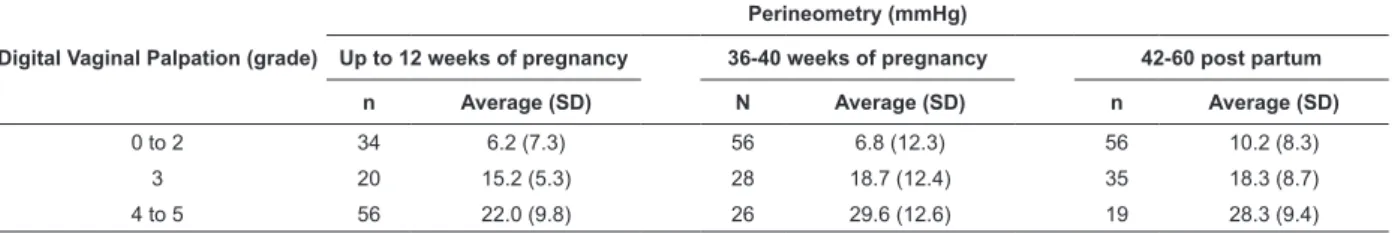
Table 1 – Average values of perineometry (in mmHg) and digital vaginal palpation (Oxford scale grades) according to
the point at which the evaluation was performed. Itapecerica da Serra, SP, Brazil. February 2007 to August 2008
Digital Vaginal Palpation (grade)
Perineometry (mmHg)
Up to 12 weeks of pregnancy 36-40 weeks of pregnancy 42-60 post partum
n Average (SD) N Average (SD) n Average (SD)
0 to 2 34 6.2 (7.3) 56 6.8 (12.3) 56 10.2 (8.3)
3 20 15.2 (5.3) 28 18.7 (12.4) 35 18.3 (8.7)
4 to 5 56 22.0 (9.8) 26 29.6 (12.6) 19 28.3 (9.4)
Table 2 presents the analysis of the correlation
between the PFMS results evaluated by the two
methods. This analysis was performed separately for
each evaluation point in time, indicating a strong positive
correlation between all them.
Table 2 – Correlation between the PFMS values obtained
through perineometry (in mmHg) and digital vaginal
palpation (Oxford scale grades) (n = 110). Itapecerica
da Serra, SP, Brazil. February 2007 to August 2008
Perineometry x palpation Spearman’s ρ p-value <
Up to 12 weeks of pregnancy 0.771 0.0001
Between 36-40 weeks of pregnancy 0.814 0.0001
Between 42-60 days postpartum 0.703 0.0001
ρ > 0.70 (strong correlation); ρ = 0.30-0.70 (moderate correlation); ρ <
0-0.30 (poor correlation)
Discussion
The variables analyzed to compare the dropout
group and the studied sample indicated homogeneity
between groups, which reduces the probability of bias
due to the loss of cohort follow-up.
The PFMS values obtained through perineometry can be classiied according to intensity: absence of contraction (0.0); mild contraction (1.6 to 16.0 mmHg); moderate
contraction (17.6 to 32.0 mmHg); normal contraction
(33.6 to 46.4 mmHg)(11). The results of the three points
of evaluation revealed that when the digital vaginal
palpation was between grades 0 and 2, perineometry
indicated mild contraction and when the digital vaginal
palpation indicated grades 3 to 5, contraction was
evaluated as moderate by perineometry.
The strong positive correlation obtained in this study
between the two methods of evaluating PFMS indicates
that perineometry can be validated by the clinical method
of digital vaginal palpation using the Oxford scales, which
is in agreement with other authors’ conclusions(11,13).
This correlation is important because in the absence of
a perineometer, a specialized professional can perform
evaluation through digital vaginal palpation(13).
On the other hand, no signiicant correlation was
found in a study carried out with 20 women using these
same methods to evaluate PFMS. The article held that
appropriate measurement of PFMS depended on the
cooperation and position of the participants as well as
the previous experience of the researcher, which hinder
the evaluation task(15).
We must take into account that even with technical
standardization, randomization of the sequence of
application of methods, previous instruction to women
and PFMS measurements performed by the same
professional, there were cases in which the Oxford scale
indicated grades incompatible with values inferred by
the perineometer.
The women participating in this study showed
positive acceptance of the PFMS evaluation with both
methods considering that, even though these are
painless procedures, they may cause embarrassment
and discomfort. It is worth noting that the evaluation
was performed in the context of a antenatal consultation
and postpartum return visit, situations in which a good
rapport has been established between the researchers
and participants. Of the total of women included in the
study, 1.8% refused to continue the study.
It is important to stress that further studies
addressing PFMS measurement are necessary to establish a proile of PFMS during pregnancy and
puerperium. It is also essential that the professional caring for these women value not only the identiication of factors related to reduced PFMS but also encourage
them to practice exercises to strengthen the PF muscles
and also to report complains related the genitourinary
tract(6,9,14-15,17).
Evaluating PFMS in the postpartum may serve
Riesco MLG, Caroci AS, Oliveira SMJV, Lopes MHBM.
disorders or aggravation in the long term. Digital vaginal
palpation is a simple method with no costs and does
not require special equipment. However, it does require
that the professional performing it to be appropriately
trained to evaluate PFMS. This method has helped in
the diagnosis of urinary, intestinal and sexual disorders.
Performing perineometry is more important in the realization of pelvic loor exercises with biofeedback for treating these disorders(2-3,5,9-13,15).
A relevant methodological aspect in this study
was the use of surface electromyography during the PFMS evaluation because women showed dificulty in distinguishing contractions of the pelvic loor and abdominal muscles. The use of this equipment avoided
registering PFMS performed simultaneously with
abdominal muscle contractions(18).
Another important aspect of how data were
collected to stress is that one researcher measured
PFMS while another read the perineometry result. This sought to avoid the result of perineometry inluencing the researcher while performing the digital vaginal
palpation.
Several devices and evaluation methods and also
a lack of standardized parameters to classify the pelvic loor function are observed in the literature, which limit comparison of results of different studies. Hence, this is
a topic that warrants further investigation and debate.
In addition to issues related to PFMS evaluation, scientiic literature also analyzes the impact of UI on women’s emotional health – suffering, diminished self-esteem, isolation, dificulties coping with the problem, among others – which provides an important contribution to nursing practice in women’s health(19).
Conclusion
This study’s results indicate that there is a positive correlation between the PFMS values obtained through
perineometry and digital vaginal palpation, which indicates
that both methods are valid measures of PFMS.
Acknowledgments
We thank the state of São Paulo Research Foundation, FAPESP, for the inancial support provided to this study, the University of São Paulo, College of Nursing, and the
Adventist University Center for institutional support.
References
1. Glazener CMA, Cooper K. Anterior vaginal repair for urinary incontinence in women (Cochrane Review). In: The Cochrane Library, Issue 3, 2003. Oxford: Update; 2003.
2. Voorham-van der Zalm PJ, Lycklama à Nijeholt GAB, Elzevier HW, Putter H, Pelger RCM. Diagnostic investigation of the pelvic floor: a helpful tool in the approach in patients with complaints of micturition, defecation, and/or sexual dysfunction. J Sex Med 2008 April; 5(4):864-71.
3. Norton C, Hosker G, Brazzelli M. Biofeedback and/or sphincter exercises for the treatment of faecal incontinence in adults (Cochrane Review). In: The Cochrane Library, Issue 3, 2003. Oxford: Update; 2003.
4. Brubaker L, Handa VL, Bradley CS, Connolly A, Moalli P, Brow MB, et al. Sexual function 6 months after first delivery. Obstet Gynecol 2008 May; 111(5):1040-4.
5. Coletti SH, Haddad JM, Barros JPF. Avaliação funcional do assoalho pélvico. In: Amaro JL, Haddad JM, Trindade JCS, Ribeiro RM. Reabilitação do assoalho pélvico nas funções urinárias e anorretais. São Paulo (SP): Segmento Farma; 2005. p. 67-75. 6. Zanetti MRD, Castro RA, Rotta AL, Santos PD, Sartori M, Girão MJBC. Impacto of supervised physiotherapeutic pelvic floor exercises for treating female stress urinary incontinence. Sao Paulo Med J 2007 September; 125(5):265-9.
7. Örnö AK, Marsál K, Herbst A. Ultrasonographic anatomy of perineal structures during pregnancy and immediately following obstetric injury. Ultrasound Obstet Gynecol 2008 September; 32(4):527-34.
8. Braekken IH, Majida M, Engh ME, Bø K. Test-retest reliability of pelvic floor muscle contraction measured by 4D ultrasound. Neurourol Urodyn 2009 January; 28(1):68-73.
9. Castro RA, Arruda RM, Zanetti RD, Santos PD, Sartori MGF, Girão MJBC. Single-blind, randomized, controlled trial of pelvic floor muscle training, electrical stimulation, vaginal cones, and no active treatment in the management of stress urinary incontinence. Clinics 2008 July-August; 63(4):465-72.
10. Fitzpatrick M, O’Herlihy C. The effects of labour and delivery on the pelvic floor. Best Pract Res Clin Obstet Gynaecol 2001 February; 15(1):63-79.
11. Barbosa AMP, Carvalho LR, Martins AMVC, Calderon IMP, Rudge MVC. Efeito da via de parto sobre a força muscular do assoalho pélvico. Rev Bras Ginecol Obstet 2005 novembro; 27(11):677-82.
12. Menta SS, Schirmer J. Relação entre a pressão muscular perineal no puerpério e o tipo de parto. Rev Bras Ginecol Obstet 2006 setembro; 28(9):523-9.
13. Oliveira C, Lopes MAB, Longo e Pereira LCL, Zugaib M. Effects of pelvic floor muscle training during pregnancy. Clinics 2007 July-August; 62(4):439-46.
www.eerp.usp.br/rlae
15. Bø K, Finckenhagen HB. Vaginal palpation of pelvic floor muscle strength: inter-test reproducibility and comparison between palpation and vaginal squeeze pressure. Acta Obstet Gynecol Scand 2001 October; 80(10):883-7.
16. Hulley SB, Cummings SR. Designing clinical research. Baltimore (MD): Williams & Wilkins; 1988.
17. Bø K, Larsen S, Kvarstein B, Hagen RH. Classification and characterization of responders to pelvic floor muscle exercise for female stress urinary incontinence. Neurourol Urodyn 1990 July; 9(4):395-7.
18. Nagib, ABL, Guirro ECO, Palauro VA, Guirro RRJ. Avaliação da sinergia da musculatura abdomino-pélvica em nulíparas com eletromiografia e biofeedback perineal. Rev Bras Ginecol Obstet 2005 abril; 27(4):210-5.
19. Higa R, Lopes MHBM, Turato ER. Significados psicoculturais da incontinência urinária feminina? Uma revisão. Rev. Latino-Am. Enfermagem [online]. 2008, 16(4):779-86. ISSN 0104-1169. doi: 10.1590/S0104-11692008000400020.
Received: Jun. 10th 2009