Evaluation of Swallowing in Infants with Congenital
Heart Defect
Karine da Rosa Pereira
1Cora Firpo
2Marisa Gasparin
3Adriane Ribeiro Teixeira
1Silvia Dornelles
1Tzvi Bacaltchuk
2Deborah Salle Levy
11Department of Speech Pathology, Universidade Federal do Rio Grande do Sul, Porto Alegre, Rio Grande do Sul, Brazil
2Department of Paediatric Cardiology, Instituto de Cardiologia do Rio Grande do Sul, Porto Alegre, Rio Grande do Sul, Brazil
3Department of Speech Pathology, Instituto de Cardiologia do Rio Grande do Sul, Porto Alegre, Rio Grande do Sul, Brazil
Int Arch Otorhinolaryngol 2015;19:55–60.
Address for correspondence Karine Pereira, AuD, Department of Psychology, Universidade Federal do Rio Grande do Sul, Ramiro Barcelos, 2600 Porto Alegre, Rio Grande do Sul 90035-003, Brazil (e-mail: [email protected]).
Introduction
Congenital heart defect is defined as a malformation of the heart or blood vessels that develops during the fetal period.1 The prevalence is 9:1,000 live births, corresponding to 1.35 million newborns per year.2Most of these children require surgical intervention, often in early childhood.3
The multiple surgeries to correct the heart defect are debilitating and often hinder the quality of life.4The presence of chromosomal abnormalities, cyanosis, and heart failure increase the complexity and challenge involved in the treat-ment.5The life expectancy of patients with congenital heart defect has increased due to advances in early diagnosis,1,3,6 care after birth, and surgical techniques.3,6 However, with
improved survival, new challenges have arisen in the growth and development of these children.6
In childhood, oral feeding is established as a reciprocal process,7one that is essential in the life experience of young children.8 Swallowing is a complex process that involves neurologic and aerodigestive coordination.9Neurobehavioral markers, such as postural control, regulation of the sleep-wake cycle, and maturation of coordination of the suck-swallow-breathe process, provide the clinical picture neces-sary for the child to advance toward oral feeding readiness.10 Newborns with congenital heart defect may require a prolonged hospital stay, well beyond the neonatal period, during the most important period of development. In the long term, this could affect somatic growth as well as cognitive and Keywords
►
congenital heart
►
defects
►
infant
►
deglutition disorders
►
respiratory aspiration
►
speech
►
language and hearing
sciences
Abstract
Introduction
Surgical repair of congenital heart disease in the
fi
rst years of life
compromises the coordination of the suction, breathing, and swallowing functions.
Objective
To describe the alterations in swallowing found in infants with congenital
heart defect during their hospitalization.
Methods
Prospective, cross-sectional study in a reference hospital for heart disease.
The sample consisted of 19 postsurgical patients who underwent an evaluation of
swallowing. The infants included were younger than 7 months and had a diagnosis of
congenital heart defect and suspected swallowing dif
fi
culties.
Results
Of the 19 infants with congenital heart defect, the median age was 3.2
months. A signi
fi
cant association was found between suction rhythm and dysphagia
(
p
¼
0.036) and between oral-motor oral feeding readiness and dysphagia (
p
¼
0.014).
Conclusions
The data suggest that dysphagia often occurs after surgery in infants
with congenital heart defect. Infants with congenital heart defect had very similar
behavior to preterm infants in terms of oral feeding readiness.
received
March 3, 2014
accepted
June 9, 2014
published online
November 5, 2014
DOI http://dx.doi.org/ 10.1055/s-0034-1384687.
ISSN 1809-9777.
Copyright © 2015 by Thieme Publicações Ltda, Rio de Janeiro, Brazil
social-emotional growth.6Among these infants, THE nutri-tional condition resulting from the inability to feed often leads to an imbalance in energy intake, resulting in impaired growth.11 Due to compromised cardiopulmonary function, these infants may need a longer time to feed or may present with a lack of appetite and food refusal.12Feeding difficulties may not be combined with oropharyngeal dysphagia.13
In pediatrics, oropharyngeal dysphagia may have a varied clinical presentation, depending on its etiology, complexity, and impact on the lives of patients with feeding difficulties.14 It is often observed in children with various medical diagno-ses,15 and it may be caused by several conditions, such as cerebral palsy, cleft lip or palate, and other structural abnor-malities,16in addition to congenital heart defect. Dysphagia not only deprives one of the pleasure of eating, but also endangers health,17because it could lead to the development of complications, including impaired growth,18chronic aspi-ration, lung disease,18,19malnutrition,17and others.
The aim of this study was to describe the swallowing alterations found in infants with congenital heart defect evaluated during their hospitalization period.
Methods
This prospective, comparative, cross-sectional study was conducted at the Pediatric Intensive Care Unit at the Institute of Pediatric Cardiology in Porto Alegre, Brazil, from Au-gust 2011 to AuAu-gust 2012.
The inclusion criteria were as follows: infants under 7 months, diagnosed with congenital heart defect and with suspected swallowing disorders were included in the study. Infants with a neurologic or syndromic medical diagnosis, or awaiting surgical correction, were excluded from the research. This study used a protocol to characterize the sample, the Preterm Oral Feeding Readiness Assessment Scale,20and a protocol for the clinical evaluation of swallowing based on the study by Weir et al.21
The protocol for the clinical evaluation of swallowing had the following information: date of birth, gender, medical diagnosis, date of the cardiac surgery, time of the mechanical ventilation.
The Preterm Oral Feeding Readiness Assessment scale was used in this study. According to Fujinaga et al,22this instrument is divided into five categories: (1) corrected gestational age (32 weeks; 32 to 34 weeks, and 34 weeks); (2) behavior state (state of conscience, posture, and global tonus); (3) oral posture (lips and tongue pos-ture); (4) oral reflexes (rooting, sucking, biting, and gag reflexes); (5) nonnutritive sucking (tongue movement, tongue cupping, jaw movement, sucking strength, sucking pauses, pause per sucking, maintenance of sucking rhythm, maintenance of alertness, and stress behavioral signs). Performance in every item is graded on a scale from 0 to 2 points, with a total score that varies from 0 to 36 points. However, thefirst category was not considered in our study.
In this evaluation, the infants were placed on their moth-er’s lap to assess oral posture, oral reflexes, and nonnutritive
sucking. Verification of oral reflexes was performed using a glovedfinger and/or a pacifier.
The clinical evaluation occurred through breast- or bottle-feeding. The infant who had previous experience of breast-feeding was evaluated at thefirst moment in breast-feeding, and if necessary afterward with a bottle. Thefirst oral assess-ment using either bottle- or breast-feeding occurred after application of the Preterm Oral Feeding Readiness Assessment Scale. The bottle evaluated wasfilled with liquid and thickened liquid consistencies, and we observed coordination of sucking, swallowing, and breathing and presence of oral stasis, cough, fatigue after feeding, cyanosis, and desaturation. After swal-lowing evaluation, the sample was divided into two groups to compare the clinicalfindings for each group. The infants with normal swallowing comprise group 1 and those who had oropharyngeal dysphagia comprise group 2.
The study was approved by the Research Ethics Committees of the Universidade Federal do Rio Grande do Sul Postgraduate Program and of Instituto de Cardiologia, under registration No. 2010064 and No. 4603/11, respectively. Consent was obtained from the guardians before the study was conducted.
The database and the analyses were performed using SPSS version 19 (Statistical Package for the Social Sciences, New York, US). Fisher exact test was used to assess the association between groups and other qualitative variables. The nonparametric Mann-Whitney test was used to compare the duration of mechanical ventilation between the groups, and the Spearman test was used to correlate oral-motor readiness with the time of mechanical ventilation. Qualitative data were described as ab-solute and relative values. Qualitative variables were expressed through median, minimum, and maximum values. The present study adopted a significance level ofp¼0.05.
Results
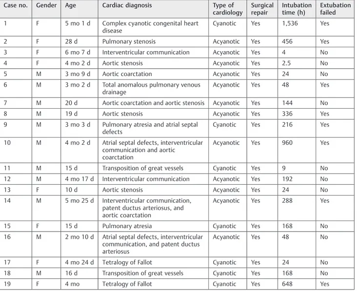
Demographic and cardiac characteristics of patients are shown in►Table 1. Out of the 19 infants in the sample, 11 (58%) were boys. The median age was 3.2 months (minimum¼0.3, maximum¼6.2). Twelve infants had acyanotic congenital heart defect, and seven had cyanotic congenital heart defect.
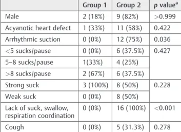
Oropharyngeal dysphagia was identified in 16 (84%) of the infants under study. The most common clinicalfinding was the lack of suck-swallow-breathe coordination (p.001), which was observed in combination with oral leaking in 5 infants; stasis in the oral cavity in 4; cough during feeding in 5; fatigue during feeding in 4; desaturation in 3; and cyanosis during feeding in 1. Descriptions of the clinical evaluation and score for Preterm Oral Feeding Readiness Assessment Scale are described in►Table 2.
The group of infants with normal swallowing and the group with oropharyngeal dysphagia had a median 24 and 48 hours in mechanical ventilation, respectively. There was no correlation between the duration of mechanical ventilation and the oral readiness score. Spearman correlation was
r 349.
►Fig. 1shows a comparison of performance in the readi-ness for oral feeding protocol between the group of infants who had normal swallowing and the group of infants who had oropharyngeal dysphagia. The group of infants with normal swallowing had a median score of 33 (minimum¼30, maximum¼36) and the group of infants with oropharyngeal dysphagia had a median score of 25 (minimum¼16, maxi-mum¼33). The correlation between oral-motor readiness and dysphagia wasp¼0.014.
Discussion
The present study showed that infants with congenital heart defect have low scores on the evaluation of readiness for oral
feeding, which are very close to those of preterm infants.22It should be underscored that the evaluation of readiness for preterm infants differs from the evaluation of infants with congenital heart defect, because the coordination of the suck-swallow-breathe process improves with the matura-tion of the central nervous system in cases of preterm birth.23
The mean values for the oral-motor readiness scores found in this survey were 25 for the group of infants with oropha-ryngeal dysphagia and 33 for the group of infants with normal swallowing. These results show that oral-motor skill is asso-ciated with oropharyngeal dysphagia and that the assessment of readiness before clinical evaluation is extremely important. By exploring possible variables that could affect the per-formance of swallowing, we found that the duration of mechanical ventilation did not influence the oral-motor readiness or the outcome of the clinical evaluation. This finding diverges from the literature, which states that oro-pharyngeal dysphagia may occur after mechanical ventila-tion24and after prolonged intubation.25
Table 1 Demographic characteristics of cardiac patients
Case no. Gender Age Cardiac diagnosis Type of cardiology
Surgical repair
Intubation time (h)
Extubation failed
1 F 5 mo 1 d Complex cyanotic congenital heart
disease
Cyanotic Yes 1,536 Yes
2 F 28 d Pulmonary stenosis Acyanotic Yes 456 Yes
3 F 6 mo 7 d Interventricular communication Acyanotic Yes 4 No
4 F 4 mo 2 d Aortic stenosis Acyanotic Yes 2.5 No
5 M 3 mo 9 d Aortic coarctation Acyanotic Yes 24 No
6 M 3 mo 2 d Total anomalous pulmonary venous
drainage
Acyanotic Yes 48 Yes
7 M 20 d Aortic coarctation and aortic stenosis Acyanotic Yes 144 No
8 M 19 d Aortic stenosis Acyanotic Yes 336 Yes
9 M 3 mo 3 d Pulmonary atresia and atrial septal
defects
Cyanotic Yes 216 Yes
10 M 4 mo 2 d Atrial septal defects, interventricular communication and aortic
coarctation
Acyanotic Yes 960 Yes
11 M 15 d Transposition of great vessels Cyanotic Yes 9 No
12 M 4 mo 17 d Interventricular communication Acyanotic Yes 192 No
13 F 10 d Aortic stenosis Acyanotic Yes 24 No
14 M 5 mo 25 d Interventricular communication,
patent ductus arteriosus, and aortic coarctation
Acyanotic Yes 288 Yes
15 F 15 d Pulmonary atresia Cyanotic Yes 168 No
16 M 2 mo 10 d Atrial septal defects, interventricular communication, and patent ductus arteriosus
Acyanotic Yes 48 No
17 F 4 mo 24 d Tetralogy of Fallot Cyanotic Yes 24 No
18 M 16 d Transposition of great vessels Cyanotic Yes 168 No
Clinical evaluation showed that in infants with congenital heart defect, oropharyngeal dysphagia is characterized by a lack of coordination of the suck-swallow-breathe process, whether or not this is combined with stasis of food in the oral cavity, cough, anterior leaking, and fatigue during breast-feeding. It is known that oropharyngeal dysphagia may occur in young children due to fatigue of the swallowing mecha-nism.25 Moreover, oral feeding requires muscle strength to extract the milk from the bottle or from the mother’s breast and coordination of the suck-swallow-breathe process; also,
infants with congenital heart defect are likely to resist ingest-ing the total prescribed volume, due to surgical intervention, which hinders weight gain and growth of these newborns and infants.26
Among the pediatric population with heart disease, risk factors for oropharyngeal dysphagia as follows: age under 3 years, preoperative intubation, intubation greater than 7 days, and operations for obstructive injuries on the left side. The use of transesophageal echocardiography in chil-dren weighing less than 5.5 kg is considered a predictor of
Table 2 Clinical evaluation and score of infants in the Preterm Oral Feeding Readiness Assessment scale
Case no. Consistencies Utensils Clinical findings Result Score
1 Liquid thickened Latex nipple, orthodontic nipple, conventionalflow (milk hole)
Incoordination of suck, swallow, and respiration
Oropharyngeal dysphagia 20
2 Liquid Breast-feeding Incoordination of suck,
swallow, and respiration; desaturation fatigue
Oropharyngeal dysphagia 23
3 Liquid thickened Latex nipple, orthodontic nipple, reducedflow (water/tea hole)
Incoordination of suck, swallow, and respiration
Oropharyngeal dysphagia 25
4 Liquid Silicone nipple, orthodontic nipple, conventionalflow (milk hole)
Incoordination of suck, swallow, and respiration; cough fatigue
Oropharyngeal dysphagia 30
5 Liquid thickened Silicone nipple, conventional nipple, conventionalflow (milk hole)
Incoordination of suck, swallow, and respiration
Oropharyngeal dysphagia 33
6 Liquid Breast-feeding; silicone nipple, or-thodontic nipple, conventionalflow (milk hole)
Incoordination of suck, swallow, and respiration; oral spillage; cough
Oropharyngeal dysphagia 25
7 Liquid Silicone nipple, conventional nipple, conventionalflow (milk hole)
Incoordination of suck, swallow, and respiration
Oropharyngeal dysphagia 33
8 Liquid Latex nipple, orthodontic nipple, conventionalflow (milk hole)
Incoordination of suck, swallow, and respiration
Oropharyngeal dysphagia 28
9 Liquid Silicone nipple, conventional nipple, conventionalflow (milk hole)
Incoordination of suck, swallow, and respiration; stasis in the oral cavity; cyanosis
Oropharyngeal dysphagia 22
10 Liquid thickened Latex nipple, orthodontic nipple, conventionalflow (milk hole)
Incoordination of suck, swallow, and respiration; fatigue
Oropharyngeal dysphagia 25
11 Liquid thickened Latex nipple, conventional nipple, conventionalflow (milk hole)
Desaturation Incoordination of suck, swallow, and respiration; fatigue; stasis in the oral cavity
Oropharyngeal dysphagia 16
12 Liquid silicone nipple, conventional nipple, conventionalflow (milk hole)
Incoordination of suck, swallow, and respiration; oral spillage; cough
Oropharyngeal dysphagia 29
13 Liquid Latex nipple, conventional nipple, conventionalflow (milk hole)
Incoordination of suck, swallow, and respiration; oral spillage; cough
Oropharyngeal dysphagia 29
14 Liquid Breast-feeding – Normal swallowing 30
15 Liquid Latex nipple, conventional nipple, conventionalflow (milk hole)
Incoordination of suck, swallow, and respiration; stasis in the oral cavity oral spillage
Oropharyngeal dysphagia 25
16 Liquid Breast-feeding Incoordination of suck,
swallow, and respiration
Oropharyngeal dysphagia 24
17 Liquid Breast-feeding – Normal swallowing 36
18 Liquid Breast-feeding – Normal swallowing 33
19 Liquid thickened Silicone nipple, conventional nipple, conventionalflow (milk hole)
Incoordination of suck, swallow, and respiration; stasis in the oral cavity; oral spillage
dysphagia.27 However, clinical evidence of oropharyngeal dysphagia in infants investigated postoperatively in this research emphasizes that dysphagia may take place with intubation times of less than 7 days.
Kohr et al aimed to determine the incidence and risk factors for oropharyngeal dysphagia in children after cardiac surgery and found that oropharyngeal dysphagia occurred in 9 (18%) of the 50 patients.27The videofluoroscopicfindings were as follows: 7 (78%) cases of delayed triggering of swallowing; 2 (22%) cases of laryngeal penetration, with direct aspiration and vocal fold paralysis; 2 (22%) cases of occasional laryngeal penetration without aspiration; 1 (11%) case of laryngeal reduction; and 2 (22%) cases of direct
aspiration resulting from vocal fold paralysis. Sachdeva et al,28whose objective was to evaluate the impact of voice disorders and feeding in children after cardiac surgery, showed that swallowing dysfunction was observed in 34 (89%) children with vocal fold alterations. The swallowing examination was performed in 29 patients with congenital heart defects, 30 days after surgery. The most common pathophysiological finding was aspiration, which was ob-served in 23 (80%) children. Laryngeal penetration occurred in 5 of them (17%), and delayed triggering of swallowing in only 1 (3%). Through instrumental evaluation, researchers were able to identify oropharyngeal dysphagia in this popu-lation of children with congenital heart defects.
Yi et al performed a retrospective study in which the objective was to evaluate the prevalence and clinical predic-tors of dysphagia and determine the videofluoroscopicfi nd-ings of swallowing in children who underwent cardiac surgery.29 Through videofluoroscopic findings, the authors concluded that 67.9% of the children had laryngeal penetra-tion and 63.6% had tracheal aspirapenetra-tion, and of these 85.7% of symptoms were silent, without the presence of cough. In our clinical study, we also observed a low incidence of cough during swallowing. Only 4 of the 16 infants identified with oropharyngeal dysphagia showed cough while swallowing.
Our results corroborate the studies by Kohr et al,27 Sach-deva et al,28and Yi et al,29which showed that oropharyngeal dysphagia often occurs postoperatively in infants, as most infants who had corrective surgery presented oropharyngeal dysphagia, even with thickened liquid. Thickening of food is a procedure adopted to minimize the pattern of dysphagia.27
It is important to note that feeding problems are common among children with congenital heart defect.6,11,27–31 Ac-cording to Arvedson,13feeding difficulties are characterized by refusal, disruptive behavior, preference, and lack of feeding competence expected for the subjects’level of development. Newborns who were born with a serious heart condition requiring heart surgery in thefirst month of life have a high risk of presenting feeding difficulties until 2 years of age.30 The speech therapist should be aware of these behaviors during the evaluation of swallowing.
The results of the present study suggest that dysphagia often occurs in infants after corrective surgery for congenital heart condition. The use of the preterm readiness for an oral feeding protocol enabled us to verify that infants with con-genital heart defect may present with very similar behavior to those of preterm newborns.
Conclusion
In the present study, the occurrence of oropharyngeal dys-phagia in infants under 7 months of age with congenital heart defect was observed, and the samefinding was detected by clinical evaluation. Infants with congenital heart defect showed a very similar behavior to that of preterm newborns. However, oropharyngeal dysphagia is a variable that still needs to be further studied to determine the epidemiologic data and identify the best clinical management outcome among this population.
Table 3 Comparative distribution of the variables between the two groups
Group 1 Group 2 pvaluea
Male 2 (18%) 9 (82%) >0.999
Acyanotic heart defect 1 (33%) 11 (58%) 0.422
Arrhythmic suction 0 (0%) 12 (75%) 0.036
<5 sucks/pause 0 (0%) 6 (37.5%) 0.427
5–8 sucks/pause 1(33%) 4 (25%)
>8 sucks/pause 2 (67%) 6 (37.5%)
Strong suck 3 (100%) 8 (50%) 0.228
Weak suck 0 (0%) 8 (50%)
Lack of suck, swallow, respiration coordination
0 (0%) 16 (100%) <0.001
Cough 0 (0%) 5 (31.3%) 0.278
Note: group 1, infants with no alterations in swallowing; group 2, infants with alterations in swallowing.
aFisher exact test.
15 20 25 30 35 40 45
G1 G2
0
R
AL
R
E
AD
IN
E
S
S
RESULT
p=0.014*
Fig. 1 Comparison between the groups of infants with congenital heart defect, according to the preterm readiness for oral feeding protocol. Abbreviations: G1, group 1 (infants with no alterations in swallowing); G2, group 2 (infants with alterations in swallowing).
References
1 Teixeira FM, Coelho RM, Proença C, et al. Quality of life experi-enced by adolescents and young adults with congenital heart disease. Pediatr Cardiol 2011;32(8):1132–1138
2 van der Linde D, Konings EE, Slager MA, et al. Birth prevalence of congenital heart disease worldwide: a systematic review and meta-analysis. J Am Coll Cardiol 2011;58(21):2241–2247 3 Gerdes M, Flynn T. Clinical assessment of neurobehavioral
out-comes in infants and children with congenital heart disease. Progress in Pediatr Cardiol 2010;29(2):97–105
4 Bruneau BG. The developmental genetics of congenital heart disease. Nature 2008;451(7181):943–948
5 Medoff-Cooper B, Naim M, Torowicz D, Mott A. Feeding, growth, and nutrition in children with congenitally malformed hearts. Cardiol Young 2010;20(3, Suppl 3):149–153
6 Sables-Baus S, Kaufman J, Cook P, da Cruz EM. Oral feeding out-comes in neonates with congenital cardiac disease undergoing cardiac surgery. Cardiol Young 2012;22(1):42–48
7 Delaney AL, Arvedson JC. Development of swallowing and feeding: prenatal throughfirst year of life. Dev Disabil Res Rev 2008;14(2): 105–117
8 Newman LA, Keckley C, Petersen MC, Hamner A. Swallowing function and medical diagnoses in infants suspected of dysphagia. Pediatrics 2001;108(6):E106
9 Weckmueller J, Easterling C, Arvedson J. Preliminary temporal measurement analysis of normal oropharyngeal swallowing in infants and young children. Dysphagia 2011;26(2):135–143 10 Silberstein D, Geva R, Feldman R, et al. The transition to oral
feeding in low-risk premature infants: relation to infant neuro-behavioral functioning and mother-infant feeding interaction. Early Hum Dev 2009;85(3):157–162
11 Jadcherla SR, Vijayapal AS, Leuthner S. Feeding abilities in neo-nates with congenital heart disease: a retrospective study. J Perinatol 2009;29(2):112–118
12 Woodward CS. Keeping children with congenital heart disease healthy. J Pediatr Health Care 2011;25(6):373–378
13 Arvedson JC. Assessment of pediatric dysphagia and feeding disorders: clinical and instrumental approaches. Dev Disabil Res Rev 2008;14(2):118–127
14 Arvedson J, Clark H, Lazarus C, Schooling T, Frymark T. The effects of oral-motor exercises on swallowing in children: an evidence-based systematic review. Dev Med Child Neurol 2010;52(11): 1000–1013
15 Sitton M, Arvedson J, Visotcky A, et al. Fiberoptic endoscopic evaluation of swallowing in children: feeding outcomes related to diagnostic groups and endoscopicfindings. Int J Pediatr Oto-rhinolaryngol 2011;75(8):1024–1031
16 Heiss CJ, Goldberg L, Dzarnoski M. Registered dietitians and speech-language pathologists: an important partnership in dysphagia management. J Am Diet Assoc 2010;110(9):1290, 1292–1293
17 Hongama S, Nagao K, Toko S, et al. MI sensor-aided screening system for assessing swallowing dysfunction: application to the repetitive saliva-swallowing test. J Prosthodont Res 2012;56(1): 53–57
18 Hewetson R, Singh S. The lived experience of mothers of children with chronic feeding and/or swallowing difficulties. Dysphagia 2009;24(3):322–332
19 Christiaanse ME, Mabe B, Russell G, Simeone TL, Fortunato J, Rubin B. Neuromuscular electrical stimulation is no more effective than usual care for the treatment of primary dysphagia in children. Pediatr Pulmonol 2011;46(6):559–565
20 Fujinaga CI, Scochi CGS, Santos CB, et al. Validação do conteúdo de um instrumento para avaliação da prontidão do prematuro para início da alimentação oral. Rev Bras Saude Mater Infant 2008;8(4): 391–399
21 Weir K, McMahon S, Barry L, Masters IB, Chang AB. Clinical signs and symptoms of oropharyngeal aspiration and dysphagia in children. Eur Respir J 2009;33(3):604–611
22 Fujinaga CI, de Moraes SA, Zamberlan-Amorim NE, Castral TC, de Almeida e Silva A, Scochi CG. Clinical validation of the Preterm Oral Feeding Readiness Assessment Scale. Rev Lat Am Enfermagem 2013;21(Spec No):140–145
23 Mizuno K, Ueda A. The maturation and coordination of sucking, swallowing, and respiration in preterm infants. J Pediatr 2003; 142(1):36–40
24 Brown CV, Hejl K, Mandaville AD, Chaney PE, Stevenson G, Smith C. Swallowing dysfunction after mechanical ventilation in trauma patients. J Crit Care 2011;26(1):e9–e13
25 Bordon A, Bokhari R, Sperry J, Testa D IV, Feinstein A, Ghaemma-ghami V. Swallowing dysfunction after prolonged intubation: analysis of risk factors in trauma patients. Am J Surg 2011; 202(6):679–682, discussion 682–683
26 Tutor JD, Gosa MM. Dysphagia and aspiration in children. Pediatr Pulmonol 2012;47(4):321–337
27 Kohr LM, Dargan M, Hague A, et al. The incidence of dysphagia in pediatric patients after open heart procedures with transesopha-geal echocardiography. Ann Thorac Surg 2003;76(5):1450–1456 28 Sachdeva R, Hussain E, Moss MM, et al. Vocal cord dysfunction and
feeding difficulties after pediatric cardiovascular surgery. J Pediatr 2007;151(3):312–315, e1–e2
29 Yi SH, Kim SJ, Huh J, Jun TG, Cheon HJ, Kwon JY. Dysphagia in infants after open heart procedures. Am J Phys Med Rehabil 2013; 92(6):496–503
30 Medoff-Cooper B, Irving SY. Innovative strategies for feeding and nutrition in infants with congenitally malformed hearts. Cardiol Young 2009;19(Suppl 2):90–95