Seminiferous Epithelium of Rats with Food Restriction and
Carbon Tetrachloride-Induced Cirrhosis
Marilise M. Horn, Ana R. Ramos, Leonardo Winkelmann, Ursula S. Matte, Helena A.
Goldani, Themis R. Silveira
Gene Therapy Center (MMH, USM), and Experimental Hepatology Laboratory (ARR, LW, HAG, TRS),
General Hospital of Porto Alegre, Federal University of Rio Grande do Sul, Porto Alegre, Brazil
ABSTRACT
Objective:Analyze the changes in the seminiferous epithelium in rats with carbon tetrachloride-induced cirrhosis (CCl4). Materials and Methods: Forty-eight male Wistar rats aged 45-50 days, weighing 150-180 grams were used. Twenty-two rats underwent CCl4-induced cirrhosis with CCl4 0.25 mL/Kg weekly intragastrically once a week, during 10 weeks. Addi-tionally, they had a 44% food restriction diet (Group 1). The control group was divided in two subgroups: 13 rats had a 44% food restriction diet and no CCl4 (Group 2) and 10 rats were not submitted to CCl4 or food restriction (Group 3). After 10 weeks, the rats were sacrificed and liver sections were collected for histological analysis. The testicular analysis was carried out to evaluate the frequency of tubules in stages VIII and XIV.
Results:The mean rates of stage VIII in animals with food restriction plus CCl4-induced cirrhosis and food restriction without CCl4 were significantly different from animals without either food restriction or CCl4 (18.1 ± 5.5%, 20.5 ± 2.5% and 13.4 ± 3.5%, respectively, p = 0.002). The mean rate of stage VIII in rats with cirrhosis was not significantly different from rats without cirrhosis (18.1 ± 5.5% and 17.4 ± 4.6% respectively). The mean frequency of stage XIV in rats with cirrhosis was significantly greater than rats without cirrhosis (4.7 ± 2.3% and 6.8 ± 1.9% respectively, p = 0.027). Conclusion:Animals with CCl4-induced cirrhosis and food restriction have shown alterations in spermatogenic cycle that were not seen in rats without CCl4-induced cirrhosis and food restriction.
Key words: rats; liver cirrhosis, experimental; carbon tetrachloride; food deprivation; spermatogenesis
Int Braz J Urol. 2006; 32: 94-9
INTRODUCTION
The presence of hypogonadism in cirrhosis was first described in 1966 (1); however, its patho-genesis has not yet been well established. Some cytokines of anabolic function in the testicles, such as IGF-I (Insulin-like growth factor-I) are reduced in cirrhotic rats. This cytokine reduces the effects of cir-rhosis in the testicle (2).
The gonadal dysfunction is common in chronic liver diseases, but most of the studies have
patients could present any spermatogenesis dysfunc-tion induced not only by liver disease but also in-duced by malnutrition secondary to liver disorder.
Spermatogenesis presents three important phases: a) proliferative phase (spermatogonia), in which cells undergo rapid successive divisions; b) meiotic phase (spermatocytes) in which genetic ma-terial is recombined and segregated; and c) differen-tiation or spermiogenic phase (spermatids) in which spermatids transform into specialized cells able to fertilize (7). The stages of spermatogenesis can be divided by morphological criteria into various devel-opment steps, based on the form and shape of the acrosome and the cells of a cell association (8).
The morphological features of the seminifer-ous epithelium in cirrhotic rats and restricted diet in-take have not yet been described. Thus, the present study aimed to check, by means of the histological analysis of the testicles, the possible changes in the seminiferous epithelium in rats with cirrhosis induced by Carbon Tetrachloride (CCl4) and food restriction.
MATERIALS AND METHODS
The study was conducted according to the guidelines for animal research (Guide for the Care and Use of Laboratory Animals) (9), and was ap-proved by the Hospital Research Ethics Commit-tee.
During a quarantine period of observation, the animals received a standard rat chow (Nuvilab CR-1®, Nuvital S.A., Colombo - PR, Brazil), based
on recommendations from the National Research Council and National Institute of Health - USA - pro-viding 290 KcaL/100g, which composition was 22% protein, 4% fat and 4% crude fiber. Based on our pre-vious data, the ad libitum food intake was established as 22g/rat/day. In order to have CCl4 full toxicity (10), a 44% food restriction intake (12 grams/rat/day) was used.
Forty-five male Wistar rats aged 45-50 days and weighing 150-180g were used.. The rats were kept in groups of five per cage, at a room temperature be-tween 18-22ºC with cycles of light-darkness of 12 hours.
Cirrhotic animals (Group 1): Twenty-two rats were used in this group. Cirrhosis was obtained by the administration of CCl4 (Merck p.a., Germany), 0.25mL/kg, diluted in 1mL of olive oil. The CCl4 was given once a week, intragastrically by gavage using a 6F polyethylene catheter for tracheal aspiration (MarkMed Ltd., São Paulo, Brazil) during 10 weeks. All rats received Phenobarbital, 350 mg/L, added to the ad libitum drinking water (10).
Control animals: The main control group con-sisted of two subgroups. Thirteen rats (Group 2) were submitted to a 44% food restriction diet and received once a week 1mL of olive oil by gavage in the same way as the animals of group 1, treated with CCl4. The other group consisted of 10 rats that were not submit-ted to any kind of procedure (Group 3).
After 10 weeks, all animals were sacrificed and liver sections were stained with hematoxylin-eosin (HE) and Sirius red. A semi-quantitative score was adapted to categorize liver damage: 0 = no fibro-sis; 1 = stellate enlargement of portal tract but with-out septa formation; 2 = enlargement of portal tract with rare septa formation; 3 = numerous septa with-out cirrhosis; 4 = cirrhosis (10). The testicles were fixed in Bouin during 12 hours to be analyzed with HE stain.
Two hundred transversal sections of semin-iferous tubules were analyzed in each testicle from all animals. The percentages of tubules in stage VIII (elongated spermatids that moved to the luminal as-pect of the seminiferous epithelium and lined to the lumen) and stage XIV (meiotic anaphase or telophase of meiosis I, secondary spermatocytes, or any of the phases of meiosis) were checked (11).
A cell association or stage is a defined group-ing of germ cell types at particular phases of devel-opment in cross-sectioned tubules (8). This classifi-cation divides the seminiferous epithelium cycle of rats in 14 stages according to changes in cell associa-tions (stages) arranged in a logical sequence of de-velopmental progression from spermatogonia through spermatozoa.
marked degeneration usually loose their normal as-sociation cells, and were not included in the classifi-cation described above. A ratio of stage XIV/stage VIII was also calculated, as the proportion of cells in each of these stages seems to be dependent on each other.
Results were analyzed by two-way analysis of variance (ANOVA) by using SPSS version 12.0 -USA. In all cases, p = 0.05 was established as statis-tically significant.
RESULTS
All 22 animals treated with CCl4 presented cirrhosis. The rats that were not submitted to CCl4 (with or without food restriction) did not show histo-logical changes of the liver.
Rats without cirrhosis and food restriction (group 3) presented all tubules without degeneration. A few number of degenerated tubules was observed
in both food restricted and cirrhotic animals (Table-1).
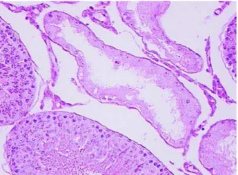
The majority of sections of seminiferous tu-bules of all animals was normal, and among them, a few number of degenerated seminiferous tubules were seen. The latter were characterized by loss of germ cells, vacuolization of germinative epithelium, inter-ruption in meiosis, or presence of Sertoli cells only, as shown in Figure-1.
The percentages of stages VIII and XIV were analyzed in 100 non-degenerated tubules (with nor-mal cell association) from each aninor-mal. Rats with cirrhosis presented lower number of cells in meio-sis (XIV) than the group without food restriction. Animals submitted only to food restriction presented intermediate frequency of cells in meiosis (Table-2). Animals with food restriction, with or without cirrhosis showed approximately the same ratio of 1:4 (0.27 ± 0.1). On the other hand, rats without food restriction showed an approximate ratio of 1:2 (0.53 ± 0.2).
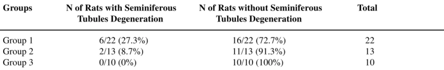
Table 1 – Frequency of rats with cirrhosis, with and without food restriction, presenting cross-sections of degenerated seminiferous tubules.
Chi-square test; p = 0.111. Group 1 = cirrhosis induced by CCl4 + 44% food restriction; Group 2 = 44% food restriction without CCl4; Group 3 = without food restriction and without CCl4.
Group 1 6/22 (27.3%) 16/22 (72.7%) 22
Group 2 2/13 (8.7%) 11/13 (91.3%) 13
Group 3 0/10 (0%) 10/10 (100%) 10
Groups N of Rats with Seminiferous N of Rats without Seminiferous Total Tubules Degeneration Tubules Degeneration
Table 2 – Frequencies and ratios of stages VIII and XIV of seminiferous epithelium in animals with CCl4-induced cirrhosis, food restriction and without food restriction.
*ANOVA and post-hoc test; Group 1 = cirrhosis induced by CCl4 + 44% food restriction; Group 2 = 44% food restriction without CCl4; Group 3 = without food restriction and without CCl4.
Group 1 Group 2 Group 3 P *
Stage VIII 18.1 ± 5.5 20.5 ± 2.5 13.4 ± 3.5 0.002
Stage XIV 04.7± 2.3 05.5± 1.4 06.8± 1.9 0.027
COMMENTS
In this study, abnormal frequencies of stages VIII (related to liberation of elongated mature sper-matids) and XIV (related to meiosis) of the spermato-genic cycle were observed in rats with food restric-tion CCl4-induced cirrhosis. Differences in animals submitted only to food restriction were also observed. Changes in other features of seminiferous epithelium disorder beyond the interruption of cell division (mitosis and meiosis), such as cell degen-eration and phagocytosis of germ cells, lack of matu-ration, and loss of germs cells. A generalized degen-eration of the seminiferous epithelium was not ob-served, only focal alterations. These results partially agree with other studies (2), which did not find a dif-fused damage to the seminiferous epithelium in rats with CCl4-induced cirrhosis.
Gonadal dysfunction is common in patients with chronic liver diseases, especially in cirrhosis induced by alcohol (3). However, there is a lack of experimental studies concerning seminiferous epithe-lium disorders that used alcohol to induce cirrhosis. Carbon Tetrachloride is a well-established hepatotoxic agent, which causes steatosis, necrosis and cirrhosis in animals. It has been extensively used as a model compound for inducing free radicals dam-age. It is bioactivated by cytochrome-P4502EI (12) into free radicals, leading to deleterious effects on
liver due to lipid peroxidation (13). Carbon Tetrachlo-ride was used in this study accordingly to previous classic experimental models of CCl4-induced cirrho-sis in rats (14). Phenobarbital can enhance the toxic-ity of this substance (6), as its main action is to in-duce the secretion of cytochrome-P4502EI (15).
The magnitude of liver injury can be influ-enced by food restriction. It has been suggested that food restriction can aggravate the toxicity of repeated oral administration of CCl4 in rats through the en-hanced metabolic activation of CCl4 by food restric-tion (10). Nevertheless, the effect of food restricrestric-tion on CCl4 toxicity is controversial as food restriction could minimize drug-related increases in peroxidation and protect the system against drug toxicity, presum-ably by induction of antioxidant potential (16,17). The expected effect of food restriction in this study was to induce more severe liver injury (18).
Seminiferous epithelium in rats is classified in 14 stages, and some tubules can present cellular characteristics of more than one stage. The classifi-cation of stage VIII followed the orientation of Leblond & Clermont (11), and is characterized by the presence of elongated spermatids aligned in the lu-men of the tubule to be liberated. In this study, for the animals without food restriction and without CCl4, the average frequency of stage VIII was 13.4 ± 3.5. For the animals with food restriction and without CCl4, it was 20.5 ± 2.5, and for the rats with CCl4-induced cirrhosis and food restriction, it was 18.1 ± 5.5. No difference was seen between the group with cirrhosis and the group with food restriction. The greater fre-quency of stages VIII in the animals with cirrhosis and with food restriction seems to be due to an accu-mulation of this stage in relation to the others. This could also be explained since stage VIII is not imme-diately affected by the changes in the testicular func-tion, as cells that no longer undergo cell division char-acterize it. As in this study, Lue et al. (19) observed that despite the accentuated loss of germ cells, after the application of heat on the testicles of rats, the elon-gated spermatids were still present in most of the se-miniferous tubules. Other examples can be mentioned (20), which using a model of testicular degeneration, by means of the implant of testosterone in rats, a four-fold reduction was observed in the conversion of sper-Figure 1 – Photomicrography of a transversal section of the
matogonia to spermatocyte compared to the conver-sion of round spermatids to elongated ones. This clearly illustrates the effect only upon germ cells in division (mitosis and meiosis).
In this study, the frequency of stage XIV (meiosis) was lower in the group with cirrhosis indi-cating a reduction in the cell division rate. However, this difference was also observed when animals with normal liver with and without food restriction are analyzed (Table-2). These results indicate that starv-ing may also have an influence on the meiosis rate, since both groups with food restriction had similar results independent of cirrhosis. Another study (7) demonstrated, in rats, that diet restriction caused re-duction of testicles and epididymis weight, lower tes-tosterone levels and copulatory efficiency. Moreover, it was observed that these effects were due to energy deficiency and not by reduced protein intake. Similar results were also observed in mice (21).
In conclusion, animals with CCl4-induced cirrhosis and food restriction have shown alterations in spermatogenic cycle that were not seen in rats with-out CCl4-induced cirrhosis and food restriction. Other studies are needed to better clarify the real role of CCl4-toxicity and food restriction on the pathogen-esis of seminiferous epithelium disorder.
ACKNOWLEDGEMENT
This work was supported by grants from the National Council for Scientific and Technological Development (CNPq – Brazil) and from the Fund for Research & Development, General Hospital of Porto Alegre, Brazil.
CONFLICT OF INTEREST
None declared.
REFERENCES
1. Schirren C, Szarvas F, Becker K: Andrological stud-ies in patients with chronic liver diseases. Hautarzt. 1966; 17: 175-8.
2. Castilla-Cortazar I, Diez N, Garcia-Fernandez M, Puche JE, Diez-Caballero F, Quiroga J, et al.: Hematotesticular barrier is altered from early stages of liver cirrhosis: effect of insulin-like growth factor 1. World J Gastroenterol. 2004; 10: 2529-34. 3. Van Thiel DH, Gavaler JS, Slone FL, Cobb CF, Smith
WI Jr, Bron KM, et al.: Is feminization in alcoholic men due in part to portal hypertension: a rat model. Gastroenterology. 1980; 78: 81-91.
4. Mooradian AD, Shamma’a M, Salti I, Cortas N: Hy-pophyseal-gonadal dysfunction in men with non-alco-holic liver cirrhosis. Andrologia. 1985; 17: 72-9. 5. Gursoy S, Baskol M, Ozbakir O, Guven K, Kelestimur
F, Yucesoy M: Hypothalamo-pituitary gonadal axis in men with chronic hepatitis. Hepatogastroenterology. 2004; 51: 787-90.
6. Hashimoto M, Kothary PC, Raper SE: Phenobarbital in comparison with carbon tetrachloride and phenobar-bital-induced cirrhosis in rat liver regeneration. J Surg Res. 1999; 81: 164-9.
7. Santos AM, Ferraz MR, Teixeira CV, Sampaio FJ, da Fonte Ramos C: Effects of undernutrition on serum and testicular testosterone levels and sexual function in adult rats. Horm Metab Res. 2004; 36: 27-33. 8. Russel LD, Ettlin RA, Sinhahikim AP, Clegg ED: The
classification and timing of spermatogenesis. In: Russel LD, Histological and histopathological evaluation of the testis. Clearwater, Cache River Press. 1990; pp. 41-58.
9. Guide for the care and use of laboratory animals, Na-tional Institutes of Health, Maryland, NaNa-tional Re-search Council/Public Health Service. 1996.
10. Seki M, Kasama K, Imai K: Effect of food restriction on hepatotoxicity of carbon tetrachloride in rats. J Toxicol Sci. 2000; 25: 33-40.
11. Leblond CP, Clermont Y: Spermiogenesis of rat, mouse, hamster and guinea pig as revealed by the pe-riodic acid-fuchsin sulfurous acid technique. Am J Anat. 1952; 90: 167-215.
12. Bruckner JV, Ramanathan R, Lee KM, Muralidhara S: Mechanisms of circadian rhythmicity of carbon tet-rachloride hepatotoxicity. J Pharmacol Exp Ther. 2002; 300: 273-81.
13. Janakat S, Al-Merie H: Optimization of the dose and route of injection, and characterisation of the time course of carbon tetrachloride-induced hepatotoxicity in the rat. J Pharmacol Toxicol Methods. 2002; 48: 41-4.
EDITORIAL COMMENT
The authors present an interesting study fo-cusing on the evaluation, by means of histology, of the seminiferous epithelium in rats with carbon tetra-chloride-induced cirrhosis.
It was observed that the animals with CCl4 -induced cirrhosis and food restriction have shown alterations in the spermatogenic cycle that had not been observed in rats without CCl4-induced cirrhosis and food restriction. However, alterations in the fre-15. Recknagel RO, Glende EA Jr, Dolak JA, Waller RL: Mechanisms of carbon tetrachloride toxicity. Pharmacol Ther. 1989; 43: 139-54.
16. Ramkumar KM, Rajesh R, Anuradha CV: Food restric-tion attenuates blood lipid peroxidarestric-tion in carbon tetra-chloride-intoxicated rats. Nutrition. 2003; 19: 358-62. 17. Pavanato A, Tunon MJ, Sanchez-Campos S, Marroni CA, Llesuy S, Gonzalez-Gallego J, et al.: Effects of quercetin on liver damage in rats with carbon tetrachlo-ride-induced cirrhosis. Dig Dis Sci. 2003; 48: 824-9. 18. Snyder R, Andrews LS: Toxic effects of solvents and
vapors. In: Klaassen CD, Amdur MO, Doull J (eds), Casarett and Doull’s Toxicology: The Basic Science of Poisons. New York, McGraw-Hill Companies. 1996; pp. 737-71.
19. Lue YH, Hikim AP, Swerdloff RS, Im P, Taing KS, Bui T, et al.: Single exposure to heat induces stage-specific germ cell apoptosis in rats: role of intratesticular testosterone on stage specificity. Endo-crinology. 1999; 140: 1709-17.
20. Sun YT, Wreford NG, Robertson DM, de Kretser DM: Quantitative cytological studies of spermatogenesis in intact and hypophysectomized rats: identification of androgen-dependent stages. Endocrinology. 1990; 127: 1215-23.
21. Wu A, Wan F, Sun X, Liu Y: Effects of dietary restric-tion on growth, neurobehavior, and reproducrestric-tion in developing Kunmin mice. Toxicol Sci. 2002; 70: 238-44.
Accepted after revision: November 30, 2005
Correspondence address: Dr. Marilise Mesquita Horn Centro de Terapia Gênica, HCPA Rua Ramiro Barcelos, 2350
Porto Alegre, RS, 90035-903, Brazil. Fax: +55 51 2101-8760
E-mail: [email protected]
quency and ratios of meiosis stages were similar be-tween cirrhosis and diet-restriction groups. Therefore, one may speculate that the effects seen in the semin-iferous epithelium may be due to starvation rather than CCl4. An additional experimental group, i.e., CCl4-induced cirrhosis without food restriction, would be very interesting to evaluate the role of starvation on the seminiferous epithelium in rats.