Venous thromboembolism prophylaxis in pregnancy
Profilaxia de tromboembolismo venoso na gestação
André Luiz Malavasi Longo de Oliveira1, Marcos Arêas Marques2
*
Abstract
Venous thromboembolism is a major cause of obstetric morbidity and mortality. During pregnancy, the risk of occurrence increases between ive and ten times when compared to women of the same age who are not pregnant. Compounding this is the fact that pregnant women present certain characteristics that make diagnosis more diicult using clinical signs (high frequency of pain and swelling in the lower limbs), echographic examination (lower sensitivity and speciicity for diagnosis of iliac vein thrombosis as pregnancy progresses), and laboratory indings (D-dimer levels progressively increase throughout pregnancy). Conducting careful stratiication of women’s venous thromboembolism risk before pregnancy could reduce the incidence of this disease, which is frequent and diicult to diagnose during pregnancy, and of its complications.
Keywords: venous thrombosis; prophylaxis; pregnancy.
Resumo
O tromboembolismo venoso é importante causa de morbidade e mortalidade obstétrica. Durante a gestação, o risco de sua ocorrência aumenta entre cinco e dez vezes quando comparado ao de mulheres não gestantes de mesma idade. Associado a esse fato, a gestante apresenta algumas limitações para o diagnóstico clínico (alta frequência de dor e edema nos membros inferiores), ecográico (menor sensibilidade e especiicidade no diagnóstico de trombose venosa de ilíaca com a evolução da gestação) e laboratorial (o D-dímero apresenta aumento progressivo no decorrer da gravidez). Uma estratiicação criteriosa de risco de tromboembolismo venoso de cada mulher antes da gestação pode diminuir a incidência dessa doença, frequente e de difícil diagnóstico na gravidez, e suas complicações.
Palavras-chave: trombose venosa; proilaxia; gravidez.
1Centro de Referência da Saúde da Mulher do Estado de São Paulo, São Paulo, SP, Brazil.
2Universidade do Estado do Rio de Janeiro – UERJ, Hospital Universitário Pedro Ernesto – HUPE, Angiologia, Rio de Janeiro, RJ, Brazil.
Financial support: None.
Conlicts of interest: he authors have served as speakers for Sanoi. Submitted: October 12, 2016. Accepted: December 14, 2016.
INTRODUCTION
Throughout history, giving birth has always been
associated with a risk of death. As hospital care
has improved, medical interventions have reduced
maternal death rates and, in countries that control
the classic direct causes of maternal death, such as
puerperal infection, eclampsia, and hemorrhage, venous
thromboembolism (VTE) igures as the number one
cause.
1,2In its most lethal form, VTE, pulmonary
embolism (PE) is subject to serious barriers that
make diagnosis during pregnancy much less likely,
which is partly the result of restrictions to the use of
imaging methods that are dependent on radiation.
3-5Pregnant women have three of the etiopathogenic
components of Virchow’s triad: a) stasis, caused by
compression of the vena cava and left common iliac
by the pregnant uterus and by reduced venous tone
resulting from the myorelaxant action of progesterone;
b) hypercoagulability, secondary to induction of hepatic
synthesis of coagulation factors VII, VIII, and X by
placental estriol, increased levels of ibrinogen and
plasminogen activator inhibitor types I and II, and
reduced synthesis of protein S; c) endothelial injury,
which occurs in implantation, endovascular remodeling
of the uterine spiral arteries, and placental delivery.
6The risk of VTE is ive to 10 times greater during
pregnancy and can be as much as 20 times greater
during the puerperium, when compared with women
of the same age who are not pregnant.
6-8After this
period, the frequency reduces rapidly, although there
is a residual risk that lasts for up to 12 weeks after
delivery.
9Deep venous thrombosis (DVT) in lower limbs is
responsible for 75 to 80% of VTE episodes during
pregnancy.
6Approximately two thirds of DVTs occur
during the prenatal period and are equally distributed
across the three trimesters. However, 43 to 60%
of PE events occur during the irst 6 weeks of the
postpartum period.
10,11Among pregnant women, DVTs
are predominately seen in the left lower limb (90%
vs. 55%) and the iliofemoral segment (72% vs. 9%),
when compared with women who are not pregnant.
This phenomenon can be attributed to the increased
compression of the left common iliac vein by the
right common iliac artery against the ifth lumbar
vertebra, caused by the pregnant uterus.
6The prevalence of VTE is 0.5 to 2.2 cases in every
1,000 births, depending on the population studied.
7,11-16The absolute incidence of VTE during pregnancy and
the puerperium was 107 per 100,000 woman‑years
in the United Kingdom (UK)
17and 175 per
100,000 woman‑years in Denmark and Canada.
18,19In Brazil there are no oficial data on maternal mortality
due to VTE.
20In the UK, PE is still the number one
direct cause of maternal deaths; but there was a
signiicant reduction in maternal mortality from PE
in vaginal deliveries (from 1.56 per 100,000 births in
2003‑2005 to 0.70 per 100,000 births in 2006‑2008).
This was the result of adoption of the irst version
(2004) of the Royal College of Obstetricians and
Gynaecologists’(RCOG) guidelines for reduction of
VTE risk during pregnancy and the puerperium.
21,22Prevention of VTE during pregnancy, by following
guidelines that consider risk factors and indicate
institution of mechanical and/or pharmacological
prophylaxis is the best strategy for reducing the rate
of this highly dangerous complication.
3-5,23METHODOLOGY
This article describes a review of literature indexed on
the PUBMED bibliographic database, with publication
dates from 2011 to 2016. The search strategy employed
the following keywords: venous thrombosis, deep
venous thrombosis, supericial venous thrombosis,
venous thrombosis prophylaxis, treatment venous
thromboembolism, disease pregnancy, pregnancy
outcome, thrombophilia pregnancy, and maternal
mortality. The criterion for selecting articles for the
study was those with at least one of these keywords.
RESULTS AND DISCUSSION
Risk factors
It is estimated that 79 to 89% of pregnant women
who die because of PE exhibit at least one identiiable
risk factor.
21-24Caesarean delivery is one signiicant
risk factor,
11,25,26but women who deliver vaginally are
also at risk.
21A previous VTE and a prior diagnosis of
thrombophilia are two risk factors for VTE in pregnant
women that can be identiied from a patient history
before pregnancy.
19,27,28Studies report that hereditary
thrombophilias are observed in 20 to 50% of cases of
VTE events in pregnancy.
24,29Pregnant women who
have had a prior VTE are at a 24.8 times greater risk
of recurrence.
24Obesity
Obesity is another important risk factor for VTE
during pregnancy
11,15,30‑32and the level of risk rises
as body mass index (BMI) increases.
33Obesity
(BMI > 30 kg/m
2) is associated with a 14.9 times increase
in the risk of PE and DVT.
32Maternal overweight
(BMI from 25 to 29.9 kg/m
2) is a very common,
The proportion of pregnant women who died from PE
in the UK between 2003 and 2008 who were obese
(BMI of 30 kg/m
2or more) was as high as 60%.
21,22Age
Data extracted from case-control studies suggest
that the risk is double for women over the age of
35.
14,15,19A study conducted in the UK with a large
cohort of women who were not pregnant found that
participants aged from 35 to 44 had a 50% higher risk
of VTE when compared with those aged 25 to 34.
Rates of prenatal VTE did not increase with age, but
recently-delivered women aged 35 to 44 exhibited a
70% higher risk when compared to those aged 25 to 34
(equivalent to an increase in absolute risk of 1.6 per
1,000 person‑years).
17A similar study conducted
in Korea observed that increases in age were not
correlated with increased risk of VTE.
25In general,
it is considered that age greater than 35 years is both
a prenatal and postnatal risk factor.
24Immobility and long-distance travel
There is limited data on immobility and
long-distance travel in pregnant women and it is necessary
to extrapolate evidence from studies of populations of
women who are not pregnant.
34-36Guidelines published
by the UK National Institute of Health and Care
Excellence (NICE)
35and the RCOG’s recommendations
on air travel during pregnancy
37state that lights that
last longer than 4 hours increase the risk of VTE.
A Norwegian case-control study indicated that there
is an increased risk of VTE among pregnant women
with BMI > 25 kg/m
2and prenatal immobilization
(deined as strict bed rest for a period of 1 week or
more before delivery or before diagnosis of VTE),
showing the multiplication effects on risk of prenatal
and postnatal VTE (risk: 40.1 and 62.3 respectively).
11Hospital admission
Hospital admission during pregnancy is associated
with an 18 times greater risk of VTE compared with
baseline risk for those not in hospital, and the risk
remains high after delivery; six times higher up to 28
days after birth. Hospital admission risk is greatest
during the third trimester of pregnancy and among
women over the age of 35.
38Other risk factors
Certain comorbidities have been associated with
increased risk of VTE during pregnancy, including
inflammatory intestinal disease,
39urinary tract
infection,
24systemic lupus erythematosus, heart
diseases,
19systemic arterial hypertension induced by
pregnancy or pre-eclampsia,
25,27and non- obstetric
prenatal surgery.
40An analysis of data from 1,475,301 discharges from
Scottish maternity units conducted by Kane et al.
27identiied risk factors associated with VTE that
included three or more previous pregnancies, obstetric
hemorrhage, and pre-eclampsia.
Hyperemesis increases the risk of postnatal VTE
by a factor of 4.4.
19Correct use of this information
has profound implications for obstetricians, since
many thromboembolic events are fatal and occur
during the irst trimester, often before the irst prenatal
consultation has been scheduled, which is when
prenatal prophylaxis will be initiated.
13,21,24,41,42Other
risk factors for VTE and their respective relative risks
are listed in Table 1.
Prophylaxis
Stratiication of risk of VTE during pregnancy is
based on case-by-case assessment of every patient
and should be conducted for all women before
pregnancy and as soon as they become pregnant, and
it is recommended that assessments should be repeated
throughout prenatal care, as new risk factors emerge.
The pregnant woman’s preferences and values should
be taken into account when thromboprophylaxis is
being chosen.
43A summary is given below of the guidelines
for diagnosis, prophylaxis, and treatment of VTE
during pregnancy published by the relevant medical
associations: the American College of Obstetricians
and Gynaecologists (ACOG),
44the Society of
Obstetricians and Gynaecologists of Canada (SOGC),
43the UK’s RCOG,
45and the American College of Chest
Physicians (ACCP).
46Table 2 lists the heparin dosages
suggested by the SOGC for prophylaxis against VTE
in pregnant women.
43Prevention of VTE recurrence
Single VTE without use of long-term anticoagulation
and with known thrombophilia
Heterozygosity for factor V Leiden or mutation of the 20210 prothrombin gene
Antepartum
ACOG: prophylactic or intermediate doses of low
molecular weight heparin (LMWH), prophylactic
doses of unfractionated heparin (UFH), or clinical
observation.
44SOGC: prophylactic doses of UFH or LMWH
(preferable).
43RCOG: prophylactic doses of LMWH for entire
ACCP: low risk of recurrence (single episode
associated with transitory risk unrelated to pregnancy
or estrogen): clinical observation;
Moderate to high risk (single unprovoked VTE
episode, VTE related to pregnancy or to use of
estrogen, or multiple unprovoked VTEs) without
use of long-term anticoagulation: prophylactic or
intermediate doses of LMWH.
46Postpartum
ACOG: intermediate doses of LMWH or UFH or
anticoagulation with vitamin K antagonists (VKA)
for 4 to 6 weeks.
44SOGC: prophylactic doses of UFH or LMWH
(preferable) for 6 weeks.
43RCOG: prophylactic doses of LMWH or
anticoagulation with VKA.
45ACCP: prophylactic or intermediate doses of
LMWH or anticoagulation with VKA for 6 weeks.
46Protein C or S deiciency
Antepartum
ACOG: prophylactic or intermediate doses of
LMWH, UFH or clinical observation.
44SOGC: prophylactic doses of UFH or LMWH
(preferable).
43RCOG: prophylactic doses of LMWH for entire
pregnancy.
45ACCP: low risk of recurrence: clinical observation;
Moderate to high risk without use of long-term
anticoagulation: prophylactic or intermediate doses
of LMWH.
46Postpartum
ACOG: anticoagulation with VKA or intermediate
doses of LMWH or UFH for 4 to 6 weeks.
44SOGC: prophylactic doses of UFH or LMWH
(preferable) for 6 weeks.
43RCOG: prophylactic doses of LMWH or
anticoagulation with VKA for 6 weeks.
45ACCP: prophylactic or intermediate doses of
LMWH for 6 weeks.
46Combined heterozygosity
Antepartum
ACOG: prophylactic, intermediate, or adjusted
doses of LMWH or UFH.
44SOGC: intermediate or therapeutic doses of UFH
or LMWH (preferable).
43RCOG: prophylactic doses of LMWH.
45ACCP: low risk of recurrence of VTE: clinical
observation.
46Postpartum
ACOG: intermediate or adjusted doses of LMWH,
UFH or anticoagulation with VKA for 4 to 6 weeks.
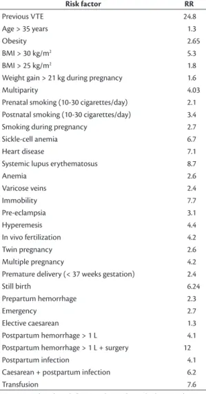
44Table 1. Risk factors for VTE during pregnancy and their associated relative risks (RR).
Risk factor RR
Previous VTE 24.8
Age > 35 years 1.3
Obesity 2.65
BMI > 30 kg/m2 5.3
BMI > 25 kg/m2 1.8
Weight gain > 21 kg during pregnancy 1.6
Multiparity 4.03
Prenatal smoking (10-30 cigarettes/day) 2.1
Postnatal smoking (10-30 cigarettes/day) 3.4
Smoking during pregnancy 2.7
Sickle-cell anemia 6.7
Heart disease 7.1
Systemic lupus erythematosus 8.7
Anemia 2.6
Varicose veins 2.4
Immobility 7.7
Pre-eclampsia 3.1
Hyperemesis 4.4
In vivo fertilization 4.2
Twin pregnancy 2.6
Multiple pregnancy 4.2
Premature delivery (< 37 weeks gestation) 2.4
Still birth 6.24
Prepartum hemorrhage 2.3
Emergency 2.7
Elective caesarean 1.3
Postpartum hemorrhage > 1 L 4.1
Postpartum hemorrhage > 1 L + surgery 12
Postpartum infection 4.1
Caesarean + postpartum infection 6.2
Transfusion 7.6
VTE: venous thromboembolism; RR: relative risk; BMI: body mass index.
Table 2. Dosages of LMWH and UFH for prophylaxis of VTE
related to pregnancy suggested b y the SOCG43. Prophylactic dose of UFH
5,000 UI SC twice a day
Intermediate dose of UFH
10,000 UI SC twice a day
Prophylactic dose of LMWH
Dalteparin: 5,000 UI once a day Enoxaparin: 40 mg once a day
Intermediate dose of LMWH
Dalteparin: 5,000 UI twice a day or 10,000 UI once a day Enoxaparin: 80 mg once a day or 40 mg twice a day
SOGC: prophylactic doses of UFH or LMWH
(preferable) for 6 weeks.
43RCOG: prophylactic doses of LMWH or
anticoagulation with VKA for at least 6 weeks.
45ACCP: prophylactic or intermediate doses of
LMWH or anticoagulation with VKA for 6 weeks.
46Antithrombin deiciency
Antepartum
ACOG: prophylactic, intermediate, or adjusted
doses of LMWH or UFH.
44SOGC: intermediate or therapeutic doses of UFH
or LMWH (preferable).
43RCOG: management should involve a physician
who is a specialist in anticoagulation or thrombosis
during pregnancy; consider serial prenatal assays
of anti-Xa factor and evaluate the possibility of
antithrombin replacement at the start of labor or before
a caesarean; if anti-Xa levels are assayed, a test should
be chosen that does not use exogenous antithrombin,
targeting a peak 4 hours after administration of
0.5 to 1.0 UI/mL: high dose LMWH (50, 75 or 100%
of full weight-adjusted dose).
45ACCP: low risk of recurrence: clinical observation;
Moderate to high risk without use of long-term
anticoagulation: prophylactic or intermediate doses
of LMWH.
46Postpartum
ACOG: prophylactic or intermediate doses of
LMWH, UFH, or anticoagulation with VKA for
4 to 6 weeks.
44SOGC: prophylactic doses of UFH or LMWH
(preferable) for 6 weeks.
43RCOG: LMWH, 50, 75 or 100% of full
weight-adjusted dose for 6 weeks or until resumption
of oral anticoagulation.
45ACCP: prophylactic or intermediate doses of
LMWH or anticoagulation with VKA.
46Antiphospholipid antibody syndrome (APS)
Antepartum
ACOG: anticoagulation with heparin for entire
pregnancy.
44SOGC: intermediate or therapeutic doses of UFH
or LMWH (preferable).
43RCOG: management should involve a physician
who is a specialist in anticoagulation or thrombosis
during pregnancy: prophylaxis with high doses of
LMWH (50, 75 or 100% of full weight‑adjusted dose).
45ACCP: low risk of recurrence: clinical observation;
Moderate to high risk of recurrence without use of
long-term anticoagulation: prophylactic or intermediate
doses of LMWH.
46Postpartum
ACOG: 6 weeks of anticoagulation with heparin.
44SOGC: prophylactic doses of UFH or LMWH
(preferable) for 6 weeks.
43RCOG: high doses of LMWH (50%, 75% or 100%
of full weight-adjusted dose) until resumption of oral
anticoagulation.
45ACCP: prophylactic or intermediate doses of
LMWH or anticoagulation with VKA.
46Previous VTE associated with a transitory risk factor
unrelated to estrogen, with no known thrombophilia
Antepartum
ACOG: clinical observation.
44SOGC: prophylactic doses of UFH or LMWH
(preferable).
43RCOG: if the VTE was provoked by major surgery,
with no other risk factors, thromboprophylaxis with
LMWH can be started at 28 weeks, if there are no
other risk factors; if the original VTE was related to
transitory risk factors other than major surgery, LMWH
should be administered for the entire pregnancy.
45ACCP: low risk of recurrence: clinical observation.
46Postpartum
ACOG: anticoagulant therapy postpartum.
44SOGC: prophylactic doses of UFH or LMWH
(preferable) for 6 weeks.
43RCOG: prophylactic doses of LMWH or
anticoagulation with VKA for at least 6 weeks.
45ACCP: prophylactic or intermediate doses of
LMWH or anticoagulation with VKA for 6 weeks,
if there is no C or S protein deiciency.
46Previous VTE associated with pregnancy or estrogen
Antepartum
ACOG: prophylactic doses of LMWH or UFH.
44SOGC: prophylactic doses of UFH or LMWH
(preferable).
43RCOG: thromboprophylaxis with LMWH.
45ACCP: moderate to high risk of recurrence without
use of long-term anticoagulation: prophylactic or
intermediate doses of LMWH.
46Postpartum
ACOG: anticoagulant therapy postpartum.
44SOGC: prophylactic doses of UFH or LMWH
(preferable) for 6 weeks.
43RCOG: prophylaxis with LMWH or anticoagulation
with VKA for at least 6 weeks.
45ACCP: prophylactic or intermediate doses of
LMWH or anticoagulation with VKA, for 6 weeks,
Previous unprovoked VTE
Antepartum
ACOG: prophylactic doses of LMWH or UFH.
44SOGC: prophylactic doses of UFH or LMWH
(preferable).
43RCOG: prophylactic doses of LMWH.
45ACCP: moderate to high risk of recurrence of VTE
without use of long-term anticoagulation: prophylactic
or intermediate doses of LMWH.
46Postpartum
ACOG: anticoagulant therapy postpartum.
44SOGC: prophylactic doses of UFH or LMWH
(preferable) for 6 weeks.
43RCOG: prophylactic doses of LMWH or AVK for
at least 6 weeks.
45ACCP: prophylactic or intermediate doses of
LMWH or anticoagulation with VKA for 6 weeks,
if there is no C or S protein deiciency.
46Two or more episodes of VTE without long-term
anticoagulation
Antepartum
ACOG: prophylactic or therapeutic doses of
LMWH or UFH.
44SOGC: prophylactic doses of UFH or LMWH
(preferable).
43RCOG: management by a physician who is a
specialist in thrombosis during pregnancy: high doses
of LMWH (50%, 75% or 100% of weight‑adjusted
dose).
45ACCP: moderate to high risk of recurrence without
use of long-term anticoagulation: prophylactic or
intermediate doses of LMWH.
46Postpartum
ACOG: anticoagulation postpartum for 4 to 6 weeks.
44SOGC: prophylactic doses of UFH or LMWH
(preferable) for 6 weeks.
43RCOG: high doses of LMWH (50%, 75% or 100%
of full weight-adjusted dose) for 6 weeks.
45ACCP: prophylactic or intermediate doses of
LMWH or anticoagulation with VKA for 6 weeks,
if there is no C or S protein deiciency.
46Two or more episodes of VTE with use of long term
anticoagulation
Antepartum
ACOG: therapeutic doses of LMWH or UFH.
44SOGC: prophylactic doses of UFH or LMWH
(preferable).
43RCOG: women should be warned of the risks to the
fetus of VKA and counseled to stop this medication and
change to LMWH as soon as pregnancy is conirmed
(the ideal is 2 weeks after a late period and before
6 weeks of pregnancy): high doses of LMWH (50%,
75% or 100% of full weight‑adjusted dose).
44ACCP: when long‑term VKA is being used and
if the patient is a candidate for substitution with
LMWH, it is suggested that pregnancy tests are
conducted frequently and VKA is only substituted
with LMWH when a pregnancy is conirmed. Adjusted
doses or 75% of the therapeutic dose of LMWH are
recommended.
46Postpartum
ACOG: resume long‑term anticoagulation.
44SOGC: resume long‑term anticoagulation.
43RCOG: high doses of LMWH (50%, 75% or 100% of
full weight-adjusted dose) for 6 weeks until resumption
of oral anticoagulation. Use of VKA can be resumed
for women who were on long-term anticoagulation
with this drug when the risk of bleeding has reduced,
usually 5 to 7 days postpartum.
45ACCP: it is suggested that resumption of long‑term
anticoagulation is preferable to administration of
prophylactic doses of LMWH.
46Prevention of VTE associated with caesarean
delivery
A Cochrane systematic review concluded that
there is not enough evidence for post-caesarean
thromboprophylaxis because of the small number of
studies and different comparison criteria.
47While the
risk of VTE associated with caesarean is low,
15,48‑50when
it is present in combination with other risk factors,
the rate of VTE occurrence becomes signiicant and
thromboprophylaxis should be initiated.
43-46ACOG: intermittent pneumatic compression
(IPC) before caesarean if the patient is not on
thromboprophylaxis.
44SOGC: postpartum pharmacological prophylaxis
is recommended in the following situations: previous
VTE; high-risk thrombophilia (APS, antithrombin
deiciency, homozygosity for factor V Leiden or
G20210A prothrombin gene mutation, or combined
thrombophilias), strict bed rest for 7 days or more
before delivery, blood loss of at least 1 L during
peripartum or postpartum, transfusion of blood
products, postpartum surgery, and infection during
peripartum or postpartum.
43Use of pharmacological
prophylaxis should be considered in the event of two
or more of the following: BMI ≥ 30 kg/m
2at irst
prenatal consultation, smoking > 10 cigarettes/day,
previa, emergency caesarean, blood loss of at least 1
L during peripartum or postpartum or transfusion
of blood products, low risk thrombophilia (C or S
protein deiciency, heterozygosity for factor V Leiden
or G20210A prothrombin gene mutation), maternal
cardiac disease, systemic lupus erythematosus,
sickle‑cell anemia, intestinal inlammatory disease,
varicose veins in lower limbs, gestational diabetes,
premature delivery, still birth, or three or more of
the following risk factors: age > 35 years, parity
≥ 2, any type of assisted reproductive technology,
multiple pregnancy, placental abruption, premature
rupture of membranes, elective caesarean, or maternal
cancer. Women with persistent risk factors should
be given thromboprophylaxis for a minimum of
6 weeks postpartum; women with transitory risk
factors during antepartum or peripartum should be
given thromboprophylaxis until hospital discharge
or up to 2 weeks after birth.
43RCOG: emergency caesarean, 10 days after
delivery with prophylactic doses of LMWH; for all
other caesarean patients, consider 10 days of LMWH
at prophylactic doses if there are other risk factors.
45ACCP: in the absence of additional risk factors,
no prophylaxis should be prescribed beyond early
mobilization; in cases with one major risk factor
or ≥ two minor risk factors (or one minor factor
if caesarean was an emergency), prophylaxis with
LMWH after delivery is suggested while the patient
is still in hospital (if there are contraindications to
anticoagulation, use mechanical prophylaxis with
elastic stockings or IPC); in extremely high‑risk
cases with additional risk factors that persist into
puerperium, combine LMWH with elastic stockings
and/or IPC; selected high‑risk patients with additional
risk factors that persist into puerperium should be
given up to 6 weeks extension of prophylaxis after
hospital discharge.
46CONCLUSIONS
The recommendations suggested here could be
subject to individual variations between different
patients and are intended to inform and not to
substitute the clinical judgment of the physician,
who is ultimately responsible for determination of
the appropriate treatment for each individual patient.
Notwithstanding, with appropriate prophylactic
management, the incidence of VTE in pregnant
women can be reduced, thereby preventing its acute
and chronic complications.
REFERENCES
1. Say L, Chou D, Gemmill A, et al. Global causes of maternal death: a WHO systematic analysis. Lancet Glob Health. 2014;2(6):e323-33. PMid:25103301. http://dx.doi.org/10.1016/S2214-109X(14)70227-X.
2. Bourjeily G, Paidas M, Khalil H, Rosene-Montella K, Rodger M. Pulmonary embolism in pregnancy. Lancet. 2010;375(9713):500-12. PMid:19889451. http://dx.doi.org/10.1016/S0140-6736(09)60996-X.
3. Chan WS, Ray JG, Murray S, Coady GE, Coates G, Ginsberg JS. Suspected pulmonary embolism in pregnancy: clinical presentation, results of lung scanning, and subsequent maternal and pediatric outcomes. Arch Intern Med. 2002;162(10):1170-5. PMid:12020189. http://dx.doi.org/10.1001/archinte.162.10.1170.
4. Shahir K, Goodman LR, Tali A, Thorsen KM, Hellman RS. Pulmonary embolism in pregnancy: CT pulmonary angiography versus perfusion scanning. AJR Am J Roentgenol. 2010;195(3):214-20. PMid:20729418. http://dx.doi.org/10.2214/AJR.09.3506.
5. Ramsay R, Byrd L, Tower C, James J, Prescott M, Thachil J. The problem of pulmonary embolism diagnosis in pregnancy. Br J Haematol. 2015;170(5):727-8. PMid:25752876. http://dx.doi. org/10.1111/bjh.13322.
6. Simcox LE, Ormesher L, Tower C, Greer IA. Pulmonary thromboembolism in pregnancy: diagnosis and management. Breathe. 2015;11(4):282-9. PMid:27066121. http://dx.doi.org/10.1183/20734735.008815.
7. Heit JA, Kobbervig CE, James AH, Petterson TM, Bailey KR, Melton LJ 3rd. Trends in the incidence of venous thromboembolism during pregnancy or postpartum: a 30-year population-based study. Ann Intern Med. 2005;143(10):697-706. PMid:16287790. http://dx.doi. org/10.7326/0003-4819-143-10-200511150-00006.
8. Greer IA. Thrombosis in pregnancy: updates in diagnosis and management. Hematology. 2012;2012:203-7. PMid:23233582.
9. Kamel H, Navi BB, Sriram N, Hovsepian DA, Devereux RB, Elkind MSV. Risk of a thrombotic event after the 6-week postpartum period. N Engl J Med. 2014;370(14):1307-15. PMid:24524551. http://dx.doi.org/10.1056/NEJMoa1311485.
10. Ray JG, Chan WS. Deep vein thrombosis during pregnancy and the puerperium: a meta-analysis of the period of risk and the leg of presentation. Obstet Gynecol Surv. 1999;54(4, Supl):265-71. PMid:10198931. http://dx.doi.org/10.1097/00006254-199911001-00026.
11. Jacobsen AF, Skjeldestad FE, Sandset PM. Incidence and risk patterns of venous thromboembolism in pregnancy and puerperium: a register-based case-control study. Am J Obstet Gynecol. 2008;198(2):233.e1-7. PMid:17997389. http://dx.doi. org/10.1016/j.ajog.2007.08.041.
12. Heit JA, Kobbervig CE, James AH, Petterson TM, Bailey KR, Melton LJ 3rd. Trends in the incidence of venous thromboembolism during pregnancy or postpartum: a 30-year population-based study. Ann Intern Med. 2005;143(10):697-706. PMid:16287790. http://dx.doi. org/10.7326/0003-4819-143-10-200511150-00006.
13. Gherman RB, Goodwin TM, Leung B, Byrne JD, Hethumumi R, Montoro M. Incidence, clinical characteristics, and timing of objectively diagnosed venous thromboembolism during pregnancy. Obstet Gynecol. 1999;94(5 Pt 1):730-4. PMid:10546719.
14. Lindqvist P, Dahlbäck B, Marŝál K. Thrombotic risk during pregnancy: a population study. Obstet Gynecol. 1999;94(4):595-9. PMid:10511366.
16. Anderson FA Jr, Wheeler HB, Goldberg RJ, et al. A population-based perspective of the hospital incidence and case-fatality rates of deep vein thrombosis and pulmonary embolism. The Worcester DVT Study. Arch Intern Med. 1991;151(5):933-8. PMid:2025141. http://dx.doi.org/10.1001/archinte.1991.00400050081016.
17. Sultan AA, West J, Tata LJ, Fleming KM, Nelson-Piercy C, Grainge MJ. Risk of first venous thromboembolism in and around pregnancy: a population-based cohort study. Br J Haematol. 2012;156(3):366-73. PMid:22145820. http://dx.doi.org/10.1111/j.1365-2141.2011.08956.x.
18. Virkus RA, Løkkegaard ECL, Bergholt T, Mogensen U, Langhoff-Roos J, Lidegaard Ø. Venous thromboembolism in pregnant and puerperal women in Denmark 1995-2005. A national cohort study. Thromb Haemost. 2011;106(2):304-9. PMid:21713323. http:// dx.doi.org/10.1160/TH10-12-0823.
19. Liu S, Rouleau J, Joseph KS, et al. Epidemiology of pregnancy-associated venous thromboembolism: a population-based study in Canada. J Obstet Gynaecol Can. 2009;31(7):611-20. PMid:19761634. http://dx.doi.org/10.1016/S1701-2163(16)34240-2.
20. Brasil. Ministério da Saúde [site na Internet]. Painel de monitoramento da mortalidade materna. Brasília; 2015. [citado 2016 abr 26]. http:// svs.aids.gov.br/dashboard/mortalidade/materna.show.mtw
21. Lewis G. Saving mothers’ lives: reviewing maternal deaths to make motherhood safer 2003-2005: the seventh report of confidential enquiries into maternal and child health in the United Kingdom. London: CEMACH; 2007.
22. Centre for Maternal and Child Enquiries (CMACE). Saving mothers’ lives. Reviewing maternal deaths to make motherhood safer: 2006–08. The eighth report on confidential enquiries into maternal deaths in the United Kingdom. BJOG. 2011;118(Supl 1):1-203.
23. Knight M, Kenyon S, Brocklehurst P, Neilson J, Shakespeare J, Kurinczuk JJ. Saving lives, improving mother’s care: lessons learned to inform future maternity care from the UK and Ireland Confidential Enquiries into Maternal Deaths and Morbidity 2009-12. Oxford: National Perinatal Epidemiology Unit, University of Oxford; 2014.
24. Nelson-Piercy C, Maccallum P, Mackillop MA. Reducing the risk of venous thromboembolism during pregnancy and the puerperium: RCOG green-top guideline. 37. ed. London: RCOG; 2015. [citado 2016 abr 27]. https://www.rcog.org.uk/globalassets/documents/ guidelines/gtg-37a.pdf
25. Won HS, Kim DY, Yang MS, Lee SJ, Shin H, Park JB. Pregnancy-induced hypertension, but not gestational diabetes mellitus, is a risk factor for venous thromboembolism in pregnancy. Korean Circ J. 2011;41(1):23-7. PMid:21359065. http://dx.doi.org/10.4070/ kcj.2011.41.1.23.
26. Liu S, Liston RM, Joseph KS, Heaman M, Sauve R, Kramer MS. Maternal mortality and severe morbidity associated with low-risk planned cesarean delivery versus planned vaginal delivery at term. CMAJ. 2007;176(4):455-60. PMid:17296957. http://dx.doi. org/10.1503/cmaj.060870.
27. Kane EV, Calderwood C, Dobbie R, Morris C, Roman E, Greer IA. A population-based study of venous thrombosis in pregnancy in Scotland 1980-2005. Eur J Obstet Gynecol Reprod Biol. 2013;169(2):223-9. PMid:23684606. http://dx.doi.org/10.1016/j. ejogrb.2013.03.024.
28. McColl MD, Walker ID, Greer IA. The role of inherited thrombophilia in venous thromboembolism associated with pregnancy. Br J Obstet Gynaecol. 1999;106(8):756-66. PMid:10453824. http:// dx.doi.org/10.1111/j.1471-0528.1999.tb08395.x.
29. Gerhardt A, Scharf RE, Zotz RB. Effect of hemostatic risk factors on the individual probability of thrombosis during pregnancy and the puerperium. Thromb Haemost. 2003;90(1):77-85. PMid:12876629.
30. Knight M. Antenatal pulmonary embolism: risk factors, management and outcomes. BJOG. 2008;115(4):453-61. PMid:18201281. http:// dx.doi.org/10.1111/j.1471-0528.2007.01622.x.
31. James AH, Jamison MG, Brancazio LR, Myers ER. Venous thromboembolism during pregnancy and the postpartum period: incidence, risk factors, and mortality. Am J Obstet Gynecol. 2006;194(5):1311-5. PMid:16647915. http://dx.doi.org/10.1016/j. ajog.2005.11.008.
32. Larsen TB, Sørensen HT, Gislum M, Johnsen SP. Maternal smoking, obesity, and risk of venous thromboembolism during pregnancy and the puerperium: a population-based nested case-control study. Thromb Res. 2007;120(4):505-9. PMid:17257657. http:// dx.doi.org/10.1016/j.thromres.2006.12.003.
33. Robinson HE, O’Connell CM, Joseph KS, McLeod NL. Maternal outcomes in pregnancies complicated by obesity. Obstet Gynecol. 2005;106(6):1357-64. PMid:16319263. http://dx.doi.org/10.1097/01. AOG.0000188387.88032.41.
34. Anderson FA Jr, Spencer FA. Risk factors for venous thromboembolism. Circulation. 2003;107(23, Supl 1):16. PMid:12814980.
35. National Collaborating Centre for Women’s and Children’s Health. National Institute for Health and Clinical Excellence. Antenatal care: routine care for the healthy pregnant woman. 2. ed. London: RCOG Press; 2008. Clinical Guideline, 2008.
36. Hezelgrave NL, Whitty CJM, Shennan AH, Chappell LC. Advising on travel during pregnancy. BMJ. 2011;342(1):d2506. PMid:21527456. http://dx.doi.org/10.1136/bmj.d2506.
37. Greer I. Air travel and pregnancy. London: RCOG; 2013. no. 1. [citado 2014 abr 29]. https://www.rcog.org.uk/en/guidelines-researchservices/ guidelines/sip1/
38. Sultan AA, West J, Tata LJ, Fleming KM, Nelson-Piercy C, Grainge MJ. Risk of first venous thromboembolism in pregnant women in hospital: population based cohort study from England. BMJ. 2013;347(15):f6099. PMid:24201164. http://dx.doi.org/10.1136/ bmj.f6099.
39. Bröms G, Granath F, Linder M, Stephansson O, Elmberg M, Kieler H. Complications from inflammatory bowel disease during pregnancy and delivery. Clin Gastroenterol Hepatol. 2012;10(11):1246-52. PMid:22922307. http://dx.doi.org/10.1016/j.cgh.2012.08.018.
40. Erekson EA, Brousseau EC, Dick-Biascoechea MA, Ciarleglio MM, Lockwood CJ, Pettker CM. Maternal postoperative complications after non obstetric antenatal surgery. J Matern Fetal Neonatal Med. 2012;25(12):2639-44. PMid:22735069. http://dx.doi.org/1 0.3109/14767058.2012.704445.
41. Blanco-Molina A, Trujillo-Santos J, Criado J, et al. Venous thromboembolism during pregnancy or postpartum: findings from the RIETE Registry. Thromb Haemost. 2007;97(2):186-90. PMid:17264945.
42. Danilenko-Dixon DR, Heit JA, Silverstein MD, et al. Risk factors for deep vein thrombosis and pulmonary embolism during pregnancy or post-partum: a population-based, case-control study. Am J Obstet Gynecol. 2001;184(2):104-10. PMid:11174488. http:// dx.doi.org/10.1067/mob.2001.107919.
43. Chan WS, Rey E, Kent NE, et al. Venous thromboembolism and antithrombotic therapy in pregnancy. J Obstet Gynaecol Can. 2014;36(6):527-53. PMid:24927193. http://dx.doi.org/10.1016/ S1701-2163(15)30569-7.
44. James A. Practice bulletin no. 123: thromboembolism in pregnancy. Obstet Gynecol. 2011;118(3):718-29. PMid:21860313. http://dx.doi. org/10.1097/AOG.0b013e3182310c4c.
37a. [citado 2016 abr 27]. https://www.rcog.org.uk/globalassets/ documents/guidelines/gtg-37a.pdf
46. Bates SM, Greer IA, Middeldorp S, Veenstra DL, Prabulos AM, Vandvik PO. VTE, thrombophilia, antithrombotic therapy, and pregnancy: Antithrombotic Therapy and Prevention of Thrombosis. Chest. 2012;141(2, Supl):e691S-736S. PMid:22315276. http://dx.doi. org/10.1378/chest.11-2300.
47. Lockwood CJ. ACOG Guidelines at a Glance: the mystery of the antibodies still awaits a solution. Ohio: Contemporary OB/GYN; 2013. [citado 2016 abr 27]. http://contemporaryobgyn.modernmedicine. com/contemporary-obgyn/content/tags/acog-practice-bulletins/ acog-guidelines-glance-mystery-antibodies-st
48. Tooher R, Gates S, Dowswell T, Davis LJ. Prophylaxis for venous thromboembolic disease in pregnancy and the early postnatal period. Cochrane Database Syst Rev. 2010;(5):CD001689. PMid:20464719.
49. Tooher R, Gates S, Dowswell T, Davis LJ. Prophylaxis for venous thromboembolic disease in pregnancy and the early postnatal period. Cochrane Database Syst Rev. 2010;(5):CD001689. PMid:20464719. 50. Jacobsen AF, Skjeldestad FE, Sandset PM. Ante- and postnatal
risk factors of venous thrombosis: a hospital-based case-control study. J Thromb Haemost. 2008;6(6):905-12. PMid:18363820. http://dx.doi.org/10.1111/j.1538-7836.2008.02961.x.
*
Correspondence
Marcos Arêas Marques Universidade do Estado do Rio de Janeiro – UERJ, Hospital Universitário Pedro Ernesto – HUPE, Angiologia Rua Barão de Lucena, 48, sala 10 - Botafogo CEP 22260-020 - Rio de Janeiro (RJ), Brazil Tel.: +55 (21) 99859-0160 E-mail: [email protected]
Author information
ALMLO - Obstetrician at Centro de Referência da Saúde da Mulher do Estado de São Paulo; MSc in Sciences from Universidade de São Paulo (USP). MAM - Angiologist at Unidade Docente Assistencial de Angiologia, Hospital Universitário Pedro Ernesto (HUPE), Universidade do Estado do Rio de Janeiro (UERJ).
Author contributions