Mechanisms linking brain insulin
resistance to Alzheimer’s disease
Maria Niures P.S. Matioli
1, Ricardo Nitrini
2ABSTRACT.
Several studies have indicated that Diabetes Mellitus (DM) can increase the risk of developing Alzheimer’s
disease (AD). This review briefly describes current concepts in mechanisms linking DM and insulin resistance/deficiency to
AD. Insulin/insulin-like growth factor (IGF) resistance can contribute to neurodegeneration by several mechanisms which
involve: energy and metabolism deficits, impairment of Glucose transporter-4 function, oxidative and endoplasmic reticulum
stress, mitochondrial dysfunction, accumulation of AGEs, ROS and RNS with increased production of neuro-inflammation
and activation of pro-apoptosis cascade. Impairment in insulin receptor function and increased expression and activation
of insulin-degrading enzyme (IDE) have also been described. These processes compromise neuronal and glial function,
with a reduction in neurotransmitter homeostasis. Insulin/IGF resistance causes the accumulation of A
β
PP-A
β
oligomeric
fibrils or insoluble larger aggregated fibrils in the form of plaques that are neurotoxic. Additionally, there is production
and accumulation of hyper-phosphorylated insoluble fibrillar tau which can exacerbate cytoskeletal collapse and synaptic
disconnection.
Key words:
Alzheimer’s disease, diabetes mellitus, insulin resistance, neurodegeneration, mechanisms.
MECANISMOS QUE LIGAM A RESISTÊNCIA INSULÍNICA CEREBRAL À DOENÇA DE ALZHEIMER: UMA BREVE REVISÃO
RESUMO.
Atualmente, muitos estudos têm indicado que o Diabetes Mellitus (DM) pode aumentar o risco de desenvolver
doença de Alzheimer (DA). Esta revisão tem o objetivo de descrever brevemente os conceitos atuais sobre os mecanismos
que associam DM, resistência/deficiência de insulina à DA. Resistência à insulina/fator de crescimento similar à insulina (IGF)
pode contribuir para a neurodegeneração através de vários mecanismos os quais envolvem: déficit metabólico e energético,
prejuízo na função do transportador de glicose-4, estresse oxidativo e do retículo endoplasmático, disfunção mitocondrial,
acúmulo de AGEs, ROS e RNS com aumento na produção da neuro-inflamação e ativação da cascata pró-apoptóptica.
Prejuízo na função do receptor de insulina, aumento na expressão e ativação da enzima de degradação da insulina (EDI)
também têm sido descritos. Esses processos comprometem a função neuronal e glial, com redução da homeostase de
neurotransmissor. Resistência à insulina/IGF causa acúmulo de fibrilas de oligômeros de PP
β
A-
β
A e grandes agregados
fibrilares insolúveis em forma de placas que são neurotóxicos. Adicionalmente, há produção e acúmulo de fibrilas insolúveis
de tau hiperfosforilada que podem exacerbar o colapso do citoesqueleto e a desconexão sináptica.
Palavras-chave:
doença de Alzheimer, diabetes mellitus, resistência insulínica, neurodegeneração, mecanismos.
INTRODUCTION
P
opulation aging is a global phenomenon
leading to an increase in chronic diseases
such as dementia and diabetes mellitus (DM),
which pose an epidemic challenge to global
health care systems. In 2012, the WHO
pub-lished that 35.6 million people had dementia
worldwide and that this number is set to reach
65.7 million by 2030.
1Alzheimer’s disease
(AD) is the most common cause of dementia,
especially in the elderly population.
1Recently,
the International Diabetes Federation
2esti-mated that 382 million people had diabetes
in 2013, where this number may rise to 592
million within less than 25 years.
2Moreover,
1Pós-graduanda, nível de Doutorado, Departamento de Neurologia da Faculdade de Medicina da Universidade de São Paulo. 2Professor Titular da Disciplina de
Neurologia da Faculdade de Medicina da Universidade de São Paulo. Orientador e Professor Responsável pela Pós-graduação do Departamento de Neurologia da Faculdade de Medicina da Universidade de São Paulo.
Maria Niures P. S.Matioli. Av. Almirante Cochrane, 123 / 101– 11040-001 Santos SP – Brasil. E-mail: [email protected]; [email protected].
Disclosure: The authors report no conflicts of interest.
Received September 11, 2014. Accepted in final form December 20, 2014.
80% of the total number afected live in low- and
mid-dle-income countries and Type 2 diabetes (T2DM) is the
most common type of DM.
2he prevalence of AD and
T2DM increases with aging.
3he AD pathology is characterized by the
accumula-tion of the following in the brain: amyloid beta precursor
protein (A
β
PP)-A
β
large insoluble ibrillar aggregates in
the form of plaques, soluble neurotoxic oligomeric
i-brils, hyper-phosphorylation of tau protein with
neuro-ibrillary tangles (NFTs) deposition, dystrophic neuritis,
and neuropil threads.
4,5In familial forms of AD, the
mu-tations in A
β
PP, presenilin 1 (PS1) and 2 (PS2) genes, or
inheritance of the Apolipoprotein E e4 (ApoE-e4) allele
can cause increased synthesis and deposition of A
β
PP-
A
β.
6,7However, the cause of A
β
PP-A
β
accumulation in
sporadic AD, the most common form of the disease,
re-mains unknown.
5However, evidence suggests that
im-pairment in insulin and insulin-like growth factor (IGF)
compromises A
β
PP expression and protein processing
which could be responsible for A
β
PP-A
β
accumulation.
8he association between DM and AD is controversial
in literature.
9,10Many studies have demonstrated a
posi-tive association between DM and AD, especially in
epide-miological research, studies in animals and cells,
11-17but
these indings have not been entirely conirmed in
neu-ropathological studies.
3,18-23Based on this positive
asso-ciation, researchers have studied DM treatments as a
tar-get to diminish or avoid AD onset and progression.
24-28he exact mechanisms by which DM afects the
brain remain unclear, but this probably occurs through
cerebrovascular and neurodegenerative changes.
29he
aim of this article was to provide a brief review on the
main mechanisms associating AD with DM due to
insu-lin resistance and deiciency.
Insulin and insulin-like growth factor actions in the central
ner-vous system.
he insulin produced by the pancreas can
cross the blood brain barrier (BBB) from the circulation
to the brain by a receptor-dependent mechanism,
30but
the levels of insulin expression in the brain are modest
compared to circulating levels.
10he transport of
pe-ripheral insulin across the BBB and the consequences
of peripheral hyperinsulinemia or hypoinsulinemia are
signiicantly important to cerebral insulin signaling.
10Insulin binding activity has been identiied in the brain
in a number of species, including humans.
31,32Further-more, insulin receptors (IR) are expressed in cerebral
vasculature and can mediate insulin traic across the
BBB.
33Insulin and IGF play an important role in brain
function and structure.
5Insulin, IGF-1 and IGF-2
poly-peptides and receptor genes are expressed in neurons
34and glia,
35,36particularly in structures that are targeted
in neurodegenerative diseases.
34,35,37IGF and insulin
are associated with regulating and maintaining
cogni-tive function,
38and participate in neuronal and glial
functions such as growth, metabolism, survival, gene
expression, protein synthesis, cytoskeletal assembly,
neurotransmitter function, synapse formation and
plasticity.
34,39Glucose transporter 4 (GLUT4) is very important
for glucose uptake and utilization in the brain.
38Insulin
stimulates GLUT4 gene expression and protein
traick-ing from the cytosol to the plasma membrane,
modulat-ing glucose uptake and utilization.
38Consequently, the
regulation of neuronal metabolism and the generation
of energy needed for cognition and memory are linked
to insulin stimulation of GLUT4.
38GLUT4 is
abundant-ly expressed along with insulin receptors, in medial
tem-poral lobe structures which are afected in AD
pathol-ogy. Nevertheless, post-mortem brain studies have not
detected signiicant reductions in GLUT4 expression
in AD.
40Deicits in brain glucose utilization and
ener-gy metabolism, and brain insulin/IGF resistance could
be mediated by impairments in GLUT4 traicking
be-tween the cytosol and plasma membrane.
38Insulin and IGF binding to their own receptors
ac-tivates some pathways, leading to phosphorylation and
activation of intrinsic receptor tyrosine kinases. he
phosphorylated receptors interact with IR substrate
molecules and promote transmission of downstream
signals that stimulate growth, survival, metabolism,
plasticity and inhibit apoptosis.
38Brain insulin/IGF resistance and AD.
AD has been associated
with deicits in insulin/IGF signaling due to the efects
of insulin/IGF resistance and deiciency.
5Deicits in
ce-rebral glucose utilization have been described in the
ear-ly stages of AD.
41-44Suzanne de la Monte and colleagues
have proposed the concept of AD as “Type 3 diabetes”.
40hey observed an inverse correlation between IR
abun-dance and the Braak score of AD brains, with 80%
re-duced IR substrates levels in the most severe cases. hey
described reduced messenger RNA levels of IGF-1 and
increased Tau protein levels regulated by IR.
40,45Stud-ies with small interfering RNA molecules showed that
molecular disruption of brain insulin and IGF
recep-tors was suicient to cause cognitive impairment and
hippocampal degeneration similar to AD molecular
abnormalities.
46Neuro-degeneration can occur by several mechanisms such as
the activation of kinases that aberrantly
phosphory-late tau, the expression of A
β
PP and accumulation of
A
β
PP-A
β
in brain insulin/IGF resistance.
38Hypergly-cemia leads to the accumulation of advanced glycation
end products (AGEs) that disrupts removal of A
β
42 and
induces A
β
and Tau glycation, promoting A
β
aggrega-tion and NFTs formaaggrega-tion in the brain.
38,47,48AGE
pro-duction is found in normal aging, but becomes highly
accelerated in diabetes.
49Recent evidence suggests
that glyceraldehyde-derived AGEs (glycer-AGE) are the
predominant modiication of the most toxic forms of
AGEs, and Glycer-AGE-modiied proteins are directly
toxic to cultured neurons. Diabetic serum enriched with
glycer-AGE modiied proteins has shown toxic efects
on neurons.
10AGEs are also linked to microvascular
al-terations in hyperglycemia and diabetes.
50Receptor for
advanced glycation end products (RAGE) expression
has been associated with pathological conditions such
as diabetic vascular disease, chronic inlammation and
AD.
51,52Studies with immunohistochemistry for RAGE
in AD brains have demonstrated that RAGE increased
expression in neurons, microglia, astrocytes and
vascu-lar endothelial cells.
53,54RAGE binds and interacts with
AGEs and also with A
β.
49RAGE interaction with
AGE-modiied proteins in either diabetes or AD, or A
β
in
AD, can produce damaging inlammatory responses
55,56and be responsible for vascular complications in DM
and AD.
57-59RAGE mediates the transport of plasma A
β
across the BBB
60and the migration of monocytes across
the human brain endothelial cells in response to A
β.
61Microvascular disease is seen as a consequence of
diabetes and can also be found in AD brains, possibly
contributing to the cognitive impairment and
neuro-degeneration seen in AD.
5,62Decreased blood low and
impairment of oxygen and nutrient delivery exacerbate
the adverse efects of insulin/IGF resistance.
63Con-sequently, there is an increase in oxidative stress and
activation of signaling mechanisms which promote
ab-errant tau phosphorylation, A
β
PP cleavage, A
β
PP-A
β
deposition, and mitochondrial dysfunction.
38,63IR function is compromised in brain insulin/IGF
resistance, leading to many adverse efects. here is
decreased signaling through IR substrate,
phosphoino-sitol-3-kinase (PI3K) and Akt, with reduced neuronal
and oligodendroglial survival, neuronal plasticity and
myelin maintenance.
38IR dysfunction increases
acti-vation of glycogen synthetase kinase 3
β
(GSK-3
β
) and
phosphatases that negatively regulate insulin signaling,
consequently producing increased tau phosphorylation,
oxidative stress, neuro-inlammation and pro-apoptosis
signaling.
38Reduced insulin-responsive gene expression
seen in IR dysfunction can lead to deicits in
acetylcho-line and glucose metabolism.
38Impairment in GLUT4 functions in brain with
in-sulin/IGF resistance results in reduced glucose uptake
and utilization, consequently compromising cell energy
and homeostatic functions, disrupting neuronal
cyto-skeleton and synaptic connection.
38Deicits in energy
metabolism lead to increased oxidative and
endoplas-mic reticulum (ER) stress, and mitochondrial
dysfunc-tion with the generadysfunc-tion of reactive oxygen (ROS) and
reactive nitrogen species (RNS).
64-66Increased
oxida-tive stress, ROS and RNS damage RNA, DNA, proteins,
and lipid peroxidation production, energy deicits, cell
death, increased A
β
PP expression, A
β
42 deposition and
ibrillarization.
38here is activation of
pro-inlammato-ry and pro-death cascades and down-regulation of
tar-get genes that mediate cholinergic homeostasis linked
to AD in brain with insulin/IGF resistance.
5,67Impair-ment of myelin maintenance also occurs and can lead
to increased neuro-inlammation, oxidative stress,
pro-apoptosis, and further insulin resistance, besides white
matter atrophy.
38he insulin-degrading enzyme (IDE) has the
proper-ty of catabolizing insulin and A
β
, and may play a critical
role in A
β
clearance in the brain as A
β
scavenger
prote-ase.
68,69IDE acts as a general regulator of amyloid
bur-den in the pancreas and brain.
70Insulin regulates IDE
expression and can directly compete with A
β
for binding
to IDE.
71In hyper-insulin states, IDE can be diverted to
degrade insulin, consequently allowing A
β
PP-A
β
accu-mulation.
70Mutations in the IDE gene in mice resulted
in reduced activity of this enzyme, lower rates of A
β
and
insulin degradation, additionally developing
hyperinsu-linaemia and accumulating A
β
species in their brains.
72Chronic hyperglycaemia, hyperinsulinaemia, oxidative
stress, accumulation of AGEs, increased expression and
activation of IDE, increased production of
pro-inlamma-tory cytokines, and cerebral microvascular disease
asso-ciated with peripheral insulin resistance could result in
mild cognitive impairment and neurodegeneration.
38,73Brain insulin/IGF resistance and Aβ pathology.
Altered
pro-teolysis with increased A
β
PP gene expression results in
the accumulation of 40 or 42 amino acid length A
β
pep-tides that can aggregate and have been described in AD
pathology. Dysregulated expression and processing of
A
β
PP leads to the accumulation of A
β
PP-A
β
oligomeric
ibrils or insoluble larger aggregated ibrils in the form
of plaques that are neurotoxic.
5he interest in the role
consequence of dysregulated A
β
PP-A
β
expression and
protein processing has grown in literature.
38Insulin can
accelerate traicking of A
β
PP-A
β
from the trans-Golgi
network to the plasma membrane as well as its
extra-cellular secretion
74and also inhibits its intracellular
degradation by IDE.
75Impaired insulin signaling can
disrupt both the processing of A
β
PP and clearance of
A
β
PP-A
β.
76Simultaneously, A
β
PP-A
β
afects insulin
sig-naling by competing with insulin, or reducing the
ain-ity of insulin for binding to its own receptor.
77A
β
PP-A
β
oligomers desensitize and reduce the surface expression
of IRs, consequently inhibiting neuronal
insulin-signal-ing.
67Additionally, intracellular A
β
PP-A
β
interferes with
PI3k activation of Akt, leading to reduced signaling,
increased activation of GSK-3
β
, and
hyper-phosphory-lation of tau. Increased levels of GSK-3 promote A
β
PP
processing and A
β
PP-A
β
accumulation.
78Brain insulin/IGF resistance and Tau pathology.
In AD, the
main neuronal cytoskeletal lesions correlated with
se-verity of dementia, including NFTs and dystrophic
neu-rites, contain aggregated and ubiquitinated insoluble
ibrillar tau.
4,38,79Tau gene expression and
phosphoryla-tion can be regulated by insulin/IGF stimulaphosphoryla-tion.
80,81Re-duced insulin/IGF signaling can impair tau gene
expres-sion and contribute to tau pathology.
82Brain insulin/IGF
resistance results in decreased signaling through PI3K,
Akt,
80,81and Wnt/
β
-catenin,
83and increased activation
of GSK-3
β
.
84,85he hyper-phosphorylation of tau, which
leads to tau misfolding and ibril aggregation in AD
pa-thology, can be partly due to GSK-3
β
overactivation.
86Tau hyper-phosphorylation is mediated by increased
activation of cyclin-dependent kinase 5 (cdk-5) and
c-Abl kinases,
87,88and inhibition of protein phosphatases
1 and 2A.
88,89Tau protein misfolds and self-aggregates
into insoluble ibrillar structures lead to neuroibrillary
tangles, dystrophic neurites, and neuropil threads.
38,90he results of generation and accumulation of
hyper-phosphorylated insoluble ibrillar tau are the
exacer-bation of cytoskeletal collapse, neurite retraction, and
synaptic disconnection.
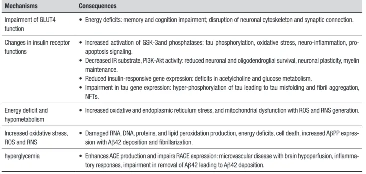
38Table 1 summarizes the main
mechanisms linking brain insulin/IGF resistance to AD
pathology.
Neurodegenerative process contributing to brain insulin
resis-tance in AD.
Interestingly, the neuropathological process
involved in AD can reinforce brain insulin resistance. A
β
toxicity, microvascular disease, oxidative stress,
tran-sition metal ion accumulations and
hyperphosphory-lated-ubiquitinated tau lead to increased brain insulin
resistance.
38A
β
42 toxicity competes with insulin and
reduces the ainity of insulin binding to its receptor.
77,91A
β
PP oligomers desensitize and reduce surface
expres-sion of insulin receptors, and interfere with PI3K
activa-tion of Akt.
38,92he A
β
toxicity disrupts insulin
signal-ing and impairs insulin stimulated neuronal survival
Table 1.
Summary of mechanisms linking brain insulin/IGF resistance to AD pathology.
Mechanisms
Consequences
Impairment of GLUT4
function
• Energy deficits: memory and cognition impairment; disruption of neuronal cytoskeleton and synaptic connection.
Changes in insulin receptor
functions
• Increased activation of GSK-3and phosphatases: tau phosphorylation, oxidative stress, neuro-inflammation,
pro-apoptosis signaling.
• Decreased IR substrate, PI3K-Akt activity: reduced neuronal and oligodendroglial survival, neuronal plasticity, myelin
maintenance.
• Reduced insulin-responsive gene expression: deficits in acetylcholine and glucose metabolism.
• Impairment in tau gene expression: hyper-phosphorylation of tau leading to tau misfolding and fibril aggregation,
NFTs.
Energy deficit and
hypometabolism
• Increased oxidative and endoplasmic reticulum stress, and mitochondrial dysfunction with ROS and RNS generation.
Increased oxidative stress,
ROS and RNS
• Damaged RNA, DNA, proteins, and lipid peroxidation production, energy deficits, cell death, increased A
β
PP
expres-sion with A
β
42 deposition and fibrillarization.
hyperglycemia
• Enhances AGE production and impairs RAGE expression: microvascular disease with brain hypoperfusion,
inflamma-tory responses, impairment in removal of A
β
42 leading to A
β
42 deposition.
and plasticity.
38Oxidative stress can produce increases
in neuro-inlammation and pro-inlammatory cytokine
inhibition of insulin signaling.
38Transition metal ion
ac-cumulations produce mitochondrial dysfunction,
oxida-tive stress, tau and A
β
PP oligomer ibrillarization, which
impair glucose uptake and utilization, and inhibit
insu-lin signainsu-ling.
38Hyperphosphorylated-ubiquitinated tau
increases oxidative stress, promotes
neuroinlamma-tion which consequently enhances insulin resistance.
38Microvascular disease exacerbates insulin resistance
through cerebral hypoperfusion and hypoxic-ischemic
injury.
38Conclusions.
A body of evidence has shown that the
structural and functional integrity of the CNS can be
compromised in the presence of brain insulin and IGF
resistance or deiciency. hese changes can contribute
to AD pathology and conversely, AD pathology can
enhance brain insulin and IGF resistance, functioning
as a positive feedback loop. However, it is necessary
to bear in mind that the majority of studies have been
conducted in the experimental ield with animal or cell
models. Elucidating the question of a connection among
DM, brain insulin resistance/deiciency and AD is very
important, especially for planning novel strategies to
prevent and treat AD in the future.
Author contribution.
Maria Niures P.S. Matioli drafted the
manuscript, and Ricardo Nitrini critically revised the
manuscript.
REFERENCES
1. World Health Organization. Dementia: a public health priority. WHO Library 2012.
2. International Diabetes Federation, sixth edition. Diabetes Atlas, 2014. www.idf.org/diabetesatlas.
3. Janson J, Laedtke T, Parisi JE, O’Brien P, Petersen RC, Butler PC. Increased risk of type 2 diabetes in Alzheimer disease. Diabetes 2004; 53:474-481.
4. Duyckaerts C, Delatour B, Potier MC. Classification and basic pathology of Alzheimer disease. Acta Neuropathol 2009;118:5-36.
5. de la Monte S M and Tong M. Brain metabolic dysfunction at the core of Alzheimer’s disease. Biochem Pharmacol 2014;88:548-559. 6. Robakis NK. Mechanisms of AD neurodegeneration may be
indepen-dent of Abeta and its derivatives. Neurobiol Aging 2011;32:372-9. 7. Wu L, Rosa-Neto P, Hsiung GY, et al. Early-onset familial Alzheimer’s
disease (EOFAD). Can J Neurol Sci 2012;39:436-445.
8. Bosco D, Fava A, Plastino M, Montalcini T, Pujia A. Possible implications of insulin resistance and glucose metabolism in Alzheimer’s disease pathogenesis. J Cell Mol Med 2011;15:1807-1821.
9. Biessels GJ, Staekenborg S, Brayne C and Scheltens P. Risk of dementia in diabetes mellitus: a systematic review. Lancet Neurol 2006; 5:64-74.
10. Arab L, Sadeghi R, Walker DG, Lue LF and Sabbagh MN. Conse-quences of Aberrant Insulin Regulation in the Brain: Can Treating Diabetes be Effective for Alzheimer’s Disease. Curr Neuropharmacol 2011;9:693-705.
11. Leibson CL, Rocca WA, Hanson VA, et al. Risk of dementia among persons with diabetes mellitus: a population-based cohort study. Am J Epidemiol 1997;145:301-308.
12. Brayne C, Gill C, Huppert FA, et al. Vascular risks and incident dementia: results from a cohort study of the very old. Dement Geriatr Cogn Disord 1998;9:175-180.
13. Ott A, Stolk RP, van Harskamp F, et al. Diabetes mellitus and the risk of dementia: The Rotterdam Study. Neurology 1999;53:1937-1942. 14. Arvanitakis Z, Wilson RS, Bienias JL, et al. Diabetes mellitus and risk
of Alzheimer disease and decline in cognitive function. Arch Neurol 2004;61:661-666.
15. Luchsinger JA, Reitz C, Honig LS, et al. Aggregation of vascular risk factors and risk of incident Alzheimer disease. Neurology 2005;654:545-551. 16. Whitmer RA, Sidney S, Selby J, et al. Midlife cardiovascular risk factors
and risk of dementia in late life. Neurology 2005;64:277-281. 17. Cheng D, Noble J, Tang MX, et al. Type 2 diabetes and late-onset
Alzheimer’s disease. Dement Geriatr Cogn Disord 2011;31:424-430. 18. Arvanitakis Z, Schneider JA, Wilson RS, at al. Diabetes is related to
cerebral infarction but not AD pathology in older persons. Neurology 2006;67:1960-5.
19. Heitner J, Dickson D. Diabetes do not have increased Alzheimer’ type pathology compared with age-matched controls subjects. A
retro-spective postmortem immunocytochemical and hisfluorescence study. Neurology 1997;49:1306-11.
20. Beeri MS, Silverman JM, Davis KL, et al. Type 2 Diabetes Is Negatively Associated with Alzheimer’s disease Neuropathology. J Gerontol A Biol Sci Med Sci 2005;60:471-475.
21. Alafuzoff I, Aho L, Helisalmi S, Mannermaat A, Soininen H. β-Amyloid deposition in brains of subjects with diabetes. Neuropathol Appl Neuro-biol 2009;35:60-68.
22. Nelson PT, Smith CD, Abner EA, et al. Human cerebral neuropathology of Type 2 diabetes mellitus. Biochimica et Biophysica Acta 2009; 1792:454-469.
23. Ahtiluoto S, Polvikoski T, Peltonen M, et al. Diabetes, Alzheimer disease, and vascular dementia: a population-based neuropathological study. Neurology, 2010;75:1195-1202.
24. Watson GS, Cholerton BA, Reger MA, et al. Preserved cognition in patients with early Alzheimer disease and amnestic mild cognitive impairment during treatment with rosiglitazone: a preliminary study. Am J Geriatr Psychiatry 2005;13:950-958.
25. Risner ME, Saunders AM, Altman JF, et al. Efficacy of rosiglitazone in a genetically defined population with mild-to-moderate Alzheimer’s disease. Pharmacogenomics J 2006;6:246-254.
26. Roses AD. Commentary on “a roadmap for the prevention of dementia: the inaugural Leon Thal Symposium.” An impending prevention clinical trial for Alzheimer’s disease: roadmaps and realities. Alzheimers Dement 2008;4:164-166.
27. Reger MA, Watson GS, Green PS, et al. Intranasal insulin improves cognition and modulates beta-amyloid in early AD. Neurology 2008;70:440-448.
28. Sato T, Hanyu H, Hirao K, et al. Efficacy of PPAR-γ agonist pioglitazone in mild Alzheimer disease. Neurobiol Aging 2011;32:1626-1633. 29. Bornstein NM, Brainin M, Guekht A, et al. Diabetes and the brain: issues
and unmet needs. Neurol Sci 2014;35:995-1001.
30. Yu Y, Kastin AJ, Pan W. Reciprocal interactions of insulin and insulin-like growth factor I in receptor-mediated transport across the blood-brain barrier. Endocrinology 2006;147:2611-2615.
31. Roth RA, Morgan DO, Beaudoin J, Sara V. Purification and character-ization of the human brain insulin receptor. J Biol Chem 1986;261:3753-3757.
32. Kotzke G, Schutt M, Missler U, et al. Binding of human, porcine and bovine insulin to insulin receptors from human brain, muscle and adipocytes and to expressed recombinant alternatively spliced insulin receptor isoforms. Diabetologia 1995;38:757-763.
expression, signaling, and malfunction in the central nervous system: relevance to Alzheimer’s disease. J Alzheimers Dis 2005;7:45-61. 35. Zeger M, Popken G, Zhang J, Xuan S, Lu QR, Schwab MH, et al.
Insulin-like Glia 2007;55:400-11.
36. Freude S, Schilbach K, Schubert M. The role of IGF-1 receptor and insulin receptor signaling for the pathogenesis of Alzheimer’s disease: from model organisms to human disease. Curr Alzheimer Res 2009; 6:213-223.
37. de la Monte SM, Longato L, Tong M, Wands JR. Insulin resistance and neurodegeneration: roles of obesity, type 2 diabetes mellitus and non-alcoholic steatohepatitis. Curr Opin Investig Drugs 2009;10:1049-1060. 38. de la Monte SM (b). Brain Insulin Resistence and Deficiency as Thera-peutic Targets in Alzheimer’s Disease. Curr Alzheimer Res 2012;9:35-66. 39. D’Ercole AJ, Ye P. Expanding the mind: insulin-like growth factor I and
brain development. Endocrinology 2008;149:5958-5962.
40. Steen E, Terry BM, Rivera EJ, et al. Impaired insulin and insulin-like growth factor expression and signaling mechanisms in Alzheimer’s disease--is this type 3 diabetes? J Alzheimers Dis 2005;7:63-80. 41. Caselli RJ, Chen K, Lee W, et al. Correlating cerebral hypometabolism
with future memory decline in subsequent converters to amnestic pre-mild cognitive impairment. Arch Neurol 2008;65:1231-1236. 42. Mosconi L, Pupi A, De Leon MJ. Brain glucose hypometabolism and
oxidative stress in preclinical Alzheimer’s disease. Ann N Y Acad Sci 2008;1147:180-95.
43. Mosconi L, Mistur R, Switalski R, et al. FDG-PET changes in brain glucose metabolism from normal cognition to pathologically verified Alzheimer’s disease. Eur J Nucl Med Mol Imaging 2009;36:811-822. 44. Langbaum JB, Chen K, Caselli RJ, et al. Hypometabolism in
Alzheimer-affected brain regions in cognitively healthy Latino individuals carrying the apolipoprotein E epsilon4 allele. Arch Neurol 2010;67:462-468. 45. Rivera EJ,GoldinA, Fulmer N, et al. Insulin and insulin-like growth factor
expression and function deteriorate with progression of Alzheimer’s disease: link to brain reductions in acetylcholine. J Alzheimers Dis 2005; 8:247-268.
46. de la Monte SM, Tong M, Bowling N, Moskal P. si-RNA inhibition of brain insulin or insulin-like growth factor receptors causes develop-mental cerebellar abnormalities: relevance to fetal alcohol spectrum disorder. Mol Brain 2011;4:13.
47. Goh SY, Cooper ME. Clinical review: role of advanced glycation end products in progression and complications of diabetes. J Clin Endo-crinol Metab 2007;93:1143-1152.
48. Takuma K, Fung F, Zhanf W, et al. RAGE-mediated signaling contributes to intraneuronal transport of amyloid-beta and neuronal dysfunction. Proc Natl Acad Sci U S A 2009;106:20021-20026.
49. Ahmad W. Overlapped Metabolic and Therapeutic Links between Alzheimer and Diabetes. Mol Neurobiol 2013;47:399-424.
50. Srikanth V, Maczurek A, Phan T, et al. Advanced glycation endprod-ucts and their receptor RAGE in Alzheimer’s disease. Neurobiol Aging. 2011;32:763-777.
51. Naka Y, Bucciarelli LG, Wendt T, et al. RAGE axis: Animal models and novel insights into the vascular complications of diabetes. Arterioscler Thromb Vasc Biol 2004;24:1342-1349.
52. Bucciarelli LG,Wendt T,Rong L, et al. RAGE is a multiligand receptor of the immunoglobulin superfamily: implications for homeostasis and chronic disease. Cell Mol Life Sci 2002;59:1117-1128.
53. Jeynes B, Provias J. Evidence for altered LRP/RAGE expression in Alzheimer lesion pathogenesis. Curr Alzheimer Res 2008;5:432-437. 54. Miller MC, Tavares R, Johanson CE, et al. Hippocampal RAGE
immu-noreactivity in early and advanced Alzheimer’s disease. Brain Res 2008; 1230:273-280.
55. Lue LF,Yan SD, Stern DM,Walker DG. Preventing activation of receptor for advanced glycation endproducts in Alzheimer’s disease. Curr. Drug Targets. CNS. Neurol Disord 2005;4:249-266.
56. Lue LF,Walker DG, Jacobson S, Sabbagh M. Receptor for advanced glycation end products: its role in Alzheimer’s disease and other neuro-logical diseases. Future Neurol 2009;4:167-177.
57. Hudson BI, Wendt T, Bucciarelli LG, et al. Diabetic vascular disease: it’s all the RAGE. Antioxid. Redox Signal 2005;7:1588-1600.
58. Jandeleit-Dahm K, Watson A, Soro-Paavonen A. The AGE/RAGE axis in diabetes-accelerated atherosclerosis. Clin Exp Pharmacol Physiol 2008;35:329-334.
59. Yamagishi S, Nakamura K, Matsui T, et al. Agents that block advanced glycation end product (AGE)-RAGE (receptor for AGEs)-oxidative stress
system: a novel therapeutic strategy for diabetic vascular complica-tions. Expert Opin Investig Drugs 2008;17:983-996.
60. Deane R, Bell RD, Sagare A, Zlokovic BV. Clearance of amyloid-beta peptide across the blood-brain barrier: implication for therapies in Alzheimer’s disease. CNS Neurol Disord Drug Targets. 2009;8:16-30. 61. Giri R, Selvaraj S, Miller CA, et al. Effect of endothelial cell polarity on
beta-amyloid-induced migration of monocytes across normal and AD endothelium. Am J Physiol Cell Physiol 2002;283:C895-C904. 62. Korf ES, White LR, Scheltens P, Launer LJ. Brain aging in very old men
with type 2 diabetes: the Honolulu-Asia aging study. Diabetes Care 2006;29:2268-2274.
63. Chen GJ, Xu J, Lahousse AS, et al. Transient hypoxia causes Alzheimer-type molecular and biochemical abnormalities in cortical neurons: poten-tial strategies for neuroprotection. J Alzheimers Dis 2003;5:209-228. 64. Hoyer S, Lannert H. Inhibition of the neuronal insulin receptor causes
Alzheimer-like disturbances in oxidative/energy brain metabolism and in behavior in adult rats. Ann N Y Acad Sci 1999;893:301-303. 65. de la Monte SM, Wands JR. Chronic gestational exposure toethanol
impairs insulin-stimulated survival and mitochondrial function in cere-bellar neurons. Cell Mol Life Sci 2002;59:882-893.
66. Hoyer S, Lee SK, Loffler T, Schliebs R. Inhibition of the neuronal insulin receptor. An in vivo model for sporadic Alzheimer disease? Ann N Y Acad Sci 2009;920:256-258.
67. de la Monte, Suzanne M. Contributions of Brain Insulin Resistance and Deficiency in Amyloid-Related Neurodegeneration in Alzheimer’s Disease. Drugs 2012;72:49-66.
68. Qiu WQ, Walsh DM, Ye Z, Vekrellis K, et al. Insulindegrading enzyme regulates extracellular levels of amyloid beta-protein by degradation. J Biol Chem 1998;273:32730-32738.
69. Ling Y, Morgan K, Kalsheker N. Amyloid precursor protein (APP) and biology of proteolytic processing: relevance to Alzheimer’s disease. International Journal of Biochemistry and Cell Biology 2003;35: 1505-1535.
70. Schuh AF, Rieder CM, Rizzi L, Chaves M, Roriz-Cruz M. Mechanisms of brain aging regulation by insulin: implications for neurodegeneration in late-onset Alzheimer’s disease. ISRN Neurol 2011;2011:306905. 71. Williamson R, McNeilly A, Sutherland C. Insulin resistence in the brain:
an old-age or new-age problem? Bioch Pharmacol 2012;84:737-45. 72. Farris W, Mansourian S, Chang Y, et al. Insulin-degrading enzyme
regu-lates the levels of insulin, amyloid beta-protein, and the beta-amyloid precursor protein intracellular domain in vivo. Proc Natl Acad Sci U S A 2003;100:4162-4167.
73. Whitmer RA. Type 2 diabetes and risk of cognitive impairment and dementia. Curr Neurol Neurosci Rep 2007;7:373-380.
74. Watson GS, Peskind ER, Asthana S, et al. Insulin increases CSF Abeta42 levels in normal older adults. Neurology 2003;60:1899-903. 75. Gasparini L, Netzer WJ, Greengard P, Xu H. Does insulin dysfunction play
a role in Alzheimer’s disease. Trends Pharmacol Sci 2002;23:288-93. 76. Messier C, Teutenberg K. The role of insulin, insulin growth factor, and insulin-degrading enzyme in brain aging and Alzheimer’s disease. Neural Plast 2005;12:311-328.
77. Xie L, Helmerhorst E, Taddei K, Plewright B, Van Bronswijk W, Martins R. Alzheimer’s beta-amyloid peptides compete for insulin binding to the insulin receptor. J Neurosci 2002;22:RC221.
78. Phiel CJ, Wilson CA, Lee VM, Klein PS. GSK-3alpha regulates produc-tion of Alzheimer’s disease amyloid-beta peptides. Nature 2003;423: 435-439.
79. Takashima A. Amyloid-beta, tau, and dementia. J Alzheimers Dis 2009;17:729-736.
80. Schubert M, Brazil DP, Burks DJ, et al. Insulin receptor substrate-2 deficiency impairs brain growth and promotes tau phosphorylation. J Neurosci 2003;23:7084-7092
81. Schubert M, Gautam D, Surjo D, et al. Role for neuronal insulin resis-tance in neurodegenerative diseases. Proc Natl Acad Sci U S A 2004; 101:3100-3105.
82. de la Monte SM, Chen GJ, Rivera E, Wands JR. Neuronal thread protein regulation and interaction with microtubule-associated proteins in SH-Sy5y neuronal cells. Cell Mol Life Sci 2003;60:2679-2691. 83. Doble BW, Woodgett JR. GSK-3: tricks of the trade for a multitasking
kinase. J Cell Sci 2003;116:1175-1186.
85. Grilli M, Ferrari Toninelli G, Uberti D, et al. Alzheimer’s disease linking neurodegeneration with neurodevelopment. Funct Neurol 2003;18: 145-148. 32.
86. Bhat R, Xue Y, Berg S, et al. Structural insights and biological effects of glycogen synthase kinase 3-specific inhibitor AR-A014418. J Biol Chem 2003;278:45937-45945.
87. Lebouvier T, Scales TM, Williamson R, et al. The microtubule-associ-ated protein tau is also phosphorylmicrotubule-associ-ated on tyrosine. J Alzheimers Dis 2009;18:1-9.
88. Morales I, Farias G, Maccioni RB. Neuroimmunomodulation in the patho-genesis of Alzheimer’s disease. Neuroimmunomodulation 2010; 17: 202-204.
89. Hanger DP, Seereeram A, Noble W. Mediators of tau phosphoryla-tion in the pathogenesis of Alzheimer’s disease. Expert Rev Neurother 2009;9:1647-1666.
90. Iqbal K, Liu F, Gong CX, Alonso Adel C, Grundke-Iqbal I. Mecha-nisms of tau-induced neurodegeneration. Acta Neuropathol 2009;118: 53-69.
91. Ling X, Martins RN, Racchi M, et al. Amyloid beta antogonizes insulin promoted secretion of the amyloid beta protein precursor. J Alzheimers Dis 2002;4:369-374.