F
ACULDADE DEE
NGENHARIA DAU
NIVERSIDADE DOP
ORTOControl of microglia morphodynamics
by the small RhoGTPase RhoA
Francisco José da Cruz Loureiro Nascimento
Integrated Masters in Bioengineering Supervisor: João Bettencourt Relvas, PhD
Co-Supervisor: Pedro Neves Melo, MSc
Control of microglia morphodynamics by the small
RhoGTPase RhoA
Francisco José da Cruz Loureiro Nascimento
Integrated Masters in Bioengineering
Resumo
A microglia são as células imunes inatas do sistema nervoso central e a sua ativação e atividade estão associadas classicamente com alterações morfológicas e de motilidade. As RhoGTPases são as principais reguladoras do citoesqueleto de actina e são, como tal, presumivelmente essenciais para a dinâmica de células microgliais. Tendo em conta o conhecimento atual reduzido, em relação a como estes reguladores control o citoesqueleto de actina e as funções celulares da microglia, tivemos como objetivo avaliar o seu papel num sistema in vitro.
Utilizando uma combinação de técnicas de microscopia de fluorescência, microscopia de célu-las vivas, manipulação genética e análise quantitativa de imagem, foi-nos possível demosntrar que a RhoA é important para a manutenção da microglia numa forma menos estendida e ramificada. Surpreendentemente observámos que a RhoA tem um baixo impacto na forma como a microglia se move, mas atua de forma a reduzir a motilidade basal destas células. Em concordância com o seu papel noutros sistemas celulares, fomos capazes de demonstrar que a RhoA continua a ser um regulador importante da dinâmica de adesões focais e da tensão intracelular, promovendo, em microglia, um equilíbrio entre a tensão do citoesqueleto de actina e pontos de adesão que favorece um forma menos ramificada e com menor motilidade.
Assim, com este estudo, revelámos um papel importante da RhoA no controlo da morfod-inâmica da microglia, com potenciais implicações no seu comportamento e função.
Abstract
Microglia are the innate immune cells of the CNS and their activation and behaviour are classi-cally associated with morphological and motility changes. RhoGTPases are the master regulators of the actin cytoskeleton and are thus expected to be essential for microglia dynamics. Given that little is known about how these regulators control the microglia actin cytoskeleton and cell function, we aimed to evaluate their role in an in vitro system.
Using a combination of fluorescence microscopy, live cell microscopy, gene-editing tech-niques and quantitative image analysis we show that RhoA is important for maintaining cells in a less extended and ramified state. Surprisingly we observed that RhoA has little impact on the way microglia cells move, but works to reduce the basal motility of these cells. In agreement with its role on other cellular systems, we also demonstrate that RhoA remains an important regulator of focal adhesion dynamics and intracellular tension, promoting, in microglia, a balance between actin cytoskeleton tension and adhesion sites that favours less ramified and less motile cells.
As such we uncovered an important role for RhoA in the control of microglia morphodynam-ics, with potential implications for their behaviour and function.
Acknowledgements
First and foremost, I would like to thank my supervisor, João Relvas, for all the support pro-vided, but mostly for his enthusiasm and how he perceives science. Second of all, I would like to thank my co-supervisor and friend, Pedro Melo, for all his teachings and how he was able to transmit his scientific knowledge to me in our many hour(s) long work sessions at the lab. I would also like to thank everyone from the glial cell biology group, for their direct or indirect support of my work at the lab. In particular, I would like to thank Pedro, Raquel, Tiago, Camila and Miguel for this last year. Since I joined GCB last year, you have all been able to support me everyday, both when things went wrong but also when things went well, thanks to you I am no longer capa-ble of cursing polyacrylamide gels just by looking at them (sincerely, I hope I will never need to polymerize any from here on out).
From here on out, I would like to advise anyone reading this section to read it at your own peril, for this will be a sincere and personal acknowledgement of all my peers and family who have endured the past 5 years of my degree alongside me in what has been a confusing, emotional, frustrating, joyous, fun and most importantly educational roller-coaster.
I would like to start of by, obviously, thanking my family for their unwavering support not only these last 5 years but all my life. My family, like and unlike many others, has pushed me to strive to attain the achievements I hope to one day cross out of my life checklist. For them and with them, I hope to, and grow everyday, knowing that yesterday I was not the same person as I am today. Their presence and support has forever led me to being who I am today. I would like to specifically thank my mother, my father and especially my brother and grandfather for shaping me into what I have become.
As for my friends I would like to thank each and everyone of you for being there everyday for me, being it good days, great days, bad days or the worst days. Your companionship has been up until now one of the things I value the most. First, some of you have taught me that Law’s db+4 is way worse than db+3 even though it catches most people online of guard. Second, even though not all of my friends play the clearly superior race in World of Warcraft, I know, that deep within your hearts you are all Taurens. For the Horde!! I would also like to thank all of the unexpected friends I’ve met over the years but most importantly some who even living in different continents are able to inspire me with their art to learn how to draw (work in progress) and create horrendous art. I would also like to thank my friends who welcomed me to their Dungeons and Dragons group, if it were not for you I would have never tried to mimic 5 different voices in the same sentence, "amigos" (read in an orcish accent). Finally, I leave this specific sentence to André Gonçalves one of my closest friends who for some reason has a problem with wind walls (whatever that is) just wait for it to be on cooldown, and know that Twitch always ganks at level 2 with red buff. I would also like to thank Mário for being present in almost all of my life, and for being able to surprise me almost weekly. Like, how have you been able to feed even more than me on Yasuo?!
Francisco Nascimento
Contents
1 Introduction 1
1.1 Microglia function . . . 1
1.1.1 Microglia activation . . . 2
1.2 Cytoskeleton, Actin Dynamics and Cell function . . . 3
1.2.1 Actin Cytoskeleton . . . 3
1.2.2 Actin-binding proteins . . . 4
1.2.3 Actin cell structures . . . 5
1.2.4 Adhesion sites . . . 5
1.3 The Cell Migration Process . . . 6
1.4 RhoGTPase Signalling and actin cytoskeleton dynamics . . . 7
1.5 Objectives . . . 8
2 Materials and Methods 11 2.1 Cell Culture . . . 11
2.2 Plasmids . . . 11
2.3 CRISPR-Cas9 mediated RhoGTPase knockout . . . 11
2.4 Western Blot Analysis . . . 12
2.5 Immunocytochemistry of cell samples . . . 12
2.6 Live cell imaging . . . 13
2.6.1 Live cell motility evaluation . . . 13
2.6.2 Live FRET imaging . . . 13
2.7 Static FRET imaging . . . 13
2.8 Image Processing and Analysis . . . 13
2.8.1 FRET Analysis . . . 13
2.8.2 Morphology assessment of RhoA KO cells vs WT cells . . . 14
2.8.3 Focal Adhesion Analysis . . . 14
2.9 Statistical Analysis . . . 14
3 Results 15 3.1 Evaluation of microglia RhoGTPase activity in response to inflammatory stimulation 15 3.2 Establishment of RhoGTPase KO cell lines . . . 17
3.3 Morphology assessment of RhoA KO cells vs WT cells . . . 18
3.4 Impact of RhoA knockout on HMC3 motility . . . 19
3.5 Role of RhoA in HMC3 actin cytoskeleton tension . . . 22
3.6 Importance of RhoA in HMC3 focal adhesions . . . 23
4 Discussion 25
viii CONTENTS
5 Conclusions and Future Work 29
5.1 Conclusions . . . 29
5.2 Future Perspectives . . . 29
List of Figures
1 Microglia process re-orientation and motility post injury with a two-photon laser taken fromDavalos et al.(2005). . . 2
2 Microglia whole cell motility post laser-induced injury taken fromDibaj et al.(2010) 3
3 RhoGTPase regulation by GEFs, GAPs and GDIs taken fromEtienne-Manneville and Hall(2002) . . . 7
4 RhoGTPase signalling pathways taken fromDráber et al.(2012) . . . 8
5 RhoGTPase FRET Ratio at timepoints: 0, 30 and 60 minutes. Plot of FRET ratio over time (mean ± SEM; nA= 5; nB= 3; nC= 4). . . 16
6 CRISPR-Cas9 mediated RhoA knockout and Western Blot KO confirmation. A gRNAs matching sequences in the 2nd and 3rd exon of the RhoA gene; B -Western Blot confirmation of RhoA expression levels, normalized for GAPDH . 17
7 Static morphology assessment of Phalloidin and DAPI double-stained cells. A - WT Unstimulated cells; B - RhoA KO Unstimulated cells; C - WT Stimu-lated cells; D - RhoA KO StimuStimu-lated cells. Plots of perimeter (E), aspect ratio (F) and area (G) of the four conditions (mean ± SD) **P<0.01; ***P<0.001; ****P<0.0001; n=150 for each condition . . . 19
8 Impact of RhoA in Motility, Phalloidin. A - RhoA KO Stimulated cells with tail detachment impairment. Plots of mean velocity, max velocity and displacement of both WT and RhoA KO cells (mean ± SD; n of at least 15 cells per condition) 20
9 Impact of RhoA in Motility, lifeactDsRed staining. Representative images of A - WT Unstimulated cells; B - RhoA KO Unstimulated cells; C - WT Stimulated cells; D - RhoA KO Stimulated cells. Arrows indicate moving cells. . . 21
10 Intracellular actin cytoskeleton tension - α-Actinin FRET sensor. Representative image of A - WT cell; B - RhoA KO cell. C - quantification of whole-cell FRET ratio (mean ± SD; nA= 14; nB= 11) ∗ ∗P < 0.0015 . . . 22
11 Adhesion sites - Paxillin and Phalloidin double-staining - analysis of morphology and quantification. Representative image of A - WT unstimulated paxillin stain-ing; B - WT unstimulated phalloidin stainstain-ing; C - RhoA KO unstimulated paxillin staining; D - RhoA unstimulated phalloidin staining; E - WT stimulated paxillin staining; F - WT stimulated phalloidin staining; G - RhoA KO stimulated pax-illin staining; H - RhoA KO stimulated phalloidin staining. I - Quantification of area (mean ± SD; n of at least 201 adhesion sites); J - Quantification of perimeter (mean ± SD; n of at least 201 adhesion sites); K - Quantifcation of aspect ratio (mean ± SD; n of at least 201 adhesion sites); L - Quantification of actin content (mean ± SD; n of at least 201 adhesion sites); M - Quantification of adhesion sites (mean ± SD; n of at least 13 cells) . . . 24
Chapter 1
Introduction
Microglia are considered the resident innate immune cells of the central nervous system (CNS). As such, and unlike the other cell types present in this system - neurons, astrocytes, oligodendro-cytes, radial glia, ependymal cells - they are of myeloid origin and share a very similar phenotype with macrophages, constituting approximately 10% of all glial cells. Early in development mi-croglia progenitors migrate from the yolk sac towards the neural plate.Pont-Lezica et al.(2011);
Herbomel et al.(2001); Ginhoux et al.(2010). Microglia progenitors enter the CNS very early during embryogenesis and are the first glial cells in the CNS. Once inside they become crucial to the guidance of neurons and axons in the formation of prenatal brain circuitsSquarzoni et al.
(2015). With time, the microglial population expands and colonizes the brain, maturing and ac-quiring mature microglia markers, like iba1, CX3CR1, CD11b or F4/80Herbomel et al.(2001). Microglia distribute in the CNS in a non-randomly fashion usually establishing themselves in zones that share the same featuresCuadros et al.(1993);Verney et al.(2010), gathering in areas of high apoptotic cell death (during development), next to developing blood vessels, axon fascicles
Herbomel et al.(2001);Dalmau et al.(1997);Cuadros et al. (1993) or in close association with radial glial cellsRezaie et al.(1999);Dalmau et al.(1997);Rigato et al.(2011). When microglia finish colonizing the developing CNS, they stop migrating and alter their morphology to a more branched stateSorokin et al.(1992).
1.1
Microglia function
Under homeostatic conditions microglia have a very distinct morphology, presenting an elon-gated shape with multiple, ramified processes around the cell body, that survey the surrounding milieu. These cells, are able to shape axonal circuits through synaptic pruning and are known to regulate synaptic plasticity, in the developing brain. Contrarily to what was once thought, they are capable of process motility, which allows for active sensing of subtle alterations in molecule con-centrations, namely ATP, chemokines or other damage-associated molecules. Therefore they are able to detect almost imperceptible changes that occur in early stages of deregulation that might
2 Introduction
potentially lead to disease Nimmerjahn et al.(2005). Microglia can have varying types of acti-vation and this is dependent on the context of the activating stimulus. In this context, microglia are known to produce a plethora of cytokines, both pro- and anti-inflammatory like IL-1α, IL-1β , IL-5, IL-6, IL-10, IL-12, tumor necrosis factor-α (TNF-α), transforming growth factor-β (TGF-β )Sawada et al.(1995);Hanisch(2002) or prostaglandinsZhang et al.(2009). Microglia is also able to produce reactive oxygen species, reactive nitrogen speciesColton and Gilbert(1987) and to act as phagocytes. Under homeostatic conditions these processes of secretion and phagocytosis enable them to modulate synaptic plasticity and pruningGehrmann et al.(1995);Neumann et al.
(2008) thereby influencing cognitive development.
Given the activities and nature of the cytokines that they produce, it is tempting to classify the activity of microglia solely as damaging (usually when pro-inflammatory species are secreted) or protective (in the case of anti-inflammatory molecules). However, such clear-cut definitions for the role of microglia are never accurate, with these cells often showing simultaneous protective or harmful responses. Alzheimer’s disease models illustrate this duality thoroughly, it has been observed that phagocytic activity exerted by microglia can ameliorate Aβ clearance and delay disease progression. Nonetheless, production of inflammatory molecules has been shown to be detrimental, and early in development microglia that have a high pro-inflammatory profile impair neurodevelopment. Furthermore, in the adult brain, exacerbated inflammatory responses by mi-croglia lead both to neuronal and astrocyte death, affecting the CNS severely and increasing the risk for neurodegenerationNayak et al.(2014).
1.1.1 Microglia activation
Facing classical stimuli, like an increase in ATP concentration as a result of dead or dying cells, microglia initially re-orient and extend their processes along the concentration gradient of this molecule towards the source of the stimulus as shown in Figure 1.
Figure 1: Microglia process re-orientation and motility post injury with a two-photon laser taken fromDavalos et al.(2005).
1.2 Cytoskeleton, Actin Dynamics and Cell function 3
A fraction of stimulated cells also displays directed whole cell motility and migrate to the site of stimulation (observed in Figure 2), facilitating a targeted response, which is also enabled by the change in cytokine production that activation triggers. Concomitantly with a change in their transcriptomic profile, activated microglia also alter their shape decreasing their number of protrusions, gaining a more amoeboid morphology and becoming more motile.
Any morphological or motility alteration rely on cytoskeleton reorganization to occur in a controlled and timely manner. This is where actin comes into play.
Figure 2: Microglia whole cell motility post laser-induced injury taken fromDibaj et al.(2010)
1.2
Cytoskeleton, Actin Dynamics and Cell function
The cytoskeleton is a complex network of filaments composed of three different members: intermediate filaments, microtubules and actin. Intermediate filaments are more stable than their counterparts, they lack polarity and are incapable of binding ATP. Their function is relatively unknown but they are thought to promote cell cohesion and prevent cell shape disruption by me-chanical forces, like compression, twisting or bendingGoldman et al.(2012). Microtubules, on the other hand ensure proper segregation of chromosomes during cell division, facilitate and guide internal trafficking of vesicles and help establishing the overall cell polarity by influencing the organization of cell componentsDesai and Mitchison(1997). They are also responsible for the motility of cilia and flagella. The last member of the cytoskeleton family, and the most relevant for this study, is actin.
1.2.1 Actin Cytoskeleton
Actin is a globular protein with ATPase activity that can assemble into filaments that are in-volved in tension support, intracellular vesicular transport, cell attachment, cytokinesis, adhesion and cell motility. Furthermore, activities such as signal transduction and immunity-related pro-cesses like phagocytosis are also greatly influenced by actin. Actin is the most dynamic constituent of the cytoskeleton enabling rapid transition between different shapes and polarities, and allowing for rapid gain of cell motilityHall(1998).
4 Introduction
Actin structures are formed by polymerisation of monomeric actin (G-actin) into double-helical filaments (F-actin). For actin to form a filament it is necessary that a large number of these molecules form an initial aggregate, in a process called nucleation, after which it is able to elongate rapidly into filaments. The addition and dissociation of new monomers is greatly influ-enced by the hydrolysis of ATP. This process preferentially leads to polymerisation of actin at one end of the filament - barbed end - and depolymerisation at the other end - pointed end. Depoly-merisation renews the pool of actin which enables further elongationPollard(1986);Carlier and Pantaloni(1997).
1.2.2 Actin-binding proteins
Even though actin polymerisation is regulated by the concentration of available actin, there are other players in the control of its dynamics. The cell’s actin pool is buffered by an actin-binding protein, thymosin-β 4, the most abundant of these proteins, which after bindingto actin with a 1:1 stoichiometry prevents exchange of the nucleotide bound to actin by blocking its dissociation, and consequently inhibiting actin polymerization and F-actin formationCarlier et al.(1993). To counter this action and promote filament extension, another actin-binding protein, profilin com-petes with thymosin for actin and binds to the opposite side of the ATP-binding cleft, blocking the site for binding to pointed ends, therefore promoting binding to the barbed end of a filament. Binding to F-actin leads to a conformational change in the actin monomer, which loses affinity to profilin and dissociatesPantaloni and Carlier(1993);Watanabe et al.(1997);Didry et al.(1998).
Aside from actin availability another mechanism that controls actin polymerisation is actin nucleation. Actin nucleators are characterized by their multiple actin-binding motifs which enable them to bind to more than one actin monomer and promote the formation of the initial actin oligomer. The main actin nucleators are the Arp 2/3 complex and formins. The Arp 2/3 complex is composed by two actin-related proteins, Arp2 and Arp3, and is mainly activated by proteins of the Wiskott-Aldrich syndrome protein family such as WASp, N-wasp, WASH and WAVE. Arp 2/3 is able to attach to the side of existing filaments and nucleate new filaments, allowing branching of filaments and the formation of a web-like structure of F-actinRessad et al.(1999);Richardson et al.(2007).
Unlike the Arp 2/3 complex, formins lead to the formation of unbranched actin filaments. Each formin molecule has a binding site for monomeric actin. Formins form dimers and induce actin polymerisation by capturing two monomers of actin and promoting a rapid elongation at the barbed end. The newly formed filaments can then be cross-linked, forming parallel bundles. Additionally, it has been observed that formins act in a simultaneous manner with profilin and polymerisation induced by formins is enhanced by this actin-binding protein. Both the Arp 2/3 complex and formins induce polymerisation preferentially near the cell membrane, allowing a fast response to external cuesCampellone and Welch(2010);Ressad et al.(1999).
Once filaments are formed, regulation of their stability and turnover to replenish the pool of monomeric actin is done by actin-binding proteins that sever filaments, severins, and by capping proteins. Severins are capable of breaking actin filaments into many smaller filaments, creating
1.2 Cytoskeleton, Actin Dynamics and Cell function 5
new filament ends that can both suffer poly- or depolymerisation. Furthermore, when these newly created ends are elongated new actin structures are able to assemble allowing a fast actin structure dynamic and, altering the physical and mechanical properties of the cell.
Cofilinis a severin with an indirect effect on F-actin. This protein binds along the length of the filament and leads to its torsion, as such it weakens the binding between each actin subunit and the filaments become more susceptible to thermal motions, generating more filament ends that are rapidly disassembledDidry et al.(1998);Sumi et al.(1999).
As previously described different nucleating proteins lead to different actin filament organiza-tion. Arp 2/3 leads to the formation of filament branching and dendritic networks, while formins lead to long, straight filaments.
1.2.3 Actin cell structures
The action of different actin nucleators allows the generation of diverse actin networks into distinct compartments in different regions of the cell. The most notable structures constituted by F-actin networks are lamellipodia, filopodia and the actin cortex. Lamellipodia are cell exten-sions that are crucial for cell motility, migration, mechano- and chemosensing. These are found at the front of motile cells and are composed by branched actin filaments, whose formation is tightly associated with the Arp2/3 complex. The growth of these structures, through the incor-poration of actin filaments underneath the cell membrane, allows the cell to protrude and probe new regions. Filopodia are thin parallel bundles of actin filaments held together by cross-linkers, such as α-actinin, and are typically located at the edge of lamellipodia. Growth of the filaments underneath the membrane is associated with the activity of formins which allows the bundle to grow towards the outside, enabling filopodia contact with external cues. Unlike lamellipodia and filopodia, which are individually localized at the periphery, the cell cortex is a thin actin network that encompasses the whole cell and is formed by dense crosslinked actin filaments and several actin binding proteins. Chief among them are myosin-2 motors. Their ability to bind and pull on actin filaments leads to the generation of contractile stress and the establishment of cortical tension, playing a vital role in the regulation of the inner mechanical forces of the cellTheriot and Mitchison(1991);Mitchison and Cramer(1996);Carlier et al.(2003);Pollard and Borisy(2003);
Le Clainche and Carlier(2008);Ponti et al.(2004);Yuan et al.(2003);Mattila and Lappalainen
(2008). In order to create sustainable forces and to ensure that tension is exerted through the entire cell, the actin network must be connected to the extracellular substrate. This is achieved at specific adhesion sites.
1.2.4 Adhesion sites
There is a large family of proteins, integrins, that establish the bridge between the cytoskeleton and the extracellular matrix (ECM), through the plasma membrane. Integrins are plasma mem-brane receptors that connect to the actin cytoskeleton via an adaptor protein, talin allowing the formation of adhesion sites. Here, the actin cytoskeleton is directly linked to the ECM, allowing
6 Introduction
sensing of the external mechanical properties but also the sustaining and generation of intracel-lular tension. Nascent adhesions are the first adhesion structures of the cell, emerging within the lamellipodia. In this early stage, adhesions contain integrins, talin, paxillin and low levels of vin-culin and focal adhesion kinase. Of these, paxillin is able to bind several signalling molecules, such as kinases and phosphatases being able to intervene in RhoGTPase regulation. Accord-ingly, these nascent adhesions small and highly transient undergoing either turnover or maturation
Alexandrova et al.(2008);Choi et al.(2008). At a later stage in maturation these adhesions form focal complexes, growing in size and requiring myosin II for their maturation and maintenance
Giannone et al. (2007);Rottner et al.(1999). In contrast, focal adhesions (FA) are formed after maturation of focal complexes, these adhesions evolve over time both in terms of size and of reg-ulatory protein content. Over the course of maturation from nascent adhesions to focal adhesions there is an enrichment of integrins and of proteins such as FAK and Src-family kinases. The addi-tion of these members to these structures shows that FAs are not only essential for cell mechanics but for controlling of cell signalling. since they act as hubs that influence processes such as cell survival and proliferationIlic et al.(1998).
1.3
The Cell Migration Process
The cell migration process is essential to all life, both during the developmental and the adult stages. This cellular function becomes even more crucial in the case of microglia which migrate early into the developing CNS and also have to be actively motile to carry out most of its immune functions.
Cell migration is characterized by a sequence of clearly cyclic defined steps, involving several distinct participants. First, the cell needs to establish a leading edge, usually in the form of a lamellipodium and in response to external cues, such as growth factors, cytokines and extracellular components. Allen et al. (1998);Nobes and Hall(1999);Knight et al.(2000). A cue may either be received at the FA level at filopodia or existing lamellipodia, leading to further protrusion in the general direction of the signal. In order for this new protrusion to become stabilized, it is followed by the establishment of new cell adhesions at the front.
After these new adhesions are formed, an increase in cell body contractility, driven by myosin-II motor activity leads to maturation of adhesions at the front and to the turnover of cell adhesions at the rear.
Finally, a further increase in contractility at the cell rear, supported by the mature adhesion sites at the leading edge, enables the cell to exert the tension required for tail retractionPalecek et al.(1998);Cox and Huttenlocher(1998) and movement of the rear towards the cell body.Ridley
(2001). At this point the cycle is complete and ready to start again.
In reality these events unfold seamlessly, allowing cells to migrate smoothly. In order for this to happen, cells must have at their disposal molecular regulators that allow them both spatial and temporal control over the multitude of processes described above. For the actin cytoskeleton these chief regulators are RhoGTPases.
1.4 RhoGTPase Signalling and actin cytoskeleton dynamics 7
1.4
RhoGTPase Signalling and actin cytoskeleton dynamics
RhoGTPases are a family of enzymes that function as "molecular switches" and are involved in the regulation of several cell processes. These molecules alernate through an active GTP-bound state and an inactive GDP-bound state. These states are rapidly modulated through the exchange of GDP for GTP, which is mediated by nucleotide exchange factors (GEFs). RhoGTPases are also regulated by GTPase activating proteins (GAPs) that stimulate GTP hydrolysis and by guanine dissociation inhibitors (GDIs)Cherfils and Zeghouf(2013).
Figure 3: RhoGTPase regulation by GEFs, GAPs and GDIs taken fromEtienne-Manneville and Hall(2002)
The RhoGTPase family of proteins is composed by the classical and the atypical RhoGTPases. While all its members participate in different aspects of the cytoskeleton regulation, most studies have focused on the classical members of the family: Cdc42, Rac1 and RhoA. Each member has typically different downstream targets, culminating in different effector functions.
All of these RhoGTPases have a significant impact on motility processes and adhesion. Cdc42 is both associated with cell polarity and filopodia extension. This RhoGTPase is able to induce Arp2/3 activity, through WASp singalling, leading to actin polymerization at the cell edge, and, consequently to the formation of filopodia. Similarly, Rac1 is also able to activate WASp family members, and, therefore leading to Arp 2/3 activation. In this case, this polymerization induction leads to lamellipodia extension. Rac1 is also capable of inhibiting contractile activity by myosin II. As such, it stabilizes lamellipodia and inhibits the formation of stress fibersPollard et al.(2000). Additionally, Rac1 is paramount for the formation of nascent adhesions, activation of this RhoGT-Pase leads to the recruitment of talin which is a component of point adhesionsTolias et al.(1995);
Martel et al.(2001). The process of cell adhesion to the ECM itself leads to activation of Rac1 and Cdc42 and this induction is necessary for cell spreadingPrice et al.(1998).
8 Introduction
Figure 4: RhoGTPase signalling pathways taken fromDráber et al.(2012)
RhoA induces the formation of parallel actin bundles through the activation of formin pro-teins while, at the same time, activating a protein kinase - Rho-dependent kinase (ROCK) - that inhibits cofilin activity, leading to filament stabilization. ROCK is also responsible for a higher myosin motor protein activity, promoting the formation of structures dependent on tension like focal adhesions.
Additionally, the maturation of focal adhesions depends on myosin-II motor activity, and, consequently on RhoA which acts upstream of this motor. As such RhoA activity is a requirement for the maturation of these structures. Rac1 is crucial for the turnover of focal complexes, it is able to increase turnover directly and indirectly Zhao et al.(2000), and by counteracting RhoA activation.Sander et al.(1999).
1.5
Objectives
As the resident innate immune cells of the CNS, microglia must rely on a highly dynamic cytoskeleton to achieve their functions across development, homeostasis and pathology. Despite the essential role that RhoGTPases play in the control of actin dynamics, how these molecules regulate the actin cytoskeleton in microglia is still poorly understood.
As such, the main goal of this thesis is to study the role of classical RhoGTPases in the control of microglia motility and morphodynamics.
In order to achieve this goal we directed our experimental work towards 5 specific aims: • evaluate the activation levels of classical RhoGTPases in a microglia cell line after
inflam-matory stimulation;
• identify a target RhoGTPase;
• generate a RhoGTPase knockout cell line;
1.5 Objectives 9
• characterize how the knockout impacts shape and motility dynamics in the presence and abscence of stimulation.
Chapter 2
Materials and Methods
The present work was carried out using a series of molecular biology techniques such as Crispr-Cas9 Knockout, Western Blot, PCR and Fluorescence Resonance Energy Transfer Mi-croscopy, which will be further presented and described in this section.
2.1
Cell Culture
Human Microglia Clone 3 (HMC3) cells were acquired from ATCC and grown in DMEM supplemented with 10% FBS, Glutamax, Sodium Pyruvate and penicillin/streptomycin. Cultures were passaged twice weekly to avoid excessive confluency. All experiments were performed using passage numbers lower than 20, to exclude possible in vitro ageing or drifting effects.
2.2
Plasmids
The Cas9 plasmid vector used for gene editing was pSpCas9(BB)-2A-GFP obtained from Ad-dgene (adAd-dgene 48138). RhoGTPase Raichu FRET sensors Yoshizaki et al. (2003); Itoh et al.
(2002) were a kind gift from Professor Matsuda from the University of Osaka. The actin cy-toskeleton FRET tension sensor was PEG-Actinin-C-sstFRET and was also obtained from Ad-dgene (61101). The Lifeact-DsRed plasmid was a kind gift from Dr. Inês Mendes Pinto from INL-Braga.
2.3
CRISPR-Cas9 mediated RhoGTPase knockout
The protocol used was based onAU Bauer et al.(2015). Briefly, the designed single guide RNAs (gRNA1 - AATCACCAGTTTCTTCCGGA; gRNA2 - ATCGACAGCCCTGATAGTTT)
were obtained from Sigma and cloned into the Cas9 plasmid vector using a golden gate strat-egy, using the Bbs1 restriction enzyme. Insertion of the sgRNAs into the plasmid vector was confirmed by DNA sequencing, using the following primer sequence CAAGGCTGTTAGAGA-GATAATTGGA. HMC3 cells were transfected with 5µg of the cloned vectors and single-cell
12 Materials and Methods
sorting was done 24 hours later, using GFP as a marker, in order to establish clonal cultures of RhoA KO. Clones which survived this process and proliferated had their RhoA protein expression evaluated by Western Blot.
2.4
Western Blot Analysis
Protein samples were obtained from 1x10⁵ cells plated on a 6-well plate using RIPA buffer, supplemented with DTT, protease and phosphatase inhibitors. After clarification by centrifugation samples were quantified by the BCA method. 10 µg of protein per sample were denatured at 95oC for 5 minutes and subjected to electrophoresis in 10% SDS-polyacrylamide gels and further elec-trotransferred onto nitrocellulose membranes using a semi-dry Trans-Blot Turbo Transfer System (Bio-Rad Laboratories, Hercules, CA, USA). The membranes were then blocked in Tris-buffered saline (TBS) containing Tween-20 0.05% and 5% milk. Subsequently, membranes were incubated with an antibody for RhoA in blocking buffer (Cell Signalling - 1:1000 dilution) at 4oC overnight
with gentle aggitation. Following incubation with primary antibody, membranes were washed with TBS 0.05% Tween-20 three times for 10 minutes and incubated with secondary antibody conjugated with horseradish peroxidase (HRP) for 1 hour at room temperature. Membranes were again washed 10 minutes three times with TBS 0.05% Tween-20. The antigen-antibody com-plexes were visualized after incubation with SuperSignalTM West Pico PLUS Chemiluminescent Substrate using the ChemiDoc MP imaging system (Bio-Rad Laboratories, Hercules, CA, USA). As a house keeping control membranes were incubated with a antiglyceraldehyde 3-phosphate dehydrogenase (GAPDH) primary antibody (1:100000 dilution) using the same method described above.
2.5
Immunocytochemistry of cell samples
In order to double stain cells for actin and paxillin cells were seeded in glass coverslips, fixed in a 4% paraformaldehyde solution, permeabilized in a 0.5% Triton-X 100 (TX) and TBS solution for 15 minutes and washed in TBS for 10 minutes. After, cell protein was blocked in the presence of a 10% Normal Goat Serum (NGS), 1% BSA and 0.025% TX solution in TBS for 1 hour and 30 minutes. Following blocking, cells were incubated with anti-Paxillin antibody (1:200 dilution) overnight at 4oC in a 1% BSA, 0.025% TX solution in TBS. Afterwards, fixed cells were washed 3 times for 5 minutes in 0.025% TX in TBS and incubated with AlexaFluor488 labeled secondary antibody (1:500) in 1% BSA/TBS for 2 hours. Cells were incubated with DyLightTM 594 Phal-loidin for 10 minutes acquired from Cell Signalling, and washed 3 times for 5 minutes in TBS, the second washing step was done with DAPI instead of TBS, in order to stain cell nuclei. Finally, coverslips were mounted on glass coverslips using FluoroshieldTM(Sigma) mounting medium.
2.6 Live cell imaging 13
2.6
Live cell imaging
All live cell images were acquired after substitution of culture media by complete Fluorobrite DMEM with 5% FBS, since phenol red is auto-fluorescent.
2.6.1 Live cell motility evaluation
Images were acquired over 12 hour live imaging sessions, every 5 minutes, with or without LPS and IFN-γ stimulation. All imaging sessions were preceded by a 1 hour baseline acquisiton. Motility analysis was performed using the TrackMate ImageJ pluginTinevez et al.(2017).
2.6.2 Live FRET imaging
In order to utilize FRET microscopy HMC3 cells were plated on glass-bottom culture dishes (µDish 35 mm, iBidi) and further transfected with the desirable plasmid. Transfection was car-ried out using the jetPRIME kit as per manufacturer’s instructions. Imaging was performed us-ing a Leica DMI6000B inverted microscope. The excitation light source was a mercury metal halide bulb integrated with the EL6000 light attenuator. High-speed low-vibration external excita-tion/emission filter wheels (equipped with CFP/YFP excitation and emission filters) were mounted on the microscope (Fast Filter Wheels, Leica Microsystems). A 440–520 nm dichroic mirror (CG1, Leica Microsystems) and a PlanApo 63 × 1.3NA glycerol immersion objective were used for FRET imaging. Images were acquired with 2x2 binning during 10 minutes after which stim-ulation with LPS and IFN-γ (1 µg/mL and 10 ng/mL, respectively) was performed and followed by 60 minutes of image capture with an interval of 1 minute between frames.
2.7
Static FRET imaging
In order to assess cytoskeleton tension via FRET, cells were seeded in poly-D-lysine (PDL) coated coverslips at a 10 µg/mL before transfection with the FRET probes. Following 24 hours after transfection, cells were fixed in 4% paraformaldehyde (PFA) and mounted in glass slides in a FluoroshieldTM (Sigma) mounting medium. Image acquisition was performed using the same conditions for live cell imaging.
2.8
Image Processing and Analysis
All image analysis was performed using the Fiji image analysis software.
2.8.1 FRET Analysis
Cells were background subtracted and then thresholded using a Huang algorythm. FRET Ratio was calculated as Acceptor/Donor signal in the case of the RhoGTPase probes and Donor/Acceptor in the case of the α-actinin. Probe signal was normalized for the baseline frames corresponding
14 Materials and Methods
to unstimulated cells, in the case of live cell experiments. Results are presented in the same gray scale calibration for each experiment.
2.8.2 Morphology assessment of RhoA KO cells vs WT cells
Individual cells were manually drawn following their contours using a multi-point polygon selection and the regions of interest’s (ROI) morphological features were measured, using the Fiji measure tool.
2.8.3 Focal Adhesion Analysis
Focal adhesion images were processed using a method similar toHorzum et al.(2014). Back-ground was subtracted using the BackBack-ground Subtractor plugin, a median filter was applied in order to sharpen the edges of focal adhesions. Afterwards, a mask was created by thresholding and subsequent erode and dilate operations were applied to clear any small pixel structures. The focal adhesions were detected using the ImageJ analyse particle function, saved and their morpho-logical features measured using the Fiji measure function.
2.9
Statistical Analysis
Analysis of statistical significance was performed using the Kruskal-Wallis test for experi-ments with a number of four conditions. For the α-actinin FRET experiment a Mann-Whitney test was used to assess statistical significance. All statistical analysis was performed on the GraphPad Prism 6 software.
Chapter 3
Results
3.1
Evaluation of microglia RhoGTPase activity in response to
in-flammatory stimulation
In order for microglia to exert their functions successfully major morphological changes have to be undertaken. RhoGTPases as previously described are the primary control mechanism when it comes to actin cytoskeleton dynamics. Accordingly, a better understanding of the role that specific RhoGTPases play in directing changes in microglia morphodynamics is of important relevance.
As an experimental model we used an immortalized human microglia cell line (HMC3). This cell line, in contrast with primary microglia cells present several advantages that ultimately led to our choice. First, cell lines are easier to transfect which was one of the main techniques used throughout our analysis. Also, long-term genetic manipulation of primary microglia (by processes of gene knockout, using CRISPR-Cas9, for example) are not feasible, since they rely on prolif-eration of the cells for selection of mutants, while cell lines are amenable to these approaches. Furthermore, immortalized cells offer us an almost unlimited, ready to use supply of material by bypassing any tissue harvesting steps, while decreasing any ethical concerns that might arise in these steps. The chosen cell line was generated through SV40-dependent immortalization of a human fetal brain-derived primary microglia culture and is strongly positive for the microglia and macrophage marker IBA1 and positive for the endotoxin co-receptor CD14, while being neg-ative for the astrocyte marker GFAP. When activated with IFN-γ these cells, like their primary counterparts, upregulate the expression of MHCII, CD68 and CD11b, while being negative for these activation markers in a resting state. Additionally, HMC3 present mixed morphologies in a resting state, which gives way to a more rounded, amoeboid shape when and have been shown to be activated by pro-inflammatory cytokines like IFN-γ, IL-1β and the endotoxin LPS. These morphological changes are rarely seen in other microglia cell lines, hence our preference.
As a first step, and in order to assess the levels of activity of the main RhoGTPases in microglia facing classical pro-inflammatory challenge we used Fluorescence Resonance Energy Transfer (FRET). In FRET a pair of fluorophores a donor and an acceptor, most commonly, CFP and YFP, respectively, which are able to transfer energy. This energy transfer is affected by the proximity of
16 Results
the pair, and as such, when these are closer together the FRET ratio increases, whereas when these are more distant FRET ratio decreases. FRET is the technique par excellence to measure protein activity in live cells and is a well established technique in our research group. In the FRET probes used when the RhoGTPase is active (GTP-bound) the fluorescent pair comes closer together, in-creasing FRET. Using these tools, we analysed the activity of the three classical RhoGTPases (Cdc42, RhoA and Rac1) in HMC3 before and after addition of the classical inflammatory stimuli LPS and IFNγ. Interestingly, different patterns of activity were observed between the different RhoGTPases (Figure 5)
Figure 5: RhoGTPase FRET Ratio at timepoints: 0, 30 and 60 minutes. Plot of FRET ratio over time (mean ± SEM; nA= 5; nB= 3; nC= 4).
For Cdc42 it was possible to observe a constant decrease in the FRET ratio post-stimulation in an almost immediate fashion. On the other hand, RhoA activity remained constant after stimula-tion, having a similar FRET ratio throughout the time-course observed. A similar lack of variation was observed for Rac1, which showed consistent activity levels throughout imaging. In sum, Rac1 and RhoA, sustained the same levels of activity from the baseline after stimulation, while Cdc42 showed a consistent decrease in activity. As such we decided to focus on Cdc42 as our target RhoGTPase.
3.2 Establishment of RhoGTPase KO cell lines 17
3.2
Establishment of RhoGTPase KO cell lines
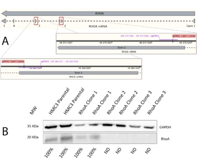
Figure 6: CRISPR-Cas9 mediated RhoA knockout and Western Blot KO confirmation. A - gRNAs matching sequences in the 2nd and 3rd exon of the RhoA gene; B - Western Blot confirmation of RhoA expression levels, normalized for GAPDH
In order to study the potential roles of Cdc42 in microglia morphodynamics, we sought to reduce its levels in HMC3 cells. Currently, there are several techniques that may be used for the experimental reduction of a target protein, either via genetic modification or by treatment with biological products (specific inhibitors or interference RNA). In our experimental setup we opted for the knockout (KO) approach, complete gene expression reduction, since it leads to a permanent manipulation of the cell line. This combined with the isolation of single cell clones allows us to work with a homogeneous population and acquire more reproducible, reliable results.
In order to obtain a RhoGTPase KO cell line, we used the CRISPR-Cas9 system. This systems consists of 2 components: Cas9 which has nuclease activity and a guide RNA (gRNA), that directs where the nuclease cuts the gene sequence. Cas9 has nuclease activity 3 nucleotides upstream of PAM sequences in the genome (any NGG sequence), and the gRNA achieves specificity through sequence complementarity in the region adjacent to the PAM sequence. After Cas9 activity, the double stranded DNA cut is repaired by non-homologous-end-joining (NHEJ). Since NHEJ occurs
18 Results
at random, causing deletion or introduction of several random nucleotides in the gap of DNA, this usually leads to a nonfunctional DNA sequence, and consequently a KO.
To implement this strategy experimentally we selected a pair of gRNAs targeting the Cdc42 sequence and inserted them in a plasmid containing the Cas9 nuclease and the gene sequence for green fluorescent protein (GFP). Upon transfection ,single cells that were positive for this fluorescent marker were sorted, allowing us to obtain a clonal population of manipulated cells.
Unfortunately, none of the isolated Cdc42-targeted single cell clones were able to expand and survive. Therefore, we decided to change our target RhoGTPase and focus on RhoA. New sets of gRNAs were selected, targeting the second and third exons of the RhoA gene (Figure 6-A), and used the same strategy described previously to transfect HMC3 cells. This time, the single cell clones survived and proliferated, being screened for RhoA protein expression by western blot (Figure 6-B). Western blot revealed that several clones (2 clones presented) were in fact knockouts for RhoA and, as such we chose clone 3 as our knockout cell line.
3.3
Morphology assessment of RhoA KO cells vs WT cells
Given the expected role of RhoA in the control of cellular contractility and tension we per-formed a morphological characterization of the cells, both in the presence and absence of the pro-inflammatory stimulants LPS and IFN-γ. In order to observe and compare cell shape between WT cells and the RhoA KO cells these were stained with DyLightTM594 Phalloidin, which binds to the actin cytoskeleton, and then fixed and imaged as shown figure.
It was possible to observe that RhoA KO cells had significantly different morphological fea-tures than WT cells, with almost all of them presented a markedly elongated shape (Figure 7-A and B). This difference was corroborated by the cell’s aspect ratio (AR - ratio between long and short axis of a cell), with RhoA KO cells presenting higher AR values (Figure 7-F), as well as an increase in cellular perimeter (Figure 7-E) and a decrease in area (Figure 7-G). Interestingly, stimulation with LPS+IFN-γ had no overall significant effect on cell shape, both in WT and RhoA KO cells, as seen in Figure 7 C and D and quantified in Figure 7E-G. Regardless we were able to observe that RhoA KO cells have a more elongated shape than WT cells, revealing a role for this regulatory protein in microglia cell shape.
3.4 Impact of RhoA knockout on HMC3 motility 19
Figure 7: Static morphology assessment of Phalloidin and DAPI double-stained cells. A - WT Unstimulated cells; B - RhoA KO Unstimulated cells; C - WT Stimulated cells; D - RhoA KO Stimulated cells. Plots of perimeter (E), aspect ratio (F) and area (G) of the four conditions (mean ± SD) **P<0.01; ***P<0.001; ****P<0.0001; n=150 for each condition
3.4
Impact of RhoA knockout on HMC3 motility
Given the observed impact of RhoA KO on cell morphology we wondered how cell migration, an essential component of microglia function was being affected in these cells. As such we char-acterized these cells’ dynamics using live cell imaging. Cells were transfected with a plasmid for the expression of LifeAct, an F-actin binding peptide, tagged with a fluorescent protein, to be able to follow the motility of cells over the course of 12 hours, in the presence or absence of stimula-tion. In general, we were able to observe that WT cells have baseline motility (Figure 9-A) that
20 Results
increases when stimulation is present (Figure 9-C). Accordingly the distance that WT cells mi-grated was increased in stimulated cells (Figure 8), due to a tendency for increased mean velocity (Figure 8) and an increase in maximum velocity as well (Figure 8). Interestingly, when observing RhoA KO cells we saw that, over the course of migration, RhoA KO cells have difficulties in the tail detachment step of the migration cycle, in some cases even leaving plasma membrane remains behind (Figure 8-A). Despite this impairment we were surprised to find that their basal motility was still higher than the one observed for WT cells, with distance travelled, mean and maximum velocity within the same range as the ones observed for stimulated WT cells (Figure 8). Stimu-lation with LPS and IFNγ proved ineffective in terms of stimulating further motility in RhoA KO cells (Figure 8).
Figure 8: Impact of RhoA in Motility, Phalloidin. A - RhoA KO Stimulated cells with tail detach-ment impairdetach-ment. Plots of mean velocity, max velocity and displacedetach-ment of both WT and RhoA KO cells (mean ± SD; n of at least 15 cells per condition)
3.4 Impact of RhoA knockout on HMC3 motility 21
Figure 9: Impact of RhoA in Motility, lifeactDsRed staining. Representative images of A - WT Unstimulated cells; B - RhoA KO Unstimulated cells; C - WT Stimulated cells; D - RhoA KO Stimulated cells. Arrows indicate moving cells.
22 Results
3.5
Role of RhoA in HMC3 actin cytoskeleton tension
Given that absence of RhoA leads to observed differences in terms or microglia morphology and motility, and that cellular tension is essential in both aspectswe assessed how the absence of RhoA was affecting cell tension. To achieve this we again turned to FRET, this time using an α-actinin tension probe. Since α-actinin is a crosslinker of actin, and that the FRET sensor is introduced inside the α-actinin protein, this probe is able to sense tension exerted on the actin cytoskeleton. We first attempted to perform these experiments in both unstimulated and stimulated cells, however after stimulation with LPS and IFN-γ, cells were not able to survive and, therefore, we only present data for unstimulated cells. Use of this probe revealed differences in the local distribution of tension (Figure 10-A-B), with RhoA KO cells presenting a more spatially diffuse tension distribution, and with concentrated focus in a smaller area than WT cells. Additionally, we observed that there was a global decrease in the average tension in RhoA KO cells when compared with WT cells, demonstrating that absence of RhoA severely impairs microglia actin cytoskeleton tension.
Figure 10: Intracellular actin cytoskeleton tension - α-Actinin FRET sensor. Representative image of A - WT cell; B - RhoA KO cell. C - quantification of whole-cell FRET ratio (mean ± SD; nA= 14; nB= 11) ∗ ∗P < 0.0015
3.6 Importance of RhoA in HMC3 focal adhesions 23
3.6
Importance of RhoA in HMC3 focal adhesions
Taking into account that cytoskeleton tension is impaired in the absence of RhoA and that focal adhesions are closely associated with both the maturation of these structures and cell motility, we studied the distribution and properties of focal adhesions in microglia cells. To do so, we used paxillin as a marker for focal adhesions and immuno-stained, together with phalloidin, cells that were either stimulated or not.
Figure 11-A shows that WT cells display numerous adhesions in the vicinity of the plasma membrane, with an extended and conical shape. This contrasts with the adhesions seen in RhoA KO cells (Figure 11-C), which are less abundant (Figure 11-M), tended to be smaller (lower area and perimeter – Figure 11 I-J) and with a more rounded shape (determined by a decreased aspect ratio, Figure 11-K). The actin content of focal adhesions was also decreased in the absence of RhoA (Figure 11-L), further showing that their generation and maturation are impaired. Inter-estingly stimulation with LPS and IFN-γ did not change any of the properties of focal adhesions in both WT and KO cells, although a slight tendency for an increase in number and maturation parameters was seen in the case of the first ones. Nevertheless, we were able to determine that in HMC3 RhoA remains crucial for the appropriate development of focal adhesions.
24 Results
Figure 11: Adhesion sites - Paxillin and Phalloidin double-staining - analysis of morphology and quantification. Representative image of A - WT unstimulated paxillin staining; B - WT unstim-ulated phalloidin staining; C - RhoA KO unstimunstim-ulated paxillin staining; D - RhoA unstimunstim-ulated phalloidin staining; E - WT stimulated paxillin staining; F - WT stimulated phalloidin staining; G - RhoA KO stimulated paxillin staining; H - RhoA KO stimulated phalloidin staining. I - Quantifi-cation of area (mean ± SD; n of at least 201 adhesion sites); J - QuantifiQuantifi-cation of perimeter (mean ± SD; n of at least 201 adhesion sites); K - Quantifcation of aspect ratio (mean ± SD; n of at least 201 adhesion sites); L - Quantification of actin content (mean ± SD; n of at least 201 adhesion sites); M - Quantification of adhesion sites (mean ± SD; n of at least 13 cells)
Chapter 4
Discussion
Microglia are extremely dynamic cells, relying in part on their capacity to change shape and acquire motility to perform their functions within the CNS. Both these features depend heavily on the actin cytoskeleton, the main responsible structure for the morphodynamics of cells. RhoGT-Pases, the main regulators of the actin cytoskeleton, are thus expected to be of paramount impor-tance for the correct and beneficial function of microglia. Our group has been the first to study the activity of RhoGTPases in microglia function. Following classical pro-inflammatory stimulation we observed that, from the classical members of RhoGTPases, only Cdc42 changed its activity, thus being potentially important for the microglia response. This does not mean that Rac1 and RhoA aredevoid of importance in the context of stimulation, only that their activity at the whole cell level does not need to vary dramatically for the cell to respond. Unfortunately we were not able to pursue the study of the role of Cdc42 in microglia morphodynamics because of an inability to develop a stable KO cell line. It has been described that for myeloid cells, the activity of Cdc42 is not only necessary for migration and adhesion but also for survival, in which knockout of this gene led to a diminished survivability of both tissue and hematopoietic progenitorsWang et al.
(2006). This can explain why, even though we were able to obtain an initial group of single cell clones, they were not able to proliferate and survive. Aiming at our new target, RhoA we were able to successfully establish a stable KO cell line. Although RhoA absence seems to be less impactful than Cdc42 absence for cell survival, we still saw noticeable differences in cell proliferation, when compared to WT, with the doubling rate of KO cells being undoubtedly lower.
Once a stable knockout cell line was obtained we were able to see striking alterations of shape in RhoA KO cells. Interestingly, the lack of RhoA led to a more elongated phenotype at the expense of cell area. These cells appear to rearrange their morphology in a way that increases perimeter by sacrificing area, which is understandable, since we would expect cell volume to remain more or less constant. Unexpectedly,classical stimulation with LPS and IFN-γ did not lead to shape changes in both WT and KO cells. Since we expected to see rapid cytoskeleton and, thus shape changes, we used a 2 hour time point for our static analyses, however this might not be the best time point to study shape. Nevertheless, the classical morphological transition after stimulation is usually seen in vivo, but not always in vitro, even when culturing primary microglia
26 Discussion
cellsKloss et al.(2001).
This inability to see shape changes in response to stimulation prevents us from concluding anything about the possible role of RhoA on microglia morphodynamics in response to stimula-tion, since WT cells appeared unaffected by stimulation. Even so, overall, we can conclude that the absence of RhoA per se leads to striking morphological changes in these cells.
After we observed these striking differences in cell shape, we moved on to the study of their motility. As expected and, in accordance with what is the normal response of microglia to pro-inflammatory stimulation, there was a tendency for stimulated cells to be more motile. Seeing that RhoA KO cells have a strikingly different morphology in a resting state, and, considering RhoA’s role in controlling several steps of the motility cycle, we expected to see stark differences in their motile behaviour. However, and in contrast to what we expected, not only did we not see any impairment in motility but we actually saw the opposite: RhoA KO cells have an increased motility, even in the absence of stimulation. This unaffected fitness in motility may be due to compensatory mechanisms undertaken by, for example RhoC, which shares a 92% amino acid sequence identity, and also has ROCK as a downstream effectorZandvakili et al.(2015). We have not explored how directed motility might be impacted by the absence of RhoA, however we can conclude that motility in general is not strictly dependent on RhoA in these cells, since we found that RhoA KO cells behave much like WT stimulated cells.
At this point we sought to understand what could be leading to the observed differences in cell shape and motility in the absence of RhoA. Both shape and motility depend on two major factors: points of adhesion and intracellular tension. These factors, depend on each other, since tension is necessary for development and maturation of adhesion points and tension cannot be exerted on the cytoskeleton in the absence of adhesion sites. They also influence shape and motility in a non-linear fashion. In the presence of too much adhesion we would predict that the cell cannot change shape. Conversely, if there is no adhesion at all the cell would collapse to a round shape. Likewise, too much intracellular tension leads to a rounding of the cell and hinders shape change. A similar parallel can be made for cell motility: too much adhesion hinders this process, since turnover and cell detachment would be impaired, while no adhesion also hampers motility through a lack of attachment that impedes forward movement. Similarly, too much tension disables forward protrusion and the cell would be locked in place, whilst no tension and the cell is unable to detach its rear from the substrate. Thus, tension and adhesions must be tightly regulated in order to achieve an optimal balance that allows both morphological changes but also acquisition of motility.
Given that RhoA is classically a master regulator of both tension and adhesion (via ROCK and consequently myosin-II motor activity) we characterized their levels and distribution, anticipating that we would observe a state of no tension and no adhesion. Interestingly, and contrarily to ex-pectation, this is not what we saw. When analysing intracellular tension, RhoA KO cells presented an overall decrease in comparison to WT, but still displayed a pattern of tension distribution inside the cell We propose that this reduction in cell tension leads to an overall relaxation of cortical tension, allowing the cells to adopt a more elongated/spreaded shape.
Discussion 27
This decrease in tension could have negative implications for cell motility, but contrary to expectation we show that RhoA KO cells actually move more. This can be explained by either a compensation by other members of the Rho subfamily, namely RhoB or RhoC or by the RhoA-independent mechanisms of tension generation. Regardless, we can conclude that lower actin cytoskeleton tension in HMC3 cells also leads to an increase in motility. This observation is most likely explained by a facilitation of the first step of the motility cycle, in which a decrease in tension can favour protrusion extension. Unfortunately, we were not able to directly observe how intracellular tension is affected by stimulation, since cells transfected with the α-actinin probe are unable to survive stimulation. This effect is probably caused by a detrimental stiffening of the actin cortex.
When we focused on the second factor which modulates shape and motility, adhesion points, we once again noticed stark differences in RhoA KO cells, not only in number of adhesion sites, but also in their size and maturation state. It is interesting to note that these fewer and underdevel-oped adhesions are still capable of efficiently anchoring an elongated cell shape and support cell migrationThese results are in accordance with what was observed in relation to tension, since a decrease in tension is expected to lead to less developed FAs. Thus, we have confirmed that in mi-croglia RhoA remains a key regulator of FAs and showed that less developed cell adhesions favour an increase in motility, likely because FAs are matured enough to support the necessary tension for motility but also more plastic to enhance the second step of migration, increasing turnover ef-ficiency. This is in accordance with some reports describing that cells with smaller adhesion sites, such as focal complexes by opposition to focal adhesions, correlate highly with a higher motility
Hotulainen and Lappalainen(2006);Zaidel-Bar et al.(2004). Interestingly, it has been observed that in cells that have stress fibres, like fibroblasts, the high level of substrate adhesion through focal adhesions inhibits cell migrationCox and Huttenlocher(1998);Cox et al.(2001).
Finally, in all of the performed experiments RhoA KO cells did not respond to stimulation, both in terms of shape, motility and adhesion parameters. Several hypotheses might explain this phenomenon. On the one hand, RhoA may be important for the sensing of stimulants, possibly by modulation of intracellular signaling pathways via FA signalling or, on the other hand, the absence of RhoA per se may already activate cell motility to its fullest and so further stimulation has no effect whatsoever on these cells.
Chapter 5
Conclusions and Future Work
5.1
Conclusions
In this work we were able to observe a never before seen effect of the knockout of RhoA in the morphodynamics of microglia. We were able to conclude that RhoA is important in maintaining cells in a less extended and possibly ramified state. RhoA, contrary to expectation, does not affect the way microglia cells move, but still seems to be an important regulator of overall motility. In microglia, RhoA remains an important regulator of focal adhesion dynamics as expected taking into account its influence on tension. RhoA seems to promote a balance between actin cytoskeleton tension and adhesion sites that favours less ramified and less motile cells. Even though, RhoA acts in a contrary fashion to expected regarding motility, it still emerges as an essential regulator of microglia morphodynamics, with potential implications for the successful function and activity of these cells, both in homeostasis and deregulation.
5.2
Future Perspectives
Given the results obtained in our work, several questions rise up, which we aim to pursue. Since RhoA KO cells are unable to create a normal level of overall tension we expect these to be more susceptible to external mechanical forces. As such we think it would be interesting to study their behaviour in a more in vivo-like, 3D culture model or even in a full in vivo model. According to our hypothesis, being surrounded by a close mesh of ECM that exerts pressure in these cells, we would expect them to possibly adopt a more rounded shape, instead of the more elongated morphology seen in our 2D-model.
On the other hand, the absence of RhoA must have some kind of impact on actin binding pro-teins, since this RhoGTPase is a master regulator of the actin cytoskeleton. Consequently, studying the activity of cofillin and profillin, for example, would probably help us to better understand the phenotype observed. Furthermore, comprehending how the downstream effectors of RhoA are behaving would help us understand if there is still myosin type II activation through some kind of
30 Conclusions and Future Work
compensation mechanism and learn why we are still able to see tension that is capable of support-ing cell migration in these cells.
Also, given that FAs are underdeveloped in the RhoA KO is adhesion-associated signalling impaired? If so, microglia function should also be impaired in the absence of RhoA. Hence, it would be relevant to study classic cellular processes associated with microglia activation such as phagocytosis or cytokine production and release. Studying these processes would help us under-stand if, other than shape and motility, these processes are also unaffected by stimulation in RhoA KO cells.
Finally, we believe it would be important to confirm all of our results and hypotheses in pri-mary cells, and, ideally, in an in vivo animal model.
References
Antonina Y. Alexandrova, Katya Arnold, Sébastien Schaub, Jury M. Vasiliev, Jean-Jacques Meis-ter, Alexander D. Bershadsky, and Alexander B. Verkhovsky. Comparative dynamics of retro-grade actin flow and focal adhesions: Formation of nascent adhesions triggers transition from fast to slow flow. PLOS ONE, 3(9):1–9, 09 2008. doi: 10.1371/journal.pone.0003234. URL
https://doi.org/10.1371/journal.pone.0003234.
William E Allen, Daniel Zicha, Anne J Ridley, and Gareth E Jones. A role for cdc42 in macrophage chemotaxis. The Journal of cell biology, 141(5):1147–1157, 1998.
Daniel E. AU Bauer, Matthew C. AU Canver, and Stuart H. AU Orkin. Generation of genomic deletions in mammalian cell lines via crispr/cas9. JoVE, (95):e52118, 2015. ISSN 1940-087X. doi: 10.3791/52118. URLhttps://www.jove.com/video/52118.
Kenneth G. Campellone and Matthew D. Welch. A nucleator arms race: cellular control of actin assembly. Nature Reviews Molecular Cell Biology, 11:237 EP –, Mar 2010. URL https: //doi.org/10.1038/nrm2867. Review Article.
M F Carlier, C Jean, K J Rieger, M Lenfant, and D Pantaloni. Modulation of the interaction between g-actin and thymosin beta 4 by the atp/adp ratio: possible implication in the regula-tion of actin dynamics. Proceedings of the Naregula-tional Academy of Sciences, 90(11):5034–5038, 1993. ISSN 0027-8424. doi: 10.1073/pnas.90.11.5034. URL https://www.pnas.org/ content/90/11/5034.
Marie-France Carlier and Dominique Pantaloni. Control of actin dynamics in cell motility11edited by n.-h. chua. Journal of Molecular Biology, 269(4):459–467, 1997. ISSN 0022-2836. URL
http://www.sciencedirect.com/science/article/pii/S0022283697910627. Marie-France Carlier, Christophe Le Clainche, Sebastian Wiesner, and Dominique Pantaloni. Actin-based motility: from molecules to movement. BioEssays, 25(4):336–345, 2003. doi: 10.1002/bies.10257. URLhttps://onlinelibrary.wiley.com/doi/abs/10.1002/ bies.10257.
Jacqueline Cherfils and Mahel Zeghouf. Regulation of small gtpases by gefs, gaps, and gdis. Physiological Reviews, 93(1):269–309, 2013. doi: 10.1152/physrev.00003.2012. URLhttps: //doi.org/10.1152/physrev.00003.2012. PMID: 23303910.
Colin K. Choi, Miguel Vicente-Manzanares, Jessica Zareno, Leanna A. Whitmore, Alex Mogilner, and Alan Rick Horwitz. Actin and a-actinin orchestrate the assembly and maturation of nascent adhesions in a myosin ii motor-independent manner. Nature Cell Biology, 10:1039 EP –, Aug 2008. URLhttps://doi.org/10.1038/ncb1763. Article.
32 REFERENCES
Carol A. Colton and Daniel L. Gilbert. Production of superoxide anions by a cns macrophage, the microglia. FEBS Letters, 223(2):284–288, 1987. doi: 10.1016/0014-5793(87) 80305-8. URL https://febs.onlinelibrary.wiley.com/doi/abs/10.1016/ 0014-5793%2887%2980305-8.
Elisabeth A Cox and Anna Huttenlocher. Regulation of integrin-mediated adhesion during cell migration. Microscopy research and technique, 43(5):412–419, 1998.
Elisabeth A Cox, Sarita K Sastry, and Anna Huttenlocher. Integrin-mediated adhesion regulates cell polarity and membrane protrusion through the rho family of gtpases. Molecular biology of the cell, 12(2):265–277, 2001.
Miguel A. Cuadros, Claude Martin, Pierre Coltey, Antonio Almendros, and Julio Navascués. First appearance, distribution, and origin of macrophages in the early development of the avian central nervous system. Journal of Comparative Neurology, 330(1):113–129, 1993. doi: 10.1002/cne.903300110. URL https://onlinelibrary.wiley.com/doi/abs/ 10.1002/cne.903300110.
Ishar Dalmau, Bente Finsen, Niels Tønder, Jens Zimmer, Berta González, and Bernardo Castel-lano. Development of microglia in the prenatal rat hippocampus. Journal of Comparative Neurology, 377(1):70–84, 1997. doi: 10.1002/(SICI)1096-9861(19970106)377:1<70:: AID-CNE7>3.0.CO;2-G. URL https://onlinelibrary.wiley.com/doi/abs/10. 1002/%28SICI%291096-9861%2819970106%29377%3A1%3C70%3A%3AAID-CNE7% 3E3.0.CO%3B2-G.
Dimitrios Davalos, Jaime Grutzendler, Guang Yang, Jiyun V. Kim, Yi Zuo, Steffen Jung, Dan R. Littman, Michael L. Dustin, and Wen-Biao Gan. Atp mediates rapid microglial response to local brain injury in vivo. Nature Neuroscience, 8(6):752–758, 2005. ISSN 1546-1726. doi: 10.1038/nn1472. URLhttps://doi.org/10.1038/nn1472.
Arshad Desai and Timothy J. Mitchison. Microtubule polymerization dynamics. Annual Review of Cell and Developmental Biology, 13(1):83–117, 1997. doi: 10.1146/annurev.cellbio.13.1.83. URLhttps://doi.org/10.1146/annurev.cellbio.13.1.83. PMID: 9442869. Payam Dibaj, Fabien Nadrigny, Heinz Steffens, Anja Scheller, Johannes Hirrlinger, Eike D.
Schomburg, Clemens Neusch, and Frank Kirchhoff. No mediates microglial response to acute spinal cord injury under atp control in vivo. Glia, 58(9):1133–1144, 2010. doi: 10.1002/glia.20993. URLhttps://onlinelibrary.wiley.com/doi/abs/10.1002/ glia.20993.
Dominique Didry, Marie-France Carlier, and Dominique Pantaloni. Synergy between actin depolymerizing factor/cofilin and profilin in increasing actin filament turnover. Journal of Biological Chemistry, 273(40):25602–25611, 1998. doi: 10.1074/jbc.273.40.25602. URL
http://www.jbc.org/content/273/40/25602.abstract.
Pavel Dráber, Vadym Sulimenko, and Eduarda Dráberová. Cytoskeleton in mast cell signaling. Frontiers in immunology, 3:130–130, May 2012. ISSN 1664-3224. doi: 10.3389/fimmu.2012. 00130. URLhttps://www.ncbi.nlm.nih.gov/pubmed/22654883.
Sandrine Etienne-Manneville and Alan Hall. Rho gtpases in cell biology. Nature, 420(6916): 629–635, 2002. ISSN 1476-4687. doi: 10.1038/nature01148. URLhttps://doi.org/10. 1038/nature01148.