CHARACTERIZATION OF THE MECHANISM OF PROTEIN
TRANSLOCATION ACROSS THE MAMMALIAN
PEROXISOMAL MEMBRANE
MARTA CRISTINA OLIVEIRA DE FREITAS
MARTA CRISTINA OLIVEIRA DE FREITAS
CHARACTERIZATION OF THE MECHANISM OF PROTEIN
TRANSLOCATION ACROSS THE MAMMALIAN
PEROXISOMAL MEMBRANE
Tese de Candidatura ao grau de Doutor em Ciências Biomédicas, submetida ao Instituto de Ciências Biomédicas de Abel Salazar da Universidade do Porto.
Orientador – Prof. Doutor Jorge Eduardo da Silva Azevedo.
Categoria – Professor Catedrático
Afiliação – Instituto de Ciências Biomédicas Abel Salazar da Universidade do Porto.
Co-orientador – Doutora Maria Clara Pereira de Sá Miranda.
Categoria – Investigadora do Instituto de Biologia Molecular e Celular, Universidade do Porto.
Afiliação – Instituto de Biologia Molecular e Celular, Universidade do Porto.
Co-orientador – Doutora Andreia Filipa Ribeiro de Carvalho
Categoria – Investigadora do Instituto de Biologia Molecular e Celular, Universidade do Porto.
Afiliação – Instituto de Biologia Molecular e Celular, Universidade do Porto.
PRECEITOS LEGAIS ... i AGRADECIMENTOS/ACKNOWLEDGMENTS ... iii ABSTRACT... v Abstract... vi Resumo ... vii ABBREVIATIONS... viii I. INTRODUCTION ... 1
1 - Peroxisome Structure and Function ... 2
2 - Peroxisomal Disorders ... 3
3 - Peroxisomal Biogenesis ... 5
3.1 - Peroxisomal Membrane Biogenesis ... 5
3.2 - Peroxisome Proliferation ... 7
3.3 - Peroxisomal Matrix Protein Import... 7
3.3.1 – PEX5... 8
3.3.2 – The cycling receptor model... 11
4 – Oligomeric matrix protein import ... 14
II. AIMS ... 17
III. EXPERIMENTAL PROCEDURES... 19
1 – Primer List... 20
2 – Production and expression of recombinant proteins... 21
3 – Plasmids for the synthesis of 35S-radiolabeled proteins... 21
4 - Synthesis of 35S-labeled-radiolabeled proteins... 22
5 – Native Polyacrylamide Gel Electrophoresis ... 22
6 – Size-Exclusion Chromatography ... 23
7 - Sucrose gradient centrifugation... 23
10 – Density gradient centrifugation analysis ... 24
11 – Miscellaneous ... 25
IV. RESULTS... 27
1- Characterization of the PEX5-cargo protein interaction ... 28
1.1 – In vitro tetramerization of 35S-catalase ... 28
1.2 – PEX5 binds monomeric catalase ... 30
1.3 – Characterization of the PEX5-catalase interaction ... 32
1.4 – The N-terminal domain of PEX14 disrupts the mCat-PEX5 interaction ... 34
2- The interaction of monomeric/oligomeric proteins with the PIM ... 40
2.1 – 35S-AOX dimerizes in vitro and its dimerization is inhibited by PEX5 ... 40
2.2 – mAOX is a better substrate than dAOX for the peroxisomal matrix protein import machinery ... 44
2.3 - PEX5 inhibits UOX tetramerization and mUOX is the preferred substrate for the PIM ... 48
V. DISCUSSION ... 51
VI. COCLUDING REMARKS ... 56
VII. REFERENCES... 58
Nesta dissertação foram utilizados os resultados do trabalho publicado abaixo indicado. A autora desta dissertação declara que interveio na concepção e execução do trabalho experimental, na interpretação de resultados e na redacção dos resultados publicados, sob o nome de “Freitas, M. O.”.
The published experimental results stated below were used in this thesis. The author of this thesis declares that she participated in the planning and execution of the experimental work, in the data interpretation, and in the preparation of the published results, under the name “Freitas, M. O.”.
Freitas M. O., Francisco T., Rodrigues T. A., Alencastre I. S., Pinto M. P. , Grou C. P., Carvalho A. F., Fransen M., Sá-Miranda C., Azevedo J. E. (2011) PEX5 Protein Binds Monomeric Catalase Blocking Its Tetramerization And Releases It Upon Binding the N-terminal Domain of PEX14. J Biol Chem. 286(47):40509-19.
Este trabalho foi financiado pela Fundação para a Ciência e Tecnologia (FCT) através de uma bolsa de doutoramento (SFRH/BD/44285/2008), pelo programa PTDC/BIA-BCM/64771/2006, pelo Fundo Europeu de Desenvolvimento Regional (PTDC/BIA-BCM/118577/2010FCOMP-01-0124-FEDER-019731) e pelo European Union VI Framework program Grant LSHGCT-2004-512018, Peroxisomes in Health and Disease.
This work was supported by Fundação para a Ciência e Tecnologia through a PhD fellowship (SFRH/BD/44285/2008), by the PTDC/BIABCM/64771/2006 program, by the Fundo Europeu de Desenvolvimento Regional, Portugal (PTDC/BIA-BCM/118577/2010FCOMP-01-0124-FEDER-019731), and by the European Union VI Framework program Grant LSHG-CT-2004-512018, Peroxisomes in Health and Disease.
Gostaria de aproveitar para agradecer a quem esteve ao meu lado durante esta etapa.
Em primeiro lugar, agradeço ao Prof. Doutor Jorge Azevedo por tudo aquilo que me ensinou e pela sua orientação e disponibilidade. Este trabalho não seria possível sem a sua dedicação.
À Doutora Clara Sá Miranda agradeço pelo apoio e preocupação em proporcionar as melhores condições de trabalho possíveis.
À Doutora Andreia Carvalho pela sua co-orientação e apoio no laboratório. Ao Doutor Marc Fransen, por toda a sua ajuda neste trabalho.
Agradeço à Fundação para a Ciência e Tecnologia (FCT), ao Fundo Europeu de Desenvolvimento Regional e ao European Union VI Framework program, Peroxisomes in Health and Disease pelo financiamento para este trabalho.
A todos os ex- e actuais membros do grupo OBF, com quem partilhei todos estes dias: à Inês, por todo o carinho e amizade; ao Tony, por me fazer pensar no futuro e destino do meu primeiro filho; ao Manel…ao Manelz, por fazer os comentários mais inesperados e cómicos num dia de trabalho; à Cláudia, por ser a melhor companheira de bancada que alguma vez terei, por todas as gargalhadas e por todas as tentativas de tomar conta do meu espaço. Aprendi muito convosco. Obrigada!
Agradeço também aos membros da UNILIPE, os vizinhos mais porreiros que a malta podia ter: Fátima, Ana Filipa, Rui, Daniel, Andrea e Lorena.
À Cátia, agradeço por comer tão lentamente que nos alarga sempre a hora de almoço.
Ao Paulo, agradeço as gargalhadas e cantorias pelo corredor…até mesmo os valentes sustos quando aparece sorrateiramente na sala de estudo.
nunca deixou de estar presente na minha vida. Uma amizade para a vida. Obrigada por me ouvires, por todos os sorrisos e preocupações.
À Tânia e à Marisa…as minhas meninas!!! Obrigada por todos os momentos que passamos juntas, por todas as parvoíces e momentos mais sérios, pela vossa amizade. Sei que posso contar sempre convosco e que, sem dúvida, ter-vos conhecido foi dos maiores benefícios que estes anos me trouxeram.
Ao Nuno…sem palavras!!! Obrigada por seres o meu “mano” mais velho, por estares sempre do meu lado e vibrares comigo nos momentos bons e menos bons. A tua preocupação e apoio constantes foram muito importantes neste percurso.
Por fim, resta-me agradecer às duas pessoas mais importantes da minha vida…os meus pais. Obrigada pelos sorrisos a cada resultado, a cada conquista. Obrigada por estarem sempre do meu lado e por me apoiarem em tudo. Espero que sintam orgulho em mim e neste trabalho…o vosso apoio e contribuição foram muito importantes para toda esta etapa.
PEX5 interacts with de novo synthesized cargo-proteins in the cytosol, transports them to the peroxisomal membrane, and after release of the cargo into the matrix of the organelle is recycled back to the cytosol. The mechanism behind the interaction of PEX5 with cargoes remains poorly understood. Considering that matrix proteins are synthesized in the cytosol in the presence of PEX5, we asked whether or not PEX5 binds the monomeric forms of these proteins. We provide data on the interaction of PEX5 with catalase, a homotetrameric enzyme in its native state. PEX5 interacts with monomeric catalase inhibiting its tetramerization, a feature that requires both the N- and C-terminal halves of PEX5. Interestingly, the PEX5-catalase interaction is disrupted by the N-terminal domain of PEX14, a component of the docking/translocation machinery. One or two of the seven PEX14-binding diaromatic motifs present in the N-terminal half of PEX5 are probably involved in this phenomenon. These observations were extended to two other major matrix proteins, acyl-CoA oxidase 1 and urate oxidase, showing that the interaction with PEX5 also inhibits their oligomerization. Furthermore, we found that the monomeric version of these proteins is more efficiently imported into the organelle matrix. Taken together, these results strongly suggest that PEX5 sequesters the newly synthesized peroxisomal proteins en route to the organelle, resembling a chaperone. We propose that this binding mode prevents premature oligomerization/non-specific interactions of cargo proteins, leading to a more efficient peroxisomal import.
As proteínas da matriz peroxissomal são sintetizadas no citosol, onde posteriormente interactuam com o receptor PEX5. A PEX5 transporta estas proteínas para a membrana do organelo e, após libertação das mesmas para a matriz, é reciclada de volta ao citosol. O mecanismo de interacção do receptor PEX5 com as proteínas da matriz peroxissomal permanece por esclarecer. Tendo em consideração que as proteínas matriciais são sintetizadas no citosol na presença da PEX5, é lícito questionar se o receptor interactua ou não com a forma monomérica destas mesmas proteínas. Neste estudo fornecemos dados sobre a interacção da PEX5 com a catalase, uma enzima homotetramérica no seu estado nativo. A PEX5 interage com a catalase monomérica, inibindo a sua tetramerização, uma característica que requer ambos os domínios N- e C-terminais da PEX5. Na presença do domínio N-terminal da PEX14, um componente da maquinaria peroxissomal de docking/translocação, a PEX5 dissocia-se da catalase. Dos sete domínios de interacção com a PEX14 presentes no N-terminal da PEX5, apenas um ou dois parecem estar envolvidos neste evento. Estes resultados foram confirmados com duas outras proteínas abundantes da matriz peroxisomal, a acil-CoA oxidase 1 e a urato oxidase, mostrando que a PEX5 também inibe a sua oligomerização. Os nossos resultados mostram que a versão monomérica destas proteínas é mais eficientemente importada para a matriz do organelo. Em conjunto, estes resultados sugerem que a PEX5 sequestra as proteínas da matriz peroxissomal após a sua síntese, apresentando características de uma chaperone. Propomos que esta ligação previne a oligomerização e interacções não específicas prematuras das proteínas matriciais, levando a um importe peroxissomal mais eficiente.
AAA ATPases associated with diverse cellular activities
AOX Acyl-CoA oxidase 1
At Arabidopsis thaliana
ATP Adenosine triphosphate
BSA Bovine serum albumin
Cat Catalase
cDNA Complementar deoxyribonucleic acid Ce Caenorhabditis elegans
CG Complementation group
CP Cargo Protein
CoA Coenzyme A
Cyt c Cytochrome c
Cys11 Cysteine residue at position 11 of mammalian PEX5
DHAP Dihydroxyacetone phosphate
Dr Danio rerio
DTM Docking/translocation machinery
DTT Dithiothreitol
DUB Deubiquitinating enzyme
E. coli Escherichia coli
EDTA Ethylenediaminetetraacetic acid
ER Endoplasmic reticulum
E1 Ubiquitin-activating enzyme
E2 Ubiquitin-conjugating enzyme
E-64 N-(trans-epoxysuccinyl)-L-leucine 4-guanidinobutylamide
Fw Forward Primer GSH Glutathione HA Hemaglutinin Hb Hemoglobin Hs Homo sapiens IAA Iodoacetamide IgG Immunoglobulin
IRD Infantile Refsum disease
Kd Dissociation constant
Ks Rate of total peroxisomal matrix protein synthesis
LBP L-bifunctional protein
MS Mass spectrometry
NALD Neonatal adrenoleukodystrophy
OA Ovalbumin
ORF Open Reading Frame
PAGE Polyacrylamide gel electrophoresis PBD Peroxisomal biogenesis disorders
PCR Polimerase Chain Reaction
PerCat Peroxisomal Catalase
PEX Peroxin
PIM Peroxisomal import machinery
PK Proteinase K
PMP Peroxisomal membrane protein
PMSF Phenylmethylsulfonyl fluoride
PNS Postnuclear supernatant
PTS1 Peroxisomal targeting sequence type 1 PTS2 Peroxisomal targeting sequence type 2 RCDP1 Rhizomelic chondrodysplasia punctata type 1
REM Receptor Export Module
RING Really interesting new gene
Rv Reverse Primer
S. cerevisiae Saccharomyces cerevisae
SCP2 Sterol carrier protein 2
SDS Sodium dodecylsulfate
SEC Size-Exclusion Chromatography
SEM Buffer containing sucrose, EDTA and MOPS
SH3 Src homology 3 domain
STI Soybean trypsin inhibitor
t1/2 Half-time
TCA Trichloroacetic acid
TPR Tetratricopeptide repeat
Tris Tris(hidroximetil)aminomethane
Ub Ubiquitin
Ub-PEX5 Monoubiquitinated PEX5 species
UOX Urate oxidase
WD Tryptophan-aspartate repeat
X-ALD X-linked adrenoleukodistrophy
1. Peroxisome Structure and Function
Peroxisomes are multifunctional organelles of eukaryotic cells. These structures were first described by Rhodin as microbodies [1], but in 1966 de Duve and Baudhuin discovered that they contained H2O2-producing oxidases and catalase, naming them
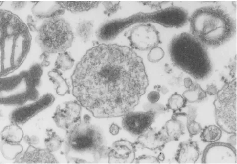
peroxisomes [2]. Peroxisomes are ubiquitous and essential organelles delimited by a single membrane and, unlike mitochondria and chloroplasts, do not contain endogenous DNA [3-5], (Figure 1). This organelle may appear as elongated, tubular and reticular structures [3, 6-8]. One important feature of peroxisomes is their metabolic plasticity. Indeed, their number, morphology and function can vary widely depending on the organism, cell- or tissue-type and environmental conditions [3, 4, 9, 10].
Figure 1: Peroxisomes. Electron micrograph of an organelle pellet from a rat liver post-nuclear
supernatant, kindly taken by Prof. Dr. Manuel Teixeira da Silva (IBMC). P, peroxisome; the arrow indicates the urate oxidase paracrystalline structure; scale bar = 1μM.
Peroxisomes carry out several metabolic functions, being hydrogen peroxide metabolism [2, 11] and β-oxidation of fatty acids (long chain and very long chain) the main ones [12-14]. In animal cells they are also involved in several anabolic processes such as biosynthesis of plasmalogens and bile acids, catabolism of purines, polyamines and prostaglandins, α-oxidation of long chain and very long chain fatty acids and in the metabolism of some amino acids and glyoxylate [4, 15-21].
The functional diversity of peroxisomes is so extensive that in some organisms they are known by different names. For example, glyoxysomes are plant
peroxisome-like organelles containing enzymes responsible for the glyoxylate cycle [22-25]; glycosomes in trypanosomatids possess most of the enzymes of the glycolytic pathway [26-28]; and in filamentous ascomycete fungi (e.g. Neurospora crassa), the Woronin bodies are specialized peroxisomes that function to seal the septal pores in response to cellular damage [29].
2. Peroxisomal Disorders
Since peroxisomes are involved in numerous metabolic processes, it is not surprising that these organelles have an important and crucial role in human physiology. In fact, a number of distinct genetic diseases are associated with this organelle and are denominated peroxisomal disorders. These disorders have devastating consequences and are caused by the malfunction or even the absence of the peroxisome. Peroxisomal disorders are frequently divided into two groups: the single peroxisomal enzyme deficiencies and the peroxisomal biogenesis disorders [30-34].
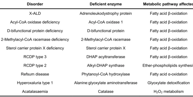
The single peroxisomal enzyme deficiencies are characterized by defects in a single peroxisomal enzyme/transporter, affecting a specific metabolic pathway such as ether-phospholipid synthesis, α- or β-oxidation of fatty acids or glyoxylate detoxification (Table 1) (reviewed in [33]).
Table 1 – List of Single Peroxisomal Enzyme Deficiencies*.
Disorder Deficient enzyme Metabolic pathway affected
X-ALD Adrenoleukodystrophy protein Fatty acid β-oxidation Acyl-CoA oxidase deficiency Acyl-CoA oxidase 1 Fatty acid β-oxidation D-bifunctional protein deficiency D-bifunctional protein Fatty acid β-oxidation 2-Methylacyl-CoA racemase deficiency 2-Methylacyl-CoA racemase Fatty acid β-oxidation Sterol carrier protein X deficiency Sterol carrier protein X Fatty acid β-oxidation
RCDP type 3 DHAP acyltransferase Fatty acid β-oxidation
RCDP type 2 Alkyl-DHAP synthase Ether-phospholipids synthesis Refsum disease Phytanoyl-CoA hydroxylase Fatty acid α-oxidation Hyperoxaluria type 1 Alanine:glyoxylate aminotransferase Glyoxylate detoxification
Acatalasaemia Catalase H2O2 metabolism
* Adapted from [33]. X-ALD, X-linked adrenoleukodystrophy; RCDP, rhizomelic chondrodysplasia punctata; CoA, coenzyme A; DHAP, dihydroxyacetone phosphate.
Peroxisomal Biogenesis Disorders (PBDs) are characterized by mutations in one of the many genes involved in proper peroxisome biogenesis, either in the membrane biogenesis, the matrix protein import pathway or division and proliferation [35, 36]. PBDs reflect a series of abnormalities due to the low number and size of peroxisomes or even the absence of normal organelles [10]. In cells of many PBD patients it is often possible to detect the so-called peroxisome “ghosts” [37]. These structures represent organelles with a normal repertoire of membrane proteins but possessing little or no matrix content [37]. Presently, the PEX genes responsible for PBDs are all known (Table 2). The association between the 13 genes and these disorders was possible due to cell fusion experiments using primary cultures of fibroblasts from PBD patients and also complementation analysis by transfection of fibroblasts with plasmids encoding different PEX proteins to restore peroxisome assembly [38-40].
Table 2 – PEX gene affected and clinical phenotypes*.
Gene Clinical phenotypes PEX1 ZS, NALD, IRD
PEX2 ZS, IRD
PEX3 ZS
PEX5 ZS, NALD
PEX6 ZS, NALD, IRD
PEX7 RCDP type 1
PEX10 ZS, NALD
PEX11β Mild ZS
PEX12 ZS, NALD, IRD
PEX13 ZS, NALD
PEX14 ZS
PEX16 ZS
PEX19 ZS
PEX26 ZS, NALD, IRD
* Adapted from [40]. ZS, Zellweger syndrome; NALD, neonatal adrenoleukodystrophy; IRD, infantile Refsum disease; RCDP, rhizomelic chondrodysplasia punctata.
The PBDs group includes disorders of the Zellweger spectrum and Rhizomelic Chondrodysplasia Punctata (RCDP) type 1 [32, 34]. The Zellweger spectrum includes the Zellweger syndrome (ZS), the neonatal adrenoleukodystrophy (NALD) and infantile Refsum disease (IRD), and these disorders have some overlapping symptoms such as liver disease, retinopathy and variable neurodevelopmental delay. One major difference between the three conditions regards the survival and phenotype severity of
IRD is the milder one, with patients living over 30 years [30, 32]. In RCDP type 1 only some metabolic functions are lost, differing from the Zellweger spectrum in its biochemical and molecular basis and clinical presentation. In fact, this disease is characterized by mutations in only one gene that encodes PEX7, whereas the Zellweger spectrum is caused by mutations in any of the remaining PEX genes [32, 41].
3. Peroxisome Biogenesis
As refered earlier, peroxisomes have no DNA and therefore peroxisomal proteins are encoded by nuclear DNA, synthesized on cytosolic ribosomes and post-translationally targeted to the organelle [3, 42-44].
Identification of PEX genes started with studies in Saccharomyces cerevisiae and were applied to a variety of organisms such as Pichia pastoris, Hansenula
polymorpha and Yarrowia lipolytica [45-50]. These studies allowed the identification of
the majority of known peroxisome biogenesis-associated genes. The mammalian homologs were then identified by homology probing approaches [51]. Over 30 peroxins have been identified among several organisms [52, 53], but only sixteen were reported in mammals (Table 3) [54]. Most of these proteins are conserved between species but depending on the organism some specific roles were embraced by a single peroxin [53, 54].
Peroxisomal biogenesis is generally divided into three processes: peroxisome membrane biogenesis, import of matrix proteins into the organelle and peroxisome proliferation. A brief description of each of these processes is provided below.
3.1 Peroxisomal Membrane Biogenesis
The origin of the peroxisomal membrane is a subject of some controversy in the field [55, 56]. Several studies support a model in which peroxisomes are regarded as autonomous organelles deriving from pre-existing ones by growth and division [57-59]. The phospholipid requirements are fulfilled by the endoplasmic reticulum (ER) [55]. However, some authors claim that the ER is involved in the de novo formation and maintenance of peroxisomes, producing vesicles (protoperoxisomes) that eventually mature into peroxisomes or fuse with pre-existing organelles [57].
Targeting of newly synthesized peroxisomal intrinsic membrane proteins (PMPs) to the organelle requires signals, designated membrane peroxisomal targeting signals (mPTS), which are characterized by a cluster of basic aminoacids and a
transmembrane domain anchoring the protein to the peroxisomal membrane [60, 61]. These signals are quite different from the ones in proteins to be imported into the matrix of the organelle (see section 3.3).
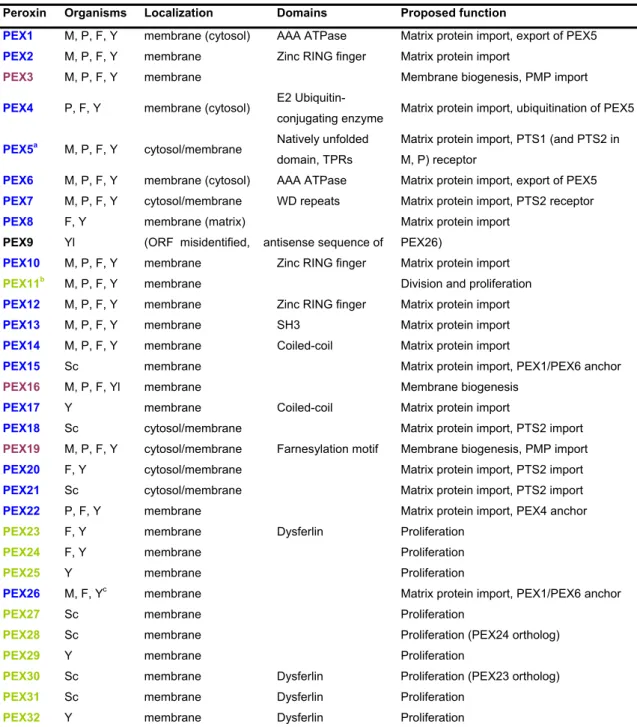
Table 3 – Proteins implicated in peroxisomal biogenesis (peroxins).
Peroxin Organisms Localization Domains Proposed function
PEX1 M, P, F, Y membrane (cytosol) AAA ATPase Matrix protein import, export of PEX5
PEX2 M, P, F, Y membrane Zinc RING finger Matrix protein import
PEX3 M, P, F, Y membrane Membrane biogenesis, PMP import
PEX4 P, F, Y membrane (cytosol) E2 Ubiquitin-
conjugating enzyme Matrix protein import, ubiquitination of PEX5
PEX5a M, P, F, Y cytosol/membrane Natively unfolded domain, TPRs
Matrix protein import, PTS1 (and PTS2 in M, P) receptor
PEX6 M, P, F, Y membrane (cytosol) AAA ATPase Matrix protein import, export of PEX5
PEX7 M, P, F, Y cytosol/membrane WD repeats Matrix protein import, PTS2 receptor
PEX8 F, Y membrane (matrix) Matrix protein import
PEX9 Yl (ORF misidentified, antisense sequence of PEX26)
PEX10 M, P, F, Y membrane Zinc RING finger Matrix protein import
PEX11b M, P, F, Y membrane Division and proliferation
PEX12 M, P, F, Y membrane Zinc RING finger Matrix protein import
PEX13 M, P, F, Y membrane SH3 Matrix protein import
PEX14 M, P, F, Y membrane Coiled-coil Matrix protein import
PEX15 Sc membrane Matrix protein import, PEX1/PEX6 anchor
PEX16 M, P, F, Yl membrane Membrane biogenesis
PEX17 Y membrane Coiled-coil Matrix protein import
PEX18 Sc cytosol/membrane Matrix protein import, PTS2 import
PEX19 M, P, F, Y cytosol/membrane Farnesylation motif Membrane biogenesis, PMP import
PEX20 F, Y cytosol/membrane Matrix protein import, PTS2 import
PEX21 Sc cytosol/membrane Matrix protein import, PTS2 import
PEX22 P, F, Y membrane Matrix protein import, PEX4 anchor
PEX23 F, Y membrane Dysferlin Proliferation
PEX24 F, Y membrane Proliferation
PEX25 Y membrane Proliferation
PEX26 M, F, Yc membrane Matrix protein import, PEX1/PEX6 anchor
PEX27 Sc membrane Proliferation
PEX28 Sc membrane Proliferation (PEX24 ortholog)
PEX29 Y membrane Proliferation
PEX30 Sc membrane Dysferlin Proliferation (PEX23 ortholog)
PEX31 Sc membrane Dysferlin Proliferation
PEX32 Y membrane Dysferlin Proliferation
M, mammals; P, plants; F, filamentous fungi, Y, yeasts, Yl, Yarrowia lipolytica only; Sc, Saccharomyces cerevisiae only; aMammals contain two main isoforms, PEX5S and PEX5L, the later harbouring a PEX7-binding site; bMammalian cells contain three PEX11 genes encoding PEX11α, PEX11β and PEX11γ; cPEX26 is absent in Sc and related yeasts. PEX1, PEX4 and PEX6 are peripheral membrane proteins facing the cytosol. PEX8 is a peripheral membrane protein facing the peroxisomal matrix. Colour coding is according to the involvement of peroxins in peroxisomal biogenesis pathways, i.e., membrane assembly (violet), import of matrix proteins (blue) and growth and division (green). AAA ATPase, ATPase associated with several cellular activities; RING, Really interesting new gene; WD, Tryptophan-aspartate motif; TPRs, tetratricopeptide repeats; SH3, Src-homology 3 domain; ORF, open reading frame.
Membrane biogenesis in mammals and other organisms requires three peroxins: the intrinsic membrane proteins, PEX3 and PEX16, and a cytosolic/membrane protein, PEX19 [62, 63]. Most PMPs interact with their receptor PEX19 in the cytosol during or right after their translation, guaranteeing an import-competent status. Therefore, PEX19 acts as a chaperone for membrane proteins probably by protecting their hydrophobic domains [64-69]. PEX3 is the docking protein at the peroxisomal membrane for the PEX19-PMP complexes [70-72] and may also promote the insertion of PMPs into the peroxisomal membrane [73]. As for PEX16, its role remains poorly understood [63] but some authors state that PEX16 promotes the peroxisomal growth from the ER [74, 75].
3.2 Peroxisome proliferation
Peroxisomes are organelles characterized by their metabolic plasticity, being able to adjust to different physiological requirements [76, 77]. In mammals, the PEX11 family is involved in peroxisome proliferation and comprises three proteins: PEX11α, PEX11β and PEX11γ [7, 78, 79]. Loss of these proteins will lead to a reduction in peroxisomal number and presence of large peroxisomes [40, 80].
Peroxisome proliferation involves growth, elongation and a final step of division (reviewed in [81]). PEX11 is responsible for peroxisome elongation while the final membrane fission is performed by a dynamin-like protein (DLP1) [57, 58]. DLP1 anchors at the peroxisomal membrane through Fis1, a C-tail anchored protein [82]. These last two proteins, required for peroxisome proliferation, are also involved in mitochondria division [83].
3.3 Peroxisome matrix protein import
Peroxisomal matrix is the place of higher protein concentration in eukaryotic cells [84]. More than 50 different enzymes can be found in the matrix of the organelle [3, 21]. Similarly to the membrane biogenesis, the matrix protein import involves targeting sequences, cytosolic receptors and a membrane machinery to promote the internalization of these proteins.
Regarding the targeting signals, peroxisomal matrix proteins are sorted via one of two pathways. The majority of proteins present a Peroxisomal Targeting Signal type 1 (PTS1), a C-terminal tripeptide with the consensus sequence (S/A/C) – (K/R/H) –
(L/M) [85-87]. However, residues upstream from the PTS1 signal can also influence the interaction between the receptor and the cargo protein [87, 88]. PEX5 is the receptor for PTS1-containing proteins and the PTS1 recognition site is at its C-terminal half [89-92]. The second PTS signal (PTS2) is a N-terminal degenerate nonapeptide consisting of the consensus sequence (R/K) – (L/V/I) – (X)5 – (H/Q) – (L/A). Only a small number
of proteins possess a PTS2 signal and after internalization it is cleaved from the matrix protein [93-95]. PEX7 is the receptor for these PTS2 proteins [96, 97] but accessory proteins are needed to assist the delivery of these cargoes to the peroxisome [93, 98]. These proteins are species-specific: PEX18/PEX21 in S. cerevisiae [99] and PEX20 in the majority of yeast and fungi [100, 101]. In mammals and plants, PEX7 uses PEX5 itself for the targeting of PTS2-proteins to the peroxisome [102-104].
3.3.1 – PEX5
PEX5 was initially identified in P. pastoris and subsequently found in all peroxisome-containing organisms (revised in [4]). In mammals, two isoforms are found due to alternative splicing of the PEX5 transcript: a large isoform, PEX5L, and a small one, PEX5S. PEX5L comprises 639 amino acids while the smaller isoform lacks an internal sequence of 37 amino acids, encoded by exon 8 [91, 105, 106]. This small region comprises the domain that interacts with PEX7, the PTS2 receptor, but this interaction is only observed in plants and mammals [102-105, 107, 108].
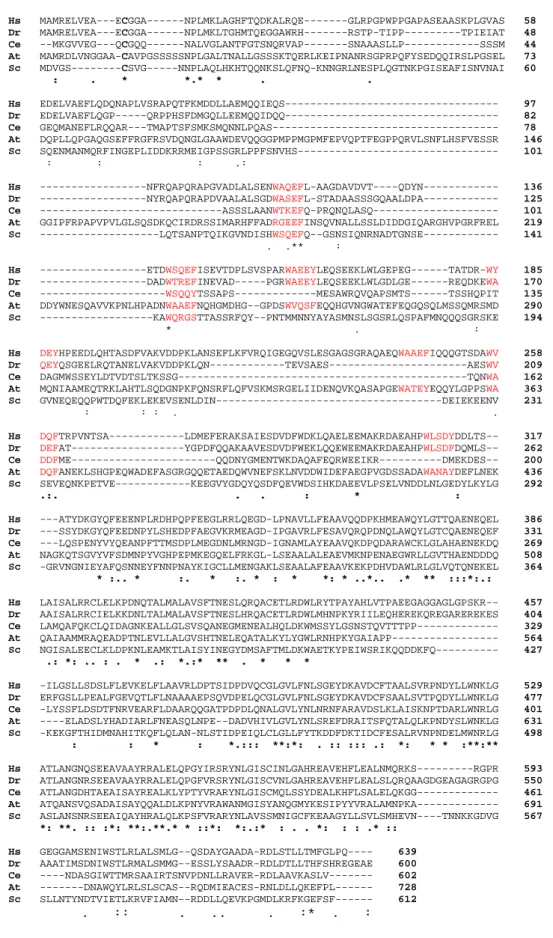
PEX5 can be divided in two distinct domains based on structural/functional data: the N-terminal and the terminal halves (Figure 2). A highly conserved C-terminal domain can be found when aligning PEX5 sequences, while the N-C-terminal half reveals low conservation between several organisms (Figure 3).
Figure 2: PEX5. Schematic presentation of the domain structure of human PEX5L and interaction sites for
known components of the peroxisomal import machinery. The purple bars indicate the diaromatic motifs (WxxxF/Y PEX14 binding sites). The blue area indicates the extra 37 aminoacid encoded by exon 8 which contains the PEX7 binding site and is absent in the small isoform of PEX5. The red boxes represent the TPR domains. TPR, tetratricopeptide repeats.
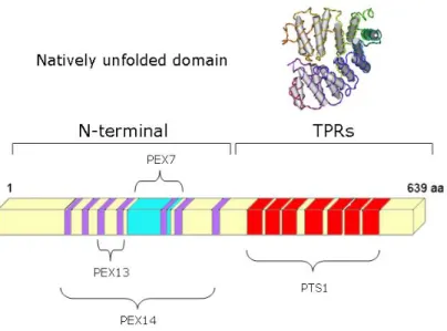
The N-terminal half of PEX5 is a natively unfolded domain [109]. PEX5, being a monomeric protein, displays a flexible N-terminal half, being characterized by an unusually large Stokes radius and a small sedimentation coefficient [110]. Natively unfolded proteins/domains are frequently involved in protein-protein interactions with multiple partners [111]. Indeed, the N-terminal domain of PEX5 is characterized by the existence of several domains of interaction with other peroxins. Seven pentapeptide diaromatic motifs WXXXF/Y are present in this PEX5 domain and are responsible for the interaction with PEX14 and PEX13, two members of the Docking/Translocation Machinery (DTM) [112-119]. As previously mentioned, the N-terminal half of PEX5 harbours the PEX7 interaction site. Besides binding to PEX7, some observations also show that the N-terminal half of PEX5 also interacts with some cargo proteins [120, 121]. The C-terminal domain of PEX5 contains seven tetratricopeptide repeats (TPRs) with a highly conserved structure, consisting of two clusters, TPRs 1-3 and TPRs 5-7, connected by a flexible hinge comprising TPR4. These TPRs are involved in the interaction with PTS1-containing proteins. This property was confirmed with studies showing PEX5 interacting with SKL-containing peptides or a PTS1 cargo protein (Figure 4) [122-126]. When PEX5 interacts with a PTS1 protein, a ring-like structure is formed by the two TPR clusters with the PTS1 peptide occupying the center of this ring-like structure. In the absence of a cargo protein there is a slight opening of the ring-like structure.
Hs MAMRELVEA---ECGGA---NPLMKLAGHFTQDKALRQE---GLRPGPWPPGAPASEAASKPLGVAS 58 Dr MAMRELVEA---ECGGA---NPLMKLTGHMTQEGGAWRH---RSTP-TIPP---TPIEIAT 48 Ce --MKGVVEG---QCGQQ---NALVGLANTFGTSNQRVAP---SNAAASLLP---SSSM 44 At MAMRDLVNGGAA-CAVPGSSSSSNPLGALTNALLGSSSKTQERLKEIPNANRSGPRPQFYSEDQQIRSLPGSEL 73 Sc MDVGS---CSVG---NNPLAQLHKHTQQNKSLQFNQ-KNNGRLNESPLQGTNKPGISEAFISNVNAI 60 : . * *.* * . . Hs EDELVAEFLQDQNAPLVSRAPQTFKMDDLLAEMQQIEQS--- 97 Dr EDELVAEFLQGP---QRPPHSFDMGQLLEEMQQIDQQ--- 82 Ce GEQMANEFLRQQAR---TMAPTSFSMKSMQNNLPQAS--- 78 At DQPLLQPGAQGSEFFRGFRSVDQNGLGAAWDEVQQGGPMPPMGPMFEPVQPTFEGPPQRVLSNFLHSFVESSR 146 Sc SQENMANMQRFINGEPLIDDKRRMEIGPSSGRLPPFSNVHS--- 101 : : : .: Hs ---NFRQAPQRAPGVADLALSENWAQEFL-AAGDAVDVT----QDYN--- 136 Dr ---NYRQAPQRAPDVAALALSGDWASEFL-STADAASSSGQAALDPA--- 125 Ce ---ASSSLAANWTKEFQ-PRQNQLASQ--- 101 At GGIPFRPAPVPVLGLSQSDKQCIRDRSSIMARHFFADRGEEFINSQVNALLSSLDIDDGIQARGHVPGRFREL 219 Sc ---LQTSANPTQIKGVNDISHWSQEFQ--GSNSIQNRNADTGNSE--- 141 . .** : Hs ---ETDWSQEFISEVTDPLSVSPARWAEEYLEQSEEKLWLGEPEG---TATDR-WY 185 Dr ---DADWTREFINEVAD---PGRWAEEYLEQSEEKLWLGDLGE---REQDKEWA 170 Ce ---WSQQYTSSAPS---MESAWRQVQAPSMTS---TSSHQPIT 135 At DDYWNESQAVVKPNLHPADNWAAEFNQHGMDHG--GPDSWVQSFEQQHGVNGWATEFEQGQSQLMSSQMRSMD 290 Sc ---KAWQRGSTTASSRFQY--PNTMMNNYAYASMNSLSGSRLQSPAFMNQQQSGRSKE 194 * . : Hs DEYHPEEDLQHTASDFVAKVDDPKLANSEFLKFVRQIGEGQVSLESGAGSGRAQAEQWAAEFIQQQGTSDAWV 258 Dr QEYQSGEELRQTANELVAKVDDPKLQN---TEVSAES---AESWV 209 Ce DAGMWSSEYLDTVDTSLTKSSG---TQNWA 162 At MQNIAAMEQTRKLAHTLSQDGNPKFQNSRFLQFVSKMSRGELIIDENQVKQASAPGEWATEYEQQYLGPPSWA 363 Sc GVNEQEQQPWTDQFEKLEKEVSENLDIN---DEIEKEENV 231 : : : . . Hs DQFTRPVNTSA---LDMEFERAKSAIESDVDFWDKLQAELEEMAKRDAEAHPWLSDYDDLTS-- 317 Dr DEFAT---YGPDFQQAKAAVESDVDFWEKLQQEWEEMAKRDAEAHPWLSDFDQMLS-- 262 Ce DDFME---QQDNYGMENTWKDAQAFEQRWEEIKR---DMEKDES-- 200 At DQFANEKLSHGPEQWADEFASGRGQQETAEDQWVNEFSKLNVDDWIDEFAEGPVGDSSADAWANAYDEFLNEK 436 Sc SEVEQNKPETVE---KEEGVYGDQYQSDFQEVWDSIHKDAEEVLPSELVNDDLNLGEDYLKYLG 292 .:. . . : * : Hs ---ATYDKGYQFEEENPLRDHPQPFEEGLRRLQEGD-LPNAVLLFEAAVQQDPKHMEAWQYLGTTQAENEQEL 386 Dr ---SSYDKGYQFEEDNPYLSHEDPFAEGVKRMEAGD-IPGAVRLFESAVQRQPDNQLAWQYLGTCQAENEQEF 331 Ce ---LQSPENYVYQEANPFTTMSDPLMEGDNLMRNGD-IGNAMLAYEAAVQKDPQDARAWCKLGLAHAENEKDQ 269 At NAGKQTSGVYVFSDMNPYVGHPEPMKEGQELFRKGL-LSEAALALEAEVMKNPENAEGWRLLGVTHAENDDDQ 508 Sc -GRVNGNIEYAFQSNNEYFNNPNAYKIGCLLMENGAKLSEAALAFEAAVKEKPDHVDAWLRLGLVQTQNEKEL 364 * :.. * :. * :. * : * *: * ..*.. .* ** :::*:.: Hs LAISALRRCLELKPDNQTALMALAVSFTNESLQRQACETLRDWLRYTPAYAHLVTPAEEGAGGAGLGPSKR-- 457 Dr AAISALRRCIELKKDNLTALMALAVSFTNESLHRQACETLRDWLMHNPKYRIILEQHEREKQREGAREREKES 404 Ce LAMQAFQKCLQIDAGNKEALLGLSVSQANEGMENEALHQLDKWMSSYLGSNSTQVTTTPP--- 329 At QAIAAMMRAQEADPTNLEVLLALGVSHTNELEQATALKYLYGWLRNHPKYGAIAPP--- 564 Sc NGISALEECLKLDPKNLEAMKTLAISYINEGYDMSAFTMLDKWAETKYPEIWSRIKQQDDKFQ--- 427 .: *: .. : . * .: *.:* ** . * * * Hs -ILGSLLSDSLFLEVKELFLAAVRLDPTSIDPDVQCGLGVLFNLSGEYDKAVDCFTAALSVRPNDYLLWNKLG 529 Dr ERFGSLLPEALFGEVQTLFLNAAAAEPSQVDPELQCGLGVLFNLSGEYDKAVDCFSAALSVTPQDYLLWNKLG 477 Ce -LYSSFLDSDTFNRVEARFLDAARQQGATPDPDLQNALGVLYNLNRNFARAVDSLKLAISKNPTDARLWNRLG 401 At ----ELADSLYHADIARLFNEASQLNPE--DADVHIVLGVLYNLSREFDRAITSFQTALQLKPNDYSLWNKLG 631 Sc -KEKGFTHIDMNAHITKQFLQLAN-NLSTIDPEIQLCLGLLFYTKDDFDKTIDCFESALRVNPNDELMWNRLG 498 : : * : *.::: **:*: . :: ::: .: *: * * :**:** Hs ATLANGNQSEEAVAAYRRALELQPGYIRSRYNLGISCINLGAHREAVEHFLEALNMQRKS---RGPR 593 Dr ATLANGNRSEEAVAAYRRALELQPGFVRSRYNLGISCVNLGAHREAVEHFLEALSLQRQAAGDGEAGAGRGPG 550 Ce ATLANGDHTAEAISAYREALKLYPTYVRARYNLGISCMQLSSYDEALKHFLSALELQKGG--- 461 At ATQANSVQSADAISAYQQALDLKPNYVRAWANMGISYANQGMYKESIPYYVRALAMNPKA--- 691 Sc ASLANSNRSEEAIQAYHRALQLKPSFVRARYNLAVSSMNIGCFKEAAGYLLSVLSMHEVN----TNNKKGDVG 567 *: **. :: :*: **:.**.* * ::*: *:.:* : . . *: : : .* :: Hs GEGGAMSENIWSTLRLALSMLG--QSDAYGAADA-RDLSTLLTMFGLPQ---- 639 Dr AAATIMSDNIWSTLRMALSMMG--ESSLYSAADR-RDLDTLLTHFSHREGEAE 600 Ce ----NDASGIWTTMRSAAIRTSNVPDNLLRAVER-RDLAAVKASLV--- 602 At ---DNAWQYLRLSLSCAS--RQDMIEACES-RNLDLLQKEFPL--- 728 Sc SLLNTYNDTVIETLKRVFIAMN--RDDLLQEVKPGMDLKRFKGEFSF--- 612 . :: . .. . :* . :
Figure 3: Sequence alignment of PEX5 proteins (ClustalW2, www.ebi.ac.uk). The long isoform of
human (Hs) PEX5 is aligned with homolog proteins from Danio rerio (Dr), Caenorhabditis elegans (Ce),
Arabidopsis thaliana (At) and Saccharomyces cerevisae (Sc). Red colour marks the WXXXF/Y diaromatic
PEX5(C) PEX5(C) PEX5(C) YQSKL SCP2
Figure 4: Structures of the C-terminal half of PEX5 (PEX5(C)). The structures were obtained in the
absence of a ligand (left, [123]) in the presence of the consensus PTS1 peptide YQSKL (Protein Data Base accession number 1FCH) (central, [122]) or in the presence of the functional PTS1 cargo protein SCP2 (right, [123]). Colour coding: TPR segments 1–3, light green; TPR4 segment, dark green; TPR segments 5–7, blue; loop connecting the TPR segment and the C-terminal helical bundle, yellow; C-terminal α-helical bundle, red; PTS1 ligands, black. Adapted from [123].
3.3.2 – The Cycling Receptor Model
PEX5 presents a dual subcellular localization. Elegant studies demonstrated that PEX5 can accumulate at the peroxisomal membrane when temperature and ATP levels are experimentally decreased [127]. Moreover, the peroxisomal accumulation of PEX5 is completely reversible suggesting that PEX5 can undergoe multiple cycles of association/dissociation. Knowing that PEX5 recognizes and binds cargo proteins, it was proposed that this protein is a cycling receptor [127]. According to this model, matrix proteins are recognized by PEX5 in the cytosol, are directed to the peroxisomal membrane, translocated to the matrix of the organelle and finally PEX5 is recycled back into the cytosol.
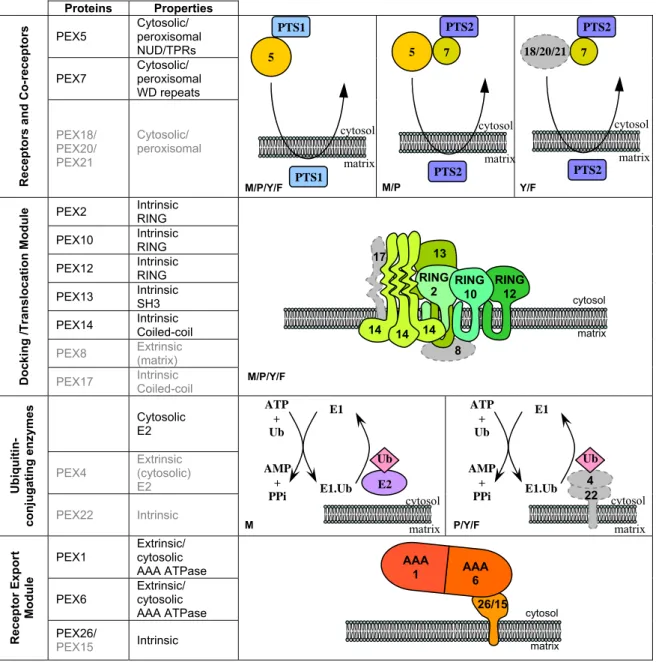
Understanding how the peroxisomal import machinery works requires knowledge on the arquitecture of its components. Over the years, numerous studies have addressed this issue. Using biochemical (e.g., pull-down assays, immunoprecipitation experiments and protein purification techniques) and genetic approaches (e.g., yeast two-hybrid) it was possible to build models on the structure of the Peroxisomal Import Machinery (PIM) (Figure 5) [128-132]. In mammals, this machinery comprises: 1) a membrane-embedded DTM that includes PEX14, PEX13 and the Really Interesting New Gene (RING)-finger peroxins PEX2, PEX10 and PEX12; and 2) a Receptor Export Module (REM) that includes the two ATPases
Associated with diverse Activities (AAA) peroxins, PEX1 and PEX6, and their membrane anchor, PEX26. Besides these peroxins, the PIM also includes ubiquitin, an ubiquitin-activating enzyme (E1) and an ubiquitin-conjugating enzyme (E2).
Proteins Properties PEX5 Cytosolic/ peroxisomal
NUD/TPRs PEX7 Cytosolic/ peroxisomal WD repeats Recep tors a nd Co-rece pt ors PEX18/ PEX20/ PEX21 Cytosolic/ peroxisomal
PEX2 Intrinsic RING PEX10 Intrinsic RING PEX12 Intrinsic RING PEX13 Intrinsic SH3 PEX14 Intrinsic Coiled-coil
PEX8 Extrinsic (matrix)
Docki ng /Tr ansl o cati on M o d u le
PEX17 Intrinsic Coiled-coil
Cytosolic E2 PEX4 Extrinsic (cytosolic) E2 Ubiq uiti n-conj uga ting enz ymes PEX22 Intrinsic
PEX1 Extrinsic/ cytosolic AAA ATPase PEX6 Extrinsic/ cytosolic
AAA ATPase Recep tor Exp o rt Modul e PEX26/ PEX15 Intrinsic
Figure 5: Components of the peroxisomal import machinery (PIM). Peroxins absent in mammals (M) are depicted in gray. Receptors and co-receptors of cargo proteins are represented. The docking/translocation module comprises mostly transmembrane proteins. PEX8 and PEX17 have been found only in lower eukaryotes [53, 54]. The first two reactions of the ubiquitin-conjugating cascade involved in the peroxisomal protein import pathway are shown. Note that in mammals the E2 component is a cytosolic protein whereas in plants, yeasts and fungi the E2 PEX4 is bound to the organelle membrane via PEX22. The precise identity of the final component of this cascade, ie, the ubiquitin-ligase catalyzing mono-ubiquitination of PEX5 remains unknown but it may be any of the 3 RING peroxins of the DTM or combinations of them. The receptor export module comprises AAA peroxins PEX1 and PEX6 and one intrinsic membrane protein anchor [133, 134], PEX26 in mammals and other organisms, and PEX15 in some yeasts. PEX26 and PEX15 are unrelated at the primary structure level [53]. Their plant functional counterpart is presently unknown [135]. Adapted from [136]. M/P Y/F matrix cytosol 7 5 PTS2 PTS2 matrix cytosol 7 18/20/21 PTS2 PTS2 matrix 5 PTS1 PTS1 cytosol matrix cytosol 26/15 AAA 1 AAA 6 matrix cytosol 8 13 17 14 14 14 RING 2 RING 12 RING 10 matrix cytosol 22 4 Ub ATP + Ub AMP + PPi E1 E1.Ub matrix cytosol E2 Ub ATP + Ub AMP + PPi E1 E1.Ub M P/Y/F M/P/Y/F M/P/Y/F
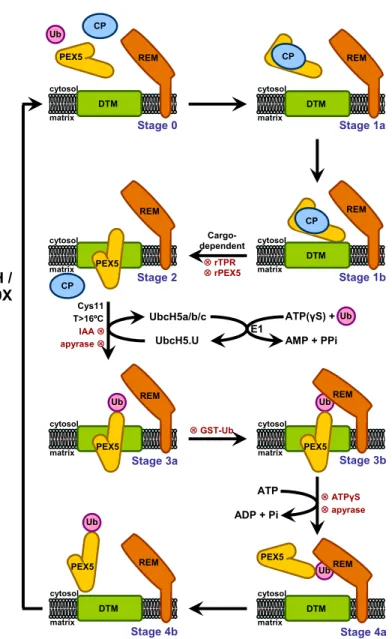
Using an in vitro import system, our laboratory has provided valuable data regarding the mammalian PEX5-mediated peroxisomal protein import pathway. This system is based in the incubation of radiolabeled PEX5 or a PTS2-containing protein with a rat liver post-nuclear supernatant (PNS) under appropriate experimental conditions. A protease-protection assay is then performed to evaluate the specificity of the process, since non-peroxisomal PEX5 or non-imported cargo protein are susceptible to proteolytic cleavage. The model presented in Figure 6 accommodates all the data gathered so far regarding this pathway. The model comprises several steps (numbered 0-4) as detailed below.
First, soluble PEX5 (stage 0) binds to cargo proteins in the cytosol yielding stage 1a PEX5. It is possible that this interaction somehow changes the conformation of PEX5 because the receptor only engages the next steps (i.e., interaction with the DTM) if cargo proteins are available [137]. The cytosolic cargo-loaded PEX5 then interacts with the DTM yielding stage 1b. There are some data suggesting that the docking site for the complex at the peroxisomal membrane is PEX13 and/or PEX14 although convincing data for these possibilities are still lacking [138, 139]. Once at the peroxisomal membrane, cargo-loaded PEX5 becomes inserted into the DTM (stage 2), acquiring a transmembrane topology with a small N-terminal domain exposed to cytosol and protease accessible [140], whereas the bulky fraction of PEX5 polypeptide chain can only be accessed by the matrix of the organelle [140]. Strikingly, the transition between stage 1b and stage 2 PEX5 is an ATP-independent process [137, 141, 142]. Next, the DTM-embedded PEX5 is monoubiquitinated at a conserved N-terminal cysteine residue (Ub-PEX5, stage 3, completely protease-resistant) [143, 144]. The evidence supporting the importance of the cysteine residue is several fold: deletion of the 17 amino acid domain containing this cysteine, its alkylation or its substitution by a serine or alanine results in PEX5 proteins still functional in the docking and membrane insertion steps but export-incompetent [142, 144, 145]. Monoubiquitination is a mandatory event for the export of PEX5, an event that is ATP-dependent [144]. PEX1/PEX6 endorse this export step, where Ubiquitin-PEX5 (Ub-PEX5) is recognized and extracted from the peroxisomal membrane back into the cytosol, yielding a soluble Ub-PEX5 conjugate (stage 4) [134, 146]. Finally, the ubiquitin moiety is removed from the Ub-PEX5 conjugate, a process that possibly occurs by a combination of enzymatic and non-enzymatic mechanisms [147, 148]. In mammals, it was recently shown that USP9X is by far the most active deubiquitinase (DUB) acting on Ub-PEX5 [149].
All the data mentioned so far was obtained using a PEX5-centered in vitro import system. However, this approach gave us no information about the cargo insertion into the DTM and its release into the organelle matrix. Recent findings from
our laboratory came to elucidate these matters. Indeed, using a PTS2-centered in vitro import system [150], it was possible to conclude that PTS2-cargo insertion into the DTM occurs at the stage 1b-to-stage 2 transition, as previously hypothesized [151] and that the release of the reporter protein to the organelle matrix occurs before ubiquitination of PEX5.
4. – Oligomeric matrix protein import
Peroxisomes can be distinguished from other organelles by their ability to import proteins in their oligomeric state (reviewed in [3]). However, this has been a controversial subject during the past years. On one hand, some work has been published stating that protein oligomerization occurs after import into the peroxisome: Lazarow and de Duve showed that catalase was imported as a monomer [152, 153] and the same was observed in cucumber glyoxysomes for malate synthethase import [154]; finally alcohol oxidase octomerization was shown to occur during or right after import to the peroxisomes [155-157]. On the other hand, several authors claim that peroxisomes have the ability to import proteins in their native state. It was shown that a peroxisomal protein lacking a PTS1 signal can form a complex with a PTS1-containing protein and enter the organelle. This phenomenon is designated “piggy-back” import.
The first evidences supporting this idea came from studies where a N-terminal truncated version of thiolase, lacking the PTS2 signal, was found to be imported in complex with a full-length version of itself [158]. Another example comes from the work with chloramphenicol acetyltransferase. Those authors proposed that subunits of this protein, lacking the PTS1 signal, were able to form heterotrimers with subunits containing a PTS1 in the cytosol and be targeted to the peroxisome [159]. The same phenomenon was reported for other proteins like Mdh3 from S. cerevisiae [85], isocitrate lyases of some plants [160] and enoyl-CoA isomerases Dci1p and Eci1p from
S. cerevisiae [161]. In a different experimental setting, Walton and collaborators
microinjected mammalian cells with gold particles (with a diameter ranging from 4-9 nm) coated with human serum albumin conjugated with a PTS1 signal [162]. They observed that these particles could be imported into the peroxisomal matrix.
Figure 6: The PEX5-mediated peroxisomal protein import pathway in mammals. The matrix protein
import pathway comprises five major steps (numbered 0-4). Stage 0, cytosolic cargo-free PEX5 (protease accessible). Stage 1, cytosolic PEX5-cargo protein complex (protease accessible). Stage 2, PEX5 embedded in the peroxisomal docking/translocation machinery (DTM) (only 2 kDa of PEX5 N-terminus are accessible to exogenously added Proteinase K). Stage 3, DTM-embedded monoubiquitinated PEX5 at Cys11 (protease protected). Stage 4, protease accessible monoubiquitinated PEX5. 8,indicates reagents that block the pathway at specific steps; CP, cargo protein; Ub, ubiquitin; IAA, iodoacetamide; rTPR, recombinant protein comprising the PTS1-binding domain of PEX5; rPEX5, recombinant full-length PEX5; REM, receptor export module. Adapted from [150].
These observations come together to illustrate the ability of peroxisomes to import: 1) proteins in their native state, 2) oligomeric proteins and 3) large structures ([158, 159], reviewed in [3]). However, the import kinetics of a given cargo protein has never been truly studied. In fact, there is only one study where this issue was
Stage 1a cytosol matrix REM DTM CP Stage 1b CP cytosol matrix REM DTM 9 GST-Ub Ub Stage 4a cytosol matrix REM PEX5 DTM Stage 4b cytosol matrix REM DTM PEX5 Ub Cargo-dependent 9 rTPR 9 rPEX5 UbcH5.U UbcH5a/b/c E1 AMP + PPi ATP(γS) + Ub Cys11 T>16ºC IAA 9 ATP ADP + Pi 9 ATPγS 9 apyrase apyrase 9 Ub CP cytosol matrix REM Stage 3a PEX5 Ub Stage 2 CP cytosol matrix CP REM PEX5 CP Ub Stage 3b cytosol matrix CP REM PEX5 Stage 0 cytosol matrix REM DTM PEX5 Ub CP GSH / USP9X
considered. Goodman and co-workers showed that the import kinetics of the trimeric chloramphenicol acetyltransferase is strangely slow, taking hours to occur [159], in contrast with the import of authentic peroxisomal proteins which, for all cases analyzed so far, occurs in minutes [152, 153, 155, 163]. The import of oligomeric proteins shows that the DTM is quite flexible, which is expected since it allows the import of monomeric proteins with different sizes, like glutathione S-transferase kappa 1 (25 kDa) or Lon-protease (105 kDa). Neverthless, it remains to be clarified if the import of oligomeric proteins represents a bona fide mechanism used under normal physiological conditions or simply a residual activity of the peroxisome import machinery.
Understanding the PEX5-mediated protein import pathway requires knowledge on how this peroxin binds cargoes in the cytosol and releases them in the peroxisomal matrix.
The aims of the work were:
- to characterize the interaction of PEX5 with cargo proteins using a biologically relevant experimental set-up;
- to determine whether the PEX5-cargo protein interaction can be disrupted by some DTM components;
- to characterize the peroxisomal import efficiency of a cargo protein in its monomeric and oligomeric state.
1. Primer list
Table 4: Primers used in DNA manipulations
Protein Primers Fw - GAGCGCGCCATATGGCAGACTTGGCCTTGTCT PEX5∆N110 Rv* - GCGTAATTAAGCTTGGCTGCAGGTC PEX5∆N147 Fw - GAGCGCGCCATATGGTTACAGACCCCTTGTCTG PEX5∆N196 Fw - GAGCGCGCCATATGACGGCCAGTGACTTTGTG PEX5∆N267 Fw - GAGCGCGCCATATGTCTGCCCTTGATATGGA PEX5∆N290 Fw - GAGCGCGCCATATGTTGCAGGCAGAGTTGGA Fw - GCTGAGGCCCACCCCGCTCTTTCTGACTATGAT W308A Rv - ATCATAGTCAGAAAGAGCGGGGTGGGCCTCAGC Fw - CCGCTCTTTCTGACGCTGATGACCTTACG PEX5∆N267-M7 ; PEX5-M7 Y312A Rv - CGTAAGGTCATCAGCGTCAGAAAGAGCGG Fw - TACATCAGATGCCGCGGTTGACCAGTTCAC W257A Rv - GTGAACTGGTCAACCGCGGCATCTGATGTA Fw - CCGCGGTTGACCAGGCCACAAGACCAGTAA PEX5M6,7** F261A Rv - TTACTGGTCTTGTGGCCTGGTCAACCGCGG Fw - GCGAACTGCATATGGCAATGCGGGAGCTGG
PEX5 (Mus musculus)
Rv - GCGGTCGACTCACTGGGGCAGGCCAAACAT Fw - GGTCTAGAGCCACCATGGCTGACAGCC Catalase Rv - GGTACCCCTCACAGATTTGCCTTCT Fw - ACTTGGCGGCAAGGGAGTAGGCAAATCTGTGA Cat∆KANL Rv - TCACAGATTTGCCTACTCCCTTGCCGCCAAGT Fw - CGGCAAGGGAGGAGGACTAACTGTGAGGTACCGA CatED Rv - TCGGTACCTCACAGTTAGTCCTCCTCCCTTGCCG Fw - GCTAATTCTAGAGCCACCATGAATCCCGATCTG AOX Rv - CGCCGTGGTACCTAGCATCAAAGCTTCGACTG Fw - GCAGCATCTAGAGCCACCATGGCCCATTACC UOX Rv - CGCGCGGGTACCTTTCACAGCCTGGAAGGCA Fw - GCGCCGTCTAGAGCCCATTACCATGACAACT 2HA-UOX Rv - CGCGCGGGTACCTTTCACAGCCTGGAAGGCA
* The same reverse primer was used for PEX5∆N110, PEX5∆N147, PEX5∆N196, PEX5∆N267 and PEX5∆N290.
** This plasmid encoding PEX5-M7 was used as the template for this mutagenesis. Cat, catalase; AOX, acyl-CoA oxidase; UOX, urate oxidase; Fw, forward primer; Rv, reverse primer.
2. Production and expression of recombinant proteins
The recombinant large isoform of human PEX5 [91, 105], a protein comprising the first 324 amino acid residues of PEX5 (ΔC1PEX5), a protein containing amino acid residues 315-639 of PEX5 (TPRs), PEX5 containing the missense mutation N526K (PEX5N526K), a protein comprising the first 80 amino acid residues of human PEX14 (NDPEX14) and full-length PEX19 (PEX19) were obtained as previously described [64, 109, 110, 145, 164].
The following His-tagged truncated versions of human PEX5 were also produced: PEX5ΔN110 (amino acid residues 111-639 of PEX5), PEX5ΔN147 (amino acid residues 148-639 of PEX5), PEX5ΔN196 (amino acid residues 197-639 of PEX5), PEX5ΔN267 (amino acid residues 268-639 of PEX5) and PEX5ΔN290 (amino acid residues 291-639 of PEX5). The cDNAs encoding these proteins were obtained by PCR using the primers listed in Table 4, and the pQE30-PEX5 [110] construct as template. The amplified DNA fragments were then digested with NdeI and SalI and cloned into the NdeI/SalI digested pET-28c vector (Novagen). Expression of these truncated versions of PEX5 was performed in the BL21 (DE3) strain of Escherichia coli (E. coli).
The QuickChange® Site-Directed Mutagenesis Kit (Stratagene) was used to replace tryptophan and phenylanaline/tyrosine residues by alanines in diaromatic motifs of PEX5 (see primers in Table 4). The three proteins obtained in this way are: PEX5ΔN267-M7 and PEX5-M7, proteins with a mutated 7th diaromatic motif; and
PEX5-M6,7, a protein possessing both the 6th and 7th diaromatic mutated. Expression
of these mutated versions of PEX5 was performed in the BL21 (DE3) strain of E. coli. In order to obtain the His-tagged mouse PEX5, its cDNA was amplified from a commercially available clone (clone MMM1013-7510385, Open Biosystems) using the primers listed in Table 4, digested with NdeI and SalI and cloned into the NdeI/SalI digested pET-28c vector. Expression of this protein was performed in the BL21 (DE3) strain of E. coli.
All plasmids were sequence verified. Purification of all PEX5 proteins was performed as described [110].
3. Plasmids for the synthesis of
35S-radiolabeled proteins
The cDNAs encoding full-length human catalase (Cat) (clone IMAGE ID 5551309, Open Biosystems), full-length mouse acyl-CoA oxidase (AOX) (clone IMAGE ID 5704873,Open Biosystems) and full-length mouse urate oxidase (UOX) (clone IMAGE ID 5136328,Open Biosystems) were amplified by PCR using the primers listed
in Table 4. The amplified sequences were digested with XbaI and KpnI and cloned into the XbaI/KpnI digested pGEM-4® vector (Promega), originating Cat,
pGEM-4-AOX and pGEM-4-UOX, respectively.
The plasmid pGEM-4-Cat was used as template to produce two other plasmids, one encoding a catalase lacking its last four C-terminal amino acid residues, the PTS1 signal (CatΔKANL), and the other encoding a catalase in which these four residues were replaced by a glutamate and aspartate sequence (CatED). These plasmids were obtained using the QuickChange® Site-Directed Mutagenesis Kit (Stratagene) and the
primer pairs described in Table 4.
The plasmid pGEM-4-2HA-AOX was kindly provided by Dr. Marc Fransen from the Katholieke Universiteit of Leuven.
A tag containing two hemaglutinin (HA) sequences (2HA-tag) was obtained by annealing of the following primers: 5’- AG CTT ACC ATG GGC TAC CCC TAT GAT GTG CCC GAT TAC GCC TAC CCA TAC GAC GTC CCA GAC TAC GCT T - 3’ and 5’-CT AGA AGC GTA GTC TGG GAC GTC GTA TGG GTA GGC GTA ATC GGG CAC ATC ATA GGG GTA GCC CAT GGT A - 3’. This linker was cloned into the HindIII/XbaI digested pGEM-4® vector, originating the pGEM-4-2HA plasmid. The UOX cDNA was
amplified from the commercial clone referred above using the primers listed in Table 4, digested with XbaI and KpnI and inserted into the previously digested pGEM-4-2HA vector. The final product (pGEM-4-2HA-UOX) encodes for a UOX with a 2HA-tag at its N-terminus.
4. Synthesis of
35S-radiolabeled proteins
35S-labeled proteins were synthesized using the TNT® T7 Quick Coupled
Transcription/Translation kit (Promega) in the presence of [35S] methionine (specific
activity >1000 Ci/mmol; PerkinElmer) following the standard conditions of the manufacturer. Unless otherwise indicated, protein synthesis was allowed to proceed for 55 min and was then blocked with 0.5 mM of cycloheximide (final concentration). Chase incubations were performed at 30 ºC for the specified periods of time. Chase reactions performed in the presence of recombinant proteins typically contained 6 μl of the translation mixture in a final volume of 10 μl.
5. Native Polyacrylamide Gel Electrophoresis
Proteins were incubated in 10 μl of 50 mM Tris-HCl, pH 8.0, 2 mM DTT for 5 min at room temperature. After addition of 1 μl of 0.17% (w/v) bromophenol blue, 50% (w/v) sucrose, the samples were loaded into Tris nondenaturating discontinuous 8%
polyacrylamide gels [165]. The gels were run at 250 V at 4 °C, for 1 hour (unless indicated otherwise), blotted onto nitrocellulose membranes, stained with Ponceau S and exposed to an x-ray film.
6. Size-Exclusion Chromatography
35S-labeled proteins (50 μl of in vitro transcription/translation reactions) or
mixtures containing recombinant proteins and 35S-labeled proteins were diluted to 250 μl with 50 mM Tris-HCl, pH 7.5, 150 mM NaCl, 1 mM EDTA-NaOH pH 8.0, 1 mM DTT and injected into a Superose 12 10/300 GL column (GE Healthcare; loop volume 200 μl) running with the same buffer at 0.5 ml/min. The column was calibrated with the following globular proteins: ferritin (440 kDa), bovine serum albumin (66 kDa), and soybean trypsin inhibitor (21.5 kDa). Fractions of 500 μl were collected, subjected to trichloroacetic acid (TCA) precipitation, and one third of each sample was analyzed by SDS-PAGE. The gels were blotted onto nitrocellulose membranes, stained with Ponceau S and exposed to an x-ray film.
Soluble mouse liver peroxisomal matrix proteins were obtained by sonicating purified peroxisomes (prepared as in Ref. [166]) in 50 mM Tris-HCl, pH 7.5, 150 mM NaCl, 1 mM EDTA-NaOH pH 8.0, 1 mM DTT and 1:500 (v/v) mammalian protease inhibitor mixture (Sigma) and centrifuging for 30 min at 100,000 x g. Two-hundred micrograms of soluble proteins, supplemented or not with 300 μg of recombinant mouse PEX5, were injected into the size-exclusion column, as above. Aliquots of 25 μl from each fraction were analyzed by SDS-PAGE/western blotting with antibodies directed to catalase (catalogue number RDICATALASEabr; Research Diagnostics, Inc) and L-bifunctional protein [167].
7. Sucrose Gradient Centrifugation
Fifty microliters of in vitro transcription/translation reactions were incubated in 200 μl of buffer A (50 mM Tris-HCl, pH 7.5, 150 mM NaCl, 1 mM EDTA-NaOH pH 7.4, 1 mM DTT) for 10 min at 37 °C. Thirty micrograms of bovine immunoglobulins G (IgGs) (156 kDa), bovine serum albumin (BSA) (66 kDa) and chicken ovalbumin (OA) (45 kDa) were added to the samples as internal standards. These mixtures were then applied onto the top of a continuous 5%-30% (w/v) sucrose gradient in buffer A. After centrifugation at 39,000 rpm for 29 h at 4 °C in a SW41 swing-out rotor (Beckman), 14 fractions of 0.8 ml were collected from the bottom of the tube. One fifth of the fractions was subjected to trichloroacetic acid precipitation and analyzed by SDS-PAGE. The
gels were blotted onto nitrocellulose membranes, stained with Ponceau S and exposed to an x-ray film.
8. Immunoprecipitations
35S-labeled proteins were diluted to 500 μl with 50 mM Tris-HCl, pH 8.0, 150
mM NaCl, 1 mM EDTA-NaOH pH 7.4, 10% (w/v) glycerol, 0.1% (w/v) Triton X-100, 0.025% of BSA and 1:500 (v/v) mammalian protease inhibitor mixture (Sigma) and subjected to immunoprecipitation using 30 μl of anti-HA antibody agarose beads (Sigma) for 3 h at 4 ºC. Beads were washed three times with the same buffer without BSA and mammalian protease inhibitor mixture. The immunoprecipitated proteins were eluted with gel loading buffer supplemented with 100 mM DTT and subjected to SDS-PAGE. The gels were blotted onto nitrocellulose membranes, stained with Ponceau S and exposed to an x-ray film.
9. In vitro import reactions
Rat liver post-nuclear supernatant (PNS) was prepared in buffer containing 0.25 M sucrose, 20 mM MOPS-KOH, pH 7.4, 1 mM EDTA-NaOH, pH 7.4, 2 μg/ml N-(trans-epoxysuccinyl)-l-leucine 4-guanidinobutylamide (E-64) (SEM buffer) as described before [140]. Where indicated, the reactions were supplemented with 10 ng or 50 ng of PEX5/PEX5N526K, 0.3 μM of TPRs/TPRsN526K or 10 μM of NDPEX14/PEX19.
In a typical import reaction, 400 μg of rat liver PNS protein and 1 μl of a rabbit reticulocyte lysate containing 35S-labeled AOX or UOX were used. Incubation was for
45 min at 37 °C in 100 μl of import buffer (0.25 M sucrose, 50 mM KCl, 20 mM MOPS-KOH, pH 7.4, 3 mM MgCl2, 20 μM methionine, 2 μg/ml E-64, and 2 mM GSH, pH 7.2)
containing 3 mM ATP. Recombinant proteins were added to some reactions, as indicated. Protease treatment of import reactions was performed on ice for 40 min using 400 μg/ml Proteinase K (PK) (final concentration). After inactivation of the protease with 500 μg/ml phenylmethylsulfonyl fluoride (PMSF) for 2 min on ice, the organelle suspensions were diluted to 1 ml with SEMK (SEM buffer containing 50 mM KCl) and isolated by centrifugation. The samples were subjected to trichloroacetic acid precipitation and analyzed by SDS-PAGE/Autoradiography.
10. Density gradient centrifugation analysis
A 4-fold scale-up of a standard import reaction was used. After PK treatment and inactivation of the protease, the complete import mixture was diluted to 1.5 ml with
SEM buffer and analyzed by Nycodenz step gradient (1.5 ml of 45% (w/v), 6 ml of 30% (w/v), 2 ml of 25% (w/v), and 2 ml of 20% (w/v) Nycodenz in 5 mM MOPS-KOH, pH 7.2, and 1 mM EDTA-NaOH, pH 7.2). The tubes were centrifuged in a vertical rotor (STEPSAVER™ 65V13; Sorvall®) at 25,000 rpm for 2 h at 4°C. Fourteen equal
fractions were collected from the bottom of the gradient, and a 250-μl aliquot of each fraction was subjected to trichloroacetic acid precipitation and SDS-PAGE. First the membranes were exposed to an x-ray film and then blotted with several antibodies. The antibodies directed to catalase (catalogue number RDI-CATALASEabr; Research Diagnostics, Inc.), KDEL (catalogue number ab12223; Abcam), and cytochrome c (catalogue number 556433; BD Pharmingen) were purchased. Rabbit and mouse antibodies were detected on Western blots using alkaline phosphatase-conjugated anti-rabbit and anti-mouse antibodies (Sigma).
11. Miscellaneous
The concentration of PEX5 in rat liver cytosol (0.75 μM) was calculated from the following data: total amount of PEX5 in liver, 4 ng/μg of total peroxisomal protein; percentage of PEX5 in cytosol, 85% [131]; peroxisomes, 2.5% (w/w) of total liver protein [168]; protein content of liver, 260 mg/g [169]; one gram of liver corresponds to 0.94 ml of which 44.4% is cytosol [170]. The weighted average molecular mass of monomeric rat liver peroxisomal proteins was estimated from the densitometric analysis of a Coomassie-stained SDS-gel loaded with a highly pure peroxisomal preparation [166]. Peak areas were divided by the corresponding apparent molecular masses and expressed as percentage of total moles. The weighted average of these values is 49 kDa. For newly synthesized peroxisomal proteins, this value may be slightly underestimated because protein maturation processes that occur in the matrix of the organelle (e.g., the cleavage of the 75 kDa acyl-CoA oxidase into the 53 and 22 kDa subunits; [171]) were not taken into account. Mole percentage for catalase (13 mole %) was calculated considering the mass percentage of the protein in rat liver peroxisomes, 15% [172], the weighted average molecular mass of rat liver peroxisomal proteins (49 kDa), the theoretical molecular mass of catalase (60 kDa), and the mass percentage of matrix proteins in total rat liver peroxisomes, 92% [166]. The amount of total peroxisomal matrix proteins in nmol/gram of rat liver was calculated from the above referred data. A value of 122 nmol/g of liver was obtained.
Because “all the major protein components of the peroxisome have the same rate of turnover” (half-life of 1.3-1.5 days; [173, 174]) one can estimate the rate of total peroxisomal matrix protein synthesis (Ks) as 30 pmol/min/g of liver. The rate of
synthesis for a particular protein is Ks times its mole fraction in the peroxisomal matrix.
For catalase (0.13 mole fraction) this corresponds to 3.9 pmol/min/g of liver, a value similar to the one reported previously (3.87 pmol/min/g of liver; [172]). The steady-state concentration of newly synthesized peroxisomal matrix proteins in the cytosol ([P]cyt) can be estimated from the expression: [P]cyt = Ks x 1.443 x t1/2, where [P]cyt is in pmol/g
of liver and t1/2 is the cytosolic half-life of the protein in min (see [152]). According to
Lazarow and colleagues [153, 175] several peroxisomal proteins display cytosolic half-lives of about seven minutes (see Figure 5 in Ref. [175]). Two outliers were noted by those authors: one was catalase, a protein presenting a cytosolic half-life of 14 min; the other was urate oxidase, a protein that after 4 min of chase was already completely found in peroxisomes, an observation suggesting that its cytosolic half-life is 2 min or less. We assume that all peroxisomal matrix proteins present a similar kinetic behavior, i.e., that on average their cytosolic half-lives is 7-8 min. The total concentration of newly synthesized peroxisomal proteins in the cytosol is thus 0.73-0.83 μM, with monomeric catalase contributing with 0.19 μM.
1. Characterization of the PEX5-cargo protein interaction
Presently, most of what we know regarding the interaction of PEX5 with cargo proteins comes from studies involving either PTS1-containing peptides or already folded peroxisomal matrix proteins. Although useful data have been obtained using these strategies, the fact remains that these studies do not reflect the in vivo situation. Indeed, in vivo, peroxisomal matrix proteins are synthesized in the cytosol in the presence of PEX5, a fact that may influence their folding/oligomerization processes and, therefore, their biogenesis pathway. For this reason, we decided to study the interaction of PEX5 with cargo proteins in a system that mimics the biological relevant situation. We selected catalase for our initial studies. Catalase, one of the most abundant peroxisomal matrix proteins, is a heme-containing enzyme containing four 60 kDa subunits, each possessing a non-canonical PTS1 (KANL) at its C-terminus [176-180]. The reason behind our choice is related to the controversy around the oligomeric state of catalase accepted by the PIM. Indeed, some authors have shown that catalase is imported still in its monomeric state, whereas other researchers propose that import occurs only after oligomerization in the cytosol [152, 153, 181-185].
1.1 – In vitro tetramerization of
35S-Catalase
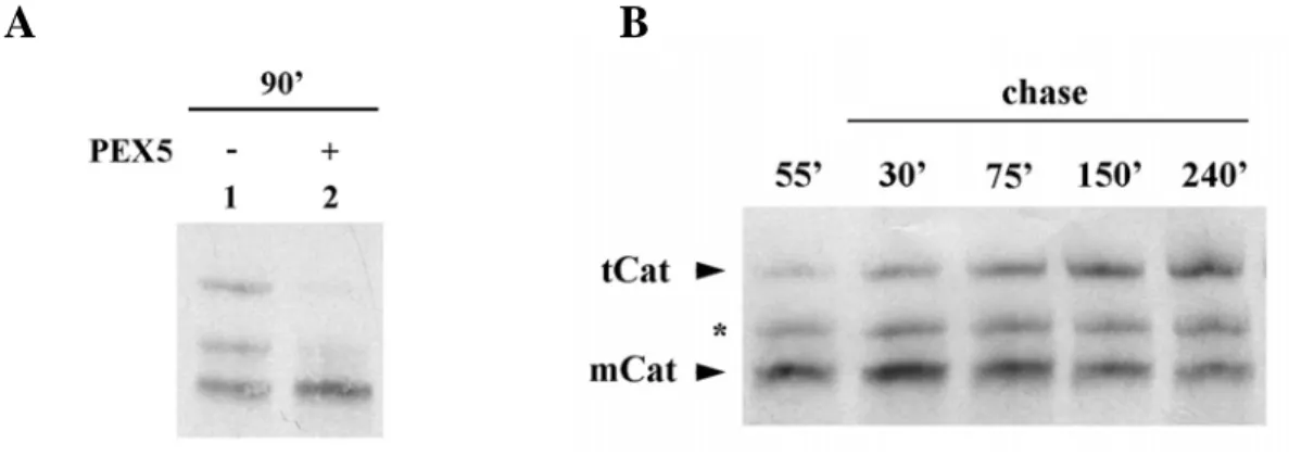
We started by synthesizing the radiolabeled human catalase for 90 min using the rabbit reticulocyte lysate-based in vitro transcription/translation system. By native-PAGE analysis we observed three distinct populations of 35S-catalase (Figure 7A, lane
-). Strikingly, when catalase is synthesized in the presence of 1 μM of recombinant PEX5, a physiological relevant concentration (see Discussion), two of these populations are no longer detected. Apparently, the presence of PEX5 during synthesis of catalase blocks some event.
To determine the nature of the three populations, we performed a pulse-chase analysis where catalase was synthesized for 55 min, cycloheximide was added to arrest protein synthesis and the reaction was further incubated for 4 h. By native-PAGE analysis (Figure 7B) we observed that after 55 min of synthesis (lane 55’) the main product was the faster migrating population, which is converted into the slower one during the 4 h chase (lane 240’). The intermediary population remains constant during the experiment, probably representing an oligomerization intermediate, the dimeric form of catalase (see below).
To better understand the properties of each catalase population observed, we performed a sedimentation analysis with catalase synthesized for just 55 min or chased for four hours. This analysis showed that the faster migrating population corresponds to a specie displaying properties of a 60 kDa globular protein, whereas the slower migrating population behaves as a 200-250 kDa protein (data not shown). These findings indicate that the faster migrating catalase population represents the monomeric form of catalase (mCat). The 200-250 kDa species might represent either tetrameric catalase (tCat) or a complex between catalase and some protein(s) from the
in vitro transcription/translation system (e.g., a chaperone).
Figure 7: 35S-catalase populations after in vitro synthesis. A) Human catalase was synthesized in vitro
in a rabbit reticulocyte lysate for 90 min at 30 ºC in the absence or presence of 1 μM human PEX5, as indicated, and analyzed by native-PAGE/autoradiography. B) 35S-labeled catalase was synthesized for 55 min. After adding cycloheximide, an aliquot was removed and frozen in liquid N2 (lane 55`). The remainder of the reaction was then incubated at 30 ºC and aliquots were removed and frozen at the indicated time points. The samples were subjected to native-PAGE/autoradiography. mCat and tCat correspond to the monomeric and tetrameric forms of catalase; the band labeled with an asterisk probably represents dimeric catalase.
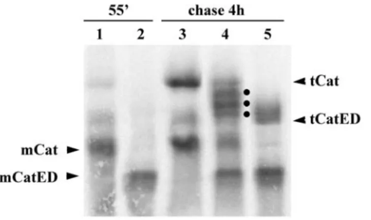
To discriminate between these two possibilities, we produced an acidic mutant version of catalase (CatED), which migrates faster than the normal protein in native gels. If, in fact, the slower migrating form corresponds to the tetrameric catalase, one should be able to form heterotetramers with CatED, containing 1, 2 or 3 molecules of the normal protein; all these heterotetramers should migrate in these gels between the homotetramers of the parental molecules. On the other hand, if the slower migrating band corresponds to a complex containing catalase and some other protein(s), then the band pattern of the protein mixture should just correspond to the sum of the
![Figure 4: Structures of the C-terminal half of PEX5 (PEX5(C)). The structures were obtained in the absence of a ligand (left, [123]) in the presence of the consensus PTS1 peptide YQSKL (Protein Data Base accession number 1FCH) (central, [122]) or in the](https://thumb-eu.123doks.com/thumbv2/123dok_br/15147552.1012512/27.892.202.736.122.399/structures-terminal-structures-obtained-presence-consensus-protein-accession.webp)