FACULDADE DE CIÊNCIAS DEPARTAMENTO BIOLOGIA ANIMAL
MODULATION OF NMDA RECEPTOR ACTIVITY
THROUGH ADENOSINE A
2ARECEPTORS IN THE
HIPPOCAMPUS
DISSERTAÇÃO
FRANCISCO MOURO
MESTRADO EM BIOLOGIA HUMANA E AMBIENTE
FACULDADE DE CIÊNCIAS DEPARTAMENTO BIOLOGIA ANIMAL
MODULATION OF NMDA RECEPTOR ACTIVITY
THROUGH ADENOSINE A
2ARECEPTORS IN THE
HIPPOCAMPUS
DISSERTAÇÃO
FRANCISCO MOURO
MESTRADO EM BIOLOGIA HUMANA E AMBIENTE
Dissertação orientada pela Doutora Raquel Alice da Silva Baptista Dias, Instituto de Medicina Molecular, Faculdade de Medicina da Universidade de Lisboa e pela
Professora Doutora Deodália Maria Antunes Dias, Faculdade de Ciências da Universidade de Lisboa
"Reza a tradição bíblica que a ausência de trabalho - a ociosidade - era a condição de felicidade do primeiro homem antes da sua queda. O amor à ociosidade permaneceu também no homem caído, mas a maldição continua a pesar sobre o homem, e não só porque temos de ganhar o nosso pão com o suor do rosto, mas também porque, pelas nossas características morais, não podemos ser ociosos e calmos. Uma voz secreta diz-nos que devemos sentir culpa da nossa ociosidade. Se o homem pudesse achar um estado em que, sendo ocioso, se sentisse útil e ciente do dever cumprido, acharia uma das facetas da felicidade primitiva."
Lev Tolstói, Guerra e Paz, 1869
"Não gostaria que alguém adoptasse o meu modo de vida por motivo nenhum; pode ocorrer que antes que o aprenda, eu já tenha descoberto outro para mim, e além disso desejo que haja no mundo tanto quanto possível pessoas diferentes. Gostaria, sim, que cada um se empenhasse em descobrir e seguir seu próprio caminho, em vez do trilhado pelo seu pai, sua mãe ou seu vizinho. Que o jovem construa, plante ou viaje, contanto que não seja impedido de fazer aquilo que, segundo ele, gostaria de fazer."
RESUMO
O hipocampo é a estrutura cerebral mais estudada na investigação neurocientífica. A subdivisão CA1 do hipocampo contém a população neuronal que apresenta menor heterogeneidade entre neurónios, encontrando-se organizada em camadas bem definidas, o que torna os neurónios fáceis de identificar e de estudar (Szilágyi et al. 2011).
O neurónio é a unidade sinalizadora individual do sistema nervoso central (SNC). Os neurónios estão inseridos numa complexa rede de circuitos neuronais que se encontra distribuída por todo o cérebro, comunicando entre si através de sinais eléctricos designados como potenciais de acção e potenciais sinápticos (Adrian, 1957).
A transmissão de informação entre um neurónio localizado pré-sinapticamente e uma célula pós-sináptica designa-se como transmissão sináptica (Breathnach, 2005). As sinapses químicas envolvem a libertação pré sináptica de neurotransmissores para a fenda sináptica (Connors & Long, 2004), activando receptores específicos localizados na membrana da célula pós-sináptica (Schoepp et al. 1999).
Os receptores NMDA (N-methyl-D-aspartate) são receptores ionotrópicos cujo principal agonista endógeno é o glutamato (Bakkar et al. 2011. As suas principais características são a dependência de voltagem, elevada permeabilidade ao cálcio e cinética de activação/desactivação lenta (Doherty & Sladek, 2011). Os receptores NMDA são moléculas heteroméricas formadas pelas subunidades GluN1, GluN2 e GluN3 (Groc et al. 2009). Na região CA1 do hipocampo, são tetra heteromeros compostos por duas subunidades GluN1 e duas subunidades GluN2 (Bakkar et al. 2011), podendo classificar-se como sinápticos ou extrasinápticos (Groc et al. 2009).
A actividade dos receptores NMDA está associada, de forma paradoxal, a fenómenos indutores de sobrevivência neuronal e de plasticidade sináptica (Larkman & Jack, 1995; Lipton & Nakanishi, 1999), e a episódios neurodegenerativos associados a morte neuronal (Xu et al. 2009). A ambivalência nas consequências da actividade dos receptores NMDA recebe o nome de “paradoxo dos receptores NMDA” (Hardingham & Bading, 2010), sendo atribuída à localização sináptica destes receptores. De facto, a actividade de receptores NMDA sinápticos aparenta estar associada a fenómenos de protecção neuronal, ao passo que a actividade de
receptores NMDA extrasinápticos conduz a fenómenos de apoptose neuronal (Hardingham & Bading, 2010; Stark & Bazan, 2011).
A actividade de receptores NMDA pode ser influenciada pela acção de neuromoduladores do SNC. A neuromodulação consiste na habilidade neuronal para modificar as propriedades eléctricas dos neurónios em resposta a mudanças bioquímicas intracelulares resultantes de estímulos sinápticos ou hormonais, permitindo ao SNC adaptar a sua capacidade de controlar funções fisiológicas num ambiente em constante mudança (Kaczmarek & Levitan, 1987).
A adenosina é um neuromodulador que medeia os seus efeitos biológicos através de quatros subtipos de receptores: receptores A1, A2A, A2B e A3 (A1Rs, A2ARs, A2BRs eA3Rs) (Tsutsui et al 2004). Grande parte das acções neuromoduladoras da adenosina são mediadas pelos receptores A1 e A2A (Gomes et al. 2010). Os receptores A1 ligam-se a proteínas Gi, cuja actividade inibe a actividade da adenilato ciclase (van Calker et al. 1979). Por seu turno, os receptores A2A ligam-se a proteínas Gs, que estimulam a expressão de adenilato ciclase (Corvol et al. 2001). Enquanto os receptores A1 se encontram amplamente disseminado pelo cérebro (Dunwiddie & Masino, 2001), os receptores A2A estão distribuídos em áreas especificas como o estriado (Haas & Selbach, 2000), embora estejam também presentes em outras áreas cerebrais(Fredholm et al. 2005).
A adenosina é uma substância neuromoduladora capaz de inibir ou de estimular a neurotransmissão no SNC (Fredholm & Dunwiddie, 1988). Os receptores A1 reduzem a libertação de neurotransmissor e inibem a transmissão sináptica. As acções inibitórias mais proeminentes dos receptores A1 estão identificadas em sistemas de transmissão glutamatérgica excitatória, através da inibição na libertação do neurotransmissor glutamato (Barrie & Nicholls, 1993).
Apesar da actividade dos receptores A2 ter um impacto limitado no controlo da transmissão sináptica basal, eles são cruciais no controlo da plasticidade sináptica (Gomes et al. 2010). No hipocampo, a utilização de agonistas (Cunha et al. 1996) e antagonistas (Li & Henry, 1998) específicos para estes receptores provocam, respectivamente, aumentos e reduções na transmissão sináptica excitatória. A nível pré-sináptico, os receptores A2A desempenham um papel facilitatório na libertação de glutamato (Lopes et al. 1999). No hipocampo, os efeitos moduladores dos receptores A2A sobre a actividade dos receptores NMDA é menos bem conhecida. Noutras estruturas cerebrais há evidências de que, a nível pós-sináptico, os receptores A2A
modulam a actividade dos receptores NMDA (Wirkner et al. 2004; Tebano et al. 2005; Rebola et al. 2008). De igual forma, sabe-se que em neurónios CA1 piramidais, os receptores A2A de adenosina controlam de forma directa a activação de receptores AMPA de glutamato (Dias et al. 2002).
Este trabalho foi desenvolvido com o objectivo de compreender se a activação farmacológica de receptores A2A de adenosina pode modular a actividade de receptores NMDA na região CA1 do hipocampo.
Utilizando fatias de hipocampo obtidas a partir de ratos Wistar jovem-adultos, os dados foram adquiridos através de técnicas de electrofisiologia, nomeadamente, procedimentos de patch-clamp utilizando a configuração whole-cell. Primeiramente, foi necessário garantir que as correntes evocadas refletiam exclusivamente actividade de receptores NMDA. Assim sendo, foi utilizado CNQX (10µM), um antagonista competitivo selectivo para receptores de glutamato AMPA e kainato. Os resultados não demonstram alterações significativas nas correntes (99%±2.9% n=3, p>0.05), o que permite concluir que as correntes registadas estavam apenas a ser evocadas pela actividade dos receptores NMDA. De igual forma, para garantir que este efeito está dependente da activação de receptores NMDA, foi utilizado um antagonista especifico destes receptores. A aplicação de DL-APV (50µM) provocou um decréscimo muito significativo nas correntes registadas (76%±4.9% n=3, p<0.005).
De seguida, com o objectivo de tentar encontrar um efeito modulador dos receptores A2A sobre a actividade dos receptores NMDA, começou-se por avaliar os efeitos do CGS 21680, um agonista selectivo dos receptores A2A, sobre a actividade dos receptores NMDA. A adição de CGS 21680 (30nM) provocou um aumento significativo nas correntes mediadas pela actividade dos receptores NMDA (23%±4,7% n=6, p<0.005). Estes dados apontam para um efeito modulador dos receptores A2A que potencia a actividade dos receptores NMDA em células CA1 do hipocampo.
Para assegurar que este efeito reflecte um efeito modulador dos receptores A2A, foi adicionado o agonista CGS 21680 na presença prévia do antagonista selectivo dos receptores A2A, o SCH 58261. A adição de SCH 58261 (100nM) não causou diferenças significativas na amplitude das correntes (3%±14,5% n=3, p>0.05), o que significa que os receptores A2A endógenos não contribuem para o efeito anteriormente registado. A adição subsequente do agonista CGS 21680 (30nM), após
significativas na amplitude das correntes registadas (2%±9,0% n=5, p>0.05). O facto do efeito potenciador do agonista ser prevenido pela presença do antagonista dos receptores A2A, permite concluir que este efeito modulador é mediado por receptores A2A.
Estes resultados sugerem que os receptores A2A modulam a actividade dos receptores NMDA, resultando numa potenciação das correntes pós-sinápticas mediadas pelos receptores NMDA. Estes resultados reflectem um efeito ainda não descrito das capacidades neuromoduladoras dos receptores A2A.
Futuramente, seria importante discriminar se as correntes mediadas reflectem a actividade de receptores NMDA localizados sinapticamente ou extrasinapticamente, uma vez que a actividade de receptores NMDA localizados em diferentes áreas sinápticas tem implicações diferentes para a saúde neuronal. A memantina, um fármaco que bloqueia preferencialmente a actividade de receptores NMDA extrasinápticos, poder-se-á revelar uma ferramenta útil para uma investigação futura sobre o tema.
ABSTRACT
Hippocampal excitatory synaptic plasticity is often considered the synaptic basis for memory formation. NMDAR activity is deeply involved in long-lasting changes in synaptic plasticity. Adenosine modulatory actions upon excitatory glutamatergic transmission are well described. However, the modulatory actions of adenosine A2ARs upon NMDARs activity in CA1 pyramidal cells have never been reported. Thus, the effect of A2AR activation on NMDARs-mediated postsynaptic currents (PSCs) was examined in CA1 pyramidal neurons of young (3-10 weeks) rat hippocampal slices, by using the whole-cell patch-clamp technique (Vh = -60mV). NMDARs-mediated currents were evoked through pressure application of NMDA (150µM) (selective NMDAR agonist) directly onto the cell soma. Bath application of A2AR agonist CGS 21680 (30nM) induced significant increases on NMDA-evoked currents (23%±4,7% n=6, p<0.005). To further address if this effect was caused by an A2AR-mediated modulation of NMDA-evoked PSCs, CGS 21680 (30nM) was superfused in the presence of SCH 58261 (100nM), a selective A2AR antagonist, which was added to the perfusion 10 minutes before the agonist. NMDA-evoked PSCs were not significantly altered by the presence of CGS 21680 (30nM) when A2ARs were previously blocked by SCH 58261 (100nM) (2%±9,0% n=5, p>0.05). To assure that the measured currents were elicited by NMDARs activity, CNQX (10µM) was used to block the activity of AMPA/kainate receptors. The results show no significant changes in NMDA-evoked PSCs in the presence of CNQX (99%±2.9% n=3, p>0.05), suggesting that these currents were mediated by NMDARs. Finally, the use of DL-APV (50µM) - selective NMDARs antagonist - served to further assure that NMDARs mediated the observed currents. In the presence of DL-APV, NMDA-evoked postsynaptic currents significantly decreased (76%±4.9% n=3, p<0.005). Together these results allow to conclude that A2ARs exert a modulatory effect over NMDARs activity at the CA1 neurons of the hippocampus, resulting in potentiation of NMDA-evoked PSCs.
LIST OF ABBREVIATIONS
A1R - adenosine A1 receptorsA2AR– adenosine A2A receptors A3R – adenosine A3 receptors A2BR – adenosine A2B receptors aCSF – Artificial Cerebrospinal Fluid ADA – Adenosine deaminase
Ag – Antigen
AMPA – α-amino-3-hydroxyl-5-methyl-4-isoxazole-propionate ATP – Adenosine triphosphate
BDNF –Brain-derived neurotropic factor CA1-CA3 – Cornu Ammonis, areas 1-3 CAM IV – Calmodulin-dependent protein cAMP – Cyclic Adenosine Monophosphate
CGS 21680 – 2-[4-(2-p-carboxyethyl) phenylamino]-5’N-ethylcarbozamidoadenosine CNQX – -cyano-7-nitroquinoxaline-2,3-dione
CNS – Central Nervous System
CREB – cAMP response element binding CPA – N6-cyclopentyladenosine
DG – Dentate Gyrus
DL-AP5 – DL-2-Amino-5-phosphonopentanoic acid DMSO – Dimethyl sulfoxide
DPCPX – 1,3-dipropyl-8-cyclopentylxanthine EC – Entorhinal Cortex
FOXO – Forkhead protein family of transcription factors GABA – γ-Aminobutiric Acid
LTP – Long-term-potentiation
MAGUKS – Membrane-associated guanylate kinases mGluR5 – Metabotropic glutamate receptor 5
NMDA – N-methyl-D-aspartic acid NMDAR – N-methyl-D-aspartate receptor PKA – Protein Kinase A
PSD-95 – postsynaptic density protein 95
PUMA - p53 upregulated modulator of apoptosis ROS – Reactive oxygen species
Sb – Subiculum
SC – Schaffer Collateral
SCH 58261 - 7-(2-phenylethyl)-5-amino-2-(2-furyl)-pyrazolo-[4,3-e]-1,2,4-triazolo[1,5 c]pyrimidine
SEM – Standard Error of the Mean
Trp53 – Transformation related protein 53 TTX – Tetrodotoxin
TABLE OF CONTENTS
1 Aims………...1
2 Introduction ………...1
2.1 Hippocampus – structure, populations and neuronal circuitry………...1
2.2 Synaptic transmission……….3
2.3 NMDA receptors……….. 6
2.3.1 NMDA receptor characterization………. 6
2.3.2 NMDA receptor structure………. 7
2.3.3 NMDA receptor activity……… 9
2.3.4 NMDA receptor paradox………. 12
2.4 Synaptic vs. Extrasynaptic NMDA receptor signalling……….. 15
2.4.1 Synaptic NMDAR-dependent neuroprotection………15
2.4.2 Extrasynaptic pro-death signalling pathways……….. 18
2.5 Neuromodulation………. 20
2.5.1 Adenosine as a neuromodulator……….. 24
2.5.2 Adenosine receptors……… 25
2.5.3 A1Rs and A2ARs modulatory actions upon NMDA receptor activity………...……….. 28
3 Techniques………. 33
3.1 Electrophysiological recordings in hippocampal slices……….. 33
3.1.1 Tissue preparation………... 33
3.1.2 Patch-clamp techniques………... 36
4 Methods……….. 38
4.1 Animals………... 38
4.2 Preparation of hippocampal slices……….. 39
4.3 Pharmacological tools………. 42
4.4 Data analysis………42
5 Results……… 43
5.1 Activation of adenosine A2A receptors facilitates NMDA receptor evoked postsynaptic currents in CA1 pyramidal neurons………... 43
5.2 Endogenous adenosine does not contribute for the measured NMDA-evoked PSCs………..………...46
5.3 Previous application of adenosine A2A receptors antagonist blocks the
effect of adenosine A2A receptors agonist.……….. 47
5.4 Application of AMPA/kainate antagonist does not alter the evoked PSCs………..…..……… 49
5.5 Application of selective NMDA receptor antagonist cancels the evoked PSCs……… 50
6 Discussion………... 52
6.1 Modulatory effect of adenosine A2A receptor upon NMDA-evoked postsynaptic currents………52
6.2 A presynaptic facilitation in the release of glutamate or a postsynaptic modulatory interaction between receptors?... 53
6.3 Postsynaptic interaction between adenosine A2A receptors and NMDA receptors……….. 54
6.4 Contribution of extrasynaptic vs. synaptic NMDA receptor population for the observed effect………. 56
6.5 The role of adenosine A2A receptors and metabotropic glutamate receptors 5 (mGluR5) in NMDA-dependent forms of LTP in CA1 hippocampal areas………58
6.6 DL-APV did not cancel NMDA-evoked PSCs completely……...59
7 General conclusions……….. 61
8 Acknowledgments………. 62
AIMS
1
The present work was designed with the main objective of identifying a possible modulatory action of adenosine A2A receptor upon NMDA receptor activity in CA1 pyramidal cells. The inhibitory modulatory role of adenosine A1 receptors has already been well described and studied. However, the adenosinergic excitatory actions are yet to be fully understood. NMDA receptors located at the CA1 hippocampal area are key components in several forms of synaptic plasticity and are involved in memory consolidation and spatial learning. Comprehending how adenosine A2A receptors modulate the activity of NMDA receptors in CA1 pyramidal cells can contribute to understand how adenosine modulates synaptic plasticity and memory.
STATE OF THE ART
2
2.1 - Hippocampus – structure, populations and neuronal circuitry
The hippocampus is, nowadays, the most widely studied brain structure in the nervous system. The interest in its study started more than a century ago sparked by the discovery of a prominent relationship between declarative memory and structures in the human medial temporal lobe, specifically the hipocampal formation (van Strien, 2009). Scoville and Milner’s were the first investigators to address memory loss in humans following removal of the hippocampal region, showing in first hand that hippocampal structures are dedicated to memory functions, independent of other cognitive abilities. Besides that, even within memory, the role of the hippocampus is selective, both concerning time window and particular domain of memory processing (Eichenbaum, 2000). The ability to repeat or recognize items just brought into consciousness, in other words the immediate memory, is intact in patients with damaged hippocampal regions (Squirel et al. 1993; Corkin, 1984). Also, general world knowledge and ancient childhood memories doesn’t seem to be impaired. These early findings suggested that the hippocampal regions play critical roles between the initial formation of memories and their final repository in the brain
(Eichenbaum, 2000). Specifically, the hippocampus plays an unavoidable role in the expression of autobiographical episodic and spatial memories (Nadel & Moscovitch, 1997).
The human ability to learn, store and recover information about daily unique and personal events is possible due to episodic memory. Generally, these memories include information about the location and time of a specific event, as well as very detailed characterization of the event itself (Dickerson & Eichenbaum, 2010). The hippocampus is widely evolved in episodic memory in both humans and animals, playing a crucial role in the functions of this particular type of memory (Deshmukh & Knierim, 2011). A vast and complex network of brain areas supports episodic memory, including widespread neocortical association areas and components of the medial temporal lobe, in which parahippocampal cortical areas and the hippocampus are included. The general organization of this neurocircuitry system is that virtually all neocortical association areas send projections that converge onto the parahippocampal areas surrounding the hippocampus. In turn, these parahippocampal areas send projections to all the subdivisions of the hippocampus (Dickerson & Eichenbaum, 2010), through a serial and unidirectional circuit that internally connects the hippocampal subdivisions. This circuit starts in the dentate gyrus and passes, subsequently and continuously, across CA1, CA3 and subiculum neuronal populations.
The most important component in the hippocampal circuitry consists in the trisynaptic loop, comprising the dentate gyrus, CA3 and CA1 neuron populations. The inflow of impulses that arrives from the entorhinal cortex and parahippocampal regions is propagated by excitatory synaptic transmissions that pass through the dentate gyrus and subsequently CA3 and CA1 regions. This information is then sent back to the entorhinal cortex areas, hence the trisynaptic loop (Wojtowicz, 2011). In the hippocampus the circuitry system is extremely important. Therefore, knowledge about hippocampal function is based upon neuronal interaction within this circuitry (Wojtowicz, 2011). Both lateral and medial areas of the entorhinal cortex separately send projections and information to all four hippocampal sub regions (Dickerson & Eichenbaum, 2010).
The various functions of the components in the trisynaptic loop are studied in the standard hippocampal slice preparation, which allows the possibility to work with
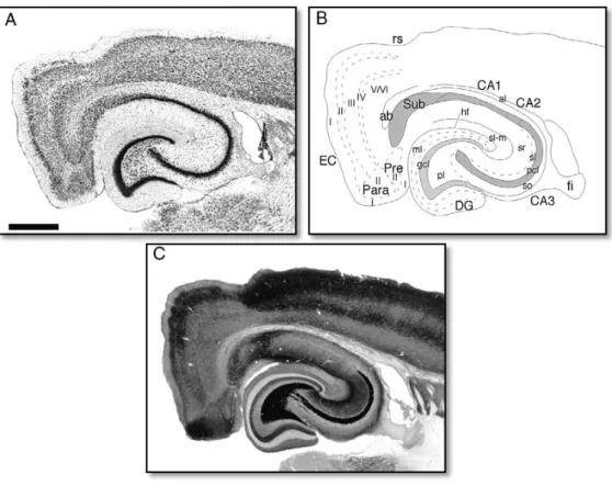
synaptic interconnections are visible, as it is possible to observe in figure 2.1.1 (Andersen et al. 1971). The CA1 subdivision of the hippocampus contains the most studied neuronal population. This population shows less heterogeneity and the pyramidal neuronal cells are organized in well-defined layers that are easy to identify and study (Szilágyi et al. 2011).
Figure 2.1.1 – Horizontal sectioning of the rat hippocampal formation. A. Nissl-stained section. B.
Line drawing showing the various regions, layers, and fiber pathways. C. Timm’s sulfide silver-stained section. CA1 - CA1 field of the hippocampus; CA2 - CA2 field of the hippocampus; CA3 - CA3 field of the hippocampus; DG - dentate gyrus; EC - entorhinal cortex. Adapted from Andersen et al. (2007).
2.2 - Synaptic transmission
Modern neuroscience, as we know it, was born more than a century ago when Santiago Ramón y Cajal found the first scientific results that supported the neuron doctrine. This doctrine consists in the idea that neurons work as the signalization functional unites in the nervous system, communicating with each other’s in a precise and specific way (Albright et al. 2001). The acceptance of this doctrine represented a radical and definitive paradigm shift to a more emphatic cellular perspective of the brain. Ramón y Cajal pertinent observations led to the development of the principle of
dynamic polarization. According to this principle, electric signalization between neurons is unidirectional; in other words, the signals propagate from the receiving pole of the neurons – the dendrites – to the presynaptic axon terminal (Albright et al. 2001). This way, the afferent and efferent elements are associated in the grey matter by unidirectional contact between the neurons (Breathnach, 2005). A few years later, Sherrington found that not all synaptic actions were excitatory and that some of them can be inhibitory (Sherrington, 1932 in Albright et al. 2000). Sherington and Eccles discoveries implied that each neuron solves the competition between excitation and inhibition using, at is initial segment, a winner-takes-all strategy. As a result, an elementary aspect of the integrative action of the brain could now be studied at the level of individual cells. One could determine how the summation of inhibition or excitation leads to an integrated, all-or-none output at the initial segment. Although the initial studies were developed in motor neurons, soon it was discovered that this results had a predictive value in all brain neurons (Albright et al. 2001).
The initial task in understanding the integrative functions of the brain consisted in twigging signal integration in individual cells. Edgar Adrian and John Langley, two contemporaries of Sherrington, developed methods of single-unit analysis within the central nervous system. With these methods, it was possible to study signalling in individual cell at any part of the nervous system. In the progression of his work, Adrian discovered that virtually all neurons use the same conserved mechanism for signalling within the cell (Albright et al. 2001). Therefore, the precise and effective communication between neurons is possible due to the transmission of information through the generation and propagation of an electric signal, acknowledged as the action potential (Adrian, 1957). In all cases, the action potential can be defined as a large, all-or-none, regenerative electric event that propagates, inexorably, from the initial segment of the axon to the presynaptic terminal (Albright
et al. 2001). They are mediated by voltage-activated Na2+ channels, which are initiated following depolarization of the membrane potential to a threshold level (Stuart et al. 1997). As all other cells, neurons have differences in potentials across the cell membrane, the inside being more negatively charged than the extracellular side. This different in potential is known as membrane resting potential.
Given the great number of existing neurons in the nervous system, it is vital the presence of appropriate and efficient communication mechanisms between
unidirectional contacts between neurons (Breathnach, 2005). Synapses can be grouped in two main clusters: electric and chemical synapses. John Langley, a Sherrington contemporary, postulated the first scientific evidence that the majority of the synapses are of chemical nature (Albright et al. 2001). In fact, the main mechanism of synaptic transmission found between neurons comprises the neurotransmitter releasing chemical synapse. However, a more simpler and fast mechanism of signalization can be achieved through electric synapses. The main components of these synapses are gap junctions, which allow the ionic current to flow directly between neurons (Connors & Long, 2004). A big majority of electric synapses are appositions of neuronal membranes entitled gap junctions. These electric synapses, also known as electrotonic synapses, allow the communicating between neuronal cells with completely different functional properties than the ones used by chemical synapses (Bennett, 1972). In electric synapses, the transmission of information depends on current flow through the gab junctions that connect the cytoplasm of pre and postsynaptic cells. In the other hand, chemical synapses are separated by an extracellular space, named synaptic cleft. The communication between neurons becomes possible due to the release and action of chemical agents known as neurotransmitters. As a result of neurotransmitters release into the synaptic cleft mediated by presynaptic cells, specific receptors located at the postsynaptic cell are activated. Figure 2.2.2 displays an example of chemical synaptic transmission involving glutamate and AMPA receptors.
Figure 2.2.2 – Chemical synaptic transmission. These steps can happen during chemical
neurotransmission at the neuromuscular junction as well as in central synapses in both vertebrates and invertebrates. This example applies to fast synaptic glutamatergic transmission, but also to others involving different neurotransmitters and corresponding postsynaptic ionotropic receptors. Adapted from Lisman et al. 2007.
The receptors located in the postsynaptic cell can belong to one of two receptor classes: ionotropic receptors and metabotropic receptors. Eccles and McGeer described in 1997, for the first time, the two main and different forms of neurotransmission. In the ionotropic transmission, the synaptic receptor located in the postsynaptic cell is an ionic channel, in which the activity is not mediated by voltage (as in the sodium and calcium channels), but instead by a chemical ligand, the neurotransmitter (Albright et al. 2000). On the other hand, the metabotropic actions of neurotransmitters mediate the production of second messengers that consequently produce metabolic alterations in the postsynaptic cell (Schoepp et al. 1999).
Depending on the type of neurotransmitter that is mainly produced in the presynaptic cell and the type of receptor mainly expressed in the postsyanptic cell, synapses can be characterized as excitatory or inhibitory. Glutamate is the major excitatory neurotransmitter in the mammalian central nervous system and mediates its actions via activation of both ionotropic and metabotropic receptors families (Arundine & Tymianski, 2003). There are three different types of ionotropic glutamate receptors, which are named after the agonists that were originally identified as the substances that selectively activate them: N-methyl-D-aspartate (NMDA)
receptors, α-amino-3-hydroxy-5-methyl-4-isoazolepropionic acid (AMPA) and 2-carboxy-3-carboxymethyl-4-isopropenylpyrrolidine (kainato) receptors. Concerning metabotropic glutamate receptors, there are eight different receptor subtypes identified (Kew and Kemp, 2005).
2.3 - NMDA receptors
2.3.1 - NMDA receptor characterization
Understanding the functional mechanisms and the expression of NMDA receptors has captured a lot of attention in the last decades. The intense study and debate about NMDA receptors has provided a great deal of knowledge about its synthesis and cellular trafficking. NMDA receptor molecular and biological proprieties, as well as the changes in their expression during synaptic transmission, evolved to two vastly studied subjects mainly due to the discovery that NMDA receptors are key components in the induction of several forms of synaptic plasticity (Groc et al. 2009). Likewise, long lasting changes in synaptic efficiency caused by
associated to damage caused by ischemia (Rothman & Olney, 1995) and to epilepsy (Rogawski, 1993), were two other important factors that led to the increased interest in studying NMDA receptor activity (Dilmore & Johnson, 1998). NMDA receptors exhibit one unique property that allows them to work as a coincident detector for correlated activity. This integrated activity appears to be required for the refinement of synaptic connections throughout brain development, and for activity-dependent potentiation or depression of synaptic inputs (Zarain-Hezerberg et al. 2005).
NMDA (N-methyl-D-aspartate) receptors are inotropic glutamate receptors (Bakkar et al. 2011). When compared with other glutamate receptors, their main characteristics are voltage-dependent activity, high calcium permeability and slow activation-deactivation kinetics (Doherty & Sladek, 2011). NMDA receptors can be found in the synapse, in extrasynaptic locations and presynaptically (Groc et al. 2009). In immature hippocampal neurons, extrasynaptic NMDA receptors represent three-quarters of all the receptors contained in these cells. Although there is an increase in the amount of synaptic NMDA receptors during brain development, a substantial amount of NMDA receptors preserve their extrasynaptic localization throughout the adult life (Hardingham & Bading, 2010). In the synapse, NMDA receptors can be found in the postsynaptic density, where they are structurally organized and spatially restricted in a large signalling macromolecular complex of synaptic scaffolding and adaptor proteins, such as the PSD-95 (postsynaptic density protein 95) family of proteins, or MAGUKS (membrane-associated guanylate kinases). Besides regulating synaptic targeting of glutamate receptors and plasticity in excitatory synapses, these proteins are responsible to physically connect NMDA receptors to kinases, phosphatases and other downstream signalling proteins, as well as to metabotropic glutamate receptors (mGluRs) (Lau & Zukin, 2007).
2.3.2 - NMDA receptor structure
NMDA receptors are heteromeric molecules formed by three subunits, namely subunits GluN1, GluN2 and GluN3. Each one of these subunits contains several variants: single GluN1 subunits with eight splice variants, that are formed through different combinations in the GRIN1 gene, four GluN2 subunits and two GluN3 subunits (Groc et al. 2009). Molecular diversity in NMDA receptor occurs due to alternate RNA splicing in GluN1 subunit (Lau & Zukin, 2007). Alternative splicing of
the same gene creates distinct isoforms responsible for NMDA receptor composition variation (for review see Doherty & Sladek, 2011; Zarain-Herzberg et al. 2005). Unlike GluN1 subunit that is constitutively expressed in NMDA receptor, GluN2 subunit expression is largely regulated during brain development (Tabish & Ticku, 2003). Through molecular screenings of NMDA receptor subunit composition, four GluN2 subunits, each encoded by a different gene, have been identified and described (Ishii et al. 1992). Besides these four GluN2 subunit-expressing genes, in mammals there is a fifth gene that encodes the GluN1 subunit, primarily identified by Moriyoshi in 1991 (Cox et al. 2005). A sixth unique GluN3 subunit receptor has been also identified and derives from a different and independent gene. The physiological signature of the receptor is determined by the co-association between a GluN1 subunit and a GluN2 (A, B, C or D), or between a GluN1 subunit and a GluN3A. It is not clearly understood how this co-assembles between subunits works, but it is believed that it reflects the genetic expression of the encoding genes in specific brain regions (for review see Goebel & Poosch, 1999).
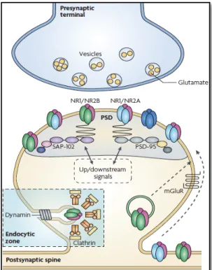
In CA1 hippocampal region, NMDA receptors are tetraheteromers, typically composed of two mandatory glycine-binding GluN1 subunits and two modulatory glutamate-biding GluN2 subunits (Doherty & Sladek, 2011). While GluN1 subunit is essential to the formation of a functional NMDA receptor, different combinations between GluN2 and GluN3 subunit types determines biophysical and pharmacological properties of NMDA channels (Takahashi, 2005). Therefore, GluN2 and GluN3 subunits award distinct properties to each receptor, such as sensitivity to glutamate, peak open probability and response kinetics (Zarain-Herzberg et al. 2005). NMDA receptor subunit composition can affect the development, maintenance and stability of synapses through various potential mechanisms (Tovar & Westbrook, 1999). As shown in figure 2.3.2.1 displayed below, in the synapse, NMDA receptors are connected to a large multi-protein complex through GluN1 and GluN2 cytoplasmic C-terminal (Collins et al. 2006). This complex facilitates NMDA receptor localization in specific areas, simplifying receptor connexions to a variety of signalling molecules that mediate many of the effects induced by NMDA receptor activity (Hardingham, 2009).
Figure 2.3.2.1 – NMDAR macromolecular signalling complex at excitatory synapses. Synaptic
NMDARs are localized in the postsynaptic density PSD (grey area), where they are structurally organized and spatially restricted in a large macromolecular signalling complex comprising scaffolding and adaptor proteins. These proteins physically link the receptors to signalling proteins and to group I mGluRs, which localize to the perisynaptic region. At mature synapses NMDARs are predominantly composed of GluN1/GluN2A or GluN1/GluN2A/GluN2B assemblies. The scaffolding proteins link NMDARs directly or indirectly to a number of signalling proteins, such as protein kinase A (PKA) and PKC, which are thought to be important in the regulation of NMDAR number and function at synaptic sites (upstream/downstream signalling molecules). Adapted from Lau & Zukin, 2007.
2.3.3 - NMDA receptor activity
The primary function of ionotropic glutamate receptors is to respond to presynaptically released transmitter by generating depolarization and excitatory postsynaptic potentials (MacDonald et al. 2007). In order to be active, NMDA receptors need to undergo membrane depolarization and the ligand binding needs to be present (Doherty & Sladek, 2011). As a result of their activity, rapid and high-resolution excitatory signals are transmitted from the presynaptic neuron to the postsynaptic one. Temporal and spatial summation of excitatory postsynaptic potentials in a single postsynaptic neuron provides convergence and integration of a large diversity of excitatory inputs from many presynaptic neurons (MacDonald et al 2007). NMDA receptor activity is essential for the regulation of almost all excitatory
activity in the brain (Doherty & Sladek, 2011). Almost every single excitatory glutamatergic synapse contains NMDA and AMPA receptors. However, excitatory postsynaptic potentials are almost entirely generated by the current flow across AMPA receptors, due to the NMDA receptor voltage dependence (MacDonald et al. 2007). In fact, the present of extracellular Mg2+ leads to a fast blockage of NMDA receptor channel, as it is represented in figure 2.3.3.1 displayed below.
Figure 2.3.3.1 – NMDA receptor at basal condition. In basal activity conditions excitatory
postsynaptic potentials are almost entirely generated by AMPA receptors due to NMDA receptor voltage dependence (MacDonald et al. 2007).
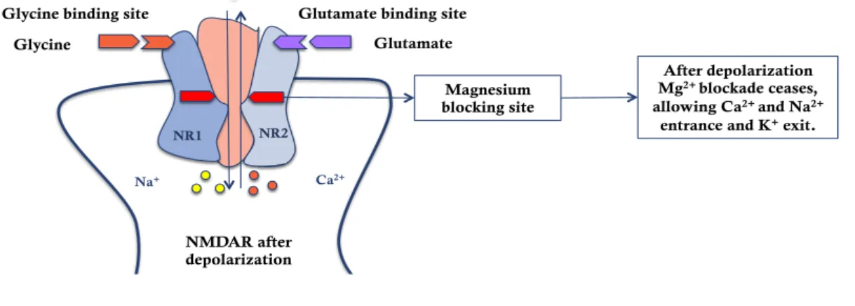
NMDA receptors only start contributing to the formation of excitatory postsynaptic potentials when occurs enough postsynaptic membrane depolarization. As it can be seen in figure 2.3.3.2, the summation of temporal and spatial excitatory postsynaptic potentials results in the ceasing of the extracellular Mg2+ blockage effect and in depolarization of the membrane (McBain & Mayer, 1994).
Figure 2.3.3.2 – NMDA receptor after depolarization of the membrane. After the summation of
temporal and spatial excitatory postsynaptic potentials the membrane is depolarized and the Mg2+ blockage ceases (McBain & Mayer, 1994; McDonald et al. 2007)
Magnesium blocking site NR1 NR2 Na+ Ca2+ After depolarization Mg2+ blockade ceases, allowing Ca2+ and Na2+
entrance and K+ exit.
NMDAR after depolarization Glycine
Glycine binding site Glutamate binding site Glutamate NMDAR before depolarization Magnesium blocking site Glycine
Glycine binding site Glutamate binding site
Glutamate extracellular MgThe presence of 2+ leads
to a fast blockage of NMDA receptor channel. These receptors
only start to contribute to the formation of excitatory postsynaptic
currents when enough postsynaptic membrane
depolarization occurs.
NMDA receptors are essential to LTP (long term potentiation), an activity dependent increase in synaptic efficiency (Larkman & Jack, 1995) mainly expressed in the hippocampus, which can underlay memory and spatial learning processes (Moriyoshi et al., 1991). This phenomenon was initially described in the seventies, when it was postulated that an evoked response in the dentate area by single test shocks to the afferent perforant pathway, often remained potentiated for a considerable time, even after relatively short periods of stimulation (Bliss & Lomo, 1973). The activity of NMDA receptors results in a significant Ca2+ influx increase. As a consequence, NMDA receptor activity is associated with a large variety of intracellular signalling pathways that are dependent on Ca2+ influx variations (MacDonald et al. 2007). For instance, the role of NMDA receptor in LTP triggering is largely associated to an increase in Ca2+ ion concentration inside the cell. This increase occurs when the postsynaptic cell is depolarized due to a large release of glutamate in the synaptic cleft. This way, the extracellular Mg2+ dissociates from the coupling location in the receptor, allowing Ca2+, as well as Na+ ions, to enter the dendritic spine. The consequent increase in Ca2+ ion concentration inside the cell is the critical element to LTP triggering (Malenka & Nicoll, 1999), as it is displayed in figure 2.3.3.3.
Figure 2.3.3.3 – NMDA receptor at basal condition and after depolarization of the membrane.
NMDA receptor activity results in a significant Ca2+ influx increase associated with a large variety of intracellular signalling pathways dependent on Ca2+ influx variations (Malenka & Nicoll, 1999).
Ca2+ increase CaM AC cAMP PKA LTP induction NMDA receptor Glutamate
Ca2+ increase inside the cell
is the critical element in LTP triggering. Due to a large release
of glutamate into the synaptic cleft the postsynaptic cell is depolarized, allowing Ca2+ to enter the cell.
Since their discovery in the seventies and the successful cloning of the receptor accomplished by Moriyoshi in 1991, NMDA receptors gain a role as one of the principal study objects in the attempt to discover the neurobiological mechanisms underlying memory and learning processes. Their study is particularly useful due to four unique NMDA receptor features: the extracellular Mg2+ voltage dependent blockage, their permeability to the passage of Ca2+, their slow kinetics and the presence of glycine as a co-agonist that offers another regulatory component that helps in the precise manipulation of these receptors (Ha et al. 2006).
2.3.4 - NMDA receptor paradox
The variations in Ca2+ concentration inside the postsynaptic cell are the main cause underlying the “NMDA receptor paradox” (Hardingham & Bading, 2010). NMDA receptors are the main responsible agents for the excessive and neurotoxic increase of Ca2+ inside the neuronal cell. When the postsynaptic cell is exposed to high levels of glutamate, it can lead to an over activation of NMDA receptors and consequently to a significant increase in Ca2+ influx inside the postsynaptic neuron. Both these process can lead to cellular signalling disruption contributing to neuronal cell death (Xu et al. 2009). In fact, Ca2+ entrance through NMDA receptors is a particular efficient way of causing neuronal cell death, when compared to its entrance through other ionic channels (Hardingham & Bading, 2010). Glutamate excitotoxicity is identified as a key component in many neurodegenerative and neurologic diseases (Xu et al. 2009). In chronic neurodegenerative diseases, where the insult is mild, excessive NMDA receptor activity leads to neuronal apoptosis. On the other hand, in cases as stroke that can lead to ischemia, the insult is fulminant and the excessive activity of NMDA receptors can lead to cellular necrosis (Lipton & Nakanishi, 1999).
However, NMDA receptors are also linked to neuronal survival molecular pathways and to synaptic plasticity processes (Xu et al. 2009). They play key roles in controlling the morphological and functional plasticity of synapses and contribute to the cellular mechanisms of memory and learning. In pathologies as Alzheimer’s disease, synaptic plasticity is severely impaired (Schimtt, 2005). Besides allowing a direct signalization between neurons, NMDA receptors are also important for the correct neuronal development and or the establishing of synaptic contacts
increase cellular apoptosis mechanisms and traumatic lesions in developing neurons (Hardingham & Bading, 2010).
During several years, the amount of Ca2+ that enters in the postsynaptic neuron through NMDA receptors was considered as the main and only accountable cause related with the different NMDA activity consequences (Lipton & Nakanishi, 1999). Therefore, moderated levels of NMDA activity were considered healthy for the neurons, while NMDA receptors over activation could lead to an overcharge of intracellular Ca2+ harmful to neuron health. According to this model, neuron responses to NMDA receptor activity can be described as a bell-shaped curve, where both a high and a low amount of receptor activity can be harmful for neuronal cell health (Hardingham & Bading, 2010).
Due to more recent discoveries, this later model of cellular behaviour responses to the changes in NMDA receptor activity has been adjusted. According to the new perspective of this paradox, synaptic NMDA receptor location is the main responsible cause for the different cellular consequences following NMDA receptor activity (Hardingham & Bading, 2010). Recent investigations have described several examples of opposing effects in neuronal signalling pathways and cellular survival concerning synaptic and extrasynaptic NMDA receptors activity (Stark & Bazan, 2011). In fact, the duality in NMDA receptor mediated processes varies accordingly to their synaptic or extrasynaptic localization. Synaptic NMDA receptors are related with the induction of signalling survival processes, while extrasynaptic NMDA receptors are associated with Ca2+ disruption and neuronal cell death (Stark & Bazan, 2011). Therefore, cellular consequences of NMDA receptor activity are directly associated to the receptor localization and not only with the amount of stimulation and Ca2+ entrance as previously mentioned (Hardingham et al. 2002). These data can be better described through an “x” shaped graph (see figure 2.3.4.1 below). The ascendant curve of the “x” represents the upsurge in neuroprotection associated with synaptic NMDA receptor activity increase. This ascendant curve is superposed to a descendent one, that illustrates the progressive decrease in neuroprotection related with the increase in extrasynaptic NMDA receptor activity (Hardingham & Bading, 2010). Accordingly to this perspective, it is not the Ca2+ overcharge per se the sole determinant of neurotoxicity but rather the Ca2+ influx through NMDA receptors localized outside the synapse that are responsible for the prejudicial cellular consequences. The influx of Ca2+ through synaptic NMDA receptors is well tolerated
by the neuronal hippocampal cells. Furthermore, it represents the required mechanism for the activation of genomic processes that can give rise to neuronal cells that are more resistant to apoptosis and oxidative processes (Hardingham et al. 2002).
Figure 2.3.4.1 – Synaptic vs. Extrasynaptic NMDA receptor activity. The schematic illustrates the
opposing effects of increasing synaptic and extrasynaptic N-methyl-D-aspartate receptor (NMDAR) activity on neuronal survival and resistance to trauma. Hypoactivity of synaptic NMDARs is harmful to neurons. Enhancing synaptic NMDAR activity triggers multiple neuroprotective pathways and this promotes neuronal survival. Low levels of activation of extrasynaptic NMDARs have no effects on neuronal survival but increasing the level of extrasynaptic NMDAR activity activates cell death pathways and exacerbates certain neurodegenerative processes, thus reducing neuronal survival (adapted from Hardingham & Bading, 2010).
Besides spatial receptor location and Ca2+ influx intensity and duration, there is a third factor that can influence the consequences of NMDA receptor activity. Subunit receptor composition may play an important role in neurotoxicity or neuronal survival processes (Hardingham, 2009). Excitotoxicity mediated by NMDA receptor activity may be triggered due to GluN2B composing subunits (Stanika et al. 2008). There is an ostensive correlation between the presence of GluN2B subunits in NMDA receptor composition, high cytosolic Ca2+ levels and susceptibility to neurotoxicity (Cheng et al. 1999).
2.4 - Synaptic vs. Extrasynaptic NMDA receptor signalling
2.4.1 - Synaptic NMDAR-dependent neuroprotection
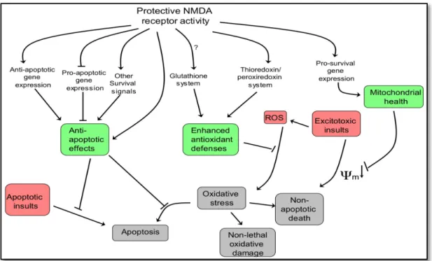
An episode of synaptic NMDA receptor activity can induce neuroprotection for a long time, even after the receptor activity as ceased (Papadia et al. 2005). Although the processes and mechanisms through which nuclear calcium-regulated genomic events increase neuronal survival activity are not fully understood (Dick & Bading, 2010), recently some light has been shed over the subject. Figure 2.4.1.1 presented below displays some of the neuroprotective pathways that are activated through synaptic NMDA receptor activity.
Figure 2.4.1.1 – Neuroprotective pathways activated by synaptic NMDA receptor activity.
Adapted from Hardingham & Bading, 2010.
Calcium influx into hippocampal neurons through synaptic NMDA receptors triggers the release of Ca2+ ions from intracellular calcium stores. Eventually, these Ca2+ ions are transduced to the cellular soma invading the nucleus (Hardingham et al. 2001). The spread of activity-induced calcium signals ending in the cell nucleus represents a main route for the communication between the synapse and the cell nucleus (Zhang et al. 2009). Nuclear Ca2+ is known to be one of the more potent inducers of neuronal gene expression (Hardingham & Bading, 2010). Changes in the
expression of pro-survival genes result in mitochondrial health improvement, the increase of antioxidant defences and the suppression of caspases.
Nuclear calcium-regulated genes are accountable for a neuroprotective shield that neurons build when the synapses are activated (Zhang et al. 2009). Among those genes, a very important target of nuclear Ca2+ is the nuclear Ca2+/ calmodulin-dependent protein (CaM) IV and the transcription factor cyclic-AMP response element binding protein (Hardingham & Bading, 2010). Synaptic NMDA receptor activity promotes neuroprotection throughout the activation of pro-survival signalling pathways, including cAMP response element-binding protein-dependent (CREB-dependent) gene expression (Soriano et al. 2006). CaM kinase IV is an important regulatory protein (Enslen et al. 1994) and CREB is the prototypical signal-regulated transcription factor. CREB is a central mediator of neuroprotection being responsible for controlling a number of pro-survival genes (Zhang et al. 2005). In hippocampal neurons, CREB-dependent gene expression is linked with long-lasting activity-dependent neuroprotective episodes against apoptotic and excitotoxic insults (Hardingham & Bading, 2010). This acquired and long-lasting form of neuroprotection depends on Ca2+ nuclear signalling (Hardingham & Bading, 2010). Through intracellular calcium level manipulations, it has been shown that Ca2+ has a main role in CREB activation (Hardingham et al. 2001). This neuroprotection arises due to the strengthening of the mitochondria against cellular stress and oxidative or toxic insults (Zhang et al. 2009).
Oxidative stress manifestation is related with an imbalance between reactive oxygen species (ROS) and cellular ability to neutralize them via intrinsic antioxidant defences. Neurons are a particularly susceptible and vulnerable type of cell concerning oxidant damage due to high formation of reactive oxygen species. The oxidative damage has a cumulative effect throughout aging and is now recognized as a major accountable factor for some neurodegenerative diseases as well as acute cerebrovascular disorders (Papadia et al. 2008). In addition to apoptosis-suppression effects, synaptic NMDA receptor activity also displays a key role in enhancing neuronal antioxidant defences (Hardingham & Bading, 2010). Synaptic NMDA receptor activity triggers signalling changes centred on the thioredoxin-peroxiredoxin system that underlie a coordinated program of gene expression changes that ultimately lead to the enhancement of antioxidant effects (Papadia et al. 2008).
Ultimately, the expression of these genes will contribute to the preservation of normal neuronal functions and in the improvement of cellular viability when facing aggressive stimuli (Hardingham & Bading, 2010). Also, they can regulate the development and function of individual synapses contributing to synaptogenesis (West et al. 2002). In the context of synaptic plasticity, CREB plays a central role in prolonging and consolidating activity dependent changes in synaptic strength (Papadia et al. 2005). Another target of nuclear Ca2+ - CREB signalling is a gene that encodes the neurotrophin brain-derived neurotrophic factor (BDNF) (Hardingham & Bading, 2010), which is strongly regulated by synaptic NMDA receptor activity (Hardingham et al. 2002). Besides showing neuroprotective proprieties, lowered BDNF expression plays an important role in neurodegeneration processes that follow NMDA receptor blockade (Hansen et al. 2004). BDNF can rescue neurons from NMDA blockade-induced neuronal cell death (Hardingham & Bading, 2010).
The suppression of neuronal death promoting genes consists in a second mechanism through which synaptic NMDA activity guarantees neuronal cell health (Hardingham & Bading, 2010). As said, long-lasting activity-dependent neuroprotection is triggered by Ca2+ entry through synaptic NMDA receptors, requiring the propagation of calcium signals into the nucleus and initiation or repression of gene transcription (Lau & Bading, 2009). Transcriptional suppression of core components of the intrinsic apoptosis cascade is regulated by nuclear Ca2+ signalling (Hardingham & Bading, 2010). Generation of neuronal apoptosis can occur through up-regulation of BH3-only genes such as Puma (Léveillé et al. 2010). It is known that synaptic NMDA receptor activity can supress the expression of this specific gene, both in vitro and in vivo. Similarly to the activation of CREB, suppression of Puma is also specifically induced by synaptic NMDA receptor activity. Ultimately, the suppression of these genes expression results in apoptosis inhibition, increasing neuronal viability (Hardingham & Bading, 2010). Also, the tumour suppression gene, trp53 and two of its targets are regulated by synaptic NMDA receptor activity. Suppression of trp53 acts mutually with nuclear activity and nuclear calcium-induced neuroprotective genes and jointly promotes neuronal surviving through inhibition of mitochondria permeability transition (Lau & Bading, 2010). Mitochondrial health improvement represents a key factor in neuroprotective synaptic NMDA receptor activity mediated pro-survival signalling (Lau & Bading, 2010). Synaptic NMDA receptor activity also subdues the expression of important
pro-death transcription factors such as the FOXO family of Forkhead transcription factors (Hardingham & Bading, 2010). FOXO dependent cellular responses include gluconeogenesis, neuropeptide secretion, atrophy, autophagy, apoptosis, cell cycle arrest and oxidative stress (Salih & Brunet, 2008). The inhibition of FOXO nuclear import is long lasting (Al-Mubarak et al. 2009), which means that it can be sustained for a considerable period of time, even after synaptic NMDA activity has ceased (Hardingham & Bading. 2010).
Also, activation of synaptic or extrasynaptic NMDA receptors that contain in their composition GluN2A subunits is correlated with neuronal survival promoting mechanisms. Furthermore, these receptors can function as a neuroprotective agent against neuronal damage provoked by NMDA receptors or other brain components (Liu et al. 2007).
2.4.2 - Extrasynaptic pro-death signalling pathways
Neuronal death dependent from NMDA receptor activity can be mediated by a variety of signalling pathways that are related to the cell type, the developmental stage or the intensity of the excitotoxic insult. Compared to synaptic NMDA receptor activity, extrasynaptic NMDA receptor activity has been shown to be highly associated with certain pro-death signalling pathways (Hardingham & Bading, 2010).
In direct contrast to what was referred just before, an episode of extrasynaptic NMDA receptors activity promotes CREB inactivation and excitotoxicity events such as mitochondrial depolarization in hippocampal neurons (Soriano et al. 2006). Moreover, CREB dephosphorylating induced by extrasynaptic NMDA receptor activity superimposes CREB activating signals (Sala et al. 2000). The extrasynaptic complexes can act as an antagonist towards nuclear signalling pathways to CREB, blocking the induction of BDNF expression resulting in mitochondrial dysfunction and neuronal cell death (Hardingham et al. 2002).
Investigation in the last two decades has proved that calcium influx into neurons through voltage-gated or ligand-gated ion channels does not imply necessarily a unique and pre-ordained response from the cell. In fact, neurons are wired in complex ways, allowing them to respond differently to calcium signals with distinct characteristics. These crucial parameters include a variety of calcium signal
through which the calcium initially flows into the neuron (Hardingham & Bading, 2002).
Different channels are associated with different signalling molecules or adaptors that can be specifically activated when calcium signals flows through those specific channels. An overwhelming example of calcium entry site-specific differences is the regulation of transcription mediated by the CREB-binding protein CREB (Hardingham & Bading, 2002). Synaptic and extrasynaptic NMDA receptors are coupled to survival and cell death pathways respectively. These differences are explained mainly due to their opposing effects on the cAMP response element binding protein (CREB) (Bengston et al. 2008). CREB regulates the expression of many proteins, including neurotrophic factor BDNF. This protein is highly studied in neuronal plasticity field of investigation due to its engagement in both early and late phases of LTP. Also, a polymorphism of the gene that encodes BDNF is linked to impaired episodic memory in humans, underlying the possible relevance for BDNF in cognitive functions (Vanhoutte & Bading, 2003). In experiments using bath glutamate it was shown that this condition fails to increase BDNF expression due to shut-off of CREB activity, triggered by extrasynaptic NMDA receptors activated by glutamate (Sala et al. 2000). CREB shut-off is induced by a rapid dephosphorylation of its active-site amino acid, serine 133 (Vanhoutte & Bading, 2003). The duration over which serine 133 remains phosphorylated affects considerably the degree of CREB-dependent transcription. The dephosphorylation of CREB on serine 133 blocks CREB-dependent reporter gene expression that is initiated by calcium influx through either L-type channels or synaptic NMDA receptor activity. This way, the observation that bath glutamate rarely promotes CREB activation being, on the other hand, highly associated with CREB dephosphorylation can be explained by the dominance of extrasynaptic NMDA receptor activity over synaptic NMDA receptors signalling (Hardingham & Bading, 2002).
Synaptic and extrasynaptic NMDA receptor signalling triggers completely different processes. In fact, synaptic and extrasynaptic NMDA receptors stimulate or inhibit the expression or suppression of the same genes or processes in an opposing way. Figure 2.4.2.1 summarizes the different signalling pathways induce by synaptic and extrasynaptic NMDA receptor activity.
Figure 2.4.2.1 – Summarizing the different signalling pathways induced by synaptic vs. extrasynaptic NMDA receptor activity. Based on Hardingham & Bading, 2010 review.
2.5 - Neuromodulation
Traditionally, the communication between neurons within a neuronal circuitry has been thought to implicate classical neurotransmission. In this classic form of communication, one neuron either inhibits or excites the other neuron with which it synapses on (Katz & Frost, 1996). Neurons communicate directly with each other by chemical synaptic transmission across synapses. Generally, neurotransmission acts through ligand-gated ion channels and the neurotransmitters involved in synaptic transmission are though to mediate brief postsynaptic responses that are spatially restricted. However, some of these agents can also modulate pre and postsynaptic responses without necessarily evoking postsynaptic potentials. Neuromodulation differs from neurotransmission in that it does not necessarily implicate excitation or inhibition from one neuron to another (Katz & Frost, 1996). Neuromodulators act through intracellular second messenger cascades that origin slower, spatially diffused and long-lasting responses (Gelinas & Nguyen, 2007), which alter the cellular or synaptic proprieties of neurons (Katz & Frost, 1996). Anatomic wired clusters of neurons in the central nervous system can be reconfigured by neuromodulators. These
Synaptic NMDARs activity
Anti-apoptotic effects Induces suppression of death Oxidative stress protection promoting genes
Extrasynaptic NMDARs activity
Apoptotic effects Mitochondrial
depolarization Induces expression of death
promoting genes CaM Kinase IV,
CREB and BDNF signaling pathways. INHIBIT S T IMULA T E
PUMA and FOXO Gene expression related with enhancement of antioxidant defenses INHIBIT S T IMULA T E
specific rhythmical patters of activity (Pflüger, 1999). In figure 2.5.1 are represented tow second messenger system that are relevant to this work.
Figure 2.5.1 – Simplified reaction schemes of two messenger systems of interest to this work. In
the left: The adenylate cyclase/cyclic AMP-dependent protein kinase system. Adenosine receptors are known to couple with G proteins that, depending of the adenosine subtype receptor, either inhibit (van Calker et al. 1979) or stimulate (Corvol et al. 2001) the expression of adenylate cyclase. In the right: Intracellular calcium ions as second level messengers. NMDA receptor activity is particularly linked with Ca2+ influx inside the postsynaptic cell being associated with a large variety of intracellular signalling pathways dependent on Ca2+ influx variations, as LTP induction (Malenka & Nicoll, 1999). Drawings adapted from Kaczmarek & Levitan, 1987.
The concept of neuromodulation was initially recognized as a mechanism where an endogenous substance, which could be released from the pre or postsynaptic component, influences the release (presynaptic modulation) or the action (postsynaptic modulation) of the neurotransmitter (Sebastião & Ribeiro, 2009). Described in a restricted way, neuromodulation can be defined as the neuronal ability to change electrical proprieties in response to intracellular biochemical changes resulting from synaptic or hormonal stimuli (Kaczmarek & Levitan, 1987). Figure 2.5.1 displays some of the changes that the neuromodulators can produce upon the electrical properties of neurons.
Figure 2.5.2 – Some of the changes that can be observed in the electrical properties of neurons.
A) Alterations in the shape of action potentials; B) Changes in frequency and pattern of firing; c) Inhibition and onset of bursting; D) Changes in response to stimulation (arrow). Taken from Kaczmarek & Levitan, 1987.
Neuromodulators can modify neural processing in many different ways regulating various neurophysiological processes (Stern et al. 2007). Therefore, they can affect the integrative proprieties of neurons, by altering the way through which they respond to synaptic drive, modify the strengths of the synaptic interconnections (Sillar et al. 2002) and reshape the plasticity of those synapses (Dayan, 2012). By changing the properties of neurons, neuromodulators can reshape the output of a circuit (Katz & Frost, 1996) and reconfigure the network, producing different output patterns, hence, making the network multifunctional (Pflüger, 1999). By doing so, neuromodulation allows the central nervous system to adapt its control of psysiological functions to a continually changing environment (Kaczmarek & Levitan, 1987). Biochemical changes that lead to modifications in the electrical features of a neuron may be triggered in one of three ways: by the action of neurotransmitters at specific synaptic junctions, by neurotransmitters secreted at less specialized release sites at a distance from the target neuron and finally, through the action of hormones (Kaczmarek & Levitan, 1987).
Figure 2.5.3 – Stimuli that can modulate neuronal activity. 1) Synaptic release of neurotransmitter;
2) Local release of agents from neuronal processes not in direct contact with the target cell; 3) Actions of hormones released from other tissues. Taken from Kaczmarek & Levitan, 1987.
Previously, neuronal interaction diagrams rarely incorporated neuromodulation, including only fast synaptic contacts. First, neuromodulation was usually viewed as being originated from a source extrinsic to the circuit and therefore not a part of the circuit itself. Secondly, even when neuromodulatory interactions are intrinsic to the neuronal circuit, these interactions can easily be overlooked because of their slow time course or because their effects are contingent upon the activity of the neurons in which they act (Katz, 1995). Yet, it is now establish that both intrinsic and extrinsic neuromodulation are important for neuronal circuits control. It is important to distinguish intrinsic from extrinsic neuromodulation to fully comprehend neuromodulatory functions in the operation of neuronal circuits. In extrinsic neuromodulation, a circuit receives modulatory inputs from neurohormones or other areas of the nervous system. Due to its location outside the circuits that it affects, a single extrinsic source can modulate different circuits simultaneously. Additionally, a single circuit can receive extrinsic neuromodulatory inputs from several different sources, allowing the circuit do be differentially modulated under different conditions (Katz, 1995). Intrinsic modulation it is not optional, rather mandatory. Moreover, by contrast with extrinsic neuromodulation, within intrinsic neuromodulation the neuromodulatory substances are released by neurons that are intrinsic to a circuit, being able to affect other neurons and synapses in the same circuit (Katz & Frost,
1996). Additionally, the amount of neuromodulatory agents release is controlled by activity within the circuit itself, not by the activity of a distant region or structure in the nervous system (Katz, 1995). Consequently, intrinsic neuromodulation occurs whenever the circuit is activated and the level of intrinsic neuromodulatory action reflects the intensity of the activity in the circuit itself (Katz & Frost, 1996). Figure 2.5.4 presented below displays the main differences between both types of neuromodulation.
Figure 2.5.4 – Extrinsic and intrinsic neuromodulation. Schematics on the left represent extrinsic
neuromodulation originated from neurons outside the local circuit. Different neuromodulatory neurons can be active at different times and in different behavioural contexts. Also, a single neuromodulatory neuron can elicit neuromodulatory effects in many different circuits simultaneously. On the other hand, the schematics on the right panel of the figure represent intrinsic neuromodulation that arises from the activity of neurons within a circuit itself. These neuromodulatory effects are always present whenever the circuit is active. Adapted from Katz & Frost, 1996.
2.5.1 - Adenosine as a neuromodulator
Among the diversity of neuromodulators existent in the nervous system, adenosine and ATP are key fine-tuners for three main reasons. First, they are among the most significant components in communication processes between neurons and glia cells. Secondly, adenosine and ATP can affect the release and action of several neurotransmitters and neuromodulators. Finally, adenosine and ATP can be release by almost all cells in the nervous system (Sebastião & Ribeiro, 2009). Purines and purine nucleotides are vital components of all living cells. ATP is used as an energy source in virtually all cellular activity and adenine is a component of nucleic acids. Due to