Rute Regina Ferreira Gonçalves
Licenciada em Bioquímica
Dissertação para obtenção do Grau de Mestre em
Genética Molecular e Biomedicina
Orientador: Helena Soares, PhD, CEDOC
Júri:
Presidente: Prof. Doutora Paula Maria Theriaga Mendes Bernardo Gonçalves Arguente: Doutora Margarida Sofia da Silva santos Saraiva
Vogal: Doutora Helena Isabel Martins Soares
Setembro 2017
Toll-like receptor 4 and its direct impact on
human CD4 T cells
iii
Rute Regina Ferreira Gonçalves
Licenciada em Bioquímica
Dissertação para obtenção do Grau de Mestre em
Genética Molecular e Biomedicina
Orientador: Helena Soares, PhD, CEDOC
Júri:
Presidente: Prof. Doutora Paula Maria Theriaga Mendes Bernardo Gonçalves Arguente: Doutora Margarida Sofia da Silva santos Saraiva
Vogal: Doutora Helena Isabel Martins Soares
Setembro 2017
Toll-like receptor 4 and its direct impact on
human CD4 T cells
v
Projeto desenvolvido em CEDOC/FCM-UNL - Centro de Estudos de Doenças Crónicas da
Faculdade de Ciências Médicas da Universidade Nova de Lisboa
CEDOC/FCM-UNL - Chronic Diseases Research Center/ NOVA Medical School, Nova University of Lisbon
Copyright, Rute Gonçalves, FCT/UNL, UNL
A Faculdade de Ciências e Tecnologia e a Universidade Nova de Lisboa têm o direito, perpétuo e sem limites geográficos, de arquivar e publicar esta dissertação através de exemplares impressos reproduzidos em papel ou de forma digital, ou por qualquer outro meio conhecido ou que venha a ser inventado, e de a divulgar através de repositórios científicos e de admitir a sua cópia e distribuição com objetivos educacionais ou de investigação, não comerciais, desde que seja dado crédito ao autor e editor.
Toll-like receptor 4 and its direct impact on
human CD4 T cells
To my mom, family and beloved friends
for always supporting, helping and standing by me
ix
Acknowledgments
I would like to express my sincere gratitude to my advisor Dra. Helena Soares for always trusting me, and for all the patience, motivation and support along the way. I would like to thank to all my lab colleagues Daniela Silva, Rita Torrão, Raquel Sousa, Sofia Cerqueira and Juliana Gonçalves, for all the helpful advices, for the fun times we shared and for being there every day. A special feeling of gratitude to Claudia Andrade for her precious support in flow cytometry techniques. Also, this project would not have been possible without the willingness of the healthy donors and Manuela Correia.
Last, but definitely not least, I would like to thank to all my family and friends, to whom I have no words for all the love, friendship and encouragement throughout this last year. This thesis is dedicated to them, but especially to my mom, my number one supporter, the one person that stands out the most, for always be driven to help me overcoming all my weaknesses, and for giving me everything, under any circumstances, so I can succeed today.
xi
The results discussed in this thesis originated:
Posters:
SINAL 2017 – 8th Meeting on Signal Transduction: “Bridging Innate and Adaptive Immunity – Toll-like Receptor 4 Signaling in human CD4 T cells”. Rute Gonçalves and Helena Soares (June 2017)
Results from this thesis supported the following successful grant
applications:
Pfizer sponsored Sociedade Portuguesa de Reumatologia Award (July 2017) – “Bridging adaptive and innate immunity through JAK signaling”.
xiii
Abstract
Toll-like receptors are mainly expressed on innate immune cells and play an important role in immune responses against pathogens by transducing signals in response to microbial products. TLR4 is well known for recognizing Lipopolysaccharide (LPS), and it has been also detected on activated human CD4 T cells at the mRNA level. However, TLR4 signaling specificities and their contribution for T lymphocyte functions remain unknown. We hypothesized that in addition to cytokines, other tissue microenvironment factors, such as the presence of LPS-bearing commensal and pathogenic bacteria can have an impact on human T cell phenotype. We showed for the first time that TLR4 is expressed in human CD4 T cells, both at the plasma membrane and in endosomal compartments. We found that direct recognition of LPS by activated CD4 T cells promotes their survival. Our results show that TLR4 engagement ensues two distinct signaling pathways driving distinct functional outcomes. At the plasma membrane, TLR4 drives pAKTS473 and p38 MAPKY182 activation, which might offer a mechanistic explanation for increased T cell survival. In addition, the plasma membrane is the site for TLR4 and TCR signaling cross-talk manifested by improved PKCθS676 phosphorylation. Surprisingly, once located to the endocytic compartment, TLR4 intersects TCR signaling by increasing the phosphorylation of a residue that has been described to be under the exclusive control of TCR engagement, p38 MAPKY323. By increasing the activation of p38 MAPKY323,TLR4 might impinge directly on CD4 T cell differentiation, due to the known role of this pathway in directing cytokine production. In this work we demonstrate how TLR4 signaling impinge on CD4 T cell functions namely manipulating cytokine production and the immune response. Our work might contribute to explain how immune plasticity is regulated by the tissue microenvironment, and how it might counteract chronic inflammations, an emerging field in immunology with elevated translational potential.
Keywords: Toll-like receptor 4; Human CD4 T cells; Helper T cell plasticity; Helper T cell
counter-regulation; Tissue protection; novel therapeutic targetsxv
Resumo
Os receptores do tipo Toll são maioritariamente expressos em células imunes inatas e desempenham um papel importante nas respostas imunitárias contra agentes patogénicos através da transdução de sinais em resposta a produtos microbianos. O TLR4 reconhece o Lipopolissacarídeo (LPS), e foi também detectado ao nível do mRNA em células T humanas ativadas. No entanto, as especificidades da sinalização do TLR4 e a sua contribuição para as funções dos linfócitos T permanecem desconhecidas. Levantou-se a hipótese que, além de citoquinas, outros fatores como a presença de bactérias comensais e patogénicas portadoras de LPS, pudessem afetar o fenótipo das células T. Mostrámos pela primeira vez que o TLR4 é expresso em células T CD4 humanas, tanto na membrana plasmática como em compartimentos intracelulares. Descobrimos que o reconhecimento direto do LPS por células T CD4 ativadas promove a sua sobrevivência. Os resultados mostram que a ativação do TLR4 segue duas vias de sinalização que geram comportamentos funcionais distintos. Na membrana plasmática, o TLR4 ativa o pAKTS473 e o p38 MAPKY182, o que pode oferecer uma explicação mecanicista para o aumento da sobrevivência das células T. Além disso, na membrana plasmática ocorre o cruzamento das sinalizações do TLR4 e do TCR manifestado pelo aumento da fosforilação do PKCθS676. Surpreendentemente, quando localizado no compartimento intracelular, o TLR4 intersecta a sinalização do TCR aumentando a fosforilação de um resíduo que foi descrito como estando sob controlo exclusivo do TCR, p38 MAPKY323. Ao aumentar a ativação do p38 MAPKY323, o TLR4 pode afetar diretamente a diferenciação de células T CD4, devido ao envolvimento desta via na produção de citoquinas. O nosso trabalho poderá contribuir para explicar o modo como a plasticidade das células T é regulada pelo microambiente, e o modo como esta pode contrariar inflamações crónicas, um novo domínio emergente na área da Imunologia com elevado potencial translacional.
Palavras-chave: Receptor do tipo Toll 4; Células T CD4 humanas; Plasticidade de células T
auxiliares; Contra-regulação de células T auxiliares; Proteção de tecidos; Alvos terapêuticosxvii
Contents
1. Introduction 1 1.1. Immune system 3 1.2. Immune recognition 4 1.2.1. Innate immunity 4 1.2.2. Adaptive immunity 6 1.3. Toll-like receptors 10 1.3.1. Toll-like receptor 4 121.4. Expression of Toll-like receptors in T cells 14
1.4.1. Toll-like receptor 4 in T cells 15
1.5. Aims 16
2. Materials and Methods 17
2.1. Reagents 19
2.1.1. Antibodies 19
2.2. Isolation of human peripheral blood mononuclear cells 20
2.3. Sorting and purification of CD4+ T cells 20
2.4. Culture and stimulation assays of CD4+ T cells 21
2.5. T cell survival assay 21
2.6. Immunofluorescence microscopy 22
2.7. T cell proliferation assay 23
2.8. Phenotyping of CD4+ T cells 23
2.9. Cytokine production 24
2.10. Cell signalling protocol – Phospho-protein analysis 24
2.11. Statistical analysis 24
3. Results and Discussion 25
3.1. Expression of TLR4 on activated human CD4+ T cells 27 3.2. Chronic engagement of TLR4 improves intracellular expression of TLR4 27 3.3. LPS promotes human CD4+ T cell survival, but not proliferation 29 3.4. CD4+TLR4+ T cells are double positive for PD1 and CD38 30
3.5. TLR4 engagement upregulates CD38 and CD45RO 31
3.6. Chronic engagement of TLR4 improved AKTS473, p38 MAPKY182 and PKCθS676 activation
32
3.7. LPS directly activates AKTS473 and p38 MAPKY182, but not PKCθS676 in CD4+ T cells
34
3.8. TLR4 internalization decreased the activation of AKTS473, PKCθS676 and p38 MAPKY182
36
3.9. LPS is capable to impinge on TCR signaling by activating p38 MAPKY323 38 3.10. TLR4 internalization improved the activation of p38 MAPKY323 39
xviii
3.11. TLR4 internalization is highly required for a full activation of p38 MAPKY323 39 3.12. Inhibition of TLR4 internalization with Chlorpromazine improves the activation ofAKTS473, PKCθS676 and p38 MAPKY182, while inhibiting the phosphorylation of p38 MAPKY323
40
3.13. TLR4 is recruited to the synaptic membrane after TCR stimulation in CD4+ T cells 44 3.14. ZAP70 as a candidate to crosstalk between TLR4 and TCR pathways 46 3.15. Rab11a-positive intracellular compartments colocalize with internalized TLR4 47 3.16. LPS causes CD4+ T cell to decrease IFNγ but upregulate IL17A production 48 3.17. Direct recognition of LPS promotes IL-10 production redirecting Th17 cells to a
tolerogenic profile
50
xix
Figure List
Figure 1.1 Dynamics of innate and adaptive immunity
Figure 1.2 Toll-like receptors: Subcellular localization and ligands Figure 1.3 Activation and priming of naïve DCs after pathogen encounter Figure 1.4 Antigen presentation in the lymph nodes
Figure 1.5 Polarized CD4+ T cell subsets
Figure 1.6 TLR signaling in mammalian antigen presenting cells Figure 1.7 TLR4 signaling in antigen presenting cells
Figure 1.8 Three required signals for T cell full activation Figure 2.1 PBMCs isolation and cell sorting strategy
Figure 3.1 TCR engagement upregulates TLR4 surface expression in CD4 T cells, and chronic TLR4 engagement improves intracellular TLR4
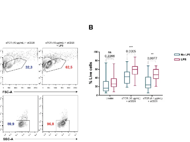
Figure 3.2 TLR4 engagement on CD4 T cells promotes their survival
Figure 3.3 TLR4 engagement on CD4 T cells does not affect their proliferation Figure 3.4 CD4+TLR4+ T cells are double positive for PD1 and CD38
Figure 3.5 LPS promotes the expression of CD38 and CD45RO in CD4+TLR4+ T cells
Figure 3.6 Chronic engagement of TLR4 improved AKTS473, p38 MAPKY182 and PKCθS676 activation
Figure 3.7 LPS can activate directly AKTS473 and p38 MAPKY182, but not PKCθS676
Figure 3.8 TLR4 internalization decreased the activation of AKTS473, PKCθS676 and p38 MAPKY182
Figure 3.9 Alternative p38 activation (p38 MAPKY323) pathway in T cells
Figure 3.10 LPS is capable to impinge TCR signaling by activating p38 MAPKY323 on CD4+ T cells Figure 3.11 TLR4 internalization improved the activation of p38 MAPKY323
Figure 3.12 TLR4 internalization is highly required for a full activation of p38 MAPKY323
Figure 3.13 Inhibition of TLR4 internalization with Chlorpromazine improves the activation of AKTS473 and PKCθS676, while inhibits the p38 MAPKY323
Figure 3.14 Inhibition of TLR4 internalization with Chlorpromazine improves the activation of PKCθS676 and p38 MAPKY182
Figure 3.15 TLR4 activates AKTS473 and p38 MAPKY182 from the plasma membrane. Similarly TLR4 and TCR signaling cross-talk occurs at the plasma membrane. Otherwise, activation of p38 MAPKY323 likely requires TLR4 signaling from the endocytic compartment
Figure 3.16 TLR4 is recruited to the synaptic membrane after TCR stimulation. At 20 minutes of stimulation, TLR4 converge to the cSMAC while being endocytosed
Figure 3.17 pZAP70Y319 is predominantly expressed closer to the plasma membrane, being a candidate signaling molecule to cross-talk between TLR4 and TCR pathways Figure 3.18 Rab11a-positive intracellular compartments colocalize with internalized TLR4
xx
Figure 3.19 Direct recognition of LPS promotes IL-17 production while inhibits IFNγFigure 3.20 Direct recognition of LPS promotes IL-10 production mostly on IL-17A-producing CD4 T cells
Figure 3.21 Direct recognition of LPS promotes IL-17 and IL-10 production, suggesting a redirection of CD4+ T cells to a tolerogenic phenotype
Figure 4.1 Proposed model for the role of TLR4 in T cells, particularly in microbe rich environments
xxi
Abbreviation List
AKT (PKB) Protein kinase B
AP1 Activator protein 1
APC Antigen presenting cell
BAD BCL-2 associated death promoter BCL-XL B-cell lymphoma extra-large
BCR B cell receptor
BFA Brefeldin A
BSA Bovine Serum Albumin
CDx Cluster of Differentiation x
cGAS Cyclic GMP-AMP synthase
CLR C-type lectin receptor
CPZ Chlorpromazine
CREB Cyclic AMP-responsive element-binding protein cSMAC Central supramolecular activation cluster DAMP Dangerous Associated Molecular Pattern
DC Dendritic cell
DMSO Dimethyl sulfoxide
DNA Deoxyribonucleic Acid
EAE Experimental autoimmune encephalomyelitis
ER Endoplasmic reticulum
ERK Extracellular signal–regulated kinase FACS Fluorescence-activated cell sorting
FBS Fetal Bovine Serum
FOXO1 Forkhead box protein O1
Foxp3 Forkhead box P3
GM-CSF Granulocyte-macrophage colony-stimulating factor IFI16 Interferon gamma inducible protein 16
IFN-γ Interferon gamma
IKKε IκB kinase ε
IL-x Interleukin-x
IRAK IL-1R-associated kinase IRFx Interferon regulatory factor x
ITAM Immune-receptor tyrosine-based activation motif iTreg Induced regulatory T cell
IU International units
JNK JUN N-terminal kinase
xxii
MAL MyD88-adapter-like
MAPK Mitogen-activated protein kinase MHC Major histocompatibility complex mTOR Mammalian target of rapamycin
MyD88 Myeloid differentiation primary response protein 88 NF-kB Nuclear factor kappa B
NOD-like receptor Nucleotide-binding oligomerization domain receptor nTreg Natural regulatory T cell
PAMP Pathogen Associated Molecular Pattern PBMC Peripheral blood mononuclear cell PBS Phosphate buffered saline
Pen/Strep Penicillin/Streptomycin
PFA Paraformaldehyde
PI3K Phosphoinositide 3-kinase PKCθ Protein kinase C-theta PLL Poly-L-Lysine hydrobromide PMA Phorbol 12-myristate 13-acetate PtdIns(3,4,5)P3 Phosphatidylinositol 3,4,5-triphosphate PtdIns(4,5)P2 Phosphatidylinositol 4,5-biphosphate qPCR Real-time polymerase chain reaction RIG-I-like receptor Retinoic-acid-inducible gene 1-like receptor RORγT RAR-related orphan receptor gamma
RPMI 1640 Roswell Park Memorial Institute medium 1640
RT Room temperature
Syk Spleen tyrosine kinase Tbet T-box transcription factor
TBK1 TANK-binding kinase 1
TCR T-cell receptor
TGF-β Transforming growth factor beta Thx Helper T cell type x
TIR Toll/interleukin-1 receptor
TIRAP TIR domain-containing adaptor protein
TLR Toll-Like Receptor
TNFα Tumour Necrosis Factor α
TR1 T regulatory type 1 cell
TRAF TNF receptor-associated factor TRAM TRIF related adaptor molecule
TRIF TIR-domain-containing adapter-inducing interferon-β VEGF Vascular endothelial growth factor
INTRODUCTION
3
1.1.
Immune system
The immune system constitutes the defense of the organism. Its main functions are to eliminate pathogenic agents and to maintain the body homeostasis. It is one of the most complex systems comprising both unspecific and specific components that, although distinct, cooperate and synergize to create a controlled protection to disease (Kennedy 2010). The immune system is an interactive network of lymphoid organs, cells, humoral factors and cytokines and, differently from other systems, it is highly dispersed throughout the organism. In fact, its elements vary from physical barriers, such as the epidermis, to the most complex and specific components, such as the lymphocytes (Parkin and Cohen 2001).
The immune system can be divided in three lines of defense:
The first line of defense are physical, chemical and biologic barriers that prevent the entry of pathogens into the body;
The second line of defense is constituted by phagocytes and other defense mechanisms (e.g. complement) that comprise the rapid response of the innate immunity;
The third line of defense consists of lymphocytes, which are highly specific cells capable to create memory and to ensue more efficient immune responses upon re-exposure.
Upon encounter with the invading pathogen, the first line of defense sets up physical, chemical and enzymatic barriers to avoid its entrance into the body. If those barriers fail or are insufficient, the innate immunity immediately comes into play, eliciting a very quick and broad immune response that often culminates in the elimination of the pathological agent. The players of the innate immunity are the monocytes, macrophages, dendritic cells (DCs), neutrophils, eosinophils, basophils and mast cells. Innate immune cells are endowed of multiple defense mechanisms capable of recognizing and neutralizing pathogens, such as
phagocytosis, defensins and the complement system. The innate immune system is capable of recognizing self from nonself through the expression of receptors that are able to recognize and bind common and preserved components present in pathogens but not in the host cells. In some cases, these receptors do not recognize microbial products directly but detect their presence indirectly through the recognition of ‘‘stress signals’’ molecules produced by cells that have become infected (Melvold and Sticca 2007). Albeit, efficient at rapidly eliminating pathogens, the innate immune response is not perfect: on one hand the strong inflammatory response ensued by the innate immune cells lead to damage of healthy
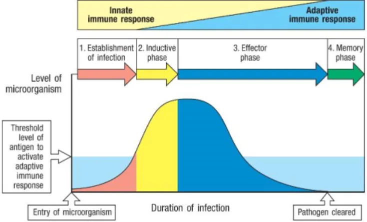
Figure 1.1 - Dynamics of innate and adaptive immunity. From (Janeway et al. 2001). After the entrance of the
microorganism, 1st and 2nd lines of defense are set up, leading to the influx of antimicrobial proteins, phagocytes, and complement to the local of infection, where they work alongside to destroy the pathogen. If they fail to clear the infection, and the threshold level of antigen is reached, the inductive phase begins and professional antigen presenting cells (APCs) migrate to the lymph nodes where they trigger the adaptive immunity. That way, innate and adaptive immunity build an efficient response against the invader. After pathogen clearance, immunological memory is mounted by the adaptive system, ensuring a faster and stronger immune response after a re-exposure to the same microorganism.
4
tissues, and on the other hand it cannot eliminate pathogens that quickly evolve to avoid their detection and clearance by the immune system.Whenever the 2nd line of defense fails to neutralize the infection, the adaptive immune system takes action and mount a latter but highly specific response against the invading pathogen (Figure 1.1). This system possesses more sophisticated defense mechanisms, it has the capability to recognize ever changing pathogens and to mount a stronger immune response upon re-exposure. Thus, the defining feature of the adaptive immune system is the memory, i.e. the capacity to mount a faster and stronger immune response, capable of avoiding reinfection with the same microorganism (Purves et al. 2000).
Along with those refined mechanisms that allow the immune system to recognize and act against non-self and/or “stress” proteins, the immune tolerance is highly required. When this discrimination fails, the immune system activates destructive immune reactions against body’s own components, and autoimmune diseases arise. This can be prevented by tolerance mechanisms to self-proteins, including: (i) Central tolerance by clonal deletion of autoreactive lymphocytes in the thymus or bone marrow. During that process, T cells undergo a positive selection that ensures T cells only recognize antigen in association with major histocompatibility complex (MHC) molecules. Finally, those previously selected T cells meet with self-peptides, and self-reactive ones are elected to die via programmed cell death and excluded from the T cell repertoire (negative selection) (Parkin and Cohen 2001); (ii) Peripheral tolerance acts after T cells are exported into the periphery. Some self-reactive T cells might escape through central tolerance mechanisms if they don’t have sufficient affinity for the self-antigen, if they only recognize tissue-specific antigens, or even if they only react at latter times of development. Peripheral tolerance resort to deletion and inactivation (anergy) of self-reacting peripheral lymphocytes; (iii) Regulatory T cells are capable to suppress or downregulate induction and proliferation of effector T cells. Those regulatory cells are known to modulate the immune system, maintain tolerance to self-antigens, thus preventing autoimmune diseases (Abramson and Husebye 2016).
Immune system hyper-responsiveness can also lead to exacerbated immune responses against foreign compounds like what happens in allergic rhinitis and asthma.
On the opposite end of the spectrum there is the immunodeficiency where the immune system is deficient in mounting an effective immune response, resulting in recurring and life-threatening infections. Altogether, the immune system, as a complex network, needs to work under a strict balance. Its overall functions are to recognize, defend and memorize, protecting the host from hazardous agents and disease, thereby preserving the integrity of the body.
1.2.
Immune recognition
1.2.1. Innate Immunity
The immune response is trigged by exposure to a foreign compound. In normal circumstances, it is a controlled response that works to eliminate pathological microbes and toxic or allergic proteins, but also, at the same time, must avoid self-destruction and the clearance of commensal and beneficial microbes. That is the reason why during the immune response there is a shift towards specialized immune receptors capable to distinguish between self- and non-self-proteins, whose specificity increases in the course of the response (Melvold and Sticca 2007). Whereas the adaptive immune
5
system uses a large repertoire of receptors encoded by rearranging genes to recognize an infinite variety of antigens, innate immunity depends upon germ-line encoded receptors to recognize pathogenic features that are common and evolutionary conserved.After exposure, cytokines produced by phagocytes [Interleukin-1 (IL-1), IL-6 and Tumour Necrosis Factor α (TNFα)] activate the acute phase response and the body generates active and unspecific proteins, such as the C-reactive protein produced by liver cells, in order to immediately act against microorganisms or to improve trauma resolution (Di Napoli et al. 2011). This acute response leads the activation of the coagulation system, and the production of the complement, lactoferrin and transferrin, lysozyme and cytokines. Those components act by neutralizing bacteria, increasing vasodilatation and vascular permeability, creating a boost and powerful influx of even more phagocytic cells. Inflammation, fever, loss of appetite and somnolence are some of the long-range effects of this stage of immune response to infection and trauma (Janeway et al. 2001).
As stated above, monocytes, macrophages and DCs are some of the key players of the innate immune system. Each one of them has its specific function in the resolution of the infection. Even though they lack the high specificity of adaptive immune cells, they still have the capability to distinguish self from nonself. They express immune receptors that allows them to recognize conserved patterns and structural motifs only presented in microorganisms - Pathogen Associated Molecular Patterns (PAMPs) – and “danger signals” that result from tissue damage - Dangerous Associated Molecular Patterns (DAMPs) (Jin et al. 2012).
The recognition of microorganisms is mediated by several families of innate immune receptors that survey the extracellular space, endolysosomal compartments and the cytoplasm for signs of infection or tissue damage and gives rise to very rapid responses. These are nucleotide-binding oligomerization domain receptors (NOD-like receptors), C-type lectin receptors (CLRs), retinoic-acid-inducible gene 1(RIG-I)-like receptors, Scavenger receptors, Toll-Like Receptors (TLRs) and the most recently described deoxyribonucleic acid (DNA) sensors Interferon gamma inducible protein 16 (IFI16) and cyclic GMP-AMP synthase (cGAS) (Joosten et al. 2016). They have a number of different functions: some stimulate ingestion of the pathogen, some are chemotactic receptors and others induce effector molecules that contribute to the priming of innate immune responses and that influence the initiation and nature of any subsequent adaptive immune response.
TLRs are the best characterized family in terms of known ligands, established localization, downstream signalling pathways and functional relevance (T Kawai and Akira 2006). TLRs are expressed in a variety of mammalian cell types, however their functional relevance is best studied in antigen presenting cells (APCs) as macrophages and DCs. They can be classified and divided depending on their ligands and subcellular sites. Some localizations are stable, whereas others can be more flexible. TLR 1, 2, 6, 5 and 4 are localized in the cell surface, thus recognizing accessible molecules in the membrane of most pathogens, like lipopolysaccharide (LPS), lipoproteins, fibronectin and flagelins. On the other hand, TLR 3, 7, 8 and 9 are localized within various endosomal compartments, where they recognize microbial nucleic acids (Kabelitz and Medzhitov 2007) (Figure 1.2).
6
Binding of TLRs on phagocytic cells triggers their activation and they become enlarged, increase their production of antimicrobial products, and start to ingest and degrade microbes. Meanwhile, some activated phagocytic cells activate the adaptive system and the adaptive immune response is finally generated. Eventually, if the innate immune response fails to neutralize the infection, the adaptive immune system takes action ensuing a highly specific and long-lasting immune response against the pathogen.1.2.2. Adaptive Immunity
The adaptive immunity is mediated by lymphocytes, specialized cells with highly specific receptors for the antigen. Lymphocytes derive from lymphoid committed progenitors, of which 25% remain in the bone marrow and differentiate into naïve B lymphocytes. The remaining ones travel to the thymus and become naïve T lymphocytes. The thymus is a specialized primary organ that provides an inductive environment for T cell development from hematopoietic progenitor cells. In addition, one of the most important roles of the thymus is the induction of central tolerance selecting a functional and self-tolerant T cell repertoire (Jin et al. 2012). Naïve B and T cells migrate into the lymph nodes where eventually they will be activated by antigen-primed DCs.
In fact, DCs are the main innate immune cells that ensue the crosstalk between the innate and the adaptive immune response. Naïve DCs, as specialized phagocytic cells, are capable to recognize exogenous pathogens and/or foreign proteins through specialized receptors such as TLRs. That induces the activation of intracellular signalling pathways and the production of inductive cytokines (Ley 2014)
(Figure 1.3 A).
Figure 1.2 - Toll-like receptors: Subcellular localization and ligands. From (Janeway et al. 2001) Toll-like receptors
are the best characterized family of innate immune receptors. Some TLRs are expressed at the plasma membrane, while others are located within endosomal compartments. They are responsible for the recognition of various microorganisms, signs of infection and/or tissue damage.
7
The internalized pathogens/proteins are processed inside the cell by acid-dependent proteases in endosomes. Meanwhile, immature MHC class II molecules are in the endoplasmic reticulum (ER) with their peptide-binding site blocked by li, an invariant chain that prevents them from binding self-peptides and/or self-peptides from intracellular pathogens. Those intracellular self-peptides follow a different endogenous pathway in which they are processed by the proteasome, loaded onto MHC class I molecules in the ER, and released through the Golgi apparatus to the plasma membrane where they will activate effector CD8+ T cells. Otherwise, immature MHC class II molecules are exported from the ER in vesicles that fuse with late endosome/lysosome compartments containing the degraded pathogenic proteins, which are then loaded onto MHC class II antigen grove. This exogenous pathway ends up by delivering the MHC class II loaded peptides to the plasma membrane and to helper CD4+ T cells activation (Kennedy 2010), (Aichinger and Lechler 1995) (Figure 1.3 B). Activated DCs migrate into the lymph nodes and they start a process called as “Antigen presentation”, activating naïve B and T lymphocytes in both B and T zones, respectively (Figure 1.4). From that moment, the adaptive immunity is initiated.Naïve T lymphocytes are activated after antigen recognition and co-stimulation leading to cell proliferation that ensures a sufficient number of lymphocytes with the same specificity, an alteration of gene expression and differentiation in cells specialized to fight the infection at task. Antigen recognition is achieved through the engagement of the T-cell receptor (TCR) by the antigen loaded MHC molecules in the APC. TCR is highly specific for the antigen-MHC complex and this specificity derives from gene recombination. The TCR cytoplasmic tail contains a sequence known as ITAM (immune-receptor tyrosine-based activation motif), essential for the initiation of signal transduction (Guy et al. 2013). For T cell full activation, besides TCR engagement, it is also required interaction between co-stimulatory molecules cluster of differentiation 28 (CD28) on the T cell, and B7 family molecules on the APC. If there are no co-stimulatory signals, T lymphocytes become anergic, a crucial mechanism in the maintenance of peripheral tolerance (Kennedy 2010).
The process of clonal expansion takes 3-7 days and ends up with the production of both effector and memory T lymphocytes, all with the same antigenic specificity.
T cells take part of the cell-mediated immunity, acting against intracellular and phagocytosed microbes. They can be divided into Helper (CD4+) and Cytotoxic (CD8+) lymphocytes. CD8+ lymphocytes are capable of killing infected cells and eliminate reservoirs of infection. CD4+ lymphocytes are not usually capable of killing cells, instead they are considered the orchestrators of the specific response (Kennedy 2010). After thymus differentiation, CD4+ T cells can be divided into Foxp3- effector T cells and Foxp3+ natural regulatory T cells (nTregs) (Fontenot, Gavin, and Rudensky 2003). Through the release of cytokines and other soluble messengers, effector CD4+ T cells can activate macrophages allowing them to degrade the internalized pathogen, they can act by inducing B cell proliferation and determining antibody secretion, complement activation and neutrophil chemotaxis. nTregs are a stable Figure 1.3 (in previous page) – Activation and priming of naïve DCs after pathogen encounter. Based on (Janeway et al. 2001). DCs recognize exogenous pathogens through specialized receptors such as TLRs. A -
TLR4 recognizes LPS in Gram-negative bacteria inducing the activation of intracellular pathways and the production of inductive cytokines. B –After TLR engagement, the pathogen is internalized and phagocytosed. Degraded pathogen-derived proteins are then loaded on MHC class II molecules and delivered to the plasma membrane. By this time, DCs are loaded with specific antigens.
8
lineage originated in the thymus, and they can modulate the immune response, maintain tolerance to self-antigens preventing autoimmune diseases.The cytokine environment at the time of antigen presentation, and the nature and quantity of the antigen itself, drive T cell polarization. During this process, CD4+ effector T cells can acquire regulatory features, therefore becoming induced regulatory T cells (iTregs) (Mayne and Williams 2013). iTregs are functional relevant in feto-maternal interfaces (Samstein et al. 2012) and to maintain tolerance to food- and microbiota-derived antigens at mucosal sites (Haribhai et al. 2009).
Each T cell subset has its specific ability to sense different inductive cytokines, and environmental cytokines can arise from the innate response and be produced by other T lymphocytes and APCs. Therefore, CD4+ T cells can be differentiated into a number of cell subsets such as iTregs, helper T cells type 1 (Th1), Th2, Th17 and others. iTregs develop in response to Transforming growth factor beta (TGF-β), Th1 develop in response to IL-12 and Interferon gamma (IFN-γ), Th2 develop in response to IL-4, IL-5 and IL-13, and Th17 develop in response to IL-6, TGF-β and IL-1.
Downstream of TCR and cytokine receptors there is the induction of a network of transcription factors that work alongside to determine the Th lineage (Zhu and Paul 2010). Consequently, each Th Figure 1.4 – Antigen presentation in the lymph nodes. Based on (Janeway et al. 2001). In the inductive phase
of the immune response, antigen primed-DCs migrate into the T-cell-zone in the lymph nodes where they activate naïve CD4 T cells to proliferate and differentiate into a respective Th subset, depending on the antigen and the cytokines produced by the APC. Meanwhile, effector CD4 T cells migrate nearby the B-cell-zone to activate naïve B cells into antibody-secreting plasma cells. Both effector T and B cells migrate to the local of infection through the efferent lymphatic system and blood plasma.
9
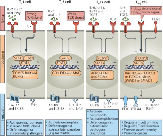
subset has its own master regulator transcription factor: Forkhead box P3 (Foxp3) induces Tregs, T-box transcription factor (Tbet) induces Th1 cells, GATA3 induces Th2 cells and RAR-related orphan receptor gamma (RORγT) is the defining transcription factor of Th17 (DuPage and Bluestone 2016).These subclasses are related with the direction in which the helper T cell pushes the response
(Figure 1.5). iTregs secrete the signature cytokines IL-10 (Eberl 2016) and TGF, essential for limiting immune responses by regulating T cell priming and function, and they also prevent autoimmunity. Th1 cells are essential for clearing intracellular bacteria and viruses, and activate macrophages. Their signature cytokine is IFN-γ. Th2 cells are highly related with the humoral response, since they are crucial to activate eosinophils and to organize the host defense against extracellular parasites (e.g. helminths), by helping B cells to produce antibodies. They secrete IL-4, IL-5 and IL-13. Th17 cells are higher secretors of IL-17A, IL-17F and IL-21. This subset is related with anti-fungal immunity, and plays a crucial role in maintaining mucosal barriers and clearing pathogens at mucosal surfaces (Hermann-Kleiter and Baier 2010). Besides their protective role, Th17 cells can play a pathogenic role particularly in chronic inflammatory conditions such as Psoriatic arthritis and Psoriasis (Mease 2015). Recent studies dissected the role of IL-17 and Th17 cells in tissue inflammation, autoimmunity and host defence, and demonstrated their contribution to local and systemic aspects of disease pathogenesis. That lead to consider these cells as potential therapeutic target for the treatment of such disorders (Miossec and Kolls 2012). Oppositely to Th1 cells, Th17 cells are very difficult to detect in inflamed tissues due to their
Figure 1.5 – Polarized CD4+ T cell subsets. Adapted from (DuPage and Bluestone 2016). Each CD4+ T cell subset can be defined by their distinct abilities to sense (red), programme (orange) and function (blue) in the control of specific pathogens or immune pathologies. The inductive cytokines, polarizing transcription factors and cytokines or chemokine receptors that are characteristic of each subset are shown, along with their association with specific forms of immune defence.
10
limited ability to proliferate after TCR engagement, but also for their tendency to shift to a Th1 phenotype in response to different stimuli (Cosmi et al. 2014). As a result from this functional plasticity, those non-classic Th1 have their pathogenic feature increased, thus opening the discussion about which subset should be targeted by therapeutic interventions.Following the topic of cell plasticity and multifunctionality, even though in a traditional view, naïve T cell activation, proliferation and differentiation are all considered as simultaneous processes, differentiated T cells can be reactivated in the local of infection/trauma or even modifying and reshaping from their “primary” function (DuPage and Bluestone 2016). Indeed, the hallmark feature of Th17 cells is their plasticity, i.e. their ability to express cytokines typical of other lineages in response to distinct microenvironments (Hirota et al. 2011),(Annunziato et al. 2007). As discussed previously, Th17 are considered to be the main population of pathogenic T cells driving autoimmunity (Hirota et al. 2007), (Langrish et al. 2005), poised to develop strong tissue-destructive properties. However, recent evidence has shown that they can adopt an IL-10 producing tolerogenic phenotype and contribute to resolution of inflammation in the central nervous system (Heinemann et al. 2014) and in the gut(Gagliani et al. 2015),(Esplugues et al. 2011). This plasticity of Th17 cells could be considered as a novel therapeutic opportunity, whereby pre-committed pro-inflammatory cells can adopt an anti-inflammatory fate.
A recent study (Heinemann et al. 2014) found that formerly Th17 cells respond to 27 and IL-12 by up-regulating Blimp1 and adopt a T regulatory type 1 (TR1) phenotype characterized by IL-10 production. While Tregs are characterized by the major transcription factor Foxp3, TR1 cells secrete huge amounts of anti-inflammatory IL-10 and express cell-surface markers CD49b and LAG-3 (Lymphocyte-activation gene 3) (Gagliani et al. 2013). Furthermore, the same group (Gagliani et al. 2015) constructed a new mouse fate-mapping mouse model to prove that CD4+ T cells formerly expressing Il17a go on to acquire an anti-inflammatory phenotype. This “transdifferentiation” of Th17 cells into TR1 cells comprises a change in their signature transcription profile and the acquisition of potent regulatory capacities.
Finally, a recent study (Yang et al. 2015) characterized a microbiota-induced hybrid population of T cells expressing both Foxp3 and RORγT and showing both transcriptional and epigenetic profiles of Th17 cells and Tregs. They proved that their specific molecular pattern provide them with stable and unique functional properties of effector Tregs that allow them to efficiently supress gut-specific inflammatory responses. Despite intense interest, the cellular and molecular cues that confer tissue-protective properties to Th17 cells have remained poorly defined.
It is crucial to understand the general cellular and molecular mechanisms that might explain how a pro-inflammatory Th17 response, which is beneficial in clearing infection, but immunopathogenic in excess, can be controlled by the tissue microenvironment.
Those mechanisms for counter-regulation of Th17 pathogenicity might be exploited to develop new and more effective therapies that restore tolerance in chronic inflammatory/autoimmune diseases without the deleterious side effects of current therapies.
1.3.
Toll-like receptors
TLRs are a family of receptors that can initiate innate immunity and inflammation in response to danger signals in the form of infection or tissue damage. The discovery of TLRs furthered our
11
understanding of how the host rapidly responds to invading pathogens. As described above, TLRs are the best known innate immune receptors which allow the activation of the second-line action against the invading pathogen. When cells encounter a pathogen, a microbial product and/or “stress signal”, TLR activation initiates signal transduction pathways that end up in a potent transcriptional response that culminates in inflammatory cytokine production (Barton and Kagan 2009).They can be classified and divided depending on their ligands and subcellular localization. TLR 1, 2, 6 and 5 are localized on the cell surface, thus recognizing accessible molecules in the membrane of most pathogens. TLR 3, 7, 8 and 9 are localized in intracellular compartments, recognizing microbial nucleic acids. TLR4 is unique among the TLRs as it is expressed both at the plasma membrane and endosomal compartments. In fact, TLR4 subcellular localization determines the distinct signalling pathways engaged and the consequent cellular cytokine profile (Barton and Kagan 2009).
TLR signal transduction has been increasingly studied through recent years, and it is now known that TLRs can be found in a huge variety of mammalian cell types and that their localization and trafficking patterns are crucial for their signalling functions (Kagan 2010).
Although those families of TLRs diverge in their distinct ligands, localization and functional relevance, most of them share a common signalling pathway to induce innate and adaptive immunity. For over 15 years studies have been focused on the downstream signalling pathways that are activated by the different TLRs in mammalian cells (Figure 1.6). TLR signalling is initiated by ligand-induced dimerization of receptors. It initiates with the activation of the adaptor protein: MyD88 (Myeloid differentiation primary response protein 88) or TRIF (TIR-domain-containing adapter-inducing interferon-β). These adaptor proteins are recruited to theToll/interleukin-1 receptor (TIR) -domain, a cytosolic tail present in all TLR receptors (O’Neill, Golenbock, and Bowie 2013).
Both the TLRs expressed on the plasma membrane and endosomal compartments, lead to the downstream cascade events involving interactions between IL-1R-associated kinases (IRAKs) and the adaptor molecules TNF receptor-associated factors (TRAFs), and that lead to the activation of the mitogen-activated protein kinases (MAPKs) JUN N-terminal kinase (JNK) and p38. Finally that will lead to the activation of the transcription factors NF-kB (Nuclear factor kappa B), CREB (cyclic AMP-responsive element-binding protein) and AP1 (activator protein 1). This cascade induces a pro-inflammatory response.
In addition, endosomal TLRs (3, 7, 8, 9 and 13) and the TLR4, due to its bipartite distribution between the plasma membrane and the endosomes, can activate a different cascade of signalling molecules that turn on an anti-inflammatory phenotype. This signalling pathway also initiates with interaction between IRAKs and TRAFs, however it ends up with the activation of different transcription factors: Interferon regulatory factor 3 (IRF3) and IRF7 (Figure 1.6).
Only TLR3 and the endocytosed TLR4 rely on TRIF to activate their downstream signaling cascades. All the other TLRs initiate their signaling pathways by activating MyD88. On both cases, some TLRs require an extra molecule to induce the activation of both MyD88- and TRIF-dependent signalling pathways. Those molecules are called the “sorting adaptors” and they promote the communication between the TLR and the respective adaptor. MAL (MyD88-adapter-like protein) also known as TIRAP (TIR domain-containing adaptor protein) is the sorting adaptor for the MyD88-dependent cascade. And
12
TRAM (TRIF related adaptor molecule) works along the TRIF molecule. The reason why some TLRs need only the signalling adaptor when others demand the sorting/signalling adaptor pair is still unknown.1.3.1. Toll-like receptor 4
TLR4 signalling is triggered by Lipopolysaccharide (LPS), a bacterial component produced by all Gram-negative bacteria (Poltorak et al. 1998). A few years ago, it was found that both TLR3 and TLR4 drive the induction of IFNα and IFNβ through a MyD88-independent pathway that ends up with the activation of the transcription factor IRF3 (Barton and Medzhitov 2003). Therefore, TLR4 was found to rely either on MyD88 or on the adaptor molecule TRIF to support, respectively, a pro- or anti-inflammatory response. Reinforcing these 2 divergent pathways for TLR4 signaling, it was demonstrated that by disrupting receptor endocytosis, MyD88-dependent activation of NF-kB could be enhanced resulting in increased LPS-induced pro-inflammatory outcome (Husebye et al. 2006). Latter, another group (Barton and Kagan 2009) found that TLR4 could be expressed at both plasma membrane and in intracellular compartments, reason why it is able to signal by those 2 distinct pathways (Figure 1.7). Figure 1.6 - TLR signaling in mammalian antigen presenting cells. From (O’Neill, Golenbock, and Bowie 2013). The TLR signalling pathway is activated by ligand-induced dimerization of receptors, followed by the binding
of MYD88 and Mal (for TLR1, TLR2, TLR5, TLR6, TLR7, TLR8 and TLR9) or TRIF and TRAM (for TLR3 and TLR4). TLR4 moves from the plasma membrane to the endosomes to switch signalling from MYD88 to TRIF. Activation of IL-1R-activating kinase (IRAK)-1, IRAK-2, IRAK-4 and TNF-receptor-associated factors (TRAFs) leads to the activation of JNK and p38. Transcription factors such as NF-κB, CREB, AP-1 and IRF proteins induce proinflammatory cytokines. Activation of the endosomal TLRs leads to the production of type I interferons.
13
Kagan lab furthered their studies to unravel the mechanism of TRIF/TRAM activation. In 2008 (Kagan et al. 2008) they suggested the new and nowadays accepted model where the TIRAP-MyD88 pathway is induced from the plasma membrane, whereas the TRAM-TRIF pathway is induced from endosomes. They demonstrated that after activation of endocytosed TLR4, the downstream signalling events improved IFN expression and a subsequent regulatory cytokine output (IFNα and IFNβ). This spatiotemporal control of the TLR signalling can be highly related with lipid second messengers incorporated in the different cell membranes. PtdIns(4,5)P2 can be modified by kinases such as PI3K (Phosphoinositide 3-kinase) to generate PtdIns(3,4,5)P3 (Phosphatidylinositol 3,4,5-triphosphate) (Li and Rudensky 2016). This depletion in PtdIns(4,5)P2 leads to a dissociation of the TIRAP/MAL sorting adaptor from the surface TLR4. As the amount of PtdIns(3,4,5)P3 arises, the TLR4 can be internalized where it will associate with the TRIF/TRAM adaptors. This TRIF-TRAM signalling pathway activates TBK1(
TANK-binding kinase 1) and IKKε (IκB kinase ε) which are responsible for type I IFN production, driving and defining the regulatory or anti-inflammatory states that will help to curtail inflammation and avoid disease (Wall et al. 2017).Husebye lab extended their work to TLR4 trafficking (Husebye et al. 2010) and they suggested a role for the small GTPase Rab11a to promote TLR4 internalization into recycling endosomes. Rab11a was also found to regulate the recruitment of TLR4 from these recycling endosomes to bacteria-containing phagosomes, where both TRAM and IRF3 are
localized, promoting TRIF-dependent TLR4 signaling and the expression of type I IFN.
Actually, in addition to the release of pro-inflammatory mediators, TLRs activation also leads to the production of IL-10. This Il10 expression is strictly regulated to ensure an effective immune response, while preventing chronic infection and tissue destruction (Teixeira-Coelho et al. 2014). Interestingly, Saraiva and collaborators (Teixeira-Coelho et al. 2014) found that the TRIF pathway previously stated regulates IL-10 production at the post-transcriptional level, discriminating between TLR2 and TLR4 activation on macrophages. In fact, they show that, unlike TLR2, TLR4 signals protected Il10 mRNA from degradation, not only due to the activation of TRIF but also due to enhanced p38 signaling. Other studies (Saraiva and O’Garra 2010), (Gabrysova et al. 2014) mention the crucial role of extracellular signal–regulated kinase (ERK) activation in the expression of IL-10 in different cells, from macrophages and
DCs to Th cells. In fact, in the previously stated paper (Teixeira-Coelho et al. 2014) they showed that inhibition of ERK does not affect Il-10 expression post-transcriptionally, but reduced the amount of IL-10 secreted after LPS-induced TLR4 activation in macrophages. Thus, ERK plays a major role in transcriptionally regulating IL-10.
Figure 1.7 - TLR4 signalling in antigen presenting cells. From (Gómez et al. 2014).
Upon ligand-induced TLR4 dimerization, the MyD88-dependent signalling pathway is activated. TLR4 can also internalize into endosomes and signal by the MyD88-independent pathway. TLR4 signalling activates multiple transcription factors, including MAPK, interferon regulatory factors and NF-κB, which promote innate immune responses.
14
Overall, this strict regulation of TLR4 signaling through compartmentalization leads to a transition between two distinct pathways related with contrasting cytokine outputs. This capacity to rapidly block one pathway and activate the other drives and defines the regulatory or anti-inflammatory state that will help to curtail inflammation and avoid disease.1.4.
Expression of TLRs in T cells
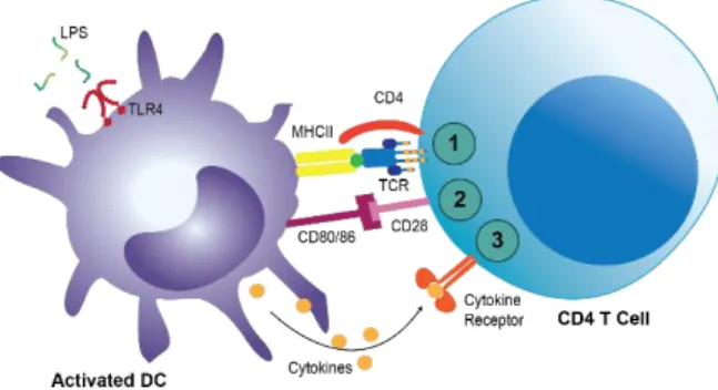
T cells are key players in the adaptive immunity. They must be activated as a response to environmental cues which initialize a complex signaling cascade and the production of immune mediators responsible for disease resolution. T cell full activation requires 3 signals, as illustrated in Figure 1.8. Even though TLRs are well-known to be expressed on innate immune cells, it was found that T cells can also express a variety of TLRs, being capable to directly respond to different TLR agonists. Thus, we wondered if TLRs could be an emerging 4th signal for not only helper T cell activation, but also for their function.
In human peripheral T cells isolated with >95% purity, low expression of TLR1-10 mRNA was detected by qPCR (Real-time polymerase chain reaction) (Hornung et al. 2002). Even though whether CD4+ T cells can express TLRs at the protein level remains controversial, T cells were found to express MyD88, the main adaptor protein that initiates TLR signaling, and its deletion affects T cell functions (Fukata et al. 2008). Studies in mouse model indicate that TCR activation and/or the presence of cytokines such as IL-2 and IFN-α upregulate TLR expression (Reba et al. 2014), (Komai-Koma et al. 2004). Moreover, these studies found that TLR ligands directly promote activated CD4+ T cell survival and/or proliferation in murine cells (Gelman et al. 2004), (Reynolds et al. 2012), (Imanishi et al. 2007), (Reynolds et al. 2010), (Zheng et al. 2008) and in human cells (Komai-Koma et al. 2004), (X. Chen et al. 2009), (Babu et al. 2006). When stimulated with Pam3Cys, a TLR2 agonist, CD4+ T cells display improved cytokine production and proliferation (Komai-Koma et al. 2004). Also, it was proved that TLR2 works as a co-stimulatory receptor for human T cells, enhancing the TCR-induced production of the cytokines, IFN-γ, IL-10, IL-13, Granulocyte-macrophage colony-stimulating factor (GM-CSF) and Vascular endothelial growth factor (VEGF), and the TCR-mediated secretion of chemokines CXCL10 and CCL5 (Chapman et al. 2013). Other co-stimulatory effects were observed with other TLR ligands. Human memory (CD45RO+) T cells, when challenged with TLR5 and TLR7/8 ligands, flagelin and resiquimod respectively, have their proliferation improved and also an increased production of IFNγ (Jeannin et al. 2005).
Figure 1.8 – Three required signals for T cell full activation. Based on (Janeway et al. 2001). To be
activated, T cells require 3 signals: 1st – The recognition of foreign antigens by the TCR in the context of the MHC; 2nd – The activation of costimulatory signals provided by the interaction of co-receptors, such as CD28 and CD40L; 3rd – The response to environmental cytokines.
15
Naïve CD4+ T cells differentiate into distinct subsets depending on the nature of the environment during TCR activation. Whether TLR signalling has a direct effect on T cell differentiation or function has remained a matter of debate.Tregs play a crucial role in the maintenance of immune tolerance and in the control of cellular immune responses. As the effector T cells, Tregs can also express different TLRs and be directly affected by them. It was previously stated that TLR2 and TLR8 stimulation can reduce the suppressive activity of Tregs (Liu et al. 2006), (Peng et al. 2005) and oppositely, TLR2 and TLR5 were able to enhance their suppressive activity (Zanin-Zhorov et al. 2006),(Crellin et al. 2005). Future studies are required to resolve the apparent discrepancies about the TLR2-induced effect on Tregs. Interestingly, a recent study (Gerriets et al. 2016) demonstrate that Treg cell metabolism was dynamically regulated by TLR signals and Foxp3. Whereas TLR signals promote Treg proliferation by increasing PI3K-AKT(protein kinase-B)-mTORC1(mammalian target of rapamycin C1) signaling and glycolysis, Foxp3 opposed PI3K-AKT-mTORC1 signaling to diminish glycolysis and anabolic metabolism while increasing oxidative and catabolic metabolism required for a suitable suppressive activity of Tregs.
Finally, it was reported that engagement of TLR7 in CD4+ T cells activates an anergic gene-expression program, preventing cell cycle, secretion of pro-inflammatory cytokines after stimulation and a final unresponsiveness state of CD4+ T cells (Dominguez-Villar et al. 2014).
As stated previously, TLRs are crucial to initiate the adaptive immunity as they mediate the recognition of the pathogen by DCs and their subsequent maturation and activation, leading to an improved antigen presentation, to polarization of T cell responses and reversal of suppressive Treg activity (Iwasaki and Medzhitov 2004). Additionally, T cell functions can be directly influenced by TLRs expressed on T cells. How TLRs can influence these functions still remain unknown, as they can lead to direct effects and cellular responses or their signaling could intersect the TCR pathway, modulating T cell responses triggered by TCR stimulation.
1.4.1. TLR4 on T cells
TLR4 has been detected at the mRNA level in CD4 T cells (Hornung et al. 2002), (Gelman et al. 2004), (Tomita et al. 2008), (Fukata et al. 2008), (Reynolds et al. 2010), (González-Navajas et al. 2010). In those studies, they definitely proved that TLR4 can modulate the development and/or function of certain Th cell subsets. However in some they used mice models, and in others they used polarizing conditions during T cell stimulation, preventing the acquisition of reliable results regarding the role of TLR4 on T cell differentiation. That is one of the reasons why there are conflicting reports about TLR4 effects on T cell functions.
Studies found that when T cells were stimulated with LPS, the bacterial ligand for TLR4, the release of the hallmark CD4+ Th2 cell cytokine, IL-4, was reduced (Watanabe et al. 1999), (Matsuguchi et al. 2000).
Furthermore, human CD4+CD28- T cells from Ankylosing Spondylitis patients show improved perforin production after LPS-induced TLR4 activation (Raffeiner et al. 2005). Following the same effect, Reynolds and collaborators demonstrated that TLR4 promotes T cell survival in a mouse model of experimental autoimmune encephalomyelitis (EAE) (Reynolds et al. 2012). Albeit LPS did not promote
16
IL-17 production, loss of TLR4 solely in CD4+ T cells almost completely abrogated disease symptoms. Implying a role for TLR4 in the survival of Th17 cells.However, the putative role of TLR4 expression in autoimmunity has yielded contradictory reports. In another autoimmune disease model, the transfer colitis model (González-Navajas et al. 2010), TLR4 has been shown to have a tonic inhibitory role on subsequent TCR-dependent CD4+ T cell responses. In fact, TLR4-deficient T cells exacerbated disease. Importantly, in this model, LPS treatment causes CD4+ T cells to lose IFNγ but upregulate IL-17 expression, promoting disease resolution. Interestingly, other studies also demonstrated a protective role from colitis in a MyD88-/- model after LPS engagement (Fukata et al. 2008), (Tomita et al. 2008). This suggests that TLR4 in a MyD88-/- model keeps signaling in a TRIF-dependent way providing a protective role associated with IL-10 production, as what happen in innate immune cells.
The apparent incongruity of these two reports might be a result of differential TLR ligand availability in the gut versus the sterile environment of the central nervous system. CD4+ T cells in the gut are continuously exposed to TLR stimuli from the microbiota, while CD4+ T cells in the central nervous system would probably be responding to host-derived danger signals.
This can be connected with the previous discussed issue of the impact of the environment on T cell differentiation. Knowing that T cells also express and respond directly through TLRs, it is easier to explain for instance, how the microbiota can influence CD4+ T cell phenotype. Thus, it is possible that, in addition to cytokines, other tissue microenvironment factors, such as LPS-bearing commensal and pathogenic bacteria can directly affect T cells through TLRs, driving a Th17 reprogramming into IL-10 secreting tolerogenic cells (Gagliani et al. 2015), (Esplugues et al. 2011), or even redirecting Tregs to a Foxp3+RORγT+ phenotype with a stable and unique functional suppressive activity during gut-specific inflammatory responses (Yang et al. 2015). I propose that this immunoregulatory mechanism might be particularly important in the microbe rich gut environment.
It is of great importance to explore in more detail the potential of defined TLR ligands to modulate adaptive immune responses in infectious diseases, cancer and autoimmunity. To manipulate T cell functions through TLRs it is required a full understanding of the molecular pathways that orchestrate the intersection between the TCR and TLRs which will depend on the T cell subset and its activation status.
1.5.
Aims
Determine how TLR4 engagement imparts on T
cell function
Unravel the crosstalk between TCR and TLR4
signaling in T cells
Determine how LPS might counter-regulate T cell inflammatory response
MATERIALS
AND METHODS
19
Reagents
Biocoll separating solution (Biochrom)
Bovine Serum Albumin (BSA) (HyClone, Thermo Scientific)
Brefeldin A (BFA, 5 mg/mL) (Penicillium brefeldianum) (Sigma- Aldrich)
CellTrace™ Far Red (Thermo Fisher)
Chlorpromazine (CPZ) (10 mM) Dimethyl sulfoxide (DMSO) (Sigma)
EBioscienceTM Foxp3/Transcription Factor (Invitrogen, Thermo Fisher) Fetal Bovine Serum (FBS) (Biochrom)
Fixable Viability Dye eFluor 780 (Affymetrix, Thermo Fisher)
Fluoromount-G (SouthernBiotech)
Interleukin-2 (IL-2) (NIH AIDS Reagent Program, Division of AIDS, NIAID, NIH from Dr. Maurice Gately, Hoffmann - La Roche Inc.)
Ionomycin (Calcium salt, Streptomyces conglobatus, 2.5 mg/mL) (Merck Millipore)
L-Glutamine (Gibco, Thermo Fisher)
Lipopolysaccharide (LPS) (Salmonella enterica serotype Minnesota, purified by gel-filtration chromatography) (Sigma-Aldrich)
LIVE/DEAD™ Fixable Aqua Dead Cell Stain (Thermo Fisher)
Paraformaldehyde (PFA) (Sigma-Aldrich)
Penicillin/Streptomycin (Pen/Strep) (Gibco, Thermo Fisher)
Phorbol 12-myristate 13-acetate (PMA, 2.5 mg/mL) (Sigma-Aldrich)
Phosphate buffered saline (PBS) (Sigma-Aldrich)
Poly-L-Lysine hydrobromide (PLL, 0.1 mg/mL) (Sigma-Aldrich)
RPMI (Roswell Park Memorial Institute) Medium 1640 (Gibco, Thermo Fisher)
Saponin 5% (Carl Roth)
UltraComp eBeads (Invitrogen, Thermo Fisher)
2.1.1. Antibodies
αCD28 [CD28.2]– 1 mg/mL (BioLegend)
αCD45 [GAP 8.3] – 2,5 mg/mL (American Type Culture Collection)
αRab11a [EPR7587(B)]– 0,3 mg/mL (Abcam)
αTCR [UCHT1]– 0,5 mg/mL (BioLegend)
αTLR4 [76B357.1]– 1 mg/mL (Abcam)
Alexa 488 αCD4 [OKT4] – 0,5 mg/mL (BioLegend)
Alexa 488 Anti-FITC [5D6.2] – 1 mg/mL (Merck Millipore)
Alexa 488 Anti-mouse IgG2b – 2 mg/mL (Thermo Fisher)
Alexa 568 Anti-mouse IgG2a – 2 mg/mL (Thermo Fisher)
20
Alexa 647 αIL10 [JES3-9D7] – 20 µg/mL (BioLegend) Alexa 647 Anti-mouse IgG2b - 2 mg/mL (Life technologies, Thermo Fisher)
Alexa 647 Anti-rabbit – 2 mg/mL (Thermo Fisher)
Anti-mouse IgG1 (crosslinking antibody) [RMG1-1]– 0,5 mg/mL (BioLegend)
APC-Cy7 αIL17-A [BL168] – 0,5 mg/mL (BioLegend)
APC-Cy7 anti-human CD38 [HIT2] – 0,4 mg/mL (Biolegend)
Brilliant Violet 421 anti-human CD279 (PD-1) [EH12.2H7] – 50 µg/mL (Biolegend)
FITC anti-mouse IgG1 – 1 mg/mL (Abcam)
Mouse IgM αTCR [MEM-93] – 1 mg/mL (Sigma-Aldrich)
Pacific Blue αIFNγ [4S.B3] - 0,5 mg/mL (BioLegend)
Pe/Cy7 anti-human CD45RO [UCHL1] – 1 mg/mL (Biolegend)
Phospho-AKT (Ser473) [Poly6490] – 0,11 mg/mL (BioLegend)
Phospho-p38 MAPK (Thr180/Tyr182) – 44 µg/mL (Cell Signaling)
Phospho-p38 MAPK (Tyr322) – 1 mg/mL (EnoGene)
Phospho-PKCθ(Protein kinase C-theta) (Ser676) – 1 mg/mL (Sigma-Aldrich)
Phospho-ZAP70(Zeta chain of T cell receptor associated protein kinase 70kDa) (Tyr319)/ Syk(Spleen tyrosine kinase) (Tyr352) – 50 µg/mL (Cell Signaling)
Rat αCD28 [low endotoxin] – 1 mg/mL (Raybiotech, Inc.)
2.2. Isolation of human peripheral blood mononuclear cells
Whole blood was collected from healthy donors by the certified CEDOC staff at the NOVA Medical School of Lisbon, in accordance with stipulated by the ethical committee. These donors provided consent for their blood cells to be used in research studies conducted at CEDOC.
Human peripheral blood mononuclear cells (PBMCs) were isolated from the blood by Ficoll density centrifugation(Fuss et al. 2009). PBMCs were frozen in FBS containing 10% DMSO, kept at −80°C for 24 hours and then stored at −150°C until use.
2.3. Sorting and purification of CD4
+T cell
PBMCs were thawed and cultured overnight in complete RPMI 1640 medium (RPMI 1640, 10% FBS, 2 mM L-glutamine, 1% Pen/Strep) with IL-2 (5 IU/mL).
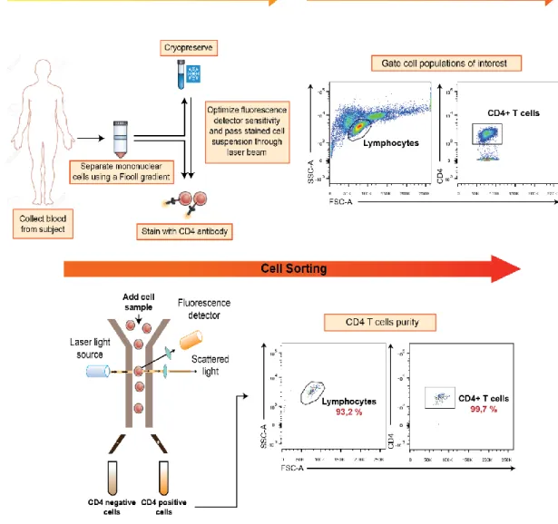
PBMCs were then washed with FACS buffer (PBS 1X + 2% FBS), labelled with αCD4 antibody A488 (1 µg/mL) and sorted in BD FACS Aria II. Cells were gated into the Lymphocyte and CD4+ population (Figure 2.1).
This step of purification is critical to obtain highly pure CD4+ T cells and exclude any possibility of contamination by professional APCs. CD4+ T cells were isolated with a purity of ~99,756%.
21
2.4. Culture and stimulation assays of CD4
+T cells
After purification, sorted cells were washed and cultured at 2x10⁶ cells/mL for 5 days in complete RPMI 1640 medium and IL-2 (20 IU/mL) in 96 round U-bottomed plates coated only with PLL or with PLL, αTCR (10 µg/mL) and αCD28 (2 µg/mL).
CD4+ T cells were left untreated or treated with LPS, the TLR4 ligand, at 1.8 µg/mL.
2.5. T cell survival assay
After 5-days in culture, unstimulated and stimulated cells were washed twice with PBS 1x and stained with either LIVE/DEAD™ Fixable Aqua Dead Cell Stain or Fixable Viability Dye eFluor 780 for 20 min at 4ºC. Cell survival was assessed by gating on lymphocytes and unlabelled cells for the dye by flow cytometry (BD FACS Canto II).
Figure 2.1 - PBMCs Isolation and Cell Sorting strategy. Adapted from (Maecker 2012). PBMCs were collected
from blood by Ficoll gradient. Sometimes, sample preparation involves cryopreservation before staining with fluorescent antibodies. Cells were stained with αCD4 antibody A488 and passed through a laser beam to record the fluorescence emission and gate cell population of interest. Cells were then sorted in BD FACS Aria II with a purity of ~99,756%.