Maria Soares
Cachide de Almeida
Estudos moleculares do BIN1 num coorte
baseado em cuidados primários
BIN1 molecular studies in a primary care-based
cohort
Maria Soares
Cachide de Almeida
Estudos moleculares do BIN1 num coorte
baseado em cuidados primários
BIN1 molecular studies in a primary care-based
cohort
Tese apresentada à Universidade de Aveiro para cumprimento dos requisitos necessários à obtenção do grau de Mestre em Biomedicina Molecular, realizada sob a orientação científica da Professora Doutora Odete Abreu Beirão da Cruz e Silva, Professora Auxiliar com Agregação do Departamento de Ciências Médicas da Universidade de Aveiro.
Este trabalho foi desenvolvido no grupo de Neurociências e Sinalização, e contou com o apoio do Instituto de Biomedicina Molecular (iBiMED), UID/BIM/04501/2013, da Universidade de Aveiro e da FCT (projeto PTDC/DTP-PIC/5587/2014 e JPND-BIOMARKAPD).
o júri
presidente Professora Doutora Ana Gabriela da Silva Cavaleiro Henriques
Professora Auxiliar Convidada do Departamento de Ciências Médicas da Universidade de Aveiro
Professora DoutoraOdete Abreu Beirão da Cruz e Silva
Professora Auxiliar com Agregação do Departamento de Ciências Médicas da Universidade de Aveiro
Professora Doutora Maria de Lourdes Gomes Pereira
agradecimentos À minha orientadora, Professora Doutora Odete da Cruz e Silva, agradeço por toda a disponibilidade, pela amizade e apoio incondicional, pela dedicação, pelos conhecimentos que transmitiu e por todas as ideias que fizeram deste trabalho um motivo de orgulho para mim. Muito obrigada pela oportunidade que me deu e por me ter deixado descobrir as maravilhas das Neurociências.
À professora Doutora Ana Gabriela Henriques, o meu mais sincero e profundo agradecimento pela co-orientação, pelo profissionalismo, pela amizade, por todo o tempo disponibilizado e pela total colaboração no solucionar de dúvidas e problemas que foram surgindo ao longo da realização deste trabalho e por todas as palavras de incentivo.
À Professora Ilka Martins Rosa, pela clareza, rigor e total disponibilidade na colaboração do tratamento estatístico dos resultados, pelo otimismo e boa disposição, assim como pelo encorajamento e motivação transmitida. À Professora Doutora Vera Afreixo, pela ajuda no tratamento estatístico de alguns dados, pela amizade, pelo apoio e pela disponibilidade.
A todos os voluntários que contribuíram para que este estudo fosse possível. A todos os meus colegas do Mestrado, em especial à Marlene Marafona e à Joana Ferreira, pela amizade, companheirismo e apoio demostrado ao longo deste percurso, por todos os momentos de partilhas e desabafos que me permitiram que cada dia e cada obstáculo fosse encarado com motivação. Um especial agradecimento à Daniela Guedes, por ter partilhado esta aventura comigo, pela ajuda sempre disponibilizada, por todos os conselhos, apoio e espírito critico. Por todas as gargalhadas que animaram o nosso trabalho e que de certa forma o enriqueceram, pelas peripécias que jamais serão esquecidas, mas sobretudo pela paciência e boa disposição que sempre me transmitiu. Um grande obrigado pela amizade e companheirismo! À Filomena Amorim, por todo o apoio, amizade e boa disposição. Obrigada pelas partilhas e pelos desabafos, pelos conselhos e pelo incentivo. Obrigada por estares sempre presente e disponível, por acreditares em mim e por me fazeres sentir orgulho no meu trabalho.
Ao Luís Ribau, por toda a paciência, carinho e incentivo, mesmo nos momentos mais difíceis. Obrigada pelas palavras doces, por nunca teres duvidado de mim e por me ensinares a acreditar mais nas minhas capacidades! Por tudo, a minha enorme gratidão!
palavras-chave Demência; Doença de Alzheimer ; BIN1; Genética Molecular; Polimorfismo; Envelhecimento; População Portuguesa
resumo Demência é uma síndrome clínica caracterizada pelo declínio progressivo das
capacidades cognitivas, estando a tornar-se cada vez mais comum, devido ao envelhecimento da população mundial. Prevê-se que o número de doentes com demência aumente em cerca de 30 milhões nos próximos 15 anos, representando grandes gastos para os sistemas de saúde e sociais. Existem vários tipos de demência, sendo que a Doença de Alzheimer (DA) é a mais comum, afetando entre 20 a 30 milhões de pessoas em todo o mundo, das quais 90.000 são portuguesas. A compreensão das características genéticas e moleculares associadas a esta doença pode constituir um meio para descobrir novos métodos de diagnóstico e tratamento. A maior parte dos casos de Alzheimer tem início tardio, afetando indivíduos com 65 ou mais anos de idade. Até recentemente apenas o gene que codifica a Apolipoproteína E (APOE) foi associado com esta forma de DA. No entanto, estudos recentes de associação genómica identificaram o gene BIN1 como sendo o loci de risco associado ao Alzheimer tardio mais significativo depois do APOE. Além disso, vários SNPs do BIN1 foram associados a este tipo de Alzheimer, sendo que o polimorfismo rs744373 foi proposto como um dos mais relevantes para a DA. Dado que os SNPs mais significativos podem variar de população para população, o objetivo principal deste trabalho foi avaliar se o polimorfismo rs744373 do gene BIN1 pode ser associado a um aumento do risco de desenvolver DA, numa população portuguesa do distrito de Aveiro, que pertence a um estudo transversal baseado em populações, realizado na Universidade de Aveiro. Analisámos 63 indivíduos Portugueses, sendo 32 doentes e 31 controlos. Neste estudo conseguimos observar que, de uma forma geral, o alelo A é o mais frequente e que o alelo G (alelo de risco) foi o menos frequente, numa razão de 3:1. Não conseguimos encontrar uma forte evidência de associação entre o rs744373 e o risco de desenvolver DA (Razão de probabilidade [RP] = 0.733 , valor p = 0.464), o que está de acordo com estudos previamente publicados. Não foi detetada significância estatística entre o rs744373 e portadores do alelo APOE-Ԑ4 (valor p = 0.467) ou indivíduos com demência (CDR≥1) (valor p = 0.269). Foi detetada uma associação entre o alelo de risco do polimorfismo de estudo e a presença de Diabetes Mellitus (RP = 6.60, valor-p = 0.035). No entanto, como a nossa amostra era pequena, deve ser feito um novo estudo para avaliar se este resultado pode ser generalizado para a população Portuguesa.
keywords Dementia; Alzheimer’s Disease; BIN1; Molecular Genetics; Polymorphism; Aging; Portuguese population
abstract Dementia is a clinical syndrome characterized by a progressive decline in
cognitive skills, and is becoming increasingly common, due to the aging of the world’s population. It is expected that the number of patients with dementia will increase by 30 million in the next 15 years, representing a major factor of costs in health care and social systems. There are several types of dementia, and Alzheimer’s Disease (AD) is the most common, affecting 20 to 30 million people worldwide, of which 90.000 are Portuguese. Understanding the genetic and molecular characteristics associated with the disease may constitute a way to discover new diagnostic methods and treatments. Most cases of AD are late-onset (LOAD), affecting individuals with 65 or more years of age. Until recently only the Apolipoprotein E gene (APOE) had been associated with this form of AD. However, recent genome-wide association studies have identified Bridging Integrator 1 (BIN1) as the most significant LOAD-associated risk loci after APOE. Furthermore, several SNPs of BIN1 have been associated to this type of AD and rs744373 was proposed to be one of the most relevant for AD. Since the most significant SNP may vary from population to population, the main aim of this work was to evaluate if BIN1 polymorphism rs744373 can be associated with the risk of AD in a Portuguese population from the Aveiro district, belonging to a cross-sectional population-based study performed in Aveiro University. We analysed 63 Portuguese individuals comprising 32 cases and 31 controls. In this study we could observe that, overall, allele A was the most frequent and allele G (risk allele) was the least frequent, in a ratio of 3:1. We didn’t find strong evidence of association for rs744373 with the AD risk (odds ratio [OR] = 0.733 , p-value = 0.464), which is in agreement with some previous published studies. No statistical significance was detected between rs744373 and APOE-Ԑ4 carriers (p-value = 0.467) or individuals with dementia (CDR≥1) (p value= 0.269). We have detected an association between the risk allele of the study polymorphism and the presence of Diabetes Mellitus (odds ratio [OR] = 6.60, p-p-value = 0.035). Nevertheless, due to our small sample size, a follow-up study is required in order to evaluate if this result can be generalized to the Portuguese population.
INDEX
ABBREVIATIONS ... iii
INTRODUCTION ... 1
1.1 Dementia ...3
1.2 Alzheimer’s Disease (AD) ...4
1.3 Neuropathological Hallmarks of AD ...5
1.3.1 Senile Plaques (SP) ...6
1.3.2 Neurofibrillary Tangles (NFTs) ...8
1.4 Genetics of AD ... 13
1.4.1 Early-Onset Alzheimer Disease (EOAD) ... 13
1.4.2 Late-Onset Alzheimer Disease (LOAD) ... 14
1.5 Bridging Integrator 1 (BIN1) ... 18
1.5.1 BIN1 domains and isoforms ... 18
1.5.2 BIN1 cellular functions ... 19
1.5.3 BIN1 in AD ... 20
1.5.4 BIN1 and other diseases... 22
1.6 Diagnosis of AD ... 23
1.7 Treatment of AD ... 24
AIMS OF THE THESIS ... 27
MATERIALS AND METHODS ... 31
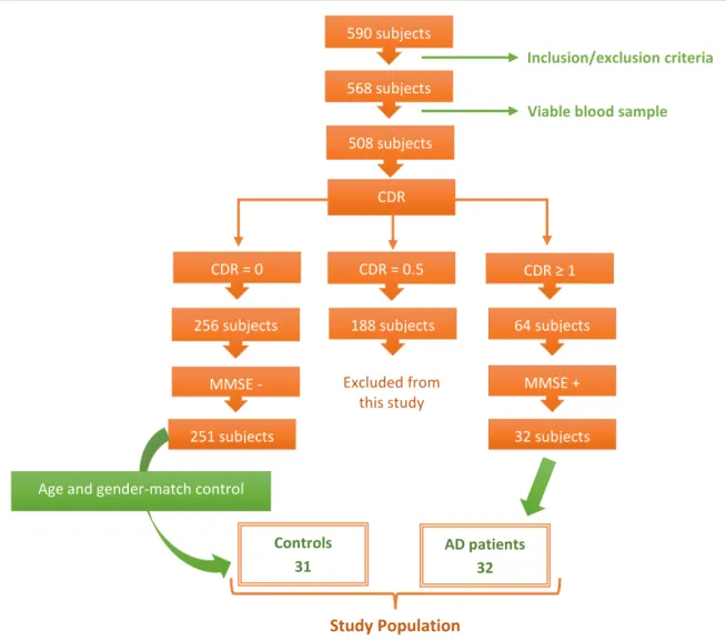
3.1 Study Group ... 33
3.2 Sample Collection ... 34
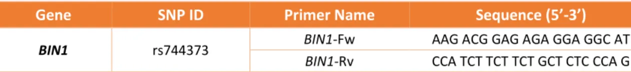
3.3 BIN1 Genotyping ... 35
3.3.1 Amplification of BIN1 gene (rs744373) by PCR ... 35
3.3.2 Precipitation of PCR products ... 36
3.3.3 Agarose Gel Electrophoresis ... 36
3.3.4 Sequencing of DNA samples ... 37
ii Maria Soares Cachide de Almeida
RESULTS ... 39
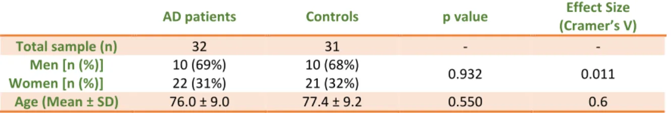
4.1 Assessment of the balance between the two study groups ... 41
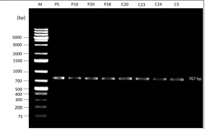
4.2 Amplification of BIN1 polymorphic region rs744373 by PCR ... 42
4.3 Correlating BIN1 polymorphism rs744373 with AD... 43
4.3.1 BIN1 (rs744373) sequencing in the study group ... 43
4.3.2 Genotype of the study population ... 44
4.3.3 Determination of the BIN1 polymorphism rs744373 genotypic frequencies in the study population ... 45
4.3.4 Determination of the BIN1 polymorphism rs744373 allelic frequencies in the study population ... 46
4.3.5 Evaluation of the statistical significance of rs744373 in the study population ... 47
4.3.6 Association of rs744373 with APOE-Ԑ4 carriers and CDR ... 49
4.3.7 Association of rs744373 with Diabetes Mellitus ... 50
DISCUSSION AND CONCLUSION ... 51
5.1 Assessment of the balance between the two study groups ... 54
5.2 Determination of BIN1 polymorphism rs744373 genotypic frequencies in the study population ... 54
5.3 Determination of BIN1 polymorphism rs744373 allelic frequencies in the study population. ... ………...55
5.4 Evaluation of the statistical significance of rs744373 in the study population ... 55
5.5 Association of rs744373 with APOE-Ԑ4 carriers and CDR ... 58
5.6 Association of rs744373 with Diabetes Mellitus ... 58
5.7 Final conclusions ... 59
REFERENCES ... 61
ANNEXES ... 75
7.1 Polymerase Chain Reaction (PCR) ... 77
7.2 Precipitation of DNA fragments ... 77
ABBREVIATIONS
AD
Alzheimer´s Disease
AGD
Angyrophilic Grain Disease
AICD
Amyloid Precursor Protein Intracellular Domain
APP
Amyloid Precursor Protein
APOE
Apolipoprotein E
Aβ
Amyloid-β peptides
BAR
BIN1/amphiphysin/RVS167
BIN1
Bridging Integrator 1
CBD
Corticobasal Degeneration
CDK5
Cyclin Dependent Kinase 5
CDR
Clinical Dementia Rate
c-MYC
c-myelocytomatosis
CNS
Central Nervous System
CSF
Cerebrospinal Fluid
DM
Diabetes Mellitus
EC
Extracellular Domain
EOAD
Early-Onset Alzheimer’s Disease
GSK-3β
Glycogen Synthase Kinase 3β
GWAS
Genome-Wide Association Studies
IC
APP Intracellular Domain
MBD
Myc-Binding Domain
MCI
Mild Cognitive Impairment
MiBD
Microtubule-Binding Domain
MRI
Magnetic Resonance Imaging
NFT
Neurofibrillary Tangle
PD
Projection Domain
iv Maria Soares Cachide de Almeida
PHF
Paired Helical Filament
PiB
Pittsburgh Compound B
PiD
Pick’s Disease
PP2A
Protein Phosphatase 2A
PSEN1
Presenilin 1
PSEN2
Presenilin 2
PSP
Progressive Supranuclear Palsy
SH3
Src Homology 3
SNP
Single Nucleotide Polymorphism
SP
Senile Plaque
2 Maria Soares Cachide de Almeida
1.1
Dementia
Dementia is a clinical syndrome usually associated with a group of symptoms and signs that culminates with a progressive decline in cognitive skills such as memory, reasoning, attention and orientation. It is also characterized by difficulty in language processing, as well as emotional and social changes, which might be linked to a state of depression, agitation, hallucinations, disinhibition or even insomnia. These changes impair daily life activities 1 like driving, shopping,
cooking or even attending to personal care, which makes it very hard for patient’s caregivers 2,3.
With the world’s population rapidly aging, it’s expected that the number of patients with dementia will increase 30 million in 15 years 3.
Depending on the cause, dementia can be reversible (rare and potentially treatable) or irreversible, being the first a result of other medical condition and the latter a consequence of a neurodegenerative process 3. Either way, most forms of dementia are progressive, becoming more
severe as the patient ages. However, it is important to highlight that dementia is not part of the natural aging process, and so not all old people will develop the symptoms associated to it. Actually, although dementia is most frequent after the age of 65, there are cases of patients in the age group of 40 to 60 years old that suffer from it 4. For these reasons, it is still unclear if dementia is only an
aging-related condition or if it tends to develop in older people just because it requires time for the pathogenic process to disclose 5.
There are several types of dementia including Parkinson’s disease 6, dementia with Lewis
bodies, Frontotemporal dementia and Alzheimer’s Disease (AD) 3. Depending on the dementia,
although they all tend to have a protracted development, there are two patterns of decline, being the first characterized by the late onset of clinical symptoms, while the other presents at an early stage.
Since Alzheimer’s disease is the most common form of age-related dementia, the next sections of the present work will be an overview of its molecular and genetic basis.
4 Maria Soares Cachide de Almeida
1.2 Alzheimer’s Disease (AD)
AD is an irreversible and progressive brain disorder, where neurodegeneration is its main feature 7,8. It is characterized by the impairment of several cognitive functions such as memory,
attention, concentration, language as well as emotional and social changes. In the final stages of the disease such symptoms are so severe that the patient has no autonomy, requiring a full-time caregiver. In these patients, pneumonia tends to be the leading cause of death 9.
It is one of the most common diseases in the developed world 10, having an incidence of
about 20 to 30 million people worldwide 11–13, with an estimated increase of more than 50 million
people by the year 2040 14. Currently, in Portugal nearly 90.000 individuals suffer from AD leading
to consider it a major driver of costs in health care and social systems 4.
The first case of AD was described in 1906 by the German psychiatrist and neuropathologist Dr. Alois Alzheimer after whom the disease was named, and it referred to an unusual disease of the cerebral cortex of a 51-year-old woman (Auguste D.). This case was an early onset, potentially caused by a PSEN2 Volga-German mutation, which led to a progressive cognitive impairment with loss of memory and language skills as well as behavioural changes 3,14.
AD is a multifactorial dementia caused by genetic, epigenetic and environmental factors and pathways, which interact among themselves leading to a complex heterogeneity of patient populations 5,15. Some of the non-genetic factors that might contribute to the development of the
disease include infections, hormones, diabetes, smoking or even emotional and social factors 16.
Although the ultimate risk factor for the appearance of AD is age 3, it has been hypothesized
that the development of this highly prevalent dementia begins 20 to 30 years before the manifestation of the first symptoms 11,17. This first stage of the disease is called Mild Cognitive
Impairment (MCI) in which people show some cognition deficits in comparison to the average individuals of their age, although it does not interfere with their daily lives in a significant way. The transition to AD occurs when the neuropathological features are more relevant, such as the spread of neurofibrillary tangles (NFTs) beyond the medial temporal lobe in cortex 7,11.
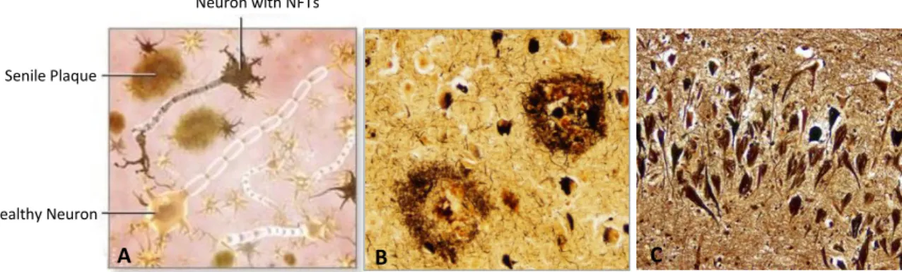
Senile Plaque
Healthy Neuron
Neuron with NFTs
1.3 Neuropathological Hallmarks of AD
Neuropathologically, AD is characterized by atypical protein aggregates 18. The main
hallmarks of the disease include the presence of senile plaques (SP) and neurofibrillary tangles (NFTs), being the first an extracellular accumulation of Amyloid- peptides (A) and the latter a intraneuronal accumulation of hyperphosphorylated TAU proteins 19,20 (Figure 1). Neurofilaments
(NFs) may also play an important role in neurofibrillary pathology and degeneration as they are one of the key components of the neuronal cytoskeleton, being the most abundant cytoskeletal protein in large myelinated axons 21.
Figure 1 – Neuropathological Hallmarks of AD. A - Schematic representation of the neuropathological hallmarks of AD. B - Senile Plaques (SP) (seen with Bielschowski silver stain). C – Neurofibrillary tangles (NFTs) (seen with Bielschowski silver stain). (Taken from22,23)
Another feature of AD is synapse loss, which may be related to changes in the normal functioning of neurons or even due to neuron death. This all culminates in brain atrophy 24,25 of
specific regions involved in memory and learning processes, namely the limbic system 26,27,
neocortical regions 28, the basal forebrain 29, and the hippocampus 30, which may partly explain the
clinical symptoms characteristic of this disease (Figure 2).
Figure 2 – Schematic comparison between a cross section of a healthy brain (left) and a brain of a patient with AD (right). In the latter it is possible to notice an overall shrinkage, mainly in the cortex and hippocampus, and an enlargement of the ventricles. (Taken from 31)
A B C Cerebral Cortex Hippocampus Severe Cortical Shrinkage Severely Enlarged Ventricles Severe Shrinkage of Hippocampus
6 Maria Soares Cachide de Almeida
Senile Plaques are spherical lesions resulting from the build-up of extracellular A, either with 40 amino acids (Aβ40) or 42 amino acids (Aβ42), which are produced through the metabolism
of Amyloid Precursor Protein (APP) after being sequential cleaved by a group of specific secretases (β- and γ-secretases) 3,32. There is a greater amount of Aβ
42 than Aβ40 within the SP due to its higher
rate of insolubility 33. This neuropathological feature of AD, unlike NFTs, is found mainly in the
isocortex 33.
1.3.1.1 Amyloid Precursor Protein (APP)
APP is a ubiquitously expressed type 1 transmembrane glycoprotein composed by 695 to 770 amino acids, encoded by a single gene located on chromosome 21 (21q21.23) with a length of about 240kb and no less than 18 exons 34. Through alternative splicing of exons 7, 8 and 15 of the
APP mRNA several isoforms are synthesized, predominantly APP695, APP751 and APP770. The 695
isoform is the only one that lacks the Kunitz Protease Inhibitor (KPI) domain.
Regarding the structure, APP has three main domains, the Extracellular Domain (EC) which includes amino acid residues 1-699 in exons 1-17, the Transmembrane Domain (TM) which is composed by residues 700-723 in exons 17 and 18, and the APP Intracellular Domain (AICD) which comprises residues 724-770 in exons 17 and 18. The A sequence, which originates the A peptide of particular interest in AD, is located in exons 16 and 17 (amino acid residues 672-713), being part of both EC and TM 16.
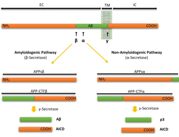
APP can undergo two different proteolytic pathways: a non-amyloidogenic and an amyloidogenic 18. In the non-amyloidogenic pathway, APP starts to suffer the action of α-secretase,
resulting APPsα (a soluble APP derivative) and a α-carboxyl-terminal fragment (APP-CTFα). This latter fragment is subsequently cleaved by -secretase generating a non-toxic peptide (p3) and the Amyloid Precursor Protein Intracellular Domain (AICD). In this pathway, as α-secretase action takes place in the A peptide sequence, it prevents the subsequent formation of A peptide, whose aggregation causes SPs. On the other hand, in the amyloidogenic pathway, APP is first cleaved by -secretase, releasing APPs and -carboxyl-terminal fragment (APP-CTF). The APP-CTF is afterwards cleaved by -secretase producing A peptide and the AICD 16,35 (Figure 3). BACE1 and
acts as a key β-secretase in the brain, since it is the rate-limiting enzyme that starts the Aβ formation
36.
Figure 3 – Schematic diagram of APP processing pathways (not drawn to scale). In the Amyloidogenic pathway APP is cleaved by β-Secretase, originating APPsβ and APP-CTFβ. The latter is subsequently cleaved by γ-Secretase originating Aβ peptide and AICD fragment. On the other hand, in the Non-Amyloidogenic pathway, APP is first cleaved by α-Secretase, giving rise to APPsα and APP-CTFα, being the last cleaved by γ-Secretase, leading to the formation of p3 peptide and the AICD fragment. Abbreviations: EC, Extracellular Domain; TM, Transmembrane Domain; IC, Intracellular Domain. (Adapted from 16).
1.3.1.2 Amyloid-β peptide (Aβ)
Amyloid is a peptide with an approximate length of 40 to 42 amino acids 11, that is
produced by proteolytic cleavage of APP, first identified in 1984 by Glenner and Wong 37.
Depending on the -secretase that cleaves APP, a variety of peptides can be generated, including A42 and A40. Of these, the A40 is the most abundant variant (about 80%)35 present in
both healthy brains and brains of people who suffer from AD. On the other hand, A42, which
aggregates much easier when compared to A40, this is found to be augmented in neuronal cells
which have suffered mutations in APP or Presenilin 1 (PSEN1) and Presenilin 2 (PSEN2) genes, which are associated with Early-Onset Alzheimer’s Disease (EOAD) 38.
NH2 COOH COOH COOH NH2 NH2 Aβ APP-CTFβ APP-CTFα
α
β APPsβ Amyloidogenic Pathway (β-Secretase) Non-Amyloidogenic Pathway (α-Secretase) APPsα EC TM IC γ-Secretase γ-Secretase COOH COOH Aβ AICD p3 AICD
γ COOH COOH COOH NH2 NH2 NH2 Aβ8 Maria Soares Cachide de Almeida
A is normally secreted in physiological processes, which indicates that it has a physiological function 35. Further, it has been discovered through several studies that A is important in
neurogenesis, producing neuroprotective effects and increasing cell viability when growth factors, neurotrophins or excitotoxic conditions are not present 39. It also plays an important role in
strengthening or weakening synapses, is relevant in synaptic plasticity 40 and memory formation 41.
In addition, it influences the metal sequestration and antioxidant activity with metals such as copper, zinc or iron. The A peptide is also important for the maintenance of homeostasis and so it is particularly important in keeping the nervous system healthy 35. The problem with A occurs
when there is an increase in the A42/A40 ratio, since it eventually contributes to the build-up of
protein aggregates that accumulate in the brain, leading to neurodegeneration and the development of AD 16,35. This is accordingly to amyloid hypothesis, which is well-known and
accepted among the scientific community.
In addition, A peptide accumulation might be toxic in a variety of ways, as it can induce oxidative stress when synchronized with redox active metals. It can also interact with membranes, leading to the formation of pores and consequently contributing to an abnormal flux of ions, culminating in neuron death. Finally, Aβ is associated with synaptic dysfunction, telomerase dysfunction (through its inhibition) or can even promote apoptosis of neuronal cells 35.
NFTs consist of intraneuronal filamentous inclusions primarily constituted by aggregates of hyperphosphorylated TAU protein which, in a regular situation, has several roles including the stabilization of microtubules and intracellular transport, both axonal and vesicular. Normally TAU is a soluble protein, however, after suffering an excessive phosphorylation by several kinases eventually becomes insoluble, losing affinity to microtubules and self-associating into paired helical filament (PHF) structures and straight filaments, which ultimately evolve into NFTs 42. The PHF
breadth ranges from 8 to 20nm, with a spacing between crossovers of approximately 80 nm, while straight filaments do not have this helical periodicity 43. The presence of NFTs can lead to
impairment of the normal axonal transport of some components required for function and survival of neuronal cells, namely vesicles with neurotransmitters, mitochondria and neurotrophic factors, and may result in neurodegeneration 42.
NFTs are found in many other neurodegenerative diseases, of which the most predominant neuropathologic feature in Progressive Supranuclear Palsy (PSP), corticobasal degeneration (CBD), Pick’s disease (PiD), Angyrophilic Grain Disease (AGD) and Dementia Pugilistica (DP), among others
44.
1.3.2.1 TAU protein
TAU is a microtubule-associated protein first discovered and isolated by Weingarten and colleagues in 1975 45, having a major role in the assembly and stabilization of microtubules 46. It is
the major protein associated with the formation of NFTs.
Expression and isoforms of TAU
Human TAU is encoded by the Microtubule-Associated Protein TAU gene (MAPT), which is located over 100kb on the long arm of chromosome 17, at position 17q21.1, comprising 16 exons
47. Importantly, exon 1 belongs to the promoter and so, although transcribed it is not translated 48.
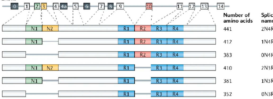
In normal adult human brain, alternative splicing of exon 2 (E2), exon 3 (E3) and exon 10 (E10) generates six TAU isoforms ranging from 352 to 441 amino acids in length. The size of those isoforms depends on the presence or absence of one or two 29- amino acid insert in the N-terminal, which are encoded by E2 and E3, and the inclusion or not of either three (R1, R3 and R4) or four (R1-R4) repeat regions in the C-terminal, which are encoded by E10. Therefore isoforms may be categorized according to the number of inserts encoded by E2 and E3 (0, 1 or 2), called 0N, 1N or 2N, and according to the number of repeat domains of the C-terminal encoded by E10 (3 or 4), called 3R or 4R 49 (Figure 4). The peripheral nervous system has neurons that often project a very
long axon with a large diameter, due to the inclusion of a N-terminal sequence encoded by exon 4a, resulting in the expression of a specific TAU isoform named “big TAU” 50,51.
Throughout the development of the human brain, TAU isoforms are differentially expressed, whereby during the foetal stages only the shortest TAU isoform is present (0N3R), while during adulthood all six isoforms are found in the brain 50. Also, foetal forms of TAU promote
assembly of microtubules less actively than adult forms, since it is in the inter-region between R1 and R2 that relies the most potent part that induces microtubule polymerization. Notably this R1-R2 inter-region is characteristic of adult TAU isoforms 4R, being responsible for the difference in the binding affinities between 3R and 4R TAU 52.
10 Maria Soares Cachide de Almeida
Importantly, the alternative splicing of E10 is linked to different tauopathies according to the isoforms found in the protein aggregates, namely 4R tauopathies (comprising PSP, CBD and AGD), 3R tauopathies (such as PiD) and 3R+4R tauopathies, as exemplified by AD 53.
Figure 4 – Schematic representation of the human MAPT gene and the six isoforms of the human brain. (Adapted from 54).
Domains and Structure of TAU
Structurally TAU can be subdivided into two major domains: the Projection Domain (PD) and the Microtubule-Binding Domain (MiBD). The PD (N-terminal) encompasses a proline-rich region as well as an acidic region, and is so designated since it projects from the microtubule surface where it might interact with a neural plasma membrane, in addition to cytoskeletal elements such as spectrin and actin filaments, allowing for the interconnection between TAU-stabilized microtubules and neurofilaments that limit the flexibility of the microtubule grid. The PD is also important in the determination of the axonal diameter and spacing between microtubules in axons, and it is likely involved in signal transduction pathways concerning phospholipase C gamma (PLC-γ)
50–52. On the other hand, the MiBD interacts with microtubules through some repeated domains
(R1-R4) located at the C-terminal, consisting of 18 amino acids encoded by exons 9, 10, 11 and 12, separated from each other by 13- or 14- residue spacer regions 55. The MiBD is extremely important,
since it interacts with microtubules, allowing for their polymerization and stabilization, in addition to regulating their dynamic stability, being implicated in their normal function. Recent evidence also supports a role of the MiBD in the modulation of the phosphorylation state of TAU proteins, once it allows the competitive binding of microtubules to TAU, inhibiting Protein Phosphatase 2A (PP2A) activity 52.
Subcellular localization and functions of TAU
The distribution of TAU, at a subcellular level is adjusted according to the developing brain
56, such that in the young neurons TAU is found among the cell body and neurites, while after the
formation of axons and polarization of neurons, TAU is found in greater amounts in axons and depleted in dendrites and nuclei 57. For this reason, the presence of TAU in dendrites represents
one of the first signs of neurodegeneration, although some studies suggest that its presence in such subcellular localization might be related to a role in the regulation of synaptic plasticity 54. This
relocation of TAU might be due to several factors, namely the greater affinity of this protein to microtubules in axons than in dendrites 58 and the quick axonal transport of TAU right after its
synthesis in the soma 59. Importantly, different isoforms of TAU are found in different cellular
compartments of neurons, which can mean that the protein has specific features depending on their subcellular localization. This way disturbance of the distribution of these isoforms might induce a gain of function of TAU that becomes toxic, resulting in neurodegeneration 54.
As previously stated, TAU has different functions according to its subcellular localization. Starting with axonal TAU, it is important to stabilize microtubules, leading to microtubule assembly and, more importantly, to regulate the dynamic instability of microtubules which facilitates the cytoskeleton reorganization 60. Another feature of TAU is the regulation of axonal transport of
cellular components such as mitochondria or neurotransmitter vesicles by influencing the function of dynein and kinesin, responsible for the transport of cargoes towards the cell body (minus end) and towards the axonal terminus (plus end), respectively. An example of this regulation relies on the fact that TAU has a stronger inhibitory effect on kinesin than on dynein and, for this reason, in the soma of cells overexpressing TAU there is an accumulation of cargoes such as APP, which is carried by kinesin-vesicles along axons and dendrites. Also, TAU might affect the axonal transport of other cargoes through competitive binding to kinesin 61. In addition to this, TAU seems to be
essential for axonal elongation and maturation, though such role still needs confirmation through further experiments 54. Besides the axon, it is believed that nuclear TAU is responsible for
maintaining the integrity of genomic DNA, cytoplasmic RNA and nuclear RNA 62.
Moreover, TAU may interact with actin, modifying the organization of the cytoskeleton network by inducing the alignment of bundles consisting of actin filaments 54.
12 Maria Soares Cachide de Almeida
Post-translational modifications and implications in TAU function
In tauopathies such as AD, the typically soluble TAU is present in an abnormal filamentous form, resulting from conformational changes and misfoldings in the normal structure of TAU, ultimately leading to the appearance of intraneuronal protein aggregates, thereby preventing the correct organization of the cytoskeleton 48. The mechanisms responsible for TAU’s loss of function
are not yet fully defined. However, like many proteins, TAU undergoes post-translational modifications, such as phosphorylation (hyperphosphorylation), acetylation, glycation, ubiquitination, nitration, truncation and some other modifications, which are believed to be the main cause of loss of normal function and acquisition of pathological features such as protein aggregation 63.
Regarding the phosphorylation, the longest variant of TAU (2N4R) has 85 potential phosphorylation sites (45 serines, 35 threonines and 5 tyrosines) that are phosphorylated and/or dephosphorylated by multiple kinases or phosphatases, due to their easy accessibility resulting from TAU’s natively unfolded structure 54. Most of these potential sites are clustered in regions
flanking the MiBD repeats (R1-R4), namely in the proline-rich region and in the C-terminal extreme, with the exception of Ser261 (R1), Ser285 (R1-R2 inter-repeat), Ser305 (R2-R3 inter-repeat), Ser 324 (R3), Ser352 (R4) and Ser356 (R4) 52. Since phosphorylation plays a crucial role in regulating the
physiological functions of TAU, such as the binding of TAU to microtubules and ultimately their stabilization and assembly, multiple studies have focused on the protein kinases and protein phosphatases responsible for the regulation of this microtubule-associated protein 54.
The main kinases involved are the Glycogen Synthase Kinase 3β (GSK-3β), which is a serine/threonine protein kinase widely expressed in the brain and associated with microtubules 64;
and Cyclin-Dependent Kinase 5 (CDK5), which is a serine/threonine protein kinase, member of the CDK family65. Interestingly, CDK5-mediated phosphorylation leads to further phosphorylation of
TAU by GSK-3β, favouring AD pathogenesis 66. This could be due to conformational changes that
allow the access of GSK-3β as well as other kinases to further phosphorylate TAU 48,54. As for
dephosphorylation of TAU, the phosphatases more likely involved are Protein Phosphatase 2A (PP2A), which accounts for ≈70% of the human brain TAU phosphatase activity 67, followed by and
Protein Phosphatase 1 (PP1) . Studies show that the activity of PP2A is diminished in the brain of AD patients, which could result from post-translational modifications of its catalytic domain, reduced PP2A expression, as well as increased levels of endogenous PP2A inhibitors 68,69.
1.4 Genetics of AD
As previously said, AD is a complex multifactorial dementia and, although our knowledge of the pathophysiology still remains in an initial state, it is broadly accepted by the scientific community that genes play a crucial role in both onset and development of the disease 70.
To date, more than two hundred mutations have been linked to AD and every year more are discovered 10 . Depending on the genetic cause and the age-of-onset, AD can be classified as
Early-Onset Alzheimer Disease (EOAD) or Late-Onset Alzheimer Disease (LOAD). The latter is the most common type of AD 10,71,72.
The EOAD tend to group within families, sometimes throughout generations 73. Usually it
develops before the age of 65 years old and is transmitted as an autosomal dominant trait, caused by extremely penetrant mutations. Only about 5% of AD cases fit in this category. To date, three genes have been highly related to this type of AD: APP, PSEN1 and PSEN2 3,70,72 (Table 1). The first
gene discovered was the APP gene, in 1987, which lies in the long arm of chromosome 21 74. Due
to its location, patients who suffer from Down syndrome tend to develop AD later in life, as they have an extra copy of the APP gene2.
The first missense mutation in APP was reported in 1991 75, after which nearly 20 other AD
mutations have been reported in this gene. Most of the APP-variants take place close to the recognized -secretase site, between residues 714 and 717, suggesting that -secretase cleavage of APP is critical for AD development 70. A few years later, other mutation was linked to the
development of EOAD. This mutation occurs on chromosome 14q24 and affects the PSEN1 gene, which encodes for presenilin 1 76, a highly conserved membrane protein necessary to release A
from APP during the -secretase action. Every year new mutations are discovered to affect this gene. Subsequently mutations in PSEN2 (located in chromosome 1) were identified and associated with AD 70. These three genes are all involved in the production of A peptides, which gave rise to
the Amyloid Hypothesis in the development of AD 72, whereby the pathophysiological process that
ultimately result in the appearance of the neuropathological features of AD (SPs and NFTs), as well vascular damage and inflammation, are consequences resulting from the abnormal production of Aβ, rather than causes of the disease process 77.
14 Maria Soares Cachide de Almeida
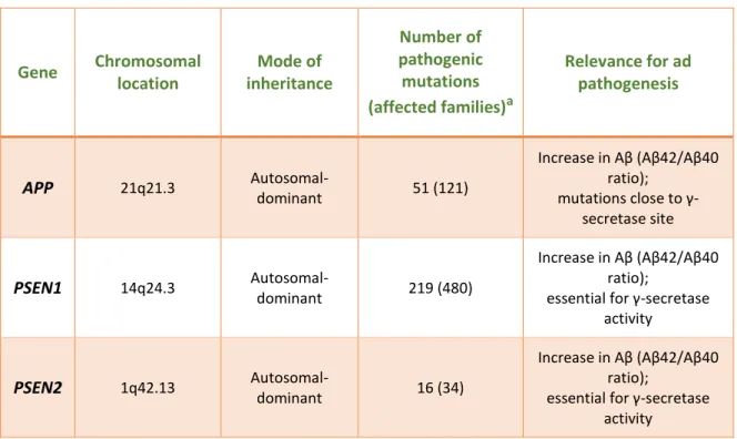
Table 1 – Overview of the stablished EOAD genes and their functional relevance for AD pathogenesis. (Adapted from 72) Gene Chromosomal location Mode of inheritance Number of pathogenic mutations (affected families)a Relevance for ad pathogenesis APP 21q21.3 Autosomal-dominant 51 (121)
Increase in Aβ (Aβ42/Aβ40 ratio);
mutations close to γ-secretase site
PSEN1 14q24.3
Autosomal-dominant 219 (480)
Increase in Aβ (Aβ42/Aβ40 ratio);
essential for γ-secretase activity
PSEN2 1q42.13
Autosomal-dominant 16 (34)
Increase in Aβ (Aβ42/Aβ40 ratio);
essential for γ-secretase activity
a(Source: “AD & FTD Mutation Database” [URL: http://www.molgen.ua.ac.be/ADMutations/] current on 6/10/2016)
Most cases of AD are Late-Onset, representing about 95% of all cases 3, affecting people
with 65 or more years of age. This type of AD is much more complex and less understood, both genetically and epigenetically, with the possible involvement of several genes. There can also be gene-gene and gene-environment interactions, which makes its appearance more unpredictable as the genes involved may contribute to or prevent the risk of developing the dementia, without causing it directly 71,73.
Until recently only one gene had been associated with this form of AD. The gene encoding apolipoprotein E (APOE), located in chromosome 19, was linked to LOAD in 1993, after A was found to bind APOE 78. This association is due to the 4 allele of APOE, which has been consistently
proved to be a risk factor of LOAD 3,72. However, studies have shown that APOE is limited to account
for approximately 50% or less of LOAD risk 79, suggesting that other genes, and therefore other
In order to identify the remaining genes for LOAD, two methods have been suggested, namely Whole Exome Sequence (WES) and Genome-Wide Association Studies (GWAS) 19,80. The
latter, allowed to overcome several technical limitations, due to the advent of microarray technology which makes it possible to evaluate several hundreds of thousands of Single-Nucleotide Polymorphisms (SNPs) in a single trial. Nevertheless, despite having several advantages once it is an approach hypothesis free, GWAS require more rigorous criteria to validate the significance of SNPs in a disease context 10. Also, since the effect sizes of the novel genes possibly related to AD
are small, it requires a remarkably big number of individuals (cases and controls) in order to identify additional genes involved in LOAD, which represents another disadvantage of this method 81.
Since 2009, and in addition to APOE, more than 20 loci have been identified by five GWAS and one meta-analysis, and significantly associated to LOAD. Among these are CLU, CR1, PICALM, BIN1, ABCA7, MEF2C, HLA-DRB1/HLA-DRB5, EPHA1, CD2AP, CD33, INPP5D, NME8, ZCWPW1, PTK2B, SORL1, CELF1, SLC24A4/RIN3, FERMT2 and CASS4 19,82–85. Depending on the possible role in
the process of AD, these genes cluster in a specific set of pathways, namely Lipid Metabolism, Immune Response or else Endocytosis, strongly highlighting the relevance of these molecular mechanisms in AD aetiology 81,86. Of all these genes, and according to the Alzgene database
(http://www.alzgene.org/), the bridging integrator 1 (BIN1) is currently recognised as the most significant LOAD-associated risk loci after APOE, and will be discussed in more detail in section 1.5.
Despite all these advances and discoveries allowed by GWAS, a significant part of the genetic contribution to AD has not yet been explained and the APOE-4 allele remains the key gene variant considered to be a LOAD risk factor.
1.4.2.1 APOE as a genetic risk factor for AD
Apolipoprotein E (APOE) is a lipoprotein of 299 amino acids and 34kDa molecular weight, first discovered in 1970s as a protein component of triglyceride-rich lipoproteins 87.
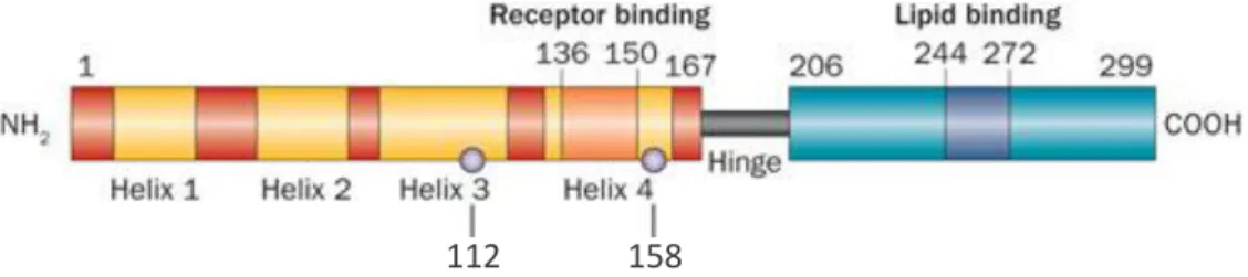
This protein has three major isoforms APOE2, APOE3 and APOE4 encoded by three alleles located on a single gene locus on position 19q13, being them 2, 3 and 4, respectively 88. The
isoforms differ by a single-amino acid substitution at residues 112 and 158 (Figure 5), which provides structural changes that influence their functions both at a cellular and molecular level, allowing the association of different isoforms to different neuropathological conditions 89.
16 Maria Soares Cachide de Almeida
Figure 5 – Schematic representation of the structure of APOE. The different isoforms APOE2, APOE3 and APOE4 differ from one another at amino acid residues 112 and 158 (grey circles) providing unique features to each isoform. (Adapted from 90).
Therefore, APOE3, which is the most common of the three isoforms and considered the normal form of APOE, has Cys-112 and Arg-158 91. On the other hand, APOE2 has 112 and
Cys-158, the latter being responsible for greater stability of this isoform and associated with a protective effect against AD 92. Finally, the APOE4 which has Arg-112 and Arg-158, is a major risk factor for AD
since the presence of one APOE-4 allele increases the risk to develop the dementia by about three times, while having two copies of the allele increases LOAD risk by 12 times 93 (Table 2).
Furthermore, APOE-4 allele shifts the age of the disease onset, with each allele lowering the age of onset by one or two decades relative to non-carriers in LOAD 94.
Importantly, while in EOAD mutations in the APP gene, the PSEN 1 gene or in the PSEN2 gene are sufficient, but not required, to cause AD, the APOE-4 allele is neither indispensable nor necessary to cause AD, being considered a risk factor that diminishes the age of onset, as previously said 95.
Table 2 – Comparison between APOE isoforms. Isoform-specific amino acid difference and allele frequency in each isoform for the general population and patients with Alzheimer’s Disease. (From 90)
Isoform-specific amino acid difference Allele frequency (%)
112 158 General AD
APOE2 Cys Cys 8.4 3.9
APOE3 Cys Arg 77.9 59.4
APOE4 Arg Arg 13.7 36.7
Functionally, APOE usually is important for lipid metabolism as well as transport, and is a protein expressed in many organs, mainly in the liver followed by the brain. APOE occurs primarily as a constituent of lipoprotein complexes alongside with other apolipoproteins and proteins in plasma as well as cerebrospinal fluid (CSF) 96. In peripheral tissues, APOE is primarily produced by
the liver and macrophages, being secreted into the circulation as a protein incorporated into very low-density lipoproteins (VLDL), chylomicron remnants, and certain subclasses of high-density lipoprotein (HDL) 86,89. APOE plays an essential role in the regulation of cholesterol as well as lipids
throughout the organism facilitating the clearance of plasma lipoproteins through the low-density lipoprotein receptor (LDLR) as well as additional LDLR-related protein family members 87. In
addition, in the central nervous system (CNS), this lipoprotein produced primarily by astrocytes 97,98,
may be involved in several physiological and pathological processes, including the metabolism and trafficking of cholesterol, resulting from neurodegeneration, to neurons lacking them for membrane repair, proliferation or remyelinization 86. Despite neuronal trafficking, APOE also plays
an essential role in synaptogenesis, blood-brain barrier integrity 97,98, and it affects glutamate
receptor function and synaptic plasticity by modulating neuronal APOE receptor recycling99.
Although the mechanism by which APOE isoforms affect the risk to develop AD is not completely understood, significant evidence of diverse neuropathological effects of APOE4 on cells within the CNS have been validated. Decreased Aβ clearance 89 and increased Aβ aggregation 100,101
has been demonstrated in 4 carriers, as well as Aβ load and plaque accumulation strongly correlated to APOE-4 dosage at autopsy 102,103. Finally, enhanced formation of
C-terminal-truncated fragments characteristic of 4 isoform stimulate TAU phosphorylation leading to preneurofibrillary tangles 104.
It is important to keep in mind the fact that APOE has different influences in AD onset and progress according to the population ethnicity 105,106 and so it is extremely important to understand
the relevance of this lipoprotein to each type of population since it might be useful for diagnosis purposes.
18 Maria Soares Cachide de Almeida
1.5 Bridging Integrator 1 (BIN1)
BIN1, also named Amphiphysin2, is a member of the BIN1/amphiphysin/RVS167 (BAR) family of genes, encoding a nucleocytoplasmatic adaptor protein highly expressed in the CNS. BIN1 maps to the long arm of the human chromosome 2 (2q14.3) and encodes no less than 20 exons 107,
which can be spliced into several isoforms. Primarily, 19 exons were identified 108, after which an
extra exon was discovered between exons 6 and 7, named exon 6a 109.
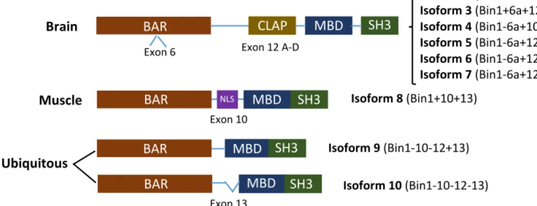
In mammals BIN1 is widely expressed, but since BIN1 transcripts undergo an extensive differential splicing, more than 10 isoforms are produced with different subcellular localization, tissue distribution, and particular functions according to specific protein interactions. Furthermore, depending on the inclusion of four exons (6a, 10, 12 and 13) different types of isoforms are produced, includingbrain specific isoforms 1-7, muscle-specific isoform 8 and isoforms 9 and 10 (ubiquitously expressed) 109. Also an aberrant isoform has been reported to be expressed
specifically in tumour cells 108. Depending on the isoform expressed, BIN1 encodes proteins ranging
from 409 amino acids (isoform 10) to 593 amino acids (isoform 1), having a predicted molecular weight of 45 kDa to 65 kDa, respectively.
Relatively to BIN1 structure, it has numerous characteristic protein domains 110. The BAR
domain (N-terminal) is ubiquitously expressed and binds lipid membranes in a dimer conformation, stimulating membrane curvature in places such as T-tubules in muscular cells, endocytic pits in neuronal and non-neuronal cells, and possibly in cytoplasmic endosomes 111,112. Importantly, the
protein-protein interaction with dynamin can be modulated through differential splicing of exon 7 in the BAR domain 113. In some brain isoforms there is also a N-terminal insert domain of 31
residues, encoded by exon 6a, located within a putative coiled-coil region in the BAR domain 114.
Another ubiquitously expressed domain is the Src Homology 3 (SH3) domain, which is encoded by exons 19 and 20 and allows the binding of proline-rich motifs 115. Interestingly, this
domain in BIN1 differ from other SH3 domains owing to a large patch of negative electrostatic potential and a remarkably prolonged n-Src loop 116. The Myc-binding domain (MBD) is present in
all isoforms, being encoded by exons 17 and 18 117, though alternative splicing of the first might
lead to the loss of the interaction between BIN1 and c-Myc 108,118. Furthermore, exon 13
and MBD, in the muscle-specific isoform (isoform 8) exon 10 translates into a small domain of 15 residues which includes a putative nuclear localization sequence (NLS), as well as a lipid-binding sequence 117,119. Finally, in brain-specific isoforms there is a clathrin and AP2 (CLAP) binding domain,
which is encoded by exon 12 (including a series of alternative brain-specific exons 12A-D) and is responsible for binding to endocytic proteins, namely clathrin and AP2/α 120,121 (Figure 6).
Figure 6 – Schematic representation of BIN1 domains and isoforms. Exons 6, 10, 12 (a-d) and 13 are alternatively spliced generating brain specific isoforms (1-7), a muscle isoform 8 and the two ubiquitously expressed isoforms (9 and 10). Abbreviations: BAR, BIN1/Amphiphysin/RVS167 domain; CLAP, clathrin-AP2 binding region; MBD, Myc-binding domain; SH3, Src homology domain; NLS, nuclear localization sequence. (Adapted from 109)
The cellular functions performed by BIN1 are regulated through specific exon splicing, thus each isoform has a particular role in the organism, also depending on subcellular localization and tissue distribution. Furthermore, BIN1 may also be regulated by some post-translational modifications such as protein phosphorylation. Additionally, epigenetic processes, such as DNA methylation, might result in the inactivation and silencing of BIN1 by affecting the promoter activity, since it contains a CpG island 108.
Several studies have associated BIN1 to the process of clathrin-mediated endocytosis as well as intracellular endosome trafficking both in neuronal and non-neuronal cells 107,122,123, through
the interaction with numerous proteins, namely dynamin 122, AP2 adaptor complexes 122,123, clathrin 121, synaptojanin 124, and endophilin 125, reflecting its ability to regulate membrane remodelling.
Furthermore, BIN1 has a potential role in regulating the actin cytoskeleton, coordinating membrane and cytoskeleton remodelling, given that it appears to link the two via the tubular membrane structure it forms. Thus, BIN1 might be involved in some AD neuropathological features
Isoform 8 (Bin1+10+13) Isoform 9 (Bin1-10-12+13) Isoform 10 (Bin1-10-12-13) Exon 6 Exon 13 Exon 10 Exon 12 A-D BAR BAR BAR BAR CLAP MBD MBD MBD MBD SH3 SH3 SH3 SH3 NLS Brain Muscle Ubiquitous Isoform 1 (Bin1+6a+12+13) Isoform 2 (Bin1+6a+12[b,c,d]+13) Isoform 3 (Bin1+6a+12d+13) Isoform 4 (Bin1-6a+10+12a+13) Isoform 5 (Bin1-6a+12[a,d]+13) Isoform 6 (Bin1-6a+12a+13) Isoform 7 (Bin1-6a+12d+13)
20 Maria Soares Cachide de Almeida
such as NFTs, since it might bind to the coiled-coil region of a plus-end protein involved in microtubules stability (CLIP170) though its BAR domain 110,126. Finally, BIN1 has numerous nuclear
functions and has been associated to DNA repair, cell cycle and apoptosis 110. In fact, the MBD of
BIN1 interacts with c-myelocytomatosis (c-MYC) oncoprotein, which has a crucial role in cell growth, apoptosis as well as malignancy 117, and several studies have shown that blocking BIN1
expression or else BIN1-Myc interaction resulted in specific inhibition of Myc-mediated apoptosis
127,128 . It is important to note that only nuclear isoforms of BIN1 can activate apoptosis. Relative to
DNA repair, BIN1, through the binding of its BAR domain to proteins which have a major role in this process, can modulate their activity 110.
Since AD is such a multifactorial disease, recently several GWAS have been performed in order to identify risk loci involved in the pathogenesis of LOAD.
After several studies, BIN1 has repeatedly been confirmed as a significant LOAD-associated risk loci, occupying the second position just after APOE 83,129–131. However, this locus was only
considered statistical significant after the study performed by Seshadri et al. 129,being subsequently
confirmed in other studies. From all the BIN1 Single Nucleotide Polymorphisms (SNPs) analysed, two were especially relevant for AD, being them rs7561528 and rs744373, both of which lie approximately 25kb to 30 kb upstream of the BIN1 gene, respectively. The first one has a risk allele “G”, while the second one has a risk allele “A” 109. Importantly, this work will be focusing on the SNP
rs744373 since it has been correlated with the rate of cognitive decline and AD progression, in addition to its risk allele “G” being linked to faster Mini-Mental State Examination (MMSE) deterioration, though this outcome does not reach statistical significance 109.
The significance of BIN1 as a risk loci to AD have been successfully replicated and confirmed in several independent candidate gene studies evaluating ethnically distinct populations, such as Caucasian and Caribbean Hispanic 132,133. Furthermore, the significance of BIN1 as a genetic risk
locus to LOAD has been replicated and confirmed in the largest family-based GWAS 134, as well as
in African American populations 135. However, this association was not detected in the Han Chinese 136 nor the Korean population 137. Importantly, the most significant SNPs may vary from study to
study, according to the datasets consisting of different ethnicities. 109 Besides, in a population, more
characterize, the best way possible, which SNPs contribute to AD in different populations. In addition, it is possible that the various SNP’s of BIN1 differentially affect the development of AD and, if this is true, it would be extremely interesting and useful to split subject groups and investigate the degree of cognitive decline separately for each BIN1 genotype 109.
Some studies have analysed the expression of BIN1 in the human brain, detecting an increase of BIN1 transcripts in brains of AD cases when compared to controls 138. However, there
are also studies that reveal a significant decrease of BIN1 in the brain of LOAD patients (87% when compared to non-demented age-matched controls) 139. In addition, although several SNPs of BIN1
have been associated to LOAD, little is known about BIN1 protein expression and its contribution to AD pathogenesis. Thus it is imperative that more studies are performed in order to help clarify the role of BIN1 in AD pathogenesis, identifying in which pathways that genetic locus may be involved, and in which way that involvement contributes to the onset of AD or its progression.
Since BIN1 has a role in the process of clathrin-mediated endocytosis, some studies have focused on the potential role of this BAR protein in APP metabolism and Aβ production, since they are both internalized through the endolysosomal trafficking pathway. Following this hypothesis, fluctuations on BIN1 expression would result in changes in APP trafficking through intracellular compartments, influencing whether APP undergoes the non-amyloidogenic or the amyloidogenic pathway, depending on the localization of the cleaving secretases. Thus, having been cleaved by β-secretase in the endosomes, if APP moved into the cell surface it would be cleaved by α-β-secretase, however, if APP moved into lysosomes it would be cleaved by γ-secretase, giving raise to Aβ 140,141.
Despite this, recent studies tried to investigate the importance of BIN1 in Aβ processing and secretion but failed to find any association between the levels of BIN1 and Aβ42 neurotoxicity 138.
Also, another approach using siRNA to knockdown endogenous BIN1, and overexpressing BIN1 in SH-SY5Y cells failed to have an impact on APP processing 139.
As mentioned previously, BIN1 has a potential role in regulating the actin cytoskeleton, coordinating membrane and cytoskeleton remodelling. Thus, it may interact with microtubule-associated proteins, like TAU. To investigate this theory a recent study was performed and identified an association between BIN1 and TAU at a biochemical, genetic and neuropathological level, strongly suggesting that increased levels of BIN1 lead to an augmented AD risk by interacting with the TAU pathway, although the exact mechanism is still not clear. Some of the possibilities lie on the fact that previous studies show that BIN1 can stabilize T-tubule structure in muscles. Thus, BIN1 might modulate microtubule stability or else TAU phosphorylation/aggregation 138. Also, BIN1
22 Maria Soares Cachide de Almeida
might be involved in NFTs formation. However, although BIN1 and TAU colocalize, there has not been detected any colocalization between BIN1 and NFTs. This suggests that BIN1 can influence the early stages of AD, being a primary contributor to the disease through the promotion of TAU aggregation, rather than influencing the late stages of the TAU-related pathology when NFTs are formed. In addition, the knockdown of BIN1 resulted in the suppression of TAU-induced neurotoxicity 138. Despite these findings, a study had some contrasting results, showing no
correlation between BIN1 and the amount of TAU pathology in AD 139. For this reason, more studies
should be performed in other populations, in order to investigate the significance of BIN1 in AD pathogenesis, potentially through the TAU pathway.
Besides AD, BIN1 has been associated to other diseases such as cancer, myopathies, namely centronuclear myopathy (CNM) and myotonic dystrophy (MD), and has also been linked to cardiac failure, though the latter requires more in-depth investigation 110.
Starting with cancer, as stated above, BIN1 interacts with c-MYC and so it can inhibit MYC-dependent transformation and tumour growth, as long as it is expressed at a standard level 117.
Also, several studies have shown that the expression of BIN1 is reduced or altered in numerous cancer types, such as colon, prostate, breast and lung cancers, as well as hepatocarcinoma and neuroblastoma 142–145. Furthermore, a study was performed, and revealed that the decrease of BIN1
expression leads to an increase of resistance to Cisplatin-based chemotherapy, which may be due to loss of control over the DNA repair mechanism 146. Therefore, augmenting the expression of
BIN1 might be an unusual and interesting strategy for the treatment of cisplatin-resistant cancers. Also, since BIN1 has been linked to cytoskeleton and membrane remodelling, it could be implicated in tumour cell migration and invasion, whereby its decrease would contribute to tumour progression.
BIN1 is greatly expressed in the brain and skeletal muscles. In the latter, several studies have shown that BIN1 has a major physiological relevance, since the increase of its expression leads to myoblast fusion and differentiation, being also essential for the positioning and remodelling of T-tubules. This BAR protein might also be responsible for sarcomere organization, once again highlighting its importance to skeletal muscle 110. Therefore, changes in BIN1 expression might
1.6 Diagnosis of AD
The assessment of AD made by an individual’s primary care physician or neurologist, requires not only the presence of memory decline but also multiple cognitive deficits. The diagnosis criteria and guidelines for AD were first established in 1984 by the Alzheimer’s Association and the National Institute of Neurological Disorders and Stroke (AANINDS) 147 and last updated almost three
decades later, in 2011, by the National Institute on Aging – Alzheimer’s Association (NIA-AA) 148.
Usually the physician starts with the medical and family history of the patient, plus psychiatric history as well as behavioural, personality and cognitive changes. During this stage of the diagnosis it is extremely important to interview a family member or a friend close to the patient, since they may be useful to detect that something is wrong even before changes are evident on tests, and can even explain how cognitive abilities, practical skills and behaviours have changed over time 149. After the first assessment, the physician also performs neuropsychological tests, such
as the Mini-mental state exam (MMSE), which allows the measurement of general cognitive function, with scores ranging from 0 (severe impairment) to 30 (no impairment), thus evaluating everyday mental skills.
Due to the progress in radiological imaging techniques, Magnetic Resonance Imaging (MRI) and other neuroimaging techniques such as Computerized Tomography (CT) and Positron Emission Tomography (PET), these are being widely used for AD diagnosis 3. MRI exams are routinely
requested, since they are three-dimensional, non-invasive and allow the characterization of the brain’s structure, just in a few minutes. There are several types of MRI, and some of the things that were possible to study using this technique go through the assessment of the integrity of white matter fiber tracts 150, to the study of local patterns of brain atrophy in individuals suffering from
MCI and AD 30,151,152, and lastly the measurement of intrinsic brain activity, which takes place
without any external stimulation, revealing changes in neuronal network activities in AD patients or individuals at risk for developing the dementia 153,154. MRI is very useful since it helps to rule out
normal pressure hydrocephalus, brain tumours, cerebral hematomas or even cerebrovascular lesions 3. Another plus of this technique relies on the fact that it allows the detection of brain
abnormalities in individuals who might be at risk to develop AD but have no apparent symptoms
24 Maria Soares Cachide de Almeida
Like MRI, PET has proven to be useful in the diagnosis of AD. Using 18F-fluorodeoxyglucose (FDG-PET) it was possible to differentiate between different forms of dementia, mainly AD and Frontotemporal Dementia (FTD), through the measurement of local brain metabolism 156,157. Recent
advances certified the use of a radioactive compound named Pittsburgh compound B (PiB) during PET assessment of AD, since it binds specifically to amyloid plaques in the brain. That way it is possible to detect one of the neuropathological hallmarks of AD, in a non-invasive way. However, there are some concerns about the use of PET-based amyloid imaging in the clinical setting, notably due to its high cost and low positive predictive value 158.
Another way of diagnosis that has been widely studied are the CSF-based protein biomarkers. Through these studies it was possible to discover that the levels of Aβ42 are reduced in AD patients, while the levels of phosphorylated TAU (P-TAU) are higher 159,160. Plasma biomarkers
have also been suggested as an option to CSF biomarkers 161.
Despite this, since AD is such a complex disease, innovative methods for AD biomarker identification as well as validation need to be established, in order to promote early AD diagnosis and therapeutic interventions 162.
1.7 Treatment of AD
Currently there is no cure for AD and scientists are just beginning to discover therapeutic methods that aid handling the symptoms of the dementia but do not delay the evolution or reverse the progress of the disease itself 163.
Therapeutically AD presents no great difference from other dementias, using drugs based on transmitter-replacement therapies. Given that, in AD, as the initial affected neurons are the glutamatergic and acetylcholinergic, the first therapeutic approach is to administer Acetylcolinesterase inhibitors that will promote cholinergic neurotransmission by preventing the breakdown of acetylcholine, thus improving memory function and attention in AD patients. Currently there are three cholinesterase inhibitors approved by U.S. Food and Drug Administration (FDA). These are galantemine, donezepil and rivastigmine. Another FDA approved drug is memantine, an NMDA (glutamate) antagonist whose function is to prevent excessive glutaminergic transmission that may result in excitotoxicity because of high intracellular calcium concentration in circumstances of overstimulation 164.