Faculdade de Farmácia
Departam ento de Farmácia Galénica e Tecnologia Farmacêutica
The link between altered glucosylceramide composition and
membrane properties:
From membrane biophysics to Gaucher Disease
Ana Raquel Pinto Varela
Orientadores: Doutora Liana C. Silva
Professor Doutor Manuel Prieto
Professor Doutor Anthony H. Futerman
Tese especialmente elaborada para a obtenção do grau de Doutor no ramo de Farmácia, especialidade de Tecnologia Farmacêutica.
Universidade de Lisboa
Faculdade de Farmácia
Departam ento de Farmácia Galénica e Tecnologia Farmacê utica
The link between altered glucosylceramide composition and membrane properties:
From membrane biophysics to Gaucher Disease Ana Raquel Pinto Varela
Orientadores: Doutora Liana C. Silva
Professor Doutor Manuel Prieto
Professor Doutor Anthony H. Futerman
Tese especialmente elaborada para a obtenção do grau de Doutor no ramo de Farmácia, especialidade de Tecnologia Farmacêutica.
Júri:
Presidente: Doutora Matilde Castro Vogais:
— Doctor Giovanni D’ Angelo,
— Doutor Vitor Manuel Vieira da Costa.
— Doutor Nuno Fernando Duarte Cordeiro Correia dos Santos, — Doutor Manuel José Estevez Prieto.
— Doutor Fñbio Monteiro Femandes.
— Doutora Helena Isabel Fialho Florindo Roque Ferreira — Doutora Liana Casquinha Silva
Trabalho financiado pela Fundação para a Ciência e a Tecnologia (FCT) através de uma bolsa de doutoramento SFRH/BDI/69982/2010., e pelas bolsas de projeto PTDC/QUI-BIQ/111411/2009 e PTDC/BBB-BQB/0506/2012
The link between altered glucosylceramide
composition and membrane properties:
From membrane biophysics to Gaucher Disease
Dissertação apresentada à Faculdade de Farmácia da Universidade de
Lisboa para prestação de provas de Doutoramento em Farmácia,
To my Mother, Father,
“baby“Brother and André.
“If you can find a path with no obstacles, it
probably don´t lead anywhere.”
I
Acknowledgments
In the end of this amazing marathon, which was the development and execution of my PhD project, I was able to enter in the world of biophysics and fluorescence. Since, this was quite a new and complex world, I needed the help of many persons and entities to be able to execute and overtake all the tasks and difficulties that I have encountered Therefore, I want to express my gratitude to all the people that I have met during my PhD and that helped me in the process, especially to the ones bellow.
First of all, I would like to express my appreciation to my advisors.
To Dr. Liana C. Silva, which was fundamental in my PhD. Thanks for giving me the opportunity to be a part of your research team, in the Intracellular Trafficking Modulation for Advanced Drug Delivery Research, for the support and guidance throughout this journey. And for pushing my limits, which made me grow as an individual and as a researcher.
I want to express my gratitude to Prof. Dr. Manuel Prieto for receiving me in the Molecular Biophysics Group of Instituto Superior Técnico (IST), for allowing me to perform the biophysical studies at the Centro de Quimica-Física Molecular (CQFM) of IST, for the good discussions, and for always keeping his good mood.
I also want to thank Prof. Dr. Anthony Futerman for receiving me in the Department of Biological Chemistry of Weizmann Institute of Science , where I was able to perform the cellular studies. Moreover, I also want to thank Professor Tony for his kindness and for all the good discussions.
An acknowledgment is also owed to Faculdade de Farmácia da Universidade de Lisboa,
Instituto Superior Técnico and Weizmann Institute of Science. For opening their facilities
to me, enabling the development of this thesis work.
I also want to acknowledge the Federation of European Biochemical Societies, which had awarded me with conference grants, which allowed my participation in more conferences, giving me the opportunity to share my work and debate it with scientists from all over the world.
II
A big thank you for the friendship off all the present and former people in the Nanomedicine and Drug Delivery Systems, which today is integrated in the Drug Development group of the iMed.ULisboa, especially to Carina, Joana Silva, Ana Saraiva, Natércia, Eva Zupančič, Ana Matos, Nuno Martinho, João Conniot, Liane Moura, Joana Marto and Helena Florindo. Thank you all for supporting me and for making me laugh in the hard times.
To my friends, colleagues, and Professors in the Centro Química-Física Molecular, in the Instituto Superior Técnico, namely to Mª João Sarmento, Joana Ricardo, Tânia Sousa, Sandra Pinto, Ana Melo, Marina Monteiro, Bruno Castro, Samuel Jacob, Paulo Caldas, Lourdes Renart, Rocio Esquembre , Fábio Fernandes and Prof. Dr. Ana Coutinho.
To Professor Hermínio Diogo, for letting me use his facilities.
To Professor Amélia M.P.S. Gonçalves da Silva, for letting me use her lab to develop part of my experiments, and for all the help in the interpretation of the results regarding the Langmuir monolayers.
To Prof. Dr. Erich Gulbins, for welcoming me in his group in the Institut für
Molekularbiologie in the Medizinische Fakultät Universitätsklinikum in Essen, were I was
able to evaluate specific interactions between sphingolipids and bacteria.
To all my dearest friends, for always keeping me up, namely Iris Santos, Pedro Pelado, Inês Ferreira, Natália Ferreira, Duarte Ferreira, João Romano, Joana Silva, Carina Peres, Ana Saraiva e Natércia.
Last, but not least I want to thank my family specialy to my Mum, Dad and baby brother, without you I would not be here, and André Carvalho, for all the support, love, and many many patiente, thank you !!!!!!
III
Financial support from FCT must be acknowledged, for the concession of my PhD grant (SFRH/BD/69982/2010), and for funding my work trought PTDC/QUI-BIQ/111411/2009, and PTDC/BQB/0506/2012 grants.
IV
This thesis was supervised by
Dr. Liana C. Silva (supervisor) Assistant Researcher
Research Institute for Medicines (iMed.ULisboa), Faculdade de Farmácia, Universidade de Lisboa, Portugal
Prof. Dr. Manuel Prieto Full Professor (co-supervisor)
Centro de Química-Física Molecular & IN - Institute of Nanoscience and Nanotechnology, Instituto Superior Técnico, Universidade de Lisboa, Lisboa, Portugal
Prof. Dr. Anthony H. Futerman Full professor (co-supervisor)
Department of Biological Chemistry, Weizmann Institute of Science, Rehovot, Israel
This work was developed at
- iMed.ULisboa – Research Institute for Medicines, Faculdade de Farmácia, Universidade de Lisboa, Av. Professor Gama Pinto, 1649-003 Lisbon, Portugal
- Centro de Química-Física Molecular & IN - Institute of Nanoscience and Nanotechnology, Instituto Superior Técnico, Universidade de Lisboa, Lisboa, Av. Rovisco Pais, 1049-001 Lisbon, Portugal
- Department of Biological Chemistry, Weizmann Institute of Science, Rehovot 76100, Israel
Ana Raquel Pinto Varela was financially supported by Fundação para a Ciência e a Tecnologia (FCT) with a PhD grant SFRH/BDI/69982/2010. The work in this thesis was supported by FCT grants PTDC/QUI-BIQ/111411/2009 and PTDC/BBB-BQB/0506/2012.
V
Abstract
Lipids are one of the most abundant molecules in a cell and are involved in the regulation of cell events. One of the mechanisms by which lipids control cell signaling is by altering membrane biophysical properties. Therefore, the biophysical properties of several lipids have been thoroughly characterized. However, some bioactive lipids like glucosylceramide (GlcCer) still have their properties poorly studied. GlcCer is a lipid involved in several cell processes. A deregulation in its metabolism triggers damaging signaling pathways ultimately leading to the development of a pathologic state, such as Gaucher Disease (GD). This disease results from the abnormal accumulation of GlcCer in cells, mainly in the lysosomes. Even though the biochemistry and biology context of GD is a subject of intensive research, the molecular mechanisms that underlie this disease remain elusive, likely due to the inherent complexity and heterogeneity of the disease. An underexplored branch of research, related to the understanding of the biophysical impact of increased levels of GlcCer in biological membranes, might provide additional insight into the mechanisms underlying this complex disease. Therefore, research that aims to characterize the biophysical features of GlcCer and its interaction with other membrane lipids is required. Using complementary techniques, including fluorescence microscopy and spectroscopy, a thorough biophysical characterization of GlcCer was performed. This comprised the study of the effect of increasing molar fractions of GlcCer in the biophysical properties and morphology in a variety of model membranes containing different lipid composition, as well as in wild type and Gaucher Disease-derived fibroblasts. Since GlcCer is present at higher abundance in the plasma membrane (PM) and in the lysosome, the studies were performed in conditions mimicking the pH environment of those cellular sites.
This detailed study revealed that: I) GlcCer increases the order of fluid membranes, II) GlcCer promotes membrane morphological alterations, such as tubules, III) GlcCer effect in membrane properties is pH sensitive, promoting an higher packing at neutral pH, IV) GlcCer impact in the membrane is modulated by cholesterol (Chol) in a concentration dependent manner, V) fibroblasts from GD patients, which are enriched in GlcCer, have a higher global membrane order in comparison to wild type fibroblasts, coherent with the observations in model membranes.
VI
These conclusions indicate that GlcCer accumulation in cells alter the biophysical properties of its membranes, possibly affecting protein activity and trafficking. These alterations could underlie some of the effects triggered by GlcCer that might contribute to the development of Gaucher Disease.
Keywords: Glucosylceramide, membrane models, lipid domains, fluorescence spectroscopy, fluorescence microscopy, membrane biophysical properties
VII
Resumo
As membranas celulares têm diversas funções, entre as quais podem identificar-se a função de interface na interação com moléculas extracelulares e a participação ativa na regulação da sinalização celular. As várias funções das membranas celulares sugerem que se trata de um organelo de estrutura complexa, no entanto, nem sempre esta foi a ideia vigente. A definição de membrana evoluiu significativamente desde que um dos mais importantes modelos de membrana, o modelo de Mosaico Fluido (FMM) de Singer e Nicholson foi proposto em 1972. Neste modelo, proteínas integrais ou periféricas estariam totalmente envolvidas ou apenas suportadas, respetivamente, por uma bicamada lipídica fluida, onde as proteínas poderiam difundir livremente. Neste modelo, os lípidos eram tidos como meras entidades estruturais sem qualquer função ativa nos processos celulares. No entanto, os desenvolvimentos tecnológicos forneceram novos dados em relação às membranas celulares que não eram compatíveis com o modelo de FMM, nomeadamente a formação de domínios lipídicos, tais como os domínios raft. Estes domínios selecionariam diferentes proteínas para diferentes zonas da membrana e, devido ao seu elevado empacotamento, poderiam também alterar a conformação proteica afetando a sua atividade. A capacidade dos lípidos membranares para segregarem e organizarem-se em domínios, afetando a difusão e a atividade proteica, conferiu aos mesmos um papel ativo na modulação de diferentes processos e sinalização celular. De acordo com a definição inicialmente proposta, os domínios raft são regiões da membrana enriquecidas em colesterol (Chol) e esfingolípidos (SLs), de entre os quais se podem nomear os glicoesfingolípidos (GSLs) (ex.glucosilceramida, GlcCer). Os SLs são uma classe lipídica com atividade biológica, sendo a sua estrutura base uma ceramida, por sua vez, formada por uma base esfingóide ligada a uma cadeia acilo de tamanho variável. Ao carbono C1 da ceramida podem ser adicionadas diferentes moléculas, como a glucose, formando-se assim a GlcCer.
A GlcCer tem diferentes funções celulares sendo, por exemplo, essencial para o desenvolvimento embrionário. O papel vital da GlcCer foi demonstrado pela inviabilidade de fetos de ratinho, que têm inibida a capacidade de sintetizarem GlcCer e consequentemente a maioria dos GSL complexos. O elevado impacto biológico da GlcCer pode em parte ser explicado, dado que a síntese de GSLs complexos, envolvidos em
VIII
diferentes processos vitais para o organismo, é dependente da GlcCer. Mais ainda, dado que a GlcCer está envolvida na formação de domínios lipídicos, como os raft, esta revela-se como um lípido que potencialmente controla diversos processos celulares, reafirmando o seu importante papel na célula. Exemplos documentados são: o papel da GlcCer no controlo das vias endocíticas, na interação célula-célula, na adesão celular e na influência da patogenicidade de vários microrganismos como o Criptococus neoformans. Contudo, este lípido apenas constitui uma pequena fração dos lípidos totais que existem na membrana, o que significa que para manter os níveis fisiológicos de GlcCer a sua síntese e degradação é altamente controlada. Consequentemente, um desequilíbrio numa via metabólica da GlcCer repercutir-se-á em efeitos negativos no funcionamento celular, o que poderá promover o desenvolvimento de patologias. De facto, o mau funcionamento de qualquer uma das moléculas envolvidas na degradação da GlcCer promove uma acumulação anormal deste lípido na célula, o que pode culminar no desenvolvimento de uma patologia designada de Doença de Gaucher (GD).
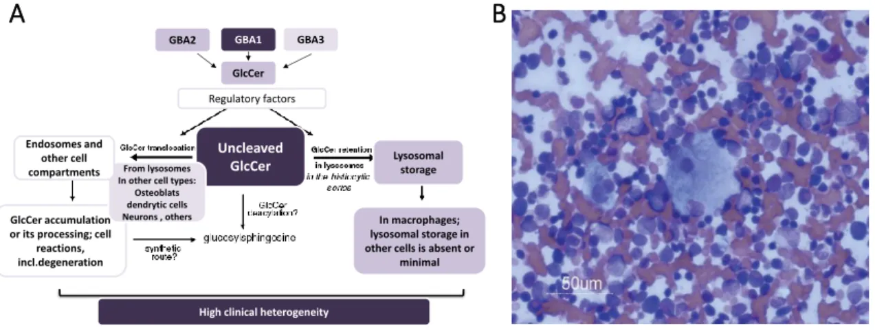
A etiologia mais frequente desta patologia deve-se a mutações no gene que codifica a enzima que degrada a GlcCer - a glucosilcerebrosidase (GCase). Na GD verifica-se uma significativa acumulação lisossomal de GlcCer, especialmente nos macrófagos. Isto deve-se à fagocitodeve-se intensa (de bactérias, fungos, diversos tipos de células, etc) que deve-se verifica nos macrófagos, aumentando muito significativamente o aporte de GlcCer nos mesmos. Esta doença manifesta-se frequentemente por uma hiperplasia do fígado e do baço, no entanto, o sistema nervoso central, os ossos e outros tecidos poderão também ser afetados. A possibilidade de envolvimento de diferentes sistemas promove uma impressionante heterogeneidade de quadros clínicos nestes doentes. Também se verifica em muitas situações que a severidade ou o fenótipo dos pacientes com GD não é passível de ser relacionado com o seu genótipo, o que muitas vezes dificulta o prognóstico desta doença por parte dos clínicos.
Apesar do contexto biológico e bioquímico da GD já ter sido exaustivamente estudado, a biofísica inerente a esta doença tem merecido pouca atenção por parte dos investigadores. Assim sendo é premente realizar estudos biofísicos que abordem o impacto da GlcCer nas propriedades de membranas modelo e naturais, tal como estudar a interação da GlcCer com outros lípidos de membrana, nomeadamente com os que tipicamente se encontram nos domínios raft.
IX
Estas questões poderão ser subdivididas em diferentes pontos, os quais irão ser desenvolvidos e esclarecidos nos capítulos II a V desta dissertação.
I) Quais são os efeitos da GlcCer, em membranas fluidas?; II) Será a GlcCer e outros esfingolípidos neutros sensíveis a alterações de pH?; III) Como é que a GlcCer interage com outros lípidos que tipicamente se encontram em domínios lipídicos, como o Chol?; IV) Como é que a GlcCer afeta as propriedades de modelos de membrana com uma composição típica de domínios raft? ; V) Haverá diferenças nas propriedades biofísicas globais das membranas de células saudáveis e de células enriquecidas em GlcCer? Para responder as estas perguntas desenhou-se um extenso estudo biofísico que explora as propriedades biofísicas da GlcCer no contexto de membranas modelo com crescente complexidade e também em membranas celulares. Neste estudo, usou-se maioritariamente modelos de membrana, tais como as vesiculas multilamelares e vesiculas unilamelares gigantes (estas com curvatura e tamanho muito semelhante a uma membrana natural). O uso destes modelos permite controlar a composição das membranas em estudo, tal como estudar os efeitos específicos de certos lípidos. Diferentes tampões, com pH distintos, foram usados para a formação das membranas modelo, permitindo analisar o efeito do fator pH nas propriedades biofísicas dos lípidos. A caracterização das membranas foi realizada através de diferentes técnicas complementares, nomeadamente: microscopia de fluorescência (confocal), espectroscopia de fluorescência, estudos em tina de Langmuir (onde se observou o comportamento das misturas em monocamadas) e de técnicas de dispersão dinâmica da luz e dispersão dinâmica electroforética.
Este detalhado estudo biofísico centrado no impacto da GlcCer em membranas com diferentes composições e a diferentes pH (7.4 e 5.5), revelou que: I) a GlcCer aumenta o empacotamento das membranas fluidas (compostas por fosfolípidos insaturados); II) promove alterações morfológicas nas membranas, como por exemplo, a formação de túbulos; III) o efeito organizador da GlcCer nas membranas é sensível ao pH, já que a pH neutro (7.4) a ordem das membranas que contêm GlcCer é maior que a pH ácido (5.5); IV) o impacto da GlcCer no empacotamento das membranas modelo depende da concentração de Chol na mesma, ou seja, o efeito da GlcCer nas propriedades biofísicas das membranas é regulado pelo Chol; VI) a ordem global das membranas de fibroblastos derivados de doentes com GD (células enriquecidas em GlcCer) é significativamente mais
X
elevada que a ordem global das membranas de fibroblastos controlo. Estes últimos dados são consistentes com os obtidos nos modelos de membrana, confirmando que as membranas artificiais são modelos adequados para prever o que ocorrerá a nível das membranas celulares.
Resumindo, neste trabalho verificou-se que a GlcCer promove um aumento da rigidez da membrana, no entanto, o seu efeito será condicionado pelos níveis de colesterol na membrana e pelo pH do meio celular. Em situações de aumento anormal dos níveis de GlcCer, como na doença de Gaucher, o elevado empacotamento e rigidez da membrana poderá afetar o transporte intracelular e a conformação de diferentes proteínas, promovendo alterações no comportamento ou até mesmo a morte celular. Os resultados obtidos nesta tese fornecem um apoio biofísico aos efeitos biológicos e patológicos promovidos pela GlcCer.
Palavras-chave: Glucosilceramida, membranas modelo, domínios, pH, espectroscopia de fluorescência, microscopia de fluorescência, propriedades biofísicas das membranas
XI
Content Index
Acknowledgments ... I
Abstract ... V
Resumo ... VII
Content Index ... XI
Figure Index ... XVI
Table Index ... XIX
List of abbreviations ... XX
Outline ... XXIV
Chapter I - Introduction ... 1
1.1BIOLOGICAL RELEVANCE OF CELL MEMBRANES... 3
1.2.MEMBRANE MODELS -HISTORICAL PERSP ECTIVE ... 4
1.3CHEMICAL COMPOSITION OF MEMBRANES ... 6
1.3.1 Carbohydrates... 6
1.3.2 Proteins ... 6
1.3.3. Lipids ... 7
2MEMBRANE LIPIDS AND THEIR BIOLOGICAL RO LE... 9
2.1 Sphingolipids ... 9
2.2 Glycosphingolipids ...10
2.3 Glucosylceramide...13
3(GLYCO)SPHINGOLIPIDS ROLE IN PATHO LOGICAL SITUATIONS...16
3.1 Gaucher Disease...17
4LIPIDS BIOPHYSICS –CORRELATION BETWEEN BIOPHYSICS AND BIOLOGY. ...19
4.1Lipid-Water systems – Lipid phases...20
4.2 Lipid phase transitions (phase diagrams)...25
4.3 Membrane dynamics– Fluidity concept ...27
5BIOPHYSICAL PROPERTIES OF SPHINGO- AND GLYCOSPHINGO LIPIDS ...29
XII
5.2 Glycosphingolipids ...31 6CEREBROSIDES BIOPHYSICAL PROPERTIES ...34
7LATERAL HETEROGENEITY IN MEMBRANES -MEMBRANE DOMAINS AND CELL REGULATION ...36
7.1 Domains Shape and size ...36 7.2 Biological relevance of lateral heterogeneity induced by SLs –Lipid Microdomains ...37 8MEMBRANE MODEL SYSTEMS ...40
8.1 Monolayers at an air-water surface or Langmuir Films. ...41 8.2 Liposomes and Vesicles ...41 9MEMBRANE CHARACTERIZATION...44
9.1 Characterization of Monolayers- Langmuir balance or trough ...44 9.2 Electrophoretic and scattered light measurements...45 9.3 Microscopy ...46 9.4 Fluorescence Spectroscopy ...48 9.5 Fluorophores ...52 10REFERENCES ...56
C
HAPTERII-
E
FFECTO
FG
LUCOSYLCERAMIDEO
NT
HEB
IOPHYSICALP
ROPERTIESO
FF
LUIDM
EMBRANES……….. 73
1ABSTRACT ...76 2INTRODUCTION ...77 3MATERIALS AND METHODS ...78 3.1 Materials...78 3.2 Fluorescence spectroscopy ...78 3.3 Confocal fluorescence microscopy ...79 3.4 Lipid monolayers and surface pressure–area measurements...80 4RESULTS...81
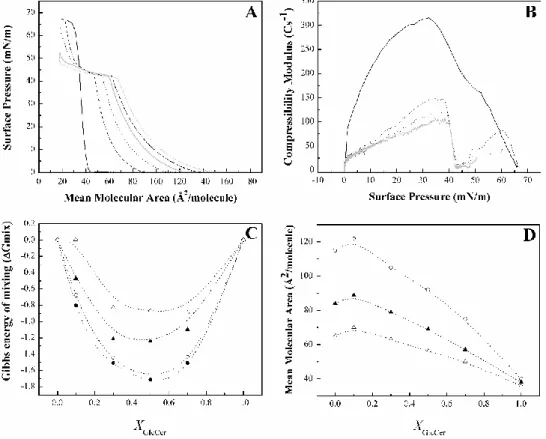
4.1 Biophysical properties of POPC/GlcCer mixtures ...81 4.2 Thermotropic characterization of POPC/GlcCer mixtures ...83 4.3 GlcCer gel domains and membrane morphology ...85 4.4 GlcCer-POPC molecular interactions in lipid monolayers ...85 5DISCUSSION...87
5.1 Properties of GlcCer...87 5.2 Effect of GlcCer on membrane biophysical properties ...88 5.3 GlcCer promotes morphological alterations...92 6CONCLUSIONS AND BIOLOGICAL IMPLICATIONS ...93
7ACKNOWLEDGMENTS...94
XIII
9SUPPORTING MATERIAL FOR:EFFECT OF GLUCOSYLCERAMIDE ON THE BIOPHYSICAL PROPERTIES OF FLUID MEMBRANES
... 100
C
HAPTERIII-
I
NFLUENCE OF INTRACELLULAR MEMBRANE PH
ON SPHINGOLIPIDORGANIZATION AND MEMBRANE BIOPHYSICAL PROPERTIES
... 103
1ABSTRACT ... 106
2.INTRODUCTION ... 107
3MATERIALS AND METHODS ... 108
3.1 Materials... 108 3.2 Fluorescence Spectroscopy ... 109 3.3 Confocal Fluorescence Microscopy ... 110 3.4 Lipid monolayers and Surface Pressure-Area Measurements... 110 4RESULTS... 111
4.1 Thermotropic studies ... 111 4.2 Spectroscopic characterization of the gel phase ... 113 4.3 Spectroscopic characterization of the fluid phase ... 115 4.4 Monolayer studies ... 117 4.5. Effect of pH on gel domain shape and size ... 122 5DISCUSSION... 123
5.1 POPC/C16-GlcCer Mixtures ... 123 5.2 POPC/Sphingomyelin and POPC/Ceramide mixtures... 125 6CONCLUSIONS AND BIOLOGICAL IMPLICATION ... 127 7ACKNOWLEDGMENTS... 128 8REFERENCES ... 128 9SUPPORTING MATERIAL FOR:INFLUENCE OF INTRACELLULAR MEMBRANE PH ON SPHINGOLIPID ORGANIZATION AND MEMBRANE BIOPHYSICAL PROPERTIES ... 132
C
HAPTERIV-
G
LUCOSYLCERAMIDE REORGANIZES CHOLESTEROL-
CONTAINING DOMAINS INA FLUID PHOSPHOLIPID MEMBRANE
... 137
1ABSTRACT ... 140
2INTRODUCTION ... 141
3MATERIALS AND METHODS ... 142
3.1 Materials... 142 3.2 Methods ... 143
XIV
4RESULTS... 145
4.1 POPC/Chol mixtures ... 145 4.2 Ternary mixtures with different C16-GlcCer/Chol ratios ... 147 4.3 Ternary mixtures with constant POPC/Chol ratio ... 150 5DISCUSSION... 154
5.1 Qualitative analysis of the interplay between GlcCer/Chol... 154 5.2 Determination of a ternary POPC/Chol/GlcCer phase diagram ... 157 6CONCLUSIONS AND BIOLOGICAL RELEVANCE ... 161
7AUTHOR CONTRIBUTIONS: ... 162
8ACKNOWLEDGMENTS... 162
9REFERENCES ... 163
10SUPPORTING MATERIAL FOR: GLUCOSYLCERAMIDE REORGANIZES CHOLESTEROL-CONTAINING DOMAINS IN A FLUID PHOSPHOLIPID MEMBRANE ... 167
10.1 Supplementary figures ... 167 10.2 Supplementary Tables ... 177 11SUPPLEMENTARY INFORMATION... 179
11.1 Determination of the fraction and composition of each phase for a three-phase situation of the POPC/Chol/C16-GlcCer ternary system ... 180 11.2 Determination of the tie-triangle boundaries ... 181 12SUPPORTING REFERENCES... 182
C
HAPTERV-
G
LUCOSYLCERAMIDE-I
NDUCEDB
IOPHYSICALC
HANGESI
NA
RTIFICIALA
NDC
ELLM
EMBRANES183
1ABSTRACT ... 185
2INTRODUCTION ... 187
3MATERIALS AND METHODS ... 189
3.1 Materials... 189 3.2 Methods ... 189 4RESULTS... 192
4.1 Studies in model membranes ... 192 4.2 Studies in living cells ... 198 5DISCUSSION... 199
5.1 Interplay between GlcCer and lipid components of model raft domains ... 199
5.2 Influence of GlcCer in cell membrane biophysical properties ………..……...202 6CONCLUSIONS AND BIOLOGICAL IMPLICATIONS ... 203
XV
8REFERENCES ... 204
9 SUPPORTING MATERIAL FOR: GLUCOSYLCERAMIDE-INDUCED BIOPHYSICAL CHANGES IN ARTIFICIAL AND CELL MEMBRANES ………..208
C
HAPTERVI-
C
ONCLUSIONS... 211
REFERENCES ………..217
C
HAPTERVII-
F
UTUREP
ERSPECTIVES... 219
XVI
Figure Index
C
HAPTERI-
I
NTRODUCTIONFIGURE 1-EUKARYOTIC ANIMAL CELL STRUCTURE. SCHEMATIC REPRESENTATION OF THE MAIN ORGANELLES OF AN ANIMAL CELL... 4 FIGURE 2-EVOLUTION OF MEMBRANE MODELS. ... 6 FIGURE 3LIPID CLASSES AND STRUCTURE. ... 8
FIGURE 4-COMPLEXITY OF GSL METABO LISM ...12
FIGURE 5-STRUCTURE OF CEREBROSIDES. ...14
FIGURE 6-CLEAVAGE OF THE Β-GLUCOSIDIC BOUND OF GLCCER BY GCASE...15
FIGURE 7-BIOLOGICAL O UTCOME OF GLCCER ABNORMAL ACCUMULATION IN THE CELL ...17
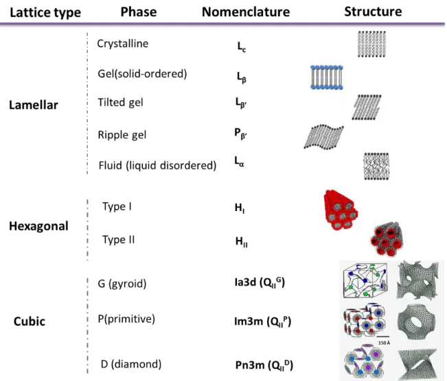
FIGURE 8-NOMENCLATURE AND SCHEMATIC REPRESENTATION OF SOME LYO TROPIC PHASES. ...22
FIGURE 9-LAMELLAR PHASE TRANSITIONS...25
FIGURE 10-LIPID PHASE DIAGRAMS ...27
FIGURE 11-TYPES OF LIPID DIFFUSION IN THE MEMBRANE...28
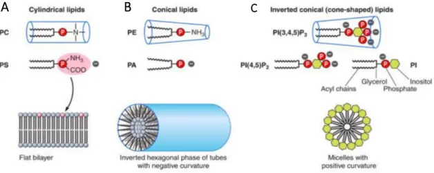
FIGURE 12-MEMBRANE INDUCED-CURVATURE BY LIPIDS ...34
FIGURE 13-DIFFERENT SHAPES OF MEMBRANE DO MAINS ...36
FIGURE 14- PH MODULATION OF DOMAINS SHAPE AND SIZE. ...37
FIGURE 15-EVOLUTION OF THE CO NCEPT OF RAFT DO MAINS AND MEMBRANE O RGANIZATION ...39
FIGURE 16-MEMBRANE MODELS ...44
FIGURE 17-CHARACTERIZATION OF MONO LAYER IN LANGMUIR THROUGH...45
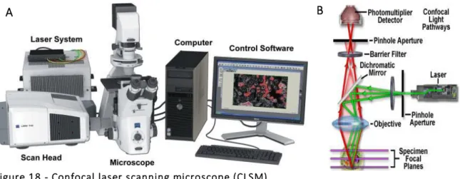
FIGURE 18-CONFOCAL LASER SCANNING MICROSCOPE (CLSM) ...47
FIGURE 19-EXCITATION (OR ABSORPTION) AND EMISSION SPECTRA ...50
FIGURE 20-ANISOTROPY MEASUREMENT ...51
FIGURE 21-LAURDAN EMISSION SPECTRA AND GP...55
C
HAPTERII-
E
FFECTO
FG
LUCOSYLCERAMIDEO
NT
HEB
IOPHYSICALP
ROPERTIESO
FF
LUIDM
EMBRANESFIGURE 1-BIOPHYSICAL BEHAVIOR OF POPC/C16:0-GLCCER MIXTURES...83
FIGURE 2-THERMOTROPIC BEHAVIOR OF POPC/C16:0-GLCCER MIXTURES. ...84
FIGURE 3-CONFOCAL FLUORESCENCE MICROSCOPY OF POPC/C16:0-GLCCER MIXTURES. ...85
FIGURE 4-POPC/C16:0-GLCCER MIXED MO NOLAYERS. ...88
FIGURE 5-VARIATION IN THE FLUID SURFACE AREA AVAILABLE FOR RHO-DOPE DISTRIBUTION...92
FIGURE S1-LAURDAN (A) EMISSION SPECTRA AND (B)GP VALUES IN POPC/GLCCER MIXTURES... 100
XVII
FIGURE S3–SOLVENT AND GLCCER EFFECT IN THE MEMBRANE PACKING………..102
C
HAPTERIII-
I
NFLUENCE OF INTRACELLULAR MEMBRANE PH
ON SPHINGOLIPIDORGANIZATION AND MEMBRANE BIOPHYSICAL PR OPERTIES
Figure 1-. Structures of the studied SLs. ... 108 FIGURE 2-THERMOTROPIC BEHAVIOR OF POPC/C16-GLCCER AND POPC/C16-SM MIXTURES IN NEUTRAL AND ACIDIC
ENVIRONMENTS. ... 111
FIGURE 3-INFLUENCE OF MEMBRANE PH ENVIRONMENT ON THE BIOPHYSICAL PROPERTIES OF POPC/GLCCER MIXTURES.114
FIGURE 4.INFLUENCE OF MEMBRANE PH ENVIRONMENT ON THE BIOPHYSICAL PROPERTIES OF THE GEL PHASE. ... 115
FIGURE 5-INFLUENCE OF MEMBRANE PH ENVIRONMENT ON THE BIOPHYSICAL PROPERTIES OF THE FLUID PHASE. ... 116
FIGURE 6.INFLUENCE OF PH ON -A ISOTHERMS OF MIXED MONO LAYERS. ... 118
FIGURE 7.CHARACTERIZATION OF THE BIOPHYSICAL BEHAVIOR OF MIXED MONO LAYERS AT PH7.4 AND 5.5. ... 121
FIGURE 8-CHARACTERIZATION OF THE EFFECT OF PH BY CO NFOCAL FLUO RESC ENCE MICROSCOPY. ... 123 FIGURE S1-STEADY-STATE FLUORESCENCE ANISOTROPY OF DPH IN MLVS COMPOSED BY INCREASING MOLAR FRACTIONS OF
C16-CER IN POPC... 132 FIGURE S2-POPC/C16-GLCCER,-C16-CER,-C16-SM AND C24:1-CER MIXED MONO LAYERS AT PH7.4 AND 5.5. ... 133 FIGURE S3–CHARACTERIZATION OF C24:1-CER MIXED MONOLAYERS... 134 Figure S4 - Confocal fluorescence microscopy of POPC/C16-GlcCer mixtures. ... 135
C
HAPTERIV-
G
LUCOSYLCERAMIDE REORGANIZES CHOLESTEROL-
CONTAINING DOMAINS INA FLUID PHOSPHOLIPID MEMBRANE
FIGURE 1-BIOPHYSICAL CHARACTERIZATION OF POPC/CHOL MIXTURES AT DIFFERENT PH VALUES……….145
FIGURE 2 - BIOPHYSICAL BEHAVIOR OF POPC/CHOL/C16-GLCCER MIXTURES CONTAINING A CONSTANT POPC
COMPOSITION... 147
FIGURE 3- CONFOCAL FLUORESCENCE MICROSCOPY OF POPC/CHOL/C16-GLCCER MIXTURES CONTAINING CONSTANT
POPCLEVELS. ... 149
FIGURE 4 - BIOPHYSICAL CHARACTERIZATION OF POPC/CHOL/C16-GLCCER MIXTURES CONTAINING A CONSTANT
POPC/CHOL RATIO... 152
FIGURE 5 - CONFOCAL FLUORESCENCE MICROSCOPY OF POPC/CHOL/C16-GLCCER GUVS CONTAINING A CONSTANT
POPC/CHOL RATIO... 153
FIGURE 6-TERNARY PHASE DIAGRAM OF POPC/CHOL/C16-GLCCER MIXTURES... 159
FIGURE S1-CHARACTERIZATION OF POPC/CHOL BINARY MIXTURES BY ELECTROPHORETIC AND DYNAMIC LIGHT SCATTERI NG
MEASUREMENTS. ... 167 FIGURE S2-CONFOCAL FLUO RESCENCE MICROSCOPY OF POPC/CHOL MIXTURES. ... 168 FIGURE S3-LIFETIME COMPONENTS OF T-PNA FLUORESCENCE INTENSITY DECAY IN MIXTURES OF POPC/CHOL/C16-GLCCER
XVIII
FIGURE S4-MORPHOLOGICAL ALTERATIONS OF POPC/CHOL/C16-GLCCER MIXTURES AT ACIDIC PH. ... 170
FIGURE S5 - CHARACTERIZATION OF POPC/CHOL/C16-GLCCER MIXTURES BY ELECTROPHORETIC AND DYNAMIC LIGHT SCATTERING MEASUREMENTS. ... 171
FIGURE S6.CHARACTERIZATION OF POPC/C16-GLCCER BINARY MIXTURES BY ELECTROPHORETIC AND DYNAMIC LIGHT SCATTERING MEASUREMENTS. ... 172
FIGURE S7.ANALYSIS OF T-PNA FLUORESCENCE INTENSITY DECAY IN POPC/CHOL/C16-GLCCER MIXTURES WITH CONSTANT
POPC/CHOL RATIO. ... 173
FIGURE S8- PH INFLUENCE IN THE BIOPHYSICAL BEHAVIOR OF POPC/CHOL/GLCCER MEMBRANES CONTAINING DIFFERENT LO
FRACTIONS... 174
FIGURE S9 - CHARACTERIZATION OF POPC/CHOL/C16-GLCCER LUVS CONTAINING CONSTANT POPC/CHOL RATIO BY ELECTROPHO RETIC AND DYNAMIC LIGHT SCATTERING MEASUREMENTS. ... 175
FIGURE S10-INFLUENCE OF PH IN THE BIOPHYSICAL PROPERTIES OF POPC-C16GLCCER LIPID MIXTURES ... 176
FIGURE S11-DETERMINATION OF THE PHASE FRACTIONS AND PHASE BOUNDARIES OF POPC/CHOL/C16-GLCCER TERNARY PHASE DIAGRAM. ... 182
C
HAPTERV-
G
LUCOSYLCERAMIDE-I
NDUCEDB
IOPHYSICALC
HANGESI
NA
RTIFICIALA
NDC
ELLM
EMBRANESFIGURE 1-BIOPHYSICAL PROPERTIES OF OF POPC/C16-SM/CHOL/C16-GLCCER AT DIFFERENT PH. ... 194
FIGURE 2-CONFOCAL FLUORESCENCE MICROSCOPY OF POPC/C16-SM/CHOL AND POPC/C16-SM/CHOL/C16-GLCCER
MIXTURES... 195
FIGURE 3-CHARACTERIZATION OF POPC/C16-SM/CHOL/C16-GLCCER MIXTURES BY ELECTROPHORETIC AND DYNAMIC
LIGHT SCATTERING MEASUREMENTS, UNDER NEUTRAL CONDITIONS. ... 197
FIGURE 4-CHARACTERIZATION OF POPC/C16-SM/CHOL/C16-GLCCER MIXTURES BY ELECTROPHORETIC AND DYNAMIC
LIGHT SCATTERING MEASUREMENTS, AT ACIDIC PH. ... 198
FIGURE 5-BIOPHYSICAL CHARACTERIZATION OF W T AND GD TYPE I MUTANT FIBROBLASTS. ... 199
FIGURE S1-POPC/C16-SM/CHOL TERNARY PHASE DIAGRAM... 208
FIGURE S2-ANALYSIS OF T-PNA FLUORESCENCE INTENSITY DECAY COMPONENTS IN POPC/C16-SM/CHOL AND
POPC/C16-SM/CHOL/C16-GLCCER MIXTURES. ... 209
FIGURE S3-CONFOCAL FLUORESCENCE MICROSCOPY OF POPC/C16-SM/CHOL MIXTURES WITH AND WITHOUT
XIX
Table Index
C
HAPTERIII-
I
NFLUENCE OF INTRACELLULAR MEMBRANE PH
ON SPHINGOLIPIDORGANIZATION AND MEMBRANE BIOPHYSICAL PR OPERTIES
Table 1 EFFECT OF THE SL STRUCTURE AND PH ENVIRONMENT ON THE MAXIMUM COMPRESSIBILITY MODULUS (CS-1)AND
RESPECTIVE MMA……….119
C
HAPTERIV-
G
LUCOSYLCERAMIDE REORGANIZES CHOLESTEROL-
CONTAINING DOMAINS INA FLUID PHOSPHOLIPID MEMBRANE
TABLE 1-PARTITION COEFFICIENT OF T-PNA IN DIFFERENT LIPID MIXTURES AND BETWEEN DIFFERENT PHASES. ... 158 TABLE S1-IONS CO NCENTRATIONS AND IO NIC STRENGTH OF THE DIFFERENT BUFFERS USED IN THE STUDY. ... 177 Table S2 -FRACTIONS OF POPC-RICH,CHOL-RICH AND GLCCER-RICH PHASES (LD,LO AND GEL,RESPECTIVELY)IN TERNARY
POPC/CHOL/C16-GLCCER MIXTURES………..………178
C
HAPTERV-
G
LUCOSYLCERAMIDE-I
NDUCEDB
IOPHYSICALC
HANGESI
NA
RTIFICIALA
NDC
ELLM
EMBRANESXX
List of abbreviations
<r> Fluorescence anisotropy ΔH Transition Enthalpy π Pressure π-A Pressure-area τ Fluorescence lifetime λem Emission wavelength λex Excitation wavelengthAFM Atomic Force Microscopy
C16-Cer N-palmitoyl-D-erythro-sphingosine C16:0-Cer* N-palmitoyl-D-erythro-sphingosine C18:1-Cer N-oleoyl-D-erythro-sphingosine C24-Cer N-lignoceroyl-D-erythro-sphingosine C24:0-Cer N-lignoceroyl-D-erythro-sphingosine C24:1-Cer N-nervonoyl-D-erythro-sphingosine
C18-GlcCer D-glucosyl-ß-1,1'-N-stearoyl-D-erythro-sphingosine C16-GlcCer D-glucosyl-ß-1,1′ N-palmitoyl-D-erythro-sphingosine C16:0-GlcCer* D-glucosyl-ß-1,1′ N-palmitoyl-D-erythro-sphingosine C16-SM N-palmitoyl-D-erythro-sphingosylphosphorylcholine
Cp Heat capacity
Cs-1 Compressibility modulus
Cer Ceramide
CerS Ceramide synthase
CERT Ceramide transfer protein
Chol Cholesterol
CLSM Confocal Laser Scanning Microscope
CNS Central Nervous System
DLS Dynamic Light Scattering
DOPE-Biotin 1,2-dioleoyl-sn-glycero-3-phosphoethanolamine-N-(biotinyl)
XXI
DSC Differential Scanning Calorimetry
ER Endoplasmic Reticulum
FAPP2 4-phosphate adaptor protein 2
FCS Fluorescence Correlation Spectroscopy
FMM Fluid Mosaic Membrane
FRET Förster Resonance Energy Transfer
GalCer Galactosylceramide
Gb3 Globotriaosyl ceramide
GBA2 Non-lysosomal β-glucosidade 2
GBA3 Neutral β-Glucosidase 3
GCase β-Glucosidase
GCS Glucosylceramide Synthase
GD Gaucher Disease
GlcCer Glucosylceramide
GLTP Glycolipid Transfer Protein
GM1 Monosialotetrahexosylganglioside
GM3 Monosialodihexosylganglioside
GP Generalized Polarization
GSL Glycosphingolpid
GUV Giant Unilamellar Vesicles
HI Micellar Hexagonal
HII Inverted Hexagonal
OH Hydroxyl group
LacCer Lactosylceramide
Laurdan 6-dodecanoyl-2-dimethylaminonaphthalene
Lα Lamellar liquid crystalline
Lβ Lamellar gel Lβ’ Tilted gel Lc Lamellar crystalline ld Liquid disordered lo Liquid ordered LM Lysosomal Membrane
XXII
LUV Large Unilamellar Vesicles
MLV Multilamellar Vesicles
MMA Mean Molecular Area
NBD-DPPE 1,2-dipalmitoyl-sn-glycero-3-phosphoethanolamine-N-(7-nitro-2-1,3-benzoxadiazol-4-yl)
P Packing parameter
PALM Photo-Activated Localization Microscopy
PC Phosphatidylcholine
PdI Polydispersity index
PE Phosphatidyletanolamine
PI Phosphatidylinositol
PIP2 Phosphatidylinositol 4, 5-bisphosphate
PL Phospholipids PM Plasma Membrane POPC 1-palmitoyl-2-oleoyl-sn-glycerol-3-phosphocholine PS Phosphatidylserine Pβ Rippled gel Rho Rhodamine Rho-DOPE N-rhodamine-dipalmitoylphosphatidylethanolamine
SIM Structure Ilumination Microscopy
so Solid ordered
SOPC 1-stearoyl-2-oleoyl-sn-glycero-3-phosphocholine
SL Sphingolipids
SM Sphingomyelin
SNARE Soluble N-ethylmaleimide-sensitive-factor attachment protein receptor
Sph Sphingosine
STED Stimulated Emission Depletion
SUV Small Unilamellar Vesicles
Tm Main Transition Temperature
TMA-DPH Trimethylammonium-diphenyhexatriene
XXIII
UDP Uracil-Diphosphate
V-ATPase Proton pumping vacuolar-type ATPase
XXIV
Outline
The work herein described is focused on the characterization of the impact of GlcCer in the biophysical properties of model and cell membranes, and also in the investigation of GlcCer interaction with other important membrane lipids. The final goal of this study is to reveal some of the biophysical properties that might be underneath the biologic effects of GlcCer, namely in the development of Gaucher Disease.
Therefore the aims of this work included the characterization of:
I. Binary lipid mixtures containing GlcCer and a common unsaturated phospholipid - Developed in Chapter II
II. Binary lipid mixtures formed by GlcCer or other uncharged sphingolipids and a fluid phospholipid in an environment mimicking the plasma and lysosomal membrane (pH 7.4 and 5.5, respectively) – Developed in Chapter III
III. Ternary mixtures containing GlcCer and Chol, at neutral and acidic pH – Developed in Chapter IV
IV. Quaternary mixtures with a composition typical of a raft membrane (fluid phospholipid, Chol and sphingomyelin (SM)) and GlcCer – Developed in Chapter V
V. Global membrane properties of cells enriched in GlcCer (derived from Gaucher Disease patients) – Developed in Chapter V
This was achieved by analyzing the membranes with different and complementary techniques, such as fluorescence microscopy and spectroscopy, studies in Langmuir trough and with electrophoretic and dynamic light scattering. The detailed characterization of GlcCer biophysical properties performed in the framework of this thesis, allows to predict the biophysical impact of this lipid in biological membranes and might shed light into some of the mechanisms that underlie the biological effects of GlcCer, namely in the development of pathologies, such as Gaucher Disease.
This dissertation is divided into 6 chapters:
Chapter I contains a short review of the topics relevant for a better understating of the work described in this thesis. It includes references to the historical perspective, the concept and definition of biomembranes. The lipids biology and known biophysics are
XXV
described, namely those regarding GlcCer. Moreover, an extensive description concerning membrane lateral heterogeneity and the existence of specialized domains, such as rafts of which GlcCer is a part of, is presented. The last part of the introduction lists the membrane models used in biophysical studies and the techniques employed in the experimental work developed in the framework of this dissertation.
Chapter II describes the biophysical properties of 1-palmitoyl-2-oleoyl-sn-glycerol-3-phosphocholine (POPC)/ D-glucosyl-ß-1,1′ N-palmitoyl-D-erythro-sphingosine (C16-GlcCer) systems with increasing molar fractions of the GSL. The model membranes were characterized by confocal microscopy, fluorescence spectroscopy and monolayer studies in Langmuir through. Through this study it was concluded that GlcCer increases the membrane order of fluid membranes and also alters the morphology of the membranes triggering the formation of tubules that could have a role in GlcCer biological function, like in cell to cell communication.
Chapter III addresses the issue of pH effect on the biophysical properties of neutral or zwitterionic sphingolipids like C16-GlcCer, N-palmitoyl-D -erythro-sphingosylphosphorylcholine (C16-SM), N-palmitoyl-D-erythro-sphingosine (C16-Cer) and N-nervonoyl-D-erythro-sphingosine (C24:1-Cer). It was possible to conclude that, in opposition to C16-Cer, GlcCer is sensitive to pH alterations inducing a higher packing of the membrane at pH 7.4. C16-SM and C24:1-Cer only evidence alterations induced by pH acidification, when present in high concentrations which are not biologically relevant. In Chapter IV, a characterization of GlcCer interaction with Chol is carried out, at neutral and acidic environments. Through the use of confocal microscopy, fluorescence spectroscopy, electrophoretic and light scattering measurements, it was possible to understand that GlcCer impact on the biophysical properties of membranes is conditioned by Chol levels, emphasizing the importance that Chol presents in the modulation of membranes physico-chemical properties and also the relevance of GlcCer in the formation of putative raft domains. Moreover, in these lipid systems the packing order was higher at neutral environments in comparison to acidic ones.
Chapter V is a study focused in the impact of GlcCer on the biophysical properties of membranes containing lipids that are typically present in a raft domain (POPC, Chol and SM) at different pH. It was concluded that GlcCer increases the order of such membranes, more evidently at pH 7.4, and that the GlcCer-induced packing is reduced when the levels
XXVI
of Chol in the quaternary mixtures increases. In addition, the global membrane packing was characterized in wild-type fibroblasts and in fibroblasts from patients with GD type I. It was observed that the global membrane order is higher in the fibroblasts with the GBA mutation, showing that an increase in the levels of GlcCer leads to a decrease in membrane fluidity, in agreement with data obtained in studies performed in model membranes. Altogether this supports the validity of model membranes as good predictors of cell membrane properties
Chapter
I
This Chapter partially comprises the work published in Biological Chemistry (2015) 396: 597-609 by Carreira A.C.a, Ventura A.Ea, Varela A.R.P.a, Silva L.C.
a equally contributing authors
3
Chapter I - Introduction
1.1 Biological relevance of cell membranes
Since the discovery of the cell by Antonie van Leeuwenhoek and Robert Hooke in the 17th
century, scientists try to characterize in detail this complex building block of all biologic systems.
Animal cells are constituted by an external plasma membrane (PM), the cytoskeleton and several subcellular compartments limited by membranes, such as the endoplasmic reticulum, mitochondria, lysosomes, Golgi apparatus and the nucleus1. It is evident that membranes have a central role in cell structure and functions. Cell membranes allow cell compartmentalization, individualization, and allow cell movement1,2. In addition membranes are involved in the majority of cell biochemical functions, since several enzymes and other proteins are located in these structures3. Nonetheless, membranes
also determine the nature of all interactions between the cell and extracellular elements, e.g. cell to cell interactions2. This control may occur through the selectiveentrance/exit
of ions and molecules of the cell or due to conformational changes induced in the membrane components3. Although the structural principles for all membranes are basically the same, they exhibit a remarkable diversity. Different protein and lipid composition enable membranes to have different properties and morphologies. Moreover, specific cellular membranes are altered in order to execute specific roles, such as the microvilli of the intestinal epithelium where invaginations of the membrane allow an optimized absorbance of different molecules3, 4. Besides that, cellular membranes can also present specific sites with specialized functions, characterized by significantly different properties comparing with the membrane bulk (e.g. tight junctions and desmosomes).3
4
1-Nucleus; 2- Nucleolus; 3- Lysosomes; 4-Endoplasmic Reticulum; 5- Golgi Apparatus; 6- Mitochondria; 7- Centriols; 8-Plasmatic Membrane, adapted from1
1.2. Membrane Models - Historical perspective
In the 20thcentury the work of several scientists shed light over the structure of cellular membranes. Overton was the first to hypothesize about the lipid nature of membranes
5. Later in 1925, Gorter and Grendel described that the plasma membrane was composed
of two lipid layers6. One decade later, in 1935, Davson and Danielli proposed a membrane
model where the surface of each side of the lipid bilayer was covered by globular proteins (Tri-Layer Model, Fig. 2A)7. Due to the development of electron microscopy, Robertson was able to extend the concept of lipid bilayer to the sub-cellular compartments of the cell and propose an alternative version of the tri-layer model designated as Unit Membrane Model (Fig. 2B)8. In this model the proteins covering the membrane are unfolded in opposition to a globular conformation8. The theory of the Unit Membrane
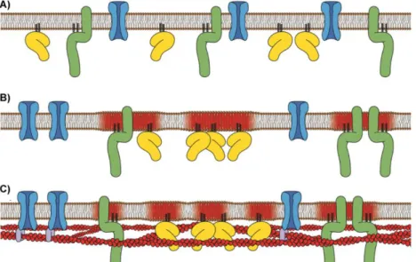
Model prevailed as the most accepted model until the 1970s, however, trans-membrane proteins were posteriorly included in the model as a consequence of new observations by Pinto da Silva and Branton9. In the 1970s, taking into account the already known facts
about membranes and new data from several authors like Cone, Poo and Frye10, 11, Singer and Nicolson proposed the Fluid-Mosaic Membrane (FMM) model (Fig. 2C). In this model, peripheral and integral proteins diffuse freely in an asymmetrical lipid matrix12.
Nevertheless, new techniques and extensive research developed in the area introduced new data that was not satisfied by the FMM original model13. Examples are the restriction
in the lateral movement of lipids and proteins – the picket fence model - described by
Figure 1 - Eukaryotic animal cell structure. Schematic representation of the main organelles of an animal cell.
5 6 3 4 3 8 7 2 1
5
Kusumi and colleagues14, 15, 16, 17, and the evidence of lipid aggregates forming specialized domains, such as the raft domains reported by Simmons and Ikonen18. The use of the
single particle tracking and single fluorescence molecule video enabled Kusumi and other authors to observe that protein membrane movement is not free due to structural membrane constraints 15, 17, 19. The movement restriction occurs through the anchoring
of transmembrane proteins to the cell cytoskeleton, forming fences that corral membrane proteins– ‘fence model’19, 20. Moreover, lipids have also their movement
limited by the compartments formed through the association between protein intracellular domains and the membrane skeleton network- ‘anchored protein picket model’, as evidenced by the works of Speroto and Mouritsen, and Fujiwara et al. 21, 22. In
addition, the formation of membrane domains can also be influenced by the cytoskeleton meshwork20, suggesting that this membrane compartmentalization could modulate cell signaling by restricting a signaling complex or specific molecules to a defined membrane compartment17.
The actual FMM model was altered in order to take into account the new data. Nowadays it includes in the membrane structure: aggregates of lipids forming domains (see section 7 for further details about membrane domains)18, protein/glycoprotein complexes23, membrane associated cytoskeletal fences19, 24, 25 and extracellular matrix structures (e.g.
collagen) (Fig. 2D). The new structures actively influence the macrostructure, dynamics and function of the biomembranes, also restricting the lateral diffusion and range of movement of membrane components13, 26. Nonetheless, there are still situations where
the FMM model does not fit properly, as in the higher levels of organization where membrane crowding and specialized domains formation are rather important structural factors. However these were not taken into account by most membrane models13.
6
A Schematic representation of the Tri-layer model. B Representation of the Unit Membrane Model. C and D Original and actual representation of the Fluid Mosaic Membrane model, adapted from13
1.3 Chemical composition of membranes
As mentioned, cellular membranes are complex structures formed by different proteins and lipids that can be conjugated with different carbohydrates.
1.3.1 Carbohydrates
These are the most abundant molecules on Earth. Chemically these compounds are polyhydroxy aldehydes, ketones, or substances that upon hydrolysis yield such compounds. There are three main classes of carbohydrates: monosaccharides (a single aldehyde or ketone unit), oligosaccharides (2 to 19 units of monosaccharides) and polysaccharides (with 20 or more units of monosaccharides). In the membranes, three types of carbohydrates are found as glycoconjugates, i.e., linked to a protein or lipid. In the plasma membrane, the carbohydrates are normally, if not exclusively, facing the external side of the membrane27.
1.3.2 Proteins
Membrane proteins are distributed either in the surface (peripheral) or immersed (integral) in the membrane matrix2.
Integral proteins are embedded in the membrane bilayer. The portion of the protein that is inside the membrane is enriched in hydrophobic amino acids (e.g. leucine, valine, etc.).
Figure 2 - Evolution of membrane models.
A
B
D
C
7
These proteins can have different number of loops, orientations (dependent on the primary structure), and size of the globular domain in contact with the aqueous environment (which can contact with one or both sides of the membrane). These proteins have distinct functions. One example is the formation of channels that are involved in the control of ions or molecules exchange with the exterior (ex. aquaporins)2. It is important
to enhance that these proteins are not rigid structures, their position and conformation are modulated by other proteins and/or lipids. In fact, lipids can directly influence the activity of a protein either by changing membrane fluidity28,29 or acting as co-factors30.
In contrast, peripheric proteins are attached to the surface of the cell membrane. Their removal does not disrupt the structure of the lipid bilayer. The membrane-protein interaction can be established directly with the membrane mediated by a covalent link with lipid anchors (such as Phosphatidylinositol 4,5-bisphosphate, PIP2) or with integral proteins2.
1.3.3. Lipids
Hundreds of different lipid molecules were identified in cell membranes. According to their structure lipids can be categorized into 3 main classes namely, glycerolipids, sphingolipids and sterols.
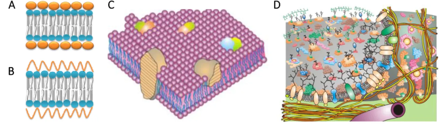
Glycerolipids are characterized by a glycerol molecule with a phosphate esterified at the α-carbon and two long-chain fatty acids esterified to the remaining carbon atoms (Fig. 3)2; examples are phosphatidylcholine (PC) (commonly representing 50% of cell lipids) 31, phosphatidyletanolamine (PE) (constitutes 20% of most membranes), phosphatidylserine (PS) and phosphatidylinositol (PI)31.
Sphingolipids (SLs), which compose 10 % of membrane lipids 32, are characterized by a sphingoid base (in mammals mainly sphingosine and dihydrosphingosine) connected by an amide linkage to a saturated or unsaturated long chain fatty acid (Fig. 3). Various substituents of the hydroxyl group (OH) of C1 of the sphingoid base are known, like phosphocholine2, 31. Ceramide is the simplest SL and constitutes the hydrophobic
backbone of all SLs and their glycosylated derivatives, the glycosphingolipids 33. These are SLs with a sugar moiety linked by a β-glycosidic bound to C1 of the OH group of ceramide, they do not have phosphate and are characterized by presenting mostly saturated acyl
8
chains 2, 34. GSLs can be categorized in sub-groups according to the number, type of sugar residues, and the presence of sialic acid or of sulfur (see Fig. 4). Cerebrosides, such as glucosyl- and galactosylceramide (GalCer), are the simplest GSLs composed solely by uncharged ceramide monohexosides. In opposition, the sulfatides may present different number of sugar residues and derive from either GalCer or GlcCer. However, the common feature within this group of GSLs is the presence of sulfur (Fig. 4). Another important group of GSLs is the globosides that may be further sub-divided into Globo-, iso-Globo-, Lacto/neoLacto- and Ganglio-series. The globosides are the product of the sequential addition of other sugars to lactosylceramide (LacCer), which in turn is formed by the addition of a β-galactose to the glucose of GlcCer. The fourth group is the gangliosides. These are the most complex GSLs in which the hydroxyl group of C1 of ceramide is substituted by an oligosaccharide chain containing hexose and sialic acid or neuraminic acid. Depending on the pH, the gangliosides are either neutral or negatively charged, i.e. amphipathic. A representative member of this group is the monosialotetrahexosylganglioside (GM1) 2, 35.
Sterols are derivatives of cyclopentanoperhydrophenanthrene and are characterized by planar and rigid nucleus (constituted by four fused cycles) with substituents above and below the plane. The most representative member of this category in mammals is Chol (Fig. 3)2 Being the most abundant lipid in the eukaryotic membranes, ranging from 25 to
50 mol % of the total lipid fraction, it plays a crucial role in the physico-chemical properties of cellular membranes36, 37.
In mammalian membranes the most abundant lipids are the glycerolipids, particularly PC lipids, which are responsible for the fluidity of the bulk membrane. Sterols constitute ≥25 % of total lipids, whereas Figure 3 Lipid Classes and Structure.
9
sphingolipids represent 10 % of the total lipids. Both SL and Chol are involved in several cellular processes 32.
2 Membrane lipids and their biological role
It is not possible to separate the evolution of the membrane model concept from the evolution of the role of lipids in the cell.
If at first lipids were thought to be structural components of the membrane, nowadays it is known that lipids have many functions in the cell. Besides from forming the crucial lipid barrier and the matrix of cellular membranes as well as providing the cell the potential for budding, fusion, and fission31, they are also anhydrous reservoirs for efficient storage of caloric reserves and essential for membrane synthesis, wherefatty acid and sterols are needed as components 33. Additionally, some lipids also have bioactive properties acting
as first and second messengers in signal transduction and molecular recognition processes33, 38. Lipids can regulate cellular processes through the modulation of
membrane biophysical properties, which affects protein sorting and conformation31, 33,
38. The properties and functions of SLs and GSLs will be further described in the following
sections.
2.1 Sphingolipids
In the past century an emphasis was made in the study of SLs, since several major signaling lipids were identified in this lipid class, such as ceramides, SM and their glycosylated derivatives. All SLs derive from the same molecule, i.e. ceramide. This lipid is formed by linking a sphingoid backbone via N-acylation to the C-2 of a fatty acid chain of variable length and unsaturation degree. Ceramide is the hydrophobic backbone of all SLs, which places this lipid in the center of SL metabolism. All other SLs are formed by the attachment of different molecules to the terminal hydroxyl of ceramide (see Fig.4). An example is the attachment of a phosphocholine headgroup, yielding SM; whereas addition of a glucose or galactose moiety is the first step in the formation of GSLs. The reason for the existence of almost countless SLs is yet to be clarified, but it is hypothesized that it might be related to a specific role of each of these lipids, or to the necessity of specific combinatorial patterns that will trigger a specific signaling pathway 39 .
10
2.1.2 Ceramides
Ceramidesare central molecules in SL metabolism, in fact, ceramides are a family of different molecules with different acyl chains characterized by different lengths and saturation degree40. Its metabolism is complex, involving more than 28 enzymes where are included: 6 different ceramide synthases (CerS), five ceramidases and at least 5 sphingomyelinases. One of the reasons for such redundancy, namely in the CerS, is the specificity that each enzyme has for different acyl chain lengths40. Commonly, mammal ceramides present acyl moieties with long chains (16 to 24 carbons) and often saturated. However, specific types of cells also express ceramides with very long and unsaturated acyl chains, such as C28 to C32 with 5 to 6 double bonds in adluminal germ cells and spermatozoa 41. Besides being the “hub” of SL metabolism, ceramide has other roles in
the cell, namely as a key lipid in the modulation of several signaling pathways. Ceramide may regulate cell events by changing membrane properties forming specialized domains which affect protein sorting, diffusion, conformation, etc.42; or acting as a second
messenger in signaling cascades43 such as in stress stimuli response 44, 45 or in the tuning of cell processes, i.e. autophagy and apoptosis46, 47.
2.2 Glycosphingolipids
GSLs are the most structurally diverse class of complex SLs. More than 500 different GSLs have been described, the main sugars being glucose, galactose, fucose, N-acetylglucosamine, N-acetylgalactosamine and sialic acid (acidic GSLs) 39. GSL are
commonly composed by a sphingoid base (mainly with 18 to 20 carbons) and a long, mostly saturated amide-linked acyl chain; the structure of the polar headgroup may vary significantly, ranging from one neutral monosaccharide moiety to big assemblies of carbohydrates and sialic acid, which gives the gangliosides their charged nature34. GSLs are normally classified as acidic or neutral48. Due to the presence of the sugar moiety, GSLs are mainly present in the extracellular leaflet of the plasma membrane3. Although
these lipids are minor constituents of the plasma membrane, contributing less than 5% to the total cellular lipid pool49, they have vital roles in the cell acting both as first and
second messengers in several signaling and regulatory pathways50, 51. In addition, GSLs
are important for membrane stability, permeability52 and are active participants in crucial cell processes including cell-to-cell adhesion50, interaction with microbial toxins39,
11
modulating immunity response53, acting as growth factors54, cell differentiation55, etc. Lipid glycosylation is also associated with the development of pathologies, namely cancer
54. The vital nature of GSLs was first identified when null mice for glucosylceramide
synthase (GCS), the first enzyme in GSL biosynthesis, died in the embryonic stage 56. Other characteristic of GSLs is that their profile changes with the age of the cell and/or organism and also with the presence of a pathological state57, 58, 59, 60.
The de novo synthesis of GSLs begins with the formation of GlcCer or GalCer, by the addition of a glucose or galactose moiety, respectively, to the ceramide backbone. Complex GSLs are further synthesized by the consecutive addition of sugar moieties, in the luminal side of the Golgi membrane, by glucosyltranferases, syalitransferases, GalNac transferases and GalCer sulfotransferases (Fig. 4). GalCer is the major precursor of sulfatides, whereas GlcCer is the major precursor of complex GSLs. After their synthesis the majority of GSLs are carried by vesicular transport to the plasma membrane, where they reside and accomplish their functions49, 61. The GSLs are afterwards recycled, passing from the PM to the early endosomes and then to the late endosomes/lysosomes, where these molecules are degraded51.
Since this Thesis is centered in the biophysical properties of GlcCer, the next section will focus on the description of GlcCer synthesis, trafficking, topology and metabolism.
12
Glycosphingolipids is a lipid class with more than 500 molecules. (A) The glycosylation of ceramide forms GalCer or GlcCer (different only by the position of one hydroxyl group in the sugar headgroup). The addition of more carbohydrates forms the complex GSLs such as gangliosides. (B) Descriptive metabolism of gangliosides. Adapted from62
Figure 4 - Complexity of GSL metabolism
B
A
Globosides
Gangliosides, see Fig. 4B Sulfatide
13
2.3 Glucosylceramide
2.3.1 Structure, metabolism and topology.
GlcCer or β-D-glucosyl-ceramide is the most abundant basic structure in GSLs and it is
ubiquitous in mammalian tissues. GlcCer is formed in the cytosolic leaflet of Golgi apparatus by the glycosylation of ceramide via GCS, also known as uracil-diphosphate (UDP)-Glucose ceramide glucosyltransferase. GCS is a 45KDa type III membrane bound protein located in the cytosolic leaflet of cis-Golgi63,64. In mammals, the ceramide
backbone of GlcCer has commonly an acyl chain length between 16 and 26 carbon atoms, however ultralong-chain hydroxyl fatty acids with up to 36 carbon atoms are present in the epidermis62.
It is worthy to mention that GlcCer is the only GSL that is synthesized in the cytosolic leaflet of the Golgi apparatus, having its carbohydrate moiety extended into the cytosol
65, 66. After its synthesis, GlcCer is transported to the luminal side of the Golgi, where it is
converted into more complex GSLs by two proposed pathways. According to one model, GlcCer is transported from the Golgi to the ER by 4-phosphate adaptor protein-2 (FAPP2) and is flipped to the luminal side by low-specificity phospholipid flippases. From the ER GlcCer returns to the Golgi by vesicular transport. The second model also involves FAPP2, however, in this hypothesis FAPP2 transports GlcCer from the cis-Golgi to the trans-Golgi network (TGN), to be flipped by an unidentified protein into the luminal side of the Golgi67. In addition, GlcCer can be carried to the cytoplasmic leaflet of the PM via a nonvesicular-transport that involves a glycolipid transfer protein (GLTP) and FAPP265, 67.
In fact, 45% of all synthesized GlcCer is located in the PM61. In this organelle, half of GlcCer remains in the cytoplasmic leaflet and the rest is translocated to the extracellular leaflet for surface expression68.
14
Structural representation of (a) C16-GlcCer and (b) C16-GalCer. The polar head is composed, respectively, by a glucose or galactose residue attached to the hydrophobic backbone formed by ceramide (adapted from 69).
Today is accepted that GlcCer should be found in the PM and in the Golgi apparatus (in the cytosolic face). However, evidence shows that GlcCer is present in other organelles, like ER where, potentially, it can be flipped to the luminal side or degraded67. In addition,
studies made with GlcCer analogues support that this GSL can be internalized from the PM by either endocytic and non-endocytic (transbilayer movement - “flip-flop”) pathways and transported to intracellular membranes, such as the nuclear membrane, or transported to the PM of other cells 70. Furthermore, GlcCer is also internalized from the PM and transported to the lysosome lumen, via endosomal pathway for degradation. GlcCer is mainly catabolized by the complementary action of an acid β-glucosidase (GCase), a modulator protein (Saposin C) and anionic lipids 71. GCase is recruited to the
lysosome by LIMP-2 (lysosome mempbrane protein-2) 72, and in the late
endosome/lysosome, due to the drop in the pH GCase, dissociates from LIMP-2 to associate instead with Saposin C (SAP-C)72. SAP-C destabilizes the lipid membrane
exposing GlcCer and also directly activates GCase in an allosteric manner, enhancing GCase activity and promoting GlcCer degradation71. Furthermore, the presence of anionic lipids, such as phosphatidylserine and phosphatidylglicerol, in the membrane site where the complex of SAP-C and GCase are anchored substantially increases GlcCer cleavage by GCase71. Although, the lysosomal route is the mainly studied, GlcCer can also be hydrolyzed in extralysosomal locations through the activity of β-glucosidase 2 (GBA2)65,67
or of a neutral β-glycosidase (GBA3)73. GBA2 is a peripheral protein that catalyzes GlcCer degradation in the ER, in the Golgi 67 and in the PM74. The highest expression of this
glucosidase is observed in the brain and testis and, in resemblance to GCase, it requires a co-factor and/or to be associated with the membrane to have its activity optimized.