INSTITUTO DE INVESTIGAÇÃO E FORMAÇÃO AVANÇADA
ÉVORA, Setembro 2014 ORIENTADORES:Doutora Rita de Sousa Professor David H. Walker Professora Manuela Vilhena
Tese apresentada à Universidade de Évora
para obtenção do Grau de Doutor em Ciências Veterinárias
Natacha Alexandra Korni da Fonseca Milhano
Insight into the underlying immune
interactions of Rickettsia infection in the
vector-pathogen-host interface
INSTITUTO DE INVESTIGAÇÃO E FORMAÇÃO AVANÇADA
ÉVORA, Setembro 2014 ORIENTADORES:Doutora Rita de Sousa Professor David H. Walker Professora Manuela Vilhena Nome do Professor
Tese apresentada à Universidade de Évora
para obtenção do Grau de Doutor em Ciências Veterinárias
Natacha Alexandra Korni da Fonseca Milhano
Insight into the underlying immune
interactions of Rickettsia infection in the
vector-pathogen-host interface
iii
The research work for this dissertation was performed at the Center for Vectors and Infectious Diseases Research, National Institute of Health (CEVDI/INSA), Lisbon, Portugal, and at the Department of Pathology, University of Texas Medical Branch, Galveston, Texas, USA.
The project was financed by the fellowship SFRH/BD/45174/2008 granted by the Fundação para a Ciência e Tecnologia, Lisbon, Portugal.
v
This incredible life experience would not have been possible, conceivable, or made sense without the presence and constant support of many, many valuable beyond words people who, in one way or another, contributed immeasurably towards spawning this thesis into existence.
First and foremost, I would like to thank my supervisors, Dr. Rita de Sousa, Dr. David Walker and Prof. Manuela Vilhena.
Dr. Rita de Sousa, for initiating me into this fascinating rickettsial world and sharing her vast knowledge with me, for her unconditional support, patience and vivaciousness at all times, for always readily giving me very sound advice on various lab dilemmas, and also a harsh word when needed, in a sentence, for bearing with me for so long -Thank you. Dr. David Walker, for accepting to become my supervisor and opening his lab to me, for his invaluable insights regarding this project, for sharing his thoughts and wisdom on Rickettsiology during our many meetings where ideas flowed, suggestions were made, and experiments designed, for always having a kind word, it was a pleasure and a great honour to work with you.
To Prof. Manuela Vilhena, who has been limitless in her support and advice on many topics, and for always putting my mind at ease at stressing moments.
To Sherrill, for always knowing what to do in all situations and for being such a jolly person, spreading happiness wherever she goes.
To Dr. Donald Bouyer, for all his advice in many situations, and constant support when needed.
To Dr. Vsevolod Popov, for teaching me all I know about EM.
To all the lab members of the Walker and Bouyer lab at UTMB, for helping me so generously in many facets of my work, in particular Dr. Tais Saito, who helped me tremendously throughout my stay in the lab, and with whom I learned a whole world more about ticks than I could ever expect, Dr. Rong Fang, for her invaluable insights into immunology, and Jeremy, thank you so much for everything.
I would also like to thank Dr. Francisco Alarcon-Chaidez, who started me out in the direction of dissecting ticks and all analyses thereafter, with great friendship.
vi
To all lab members in Centro de Estudos de Vectores e Doenças Infecciosas, with a special thanks to my close bunker colleagues, who have always made the daily toil incredible. To my special Patricias and Catarina for being awesome friends.
To my unique Galveston family at the Yellow house, without whom life will never be the same again.
To my mother, who has been my light throughout. To all present and gone - Thank you.
I would also like to extend my acknowledgements to the institutions where the research towards the completion of this dissertation was performed, namely Instituto Nacional de Saúde Dr. Ricardo Jorge, Lisbon, Portugal, and the Department of Pathology, University of Texas Medical Branch, Galveston, Texas, USA.
Finally, I am extremely grateful to Fundação para a Ciência e Tecnologia for awarding me a PhD fellowship which allowed this work to come to fruition.
vii
Ixodid ticks are second only to mosquitos in their notorious role as vectors of pathogens to both animals and humans. Rickettsioses are among the most important tick-borne diseases in Europe, Mediterranean spotted fever in particular. To date, many studies have been performed in order to uncover the underlying mechanisms of this disease, in terms of the interactions among its constituents, i.e., the pathogen, Rickettsia conorii, its vector,
Rhipicephalus sanguineus tick, and a vertebrate host. However, important gaps remain in this knowledge, among them studies of the relationship of its vector and pathogen, and also the role of tick saliva in vector-host interactions. Thus, in order to approach these limitations, in the first part of this dissertation three studies were performed in order to analyze the vector-pathogen-host interface in natural settings (chapter 2). As a result of the first of these studies, co-infections of Borrelia lusitaniae with R. helvetica and R. slovaca were found in ticks collected from a natural safari park in the south of Portugal; new host-pathogen associations were found in the second study described, performed in Madeira Island, namely lizards infected with R. monacensis, as well as detection of R. helvetica in ticks, which was a first occurrence in this island; and in the last study a new species of Rickettsia was isolated from soft ticks collected from pig pens from Alentejo, Portugal. In chapter 3 experimental studies on the vector were performed through analysis of R. massiliae in Rh. sanguineus tick organs. We performed a quantitative analysis of R. massiliae in the salivary glands of feeding Rh. sanguineus ticks, and observed a statistically significant increase in bacterial load during the first two days, followed by a plateau up to day 6 of feeding. An ultrastructural study was also performed on the salivary glands, ovaries and midgut of R. massiliae infected-Rh. sanguineus, where we observed the reactivation phenomenon of Rickettsia in the salivary glands of fed ticks as a result of tick feeding. In the final part of this dissertation the role of tick saliva was ascertained in terms
viii
of bacterial burden and immune responses in a murine susceptible host, using uninfected
Rh. sanguineus ticks and C3H/HeJ mice. No statistically significant differences in bacterial load were observed between the two groups of R. conorii-infected animals, one of which infested with ticks. However, host cytokine analysis of both groups of animals revealed statistically significant differences, suggesting an inhibitory effect of tick saliva on host pro-inflammatory responses (chapter 4).
ix
Rickettsia spp.
Os ixodídeos desempenham um papel fundamental como vectores de agentes patogénicos tanto em animais como em humanos. As rickettsioses, com destaque para a febre escaro-nodular, encontram-se entre as doenças transmitidas por carraças mais importantes na Europa. Até à data muitos estudos foram efectuados de modo a descortinar os mecanismos subjacentes a esta doença, em termos das interacções entre os seus constituintes, i.e., agente patogénico, Rickettsia conorii, o seu vector, o ixodídeo Rhipicephalus sanguineus, e um hospedeiro vertebrado. Todavia, existem ainda lacunas neste conhecimento a nível da relação vector-agente patogénico, para além do papel da saliva do vector nas interacções agente patogénico-hospedeiro. Assim, numa tentativa de colmatar estas limitações, na primeira parte desta dissertação foram realizados três estudos para análise dos fenómenos que ocorrem naturalmente na interface vector-agente patogénico-hospedeiro ocorrentes na natureza (capítulo 2). Como resultado do primeiro destes estudos, foram encontradas co-infecções de Borrelia lusitaniae com R. helvetica e R. slovaca em ixodídeos capturados num parque safari no sul de Portugal. No segundo estudo descrito, efectuado na ilha da Madeira, foram encontradas novas associações hospedeiro-agente patogénico, nomeadamente lagartixas infectadas com R. monacensis, detectou-se R. helvetica em carraças, pela primeira vez nesta ilha, e foi possível isolar uma nova espécie de Rickettsia
a partir de carraças de corpo mole capturados em pocilgas no Alentejo, Portugal. O capítulo 3 aborda os resultados de estudos experimentais efetuados no vector, através da análise de R. massiliae em orgãos de Rh. sanguineus. Realizámos uma análise quantitativa da R. massiliae em glândulas salivares de Rh. sanguineus durante a sua refeição sanguínea, tendo-se observado um aumento estatisticamente significativo da carga bacteriana nos
x
dois primeiros dias de alimentação, seguido de um patamar até ao dia 6 da alimentação sanguínea. Foi também efectuado um estudo ultraestrutural nas glândulas salivares, ovários e intestino médio de Rh. sanguineus infectados com R. massiliae, onde observámos o fenómeno de reactivação da Rickettsia nas glândulas salivares dos ixodídeos alimentados, resultante do processo de alimentação. Na parte final desta dissertação analisámos o papel da saliva de ixodídeos em termos da carga bacteriana e respostas imunes num hospedeiro murino susceptível, usando Rh. sanguineus não infectados e ratinhos C3H/HeJ. Não verificámos diferenças estatisticamente significativas entre as cargas bacterianas de dois grupos de animais infectados com R. conorii, em que apenas um dos grupos estava infestado com carraças. Todavia, a análise de citoquinas no hospedeiro em ambos os grupos experimentais revelou diferenças estatisticamente significativas, sugerindo um efeito inibitório da saliva dos ixodídeos nas respostas pro-inflamatórias do hospedeiro (capítulo 4).
xi Fig.1.1. Fig.1.2. Fig.1.3. Fig.1.4. Fig.1.5. Fig.1.6. Fig.1.7. Fig.1.8. Fig.1.9. Fig.1.10. Fig.2.1. Fig.2.2. Fig.3.1. Fig.3.2. Fig.3.3. Fig.3.4. Fig.3.5.
Hard and soft tick morphology
Relative sizes of four different species of ticks in three life stages: larvae, nymphs and adults
Life cycle of ixodid ticks
World distribution of Rh. sanguineus
Rhipicephalus sanguineus salivary glands (SG)
Diagrams illustrating (A) General view of salivary gland; (B) Agranular acinus; (C) Granular acinus
Ixodid tick saliva constituents modulate host defence responses (itch, pain, haemostasis, inflammation, immune reactions)
Electron micrograph of binary fission of Rickettsia sp. World distribution of MSF
Life cycle of tickborne rickettsiae Lizards and tick stages found in Calheta
Molecular phylogenetic analysis of R. lusitaniae sp. nov. isolated from the tick O. erraticus from Portugal. A total of 3003 aligned sequences corresponding to gltA, htr, ompA, sca1 and ompB genes (820, 394, 488, 483, 818 nt, respectively) were concatenated and subjected to maximum-parsimony inference analysis. Bootstrap values are shown at the nodes. Bar, 10 substitutions.
2D gels of unfed versus fed adult female Rh. sanguineus ticks.
Real time qPCR of rickettsial citrate synthase (Cs) gene per tick salivary gland
Electron micrographs of R. massiliae (arrow) in unfed Rh. sanguineus
salivary
g
landElectron micrographs of R. massiliae (arrows) in fed Rh. sanguineus
salivary glands. (A) R. massiliae in interstitial cells.(B) Detail of interstitial cell showing numerous Rickettsia and mitochondria. (C) Binary fission of Rickettsia in interstitial cell. (D) R. massiliae amongst secretory vesicles
Electron micrographs of R. massiliae (arrows) and Wolbachia (bold arrow) in the ovaries of unfed Rh. sanguineus. (A) R. massiliae in
6 7 8 10 11 12 14 20 23 26 55 66 77 86 87 88
xii Fig.3.6.
Fig.4.1.
Fig.4.2.
Fig.4.3.
cytoplasm of oocyte. (B) Wolbachia in the cytoplasm of oocyte. Bar 1
m
Electron micrographs of R. massiliae in the midgut of unfed Rh. sanguineus
Real time qPCR of rickettsial citrate synthase (Cs) gene. RC/tick infestation- Group of C3H/HeJ mice infested with ticks and inoculated intradermally with R. conorii ISF; RC- Group of C3H/HeJ mice inoculated intradermally with R. conorii ISF, no tick infestation
Reverse transcriptase qPCR of mRNA relative ratios (2-𝝙𝝙Ct) of IL-1,
IL-10 and NF-B. * p≤0.05.
Photomicrograph of lung of mouse inoculated intradermally with R. conorii adjacent to feeding Rh. sanguineus ticks. Mild multifocal perivascular and alveolar septal thickening by interstitial infiltration by mononuclear cells. 20 x. Hematoxylin-eosin stain
90
91
105
106
xiii Table 1.2. Table 1.3. Table 1.4. Table 1.5. Table 1.6. Table 2.1. Table 2.2. Table 2.3. Table 2.4. Table 2.5.
Genomic and proteomic studies Tick-borne diseases of humans
Main spotted fever group Rickettsia
Rates of infection and transovarial transmission
Cytokines and chemokines produced during rickettsial infections Prevalence and isolates of Rickettsia spp and Borrelia lusitaniae
from questing ticks of a Safari Park, Portugal
Rickettsia isolates, culture conditions and GenBank accession numbers
Prevalence of Borrelia burgdorferi sensu lato, Rickettsia spp., and
Anaplasma phagocytophilum infection in lizard tissue samples and ticks collected from lizards
PCR detection of Rickettsia sp. in O. erraticus pools from Portugal in 2009 and 2010.
Accession numbers of Rickettsia species used in the phylogenetic analysis 15 18 22 25 30 45 47 56 64 67
xiv LIST OF ABBREVIATIONS
ANOVA Analysis of variance CD Cluster of differentiation
CEVDI Centro de Estudos de Vectores e Doenças Infecciosas Dr. Francisco Cambournac
CTL Cytotoxic T lymphocytes DCs Dendritic cells
DEBONEL Dermacentor-borne necrosis erythema lymphadenopathy DNA Deoxyribonucleic acid
gltA Citrate synthase
ID Intradermal
IFA Immunofluorescent assay
IFN Interferon
IGS Intergenic spacer
IL Interleukin
INSA Instituto Nacional de Saude Dr. Ricardo Jorge ISF Israeli spotted fever
ha Hectares
htr Rickettsial 17KDa protein gene
LB Lyme borreliosis
LPS Lipopolysaccharide mRNA Messenger ribonucleic acid MSF Mediterranean spotted fever NF-kB Nuclear factor
NK Natural killer
NTC Non-template control
NO Nitric oxide
OmpA Outer membrane protein A OmpB Outer membrane protein B
xv PCR Polymerase chain reaction
PI Phosphoinisitide
PTK Protein tyrosine kinase
qPCR Quantitative polymerase chain reaction
RC Rickettsia conorii
RMSF Rocky Mountain spotted fever rrs 16S ribosomal ribonucleic acid
RT-PCR Reverse transcriptase polymerase chain reaction Sca Surface cell antigen
SFG Spotted fever group SGE Salivary gland extracts s.l. sensu lato
s.s. sensu stricto sp. nov. Species nova
TEM Transmission electron microscopy
TG Typhus group
Th1/2 T helper 1 and 2 cells TIBOLA Tickborne lymphadenitis TNF Tumor necrosis factor TLR Toll-like receptor
xvi TABLE OF CONTENTS Author notes Acknowledgements Abstract Resumo List of figures List of tables List of abbreviations iii v vii ix xi xiii xiv Chapter 1 1.1. 1.2. 1.2.1. 1.2.2. 1.2.3. 1.2.3.1. 1.2.4. 1.3. 1.3.1. 1.3.2. 1.3.2.1 1.3.3. 1.3.4. 1.4. 1.5.
Literature review and aims Introduction
Ticks
Tick classification Tick morphology
Ixodid tick life cycle and feeding behavior
Rhipicephalus sanguineus tick
Ixodid tick salivary glands and role of tick saliva Ixodid ticks as vectors of disease
Rickettsia morphology
Rickettsia classification Mediterranean spotted fever Ecology of Rickettsia
Rickettsial pathogenesis and host immune response Animal models of rickettsial infection
Aims 1 3 3 3 5 6 10 11 17 20 20 22 24 27 31 32 Chapter 2 2.1. 2.2.
Rickettsial diversity in Portugal Introduction
Co-infections of Rickettsia slovaca and Rickettsia helvetica with
Borrelia lusitaniae in ticks collected in a safari park, Portugal Abstract
35 39 39 39
xvii 2.3.
2.4.
Results Discussion
The role of Teira dugesii lizard as potential host for Ixodes ricinus tick-borne pathogens
Abstract Introduction
Rickettsia lusitaniae sp. nov. isolated from the soft tick
Ornithodoros erraticus (Acarina:Argasidae) Abstract
Introduction
Materials and methods Results Discussion 44 48 54 54 54 60 60 60 61 64 67 Chapter 3 3.1. 3.2. 3.2.1. 3.2.2. 3.3.
Studies in the vector Introduction
Preliminary studies
Proteomic analysis of Rh. sanguineus salivary glands Infection of Rh. sanguineus ticks with R. conorii ISF
A quantitative study of Rickettsia massiliae dissemination in
Rhipicephalus sanguineus organs Abstract
Introduction
Materials and methods Results Discussion 71 75 75 75 78 79 79 80 81 85 91 Chapter 4 4.1 4.2.
Studies in the host Introduction
The role of Rhipicephalus sanguineus saliva in the dissemination of Rickettsia conorii in C3H/HeJ mice
Abstract Introduction 95 99 99 99 100
xviii Materials and methods
Results Discussion 101 105 107 Chapter 5 References General discussion 111 123
___________________________________________________CHAPTER 1
LITERATURE REVIEW AND AIMS
3 1. 1. Introduction
Ticks are obligate hematophagous ectoparasitic arthropods parasitizing all vertebrate classes, at some stage of their life cycle (Sonenshine 1991). Not only can they damage human and animal skin, but they also play a pivotal role in transmitting zoonotic pathogens. In fact, ticks are second only to mosquitos in their importance as vectors of human pathogens, including bacteria, viruses and protozoa, and represent the most critical group of arthropods capable of transferring pathogens between animal species (Balashov 1972). Their biomedical importance and significant impact on the economy, resulting from massive losses in the cattle industry, have been paramount in the increasing interest in the study of these arthropods (Paddock and Telford 2010).
1.2. Ticks
1.2.1. Tick classification
The first reports on tick phylogenetic trees appear to have been made by Hoogstraal and Aeschlimann (1982), inferred from intuition on relative ‘primitiveness’ of tick morphology, life cycles and of their hosts. However, preliminary hypotheses about evolutionary relationships of ticks had already been suggested long before (Pomerantsev 1948, Camicas and Morel 1977). It was only later, in the 1990s, that molecular characters were taken into account in phylogeny, paving the way to an ever increasing number of papers on molecular phylogeny and evolution of ticks (Wesson and Collins 1992, Black and Piesman 1994, Rich et al. 1997, Klompen et al. 2000, among many others).
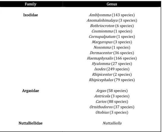
Ticks belong to the class Arachnida, subclass Acari, order Parasitiformes and suborder Ixodida (Sonenshine 1991), and are divided into three families: Ixodidae, or hard ticks, containing 720 species, Argasidae, or soft body ticks, comprising 186 species, and Nutalliellidae, represented by a single species confined to southern Africa (Table 1.1).
4
Table 1.1. Taxonomy within the order Ixodidae (adapted from Barker and Murrell 2008)
Family Genus Ixodidae Argasidae Nuttalliellidae Amblyomma (143 species) Anomalohimalaya (3 species) Bothriocroton (6 species) Cosmiomma (1 species) Cornupalpatum (1 species) Margaropus (3 species) Nosomma (1 species) Dermacentor (36 species) Haemaphysalis (166 species) Hyalomma (27 species) Ixodes (249 species) Rhipicentor (2 species) Rhipicephalus (79 species) Argas (58 species) Antricola (3 species) Carios (88 species) Ornithodoros (37 species) Otobius (3 species) Nuttalliella
The family Ixodidae comprises two major groups, the Prostriata and the Metastriata, mainly distinguished based upon their mating habits. The reproductive strategy of the Metastriate group is closely linked to their feeding, i.e., they only mate while feeding on the host, and the Prostriata mate as readily on or off the host (Sonenshine 1991). The Prostriata include 249 species in a single genus Ixodes, whereas the Metastriata are divided into four subfamilies: the Amblyomminae, including the genus Amblyomma; the Hyalomminae, including the genus Hyalomma; Haemaphysalinae, including the genus
Haemaphysalis; and the Rhipicephalinae, including the genera Dermacentor, Cosmiomma,
Margaropus, Nosomma, Anomalohimalaya, Rhipicentor and Rhipicephalus (Mullen and Durden 2002).
Argasidae are divided in two main lineages, the subfamilies Argasinae, which include the genera Argas and Antricola, and Ornithodorinae, including genera Carios, Ornithodoros
5 1.2.2. Tick morphology
Ticks are structured in two parts which are fused together: the capitulum, or gnathosoma, where the mouthparts are included, and the body, or idiosoma, which is formed by the fusion of the podosoma and opisthosoma. The podosoma contains the legs and genital pore and the opisthosoma, located on the posterior region from the leg coxae, the spiracles and the anal aperture (Sonenshine 1991). The mouthparts contain the segmented palps, the basis capituli, from which the segmented chelicerae extend, and the hypostome (Anderson and Magnarelli 2008). It is the movement of both chelicerae, resulting in the rip and tear of the host’s skin, that allows for the formation of a blood pool from which the tick then sucks the blood using the food canal in its hypostome (Sonenshine 1991).
Ixodid ticks possess a very distinct external morphology from that of Argasid ticks, in that a hard sclerotized plate, or scutum, covers the entire body in male ixodid ticks, and the dorsal anterior part of females and immature forms (Fig.1.1A). The synthesis of new alloscutum, the folded cuticle posterior to the anterior dorsal body in females, nymphs and larvae, allows their expansion upon feeding, which in females can reach up to 100 times their unfed size, depending on species (Sonenshine 1991). As a result, upon full engorgement, the scutum occupies a very small percentage on the anterior region of the largely extended body (Fig.1.1B). Males, on the other hand, are limited in their physical expansion when feeding due to the complete coverage of the idiosoma by the scutum (Fig.1.1A). Immature forms resemble adults, except they lack the external genital pores, porose areas and foveal pore clusters present in adults. In addition, larvae possess only three pairs of legs, whereas nymphs and adults possess four (Sonenshine 1991).
6
Fig.1.1. Hard and soft tick morphology. (A) Dorsal-lateral view of male (below) and female (above)
Dermacentor marginatus. (B) Engorged female Ixodes sp. (C) Dorsal view of argasid tick. Bar 1.5mm.
Argasid ticks possess a leathery and flexible cuticle, with no scutum (Fig.1.1C). Males and females are distinguished only by their genital pore. Their capitulum is located in a subterminal position, with the exception of larvae, where it is positioned anteriorly, and no dorsal shield is present (Anderson and Magnarelli 2008). In contrast to ixodid ticks, argasids do not swell immeasurably during feeding, extensive stretching does occur, owing to the folded nature of the integument; however, no additional growth takes place (Sonenshine 1991). The nymphal instar number may vary from 2-8, depending on bloodmeal sizes in preceding instars, and adults have multiple gonadotrophic cycles, as they can feed repeatedly with females laying small eggs masses after each feeding (Sonenshine 1991).
1.2.3. Ixodid tick life cycle and feeding behaviour
The ixodid tick life cycle includes four stages, namely, the embryonated egg, larva, nymphs and adults (Fig.1.2).
Scutum Legs Palp Capitulum Body A C B
7
Fig.1.2. Relative sizes of four different species of ticks in three life stages: larvae, nymphs and adults (courtesy of Mary Predny from Virginia Cooperative Extension Publication).
The majority of ixodid species require a blood meal in order for the immature stages, i.e., larvae and nymphs, to transition to the subsequent development stage, the same being true of adults for reproduction (Sonenshine 1993).
The life cycle of ixodid ticks is classified according to the number of times the development stages change hosts, and whether or not immature stages molt on their host, thus giving rise to one-host, two-host or three-host ticks (Balashov 1972, Sonenshine 1993). In general, larvae remain in areas of high humidity, usually close to the ground, where they quest for small mammals, on which they feed and drop off (or stay on, in one or two host tick cases) once engorged. After molting into nymphs the cycle begins again until they engorge and drop off (or stay on, in one host ticks). Engorged larvae and nymphs weigh about 10 to 20 times their unfed weight. Once nymphs molt into adults, they will then seek, feed and mate on their hosts, although mating may also occur on vegetation, a less
8
common occurrence, after which the engorged females drop off and lay eggs (Fig.1.3). Engorged females can weigh up to 100 times their unfed weight (Sauer et al. 1995, Goodman et al. 2005).
Fig.1.3. Life cycle of ixodid ticks (adapted from www.wwhd.org).
Depending on the species, females can lay up to 3000 eggs or more, after which they dies. The males can feed more than once, and remain on the host seeking to mate with other females. Sexual dimorphism only exists in the adult stage. The time period between feeding periods can range from weeks to several months. The complete life cycle of a tick depends on the host availability and microenvironment, i.e., temperature and humidity, and it may take between one and two years, although some species can go live to six years (Anderson and Magnarelli 2008).
Ticks depend on bloodmeals for their survival, molting and reproduction. According to Anderson and Magnarelli (2008), a successful bloodmeal depends on a series of sequential
99999
9999999
99999
9
steps, which they divided as appetence, engagement, exploration, penetration, attachment, ingestion, engorgement, detachment and disengagement. The main host seeking pattern ixodid ticks adopt is the ambush strategy, where ticks crawl onto vegetation and wait for unsuspecting hosts to pass by (Sonenshine 1993, Goodman et al. 2005). Once this occurs, host stimuli such as odor, carbon dioxide, heat and vibration trigger the tick to then cling on to the host, be it hair, fur, or cloth. Once the attachment site is selected, usually in sheltered locations of the host’s body, a process aided by tactile stimuli, the tick prepares itself by placing its body at an angle relative to the host’s and uses its chelicerae to start cutting through the epidermis and dermis, inserting its hypostome into the lesion. Cement secretion in the tick saliva occurs soon afterwards, pooling around the mouthparts and quickly hardening, thus securing the tick firmly onto the feeding site (Anderson and Magnarelli 2008). An alternating pattern of imbibing blood and injecting pharmacologically active ingredients through tick saliva then takes place, and occurs throughout the whole bloodmeal. Ixodid ticks, in contrast to argasid ticks, are slow feeders. They usually remain attached for an extended period of time, ranging from a few days to a couple of weeks, depending on their life cycle and species involved. In general larvae can feed from 2 to 4 days, nymphs up to 8 days, and adults can feed for as long as two weeks. The prolonged period of the bloodmeal is required for the production of new cuticle which permits the progressively increasing size of the tick’s body while feeding (Anderson and Magnarelli 2008). Once engorged, the ticks drop off and seek a safe haven for the molting process. Unfed ticks are able to survive long periods without feeding due to their ability to absorb water from unsaturated air (Needham and Teel 1991).
10 1.2.3.1. Rhipicephalus sanguineus tick
Rhipicephalus sanguineus, also known as the brown dog tick, is the most geographically widespread tick species in the world, particularly in tropical and subtropical regions (Fig. 1.4).
Fig.1.4. World distribution of Rh. sanguineus (Latreille, 1806) (Kolonin 2009)
It was originally described by Latreille (1806) as Ixodes sanguineus, and was later placed in the genus Rhipicephalus (Koch 1884). Many attempts to classify the species have since then been undertaken, and the taxonomic debate remains to this day (Camicas et al. 1998, Szabo et al. 2005, Dantas-Torres et al. 2013).
Rhipicephalus sanguineus ticks feed primarily on dogs, but are also known to parasitize a wide range of wild and domestic animals, including cats, rodents, birds, and also humans (Dantas-Torres 2010). Rhipicephalus sanguineus play a fundamental role in veterinary and human medicine, as it is a known vector and reservoir of many pathogens, including
11
Rickettsia, Ehrlichia, Babesia, and Coxiella burnetii (Dantas-Torres 2008). It exhibits a primarily endophilic nature, but can also survive in outdoor environments, particularly in refuges and areas where dogs are found (Demma et al. 2005, Parola et al. 2008).
Rhipicephalus sanguineus mainly displays a host-seeking behaviour; however, it also adopts the questing behaviour (Dantas-Torres 2010). As a metastriate, this species reaches sexual maturity and mates only while on the host, with females not engorging fully until mating occurs (Dantas-Torres 2010). Rhipicephalus sanguineus is a three-host tick, requiring a new host per feeding stage, upon which larvae typically feed for around 2 days, nymphs for 4-5 days, and females can feed for weeks, depending on host and environmental factors (Koch 1982, Troughton and Levin 2007). The process of oviposition may last several weeks, and the number of eggs laid, varying from 1500-4000, depends on the weight and period of oviposition (Koch 1982).
1.2.4. Ixodid tick salivary glands and role of tick saliva
The salivary glands of ixodid ticks are the largest glands in the tick’s body, and undergo remarkably complex cytological changes necessary to accommodate the tick’s physiological requirements (Bowman et al. 2008). They consist of grape-like clusters of acini, granular and agranular cells, and are localized anterolaterally on both sides of the tick’s body, occupying one third to one half of the hemocoel (Fig. 1.5). Salivary glands are composed of three acinar types in females (I-III) and four in males (I-IV), attached to a main and branching ducts
SG SG
Fig. 1.5. Rhipicephalus sanguineus
12
(Till 1961, Chinery 1965). Type I acini are localized along the main duct, near the anterior end of the salivary gland, whilst the remaining types are found posteriorly along a ramifying system of intralobular ducts (Fig. 1.6). Each acinar type consists of multiple cell types: type I acini are agranular, and possess one central cell type surrounded by pyramidal cells, a constrictor cell and peritubular cells surrounding the acinar duct; type II contain granular ‘a’, ‘b’, ‘c1-c4’, agranular ablumenal interstitial cells and an adlumenal interstitial cell, and type III has only 3 cell types, ‘d’, ‘e’ and ‘f’ (Fig.1.6). Type IV acini are present only in male ticks, containing only one type of secretory cells, type G. They are small in undifferentiated ticks, becoming hypertrophied during feeding and as large or even larger than types II and III acini (Sonenshine 1991).
Fig. 1.6. Diagrams illustrating (A) General view of salivary gland; (B) Agranular acinus; (C)
Granular acinus. SD- Main salivary duct; SGA- agranular salivary gland acini (type I); SGG- granular salivary gland acini (types II, III) (adapted from Sonenshine 1991).
The acini undergo extensive differentiation during tick feeding, with major changes in cellular differentiation.
13
Type I acini are involved in osmoregulation, particularly during the unfed periods of the tick, where the cells secrete highly concentrated solutions which are excreted via salivary ducts onto the hypostome surface (Gill and Walker 1987). Moisture from the atmosphere is then condensed on these hygroscopic salty deposits, which are then imbibed by the tick, keeping it hydrated. Various repetitions of this cycle facilitate the maintenance of the tick’s water balance (Sonenshine 1991). Types II and III granular acini are mainly involved in secretion of bioactive compounds during feeding periods, including cement and anticoagulants. The role of acini type IV in males is not clear, an involvement in the spermatophore transfer to the female genital pore has been suggested, along with observations of intense salivation occurring during this process (Feldman-Muhsam et al.
1970).
After engorgement, the female tick detaches and, after a few days, the salivary glands start to degenerate through a highly regulated process of programmed cell death. The increased levels of ecdysteroids in the salivary glands post-detachment have been associated with the degenerative process of the tick salivary gland (Harris and Kaufman 1985, Lomas et al.
1998).
The salivary glands play a fundamental role both as sites of development and subsequent transmission of infectious agents as well as secretion of bioactive products released into the saliva during the bloodmeal.
Throughout the course of a bloodmeal, ticks remain attached to a host for extended periods of time, ranging from a few days to a couple of weeks, depending on their stage in the life cycle and species involved. A normal host response to the mechanical damage caused by the tick bite would be an immediate activation of hemostasis, preventing blood loss; inflammation, producing itch or pain, thus potentially triggering defensive behavior of the host, and immunity (cellular and humoral) (Francischetti et al. 2010)(Fig.1.7). However, ticks have circumvented these potential problems by developing a wide variety
14
of pharmacologically active molecules in their saliva, which are injected into their host, allowing them to remain essentially undetected during their blood meal, feed successfully and transmit infectious agents (Ribeiro et al. 2006, Wikel 2013). In particular, tick saliva plays vital functions in the immunomodulation by the host via: a) increasing blood flow in the bite site through secretion of vasoactive agents, b) inoculating anticoagulants that keep the host’s blood in the fluid form, c) inhibiting the inflammatory process in the host, and d) immunosuppressing the host and enabling the attachment of ticks, making their rejection by the host difficult (Sauer et al. 2000).
Fig.1.7. Ixodid tick saliva constituents modulate host defence responses (itch, pain, haemostasis, inflammation, immune reactions) (adapted from Kazimirova and Stibraniova 2013).
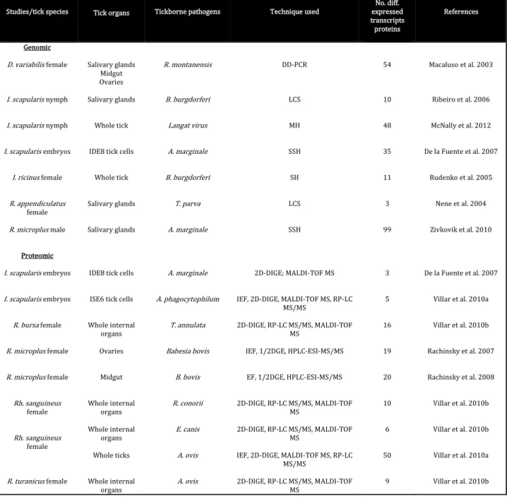
The hundreds of proteins contained in tick saliva are differentially expressed throughout the arthropod’s bloodmeal, stimulated by the continuous tick/host interplay. An increasing amount of studies have been and are being focused on genomic and proteomic
Wound healing Haemostasis Vasoconstriction Platelet aggregation Coagulation Itch, pain Inflammation Innate immunity Acquired immunity Complement Pathogen transmission (SAT)
15
approaches in order to identify and characterize these proteins, and many compounds have now been identified (Table 1.2).
Table 1.2. Genomic and proteomic studies (adapted from Liu and Bonnet 2014).
DD-PCR: differential-display polymerase chain reaction, LCS: cDNA library clones sequencing, MH: microarray hybridization, SH: subtractive hybridization, SSH: suppression-subtractive hybridization; D: dimensional, DIGE: differential in-gel electrophoresis, DGE: dimensional gel electrophoresis, ESI: tandem electrospray, HPLC: high-performance liquid chromatography, IEF: isoelectric focusing, MALDI-TOF: matrix-assisted laser desorption/ionization time-of-flight, MS: mass spectrometry, RPLC: reversed phase liquid chromatography
Studies/tick species Tick organs Tickborne pathogens Technique used
No. diff. expressed transcripts proteins References Genomic D. variabilis female I. scapularis nymph I. scapularis nymph I. scapularis embryos I. ricinus female R. appendiculatus female R. microplus male Proteomic I. scapularis embryos I. scapularis embryos R. bursa female R. microplus female R. microplus female Rh. sanguineus female Rh. sanguineus female R. turanicus female Salivary glands Midgut Ovaries Salivary glands Whole tick
IDE8 tick cells
Whole tick
Salivary glands
Salivary glands
IDE8 tick cells
ISE6 tick cells
Whole internal organs Ovaries Midgut Whole internal organs Whole internal organs Whole ticks Whole internal organs R. montanensis B. burgdorferi Langat virus A. marginale B. burgdorferi T. parva A. marginale A. marginale A. phagocytophilum T. annulata Babesia bovis B. bovis R. conorii E. canis A. ovis A. ovis DD-PCR LCS MH SSH SH LCS SSH 2D-DIGE; MALDI-TOF MS
IEF, 2D-DIGE, MALDI-TOF MS, RP-LC MS/MS
2D-DIGE, RP-LC MS/MS, MALDI-TOF MS
IEF, 1/2DGE, HPLC-ESI-MS/MS
EF, 1/2DGE, HPLC-ESI-MS/MS
2D-DIGE, RP-LC MS/MS, MALDI-TOF MS
2D-DIGE, RP-LC MS/MS, MALDI-TOF MS
IEF, 2D-DIGE, MALDI-TOF MS, RP-LC MS/MS 2D-DIGE, RP-LC MS/MS, MALDI-TOF MS 54 10 48 35 11 3 99 3 5 16 19 20 10 6 50 9 Macaluso et al. 2003 Ribeiro et al. 2006 McNally et al. 2012 De la Fuente et al. 2007 Rudenko et al. 2005 Nene et al. 2004 Zivkovik et al. 2010 De la Fuente et al. 2007
Villar et al. 2010a
Villar et al. 2010b
Rachinsky et al. 2007
Rachinsky et al. 2008
Villar et al. 2010b
Villar et al. 2010b
Villar et al. 2010a
16
Cytokines play a fundamental role in regulating immune and inflammatory responses, including innate immunity, antigen presentation, and cellular recruitment and activation, among others (Borish and Steinke 2003). Studies have shown that ticks are able to polarize murine host cytokine expression from a Th1-mediated immune reaction, predominant at the early stages of feeding, towards a Th2 profile several days into the bloodmeal (Ramachandra and Wikel 1992, Ramachandra and Wikel 1995, Ferreira and Silva 1998). Th1 cells produce interleukin-2 (IL-2) and interferon- (IFN-), for control of cell mediated immunity, and Th2 cells produce IL-4, IL-5, IL-6, IL-9, IL-10, and IL-13 for mediation of Ab responses (McGhee 2005). Tick saliva or salivary gland extracts have been shown to severely impair T-cell functions, due, in part to reduced local production of IL-2 and IFN-(Ribeiro et al. 1985, Ramachandra and Wikel 1992, Urioste et al. 1994, Inokuma et al. 1994).
Some constituents in tick saliva may have more than one biological activity, an example of which are prostaglandins. These are well known immunosuppressants, and can suppress IL-2 and IFN- production, thus inhibiting T-cell function (Betz and Fox 1991). Prostaglandin E2, contained in Boophilus microplus saliva, has been shown to inhibit T-cell proliferation (Inokuma et al. 1994). On the other hand, increased prostaglandin levels in the saliva of Amblyomma americanum ticks inhibit platelet aggregation by preventing ADP secretion during platelet activation (Ribeiro et al. 1992, Bowman et al. 1995). Another possible function of tick prostaglandins is a vasodilatory one, potentially important at the tick feeding site (Bowman et al. 1996). Apyrase is another example of a multifunctional component, inhibiting platetet aggregation (Titus and Ribeiro 1990) and also preventing neutrophil aggregation and degranulation of mast cells (Ribeiro et al.
1985).
The tick immunosuppressive effects on the host enable a favourable environment for the transmission and establishment of tick-borne pathogens (Wikel and Bergman 1997,
17
Frischknecht 2007). Early studies using insects showed that salivary gland homogenates of the sandfly Lutzomyia longipalpis increased Leishmania major up to 5000 times in cutaneous lesions, and the size of these lesions grew 10-fold after injecting of a mixture of salivary gland homogenate and promastigotes into mouse footpads (Titus and Ribeiro 1988). Further studies showed that treatment of C3H/HeJ mice with cytokines suppressed by salivary gland extracts, namely TNF-, IFN- and IL-2, conferred resistance to infestation of B. burgdorferi infected I. scapularis nymphs (Wikel et al. 1997). To date, the transmission of at least 10 tick-borne agents has been shown to be potentiated by tick saliva, in a phenomenon called saliva-assisted transmission, or SAT (Nuttall and Labuda 2008).
Even though significant advances in technology have taken place in terms of those approaches, the complexity of working with ticks, particularly infected ones, added to the limited amounts of protein obtained from ticks, have makes this area of study challenging to conduct (Valenzuela 2002, Alarcon-Chaidez and Wikel 2004, Madden et al. 2004, Oleaga
et al. 2007, Rachinsky et al. 2007, Villar et al. 2010a).
1.3. Ixodid ticks as vectors of disease
Ixodid ticks play a crucial role as vectors of human and veterinary diseases, and their importance in public health has been highlighted with the emergence of new vector-borne infectious agents as well as re-emergence of previously known ones (Gubler 1998) (Table 1.3).
18
Table 1.3. Tick-borne diseases of humans (adapted from Dantas-Torres et al. 2012).
The global number of epidemiologically important tick-borne diseases has increased dramatically in the last 30 years, examples of which are the more than ten newly
Diseases Pathogens Vectors Distribution
African tick bite fever Human granulocytic anaplasmosis Human monocytic ehrlichiosis Lyme borreliosis Mediterranean spotted fever Relapsing fever
Rocky Mountain spotted fever Tularemia Babesiosis Crimean-Congo hemorragic fever Tick-borne encephalitis Rickettsia africae Anaplasma phagocytophilum Ehrlichia chaffeensis Borrelia burgdorferi sensu lato Rickettsia conorii Borrelia spp. R. rickettsii Francisella tularensis Babesia divergens, B. microti Nairovirus Flavivirus Amblyomma hebraeum, A. variegatum Haemaphysalis concinna, H. punctata, Ixodes ricinus, I. pacificus, I. scapularis, Rhipicephalus bursa A. americanum I. hexagonus, I. pacificus, I. persulcatus, I. ricinus, I. scapularis Rh. sanguineus, R. turanicus Ornithodoros spp. A. aureolatum, A. cajennense, Dermacentor andersoni, D. variabilis, Rh. sanguineus
Many species of different genera I. ricinus, I. scapularis A. variegatum, H. punctata, Hyalomma anatolicum, H. marginatum, H. truncatum, R. bursa I. persulcatus, I. ricinus, H. concinna, H. punctata
Africa, West Indies Europe, North America,
Asia
North America Asia, Europe, North
America Africa, Asia, Europe
Africa, Asia, Europe, North America, South
America North, South and Central
America
Asia, Europe, North America Europe, North America
Africa, Asia, Europe
19
recognized spotted fever rickettsioses identified since 1984 (Raoult et al. 1986, Parola et al. 2005, Paddock et al. 2008, Shapiro et al. 2010).
Malaria and dengue are among the most important vector-borne diseases in the world; however, in Europe, tick-borne infections prevail as the main vector-borne diseases, particularly rickettsioses such as Mediterranean spotted fever, MSF (Randolph 2010). Rickettsioses are caused by obligate intracellular bacteria belonging to the order Rickettsiales, and are transmitted by arthropod vectors such ticks, lice, mites and fleas, which may act as vectors, reservoirs or/and amplifiers (Parola et al. 2005). Rickettsial diseases have a worldwide distribution and are among the oldest infectious diseases known to man, causing mild to severe or even fatal human cases. With the advances in molecular biology and cell culture techniques, new and re-emerging rickettsioses are continuously being described, many of which have been shown to play a role in human pathology (Vitale et al. 2006, Jado et al. 2007, Mediannikov et al. 2008, Nilsson 2009). In 1899, Edward E. Maxey reported the first clinical description of Rocky Mountain spotted fever. The initial description of typhus fever was published by Fracastoro in 1546. The genus Rickettsia was named after Howard T. Ricketts (1871-1910), a medical scientist trained in Pathology and Microbiology, who identified the causative agent of Rocky Mountain spotted fever, Rickettsia rickettsii, in 1906. This bacterium was identified for the first time in the blood of experimentally infected guinea pigs and monkeys, and also in tissues and eggs of Dermacentor variabilis ticks (Ricketts 1906a,b). While in Mexico City investigating the origin of an epidemic typhus outbreak, Howard T. Ricketts was infected during his attempts to isolate the organism, succumbing from the infection in 1910. Later on other Rickettsia came to be discovered, namely R. conorii, R. typhi, R. parkeri, R. montanensis, and R. rhipicephali, in 1910, 1929, 1939, 1963 and 1978, respectively, of which the last two are non-pathogenic (Parola et al. 2005).
20 1.3.1. Rickettsial morphology
Rickettsiae are small, gram-negative, obligate intracellular bacteria belonging to the order Rickettsiales, family Rickettsiaceae and genus Rickettsia. Phenotypically they grow in the cytoplasm of host cells, unbound by membranes, with some species also growing in the nucleus, and divide by binary fission (McDade 1998). Morphologically they are short, rod-like coccobacillary microorganisms, ranging from 0.3 to 0.5m in width and 0.8 to 2 m in length, with no flagella or pili (McDade 1998) (Fig. 1.8). The Rickettsia are surrounded by an outer cell wall, similar in appearance to that of other gram-negative bacteria, containing lipopolyssacharide (LPS), its precursor 2-keto-3-deoxyoctulosonic acid (KDO), and peptidoglycan (Schramek et al. 1976, Smith and Wrinkler 1979). External to the cell wall, they are often surrounded by a protein S-layer and electron lucent halo zone of undetermined composition (Silverman et al. 1978a,b).
1.3.2. Rickettsia classification
Rickettsia are phylogenetically divided into four groups, based on whole-genome analysis and antigenic characteristics: spotted fever group (SFG), includingmost of the rickettsial species, among many others; typhus group (TG), including Rickettsia typhi and R. prowazekii; an ancestral group which includes R. bellii and R. canadensis; and a transitional group, in which are included R. australis, R. felis and R. akari (Gillespie et al. 2007, 2008). Classification of Rickettsia within each group is an ever changing process, with shifts of certain Rickettsia spp. between groups occurring with the advent of new
Fig. 1.8. Electron micrograph of binary fission of Rickettsia sp.
21
evaluation methods. Traditionally Rickettsia were classified into 3 groups: typhus, spotted fever and ancestral group. Proposal of the new transitional group came with the phylogenomic and bioinformatics evaluation of nine Rickettsia spp. (Gillespie et al. 2007). The discovery of rickettsial plasmids allows for potential transfer of genetic material between groups of Rickettsia (Baldridge et al. 2010). An example of this re-classification is R. felis, formerly a member of the SFG Rickettsia, now in the transitional group (Ogata et al. 2005; Gillespie et al. 2008). Antibody cross-reactivity to LPS antigens occurs among members of the same biogroup, being uncommon to occur between groups (Vishwanath 1991).
Outer membranes proteins A (OmpA) and B (OmpB), members of the surface cell antigen (Sca) autotransporter (AT) protein family of Rickettsia, also known as Sca0 and Sca5, respectively, play a fundamental role in adhesion of Rickettsia to host cells. An additional 15 Sca orthologs (Sca1-Sca4, Sca6-Sca16) have been identified in nine rickettsial genomes (Blanc et al. 2005); however, only four of these (Sca0, Sca1, Sca2 and Sca5) have been shown to have specific functions (Li and Walker 1998, Feng et al. 2004, Uchiyama et al.
2006, Cardwell and Martinez 2009). All Rickettsia possess OmpB, but only the SFG rickettsiae possess OmpA.
Rickettsiae of the SFG possess several distinct characteristics that set them apart from other species, including expression of OmpA which, along with LPS and heat shock proteins, are recognized by the host humoral immune response; they reside in tick vectors, and use actin-based motility for intracellular locomotion (Gillespie 2007, Walker and Ismail 2008). Members of this group include both pathogenic and apparently non-pathogenic Rickettsia (Table 1.4).
22
Table 1.4. Main spotted fever group Rickettsia (adapted from Renvoise et al. 2009).
*DEBONEL: Dermacentor Borne Necrosis Erythema Lymphadenopathy; TIBOLA: TIck-Borne Lymphadenitis.
The incidence of SFG Rickettsia is determined by the geographic location of the tick vector, whose activity then determines the incidence of disease. The two main SFG diseases in Europe are MSF, caused by R. conorii, and DEBONEL-TIBOLA, caused by R. slovaca; however, the discovery of new pathogenic species is steadily increasing the list (de Sousa
et al. 2006, Vitale et al. 2006, Jado et al. 2007).
1.3.2.1. Mediterranean spotted fever
Mediterranean spotted fever is one of the oldest recognized vector-borne diseases, and its etiologic agent is R. conorii. The disease was described for the first time in Tunisia by Conor and Bruch (1910), and was from then onwards known as boutouneuse fever, due to macular-papular skin eruptions (Parola et al. 2005). The inoculation eschar at the site of
Species Disease R. aeschlimannii R. africae R. amblyommii R. conorii R. heilongjiangensis R. helvetica R. honei R. japonica R. massiliae R. monacensis R. parkeri R. raoultii R. rickettsii R. sibirica R. sibirica mongolitimonae R. slovaca R. tamurae Not designated African tick-bite fever
Not designated
Mediterranean spotted fever, Israeli spotted fever, Astrakhan fever, Indian
tick-bite typhus
Far Eastern tick-borne rickettsiosis Aneruptive fever
Flinders Island spotted fever Japanese or oriental spotted fever
Not designated Not designated Not designated DEBONEL-TIBOLA* Rocky Mountain spotted fever
Siberian tick typhus
Lymphangitis-associated rickettsiosis DEBONEL-TIBOLA*
23
the bite, the ‘tache noire’, was later described in 1925 by Boinet and Pieri in Marseilles (Olmer 1925). Later on, Durand and Conseil confirmed Rh. sanguineus, the brown dog tick, as the vector of the disease (Durand and Conseil 1930). In 1932 Brumpt described the organism in tick samples and named it R. conorii, in honor of Conor (Brumpt 1932). Mediterranean spotted fever is endemic in the Mediterranean area, including northern Africa, Middle East, India and Pakistan (Raoult and Roux 1997) (Fig. 1.9). Cases of infection by R. conorii, mostly attributed to travelling to endemic areas, have also been reported in northern and central Europe, Japan, United Kingdom and United States (Anderson et al. 1981, Lambert et al. 1984, Mc Donald et al. 1988, Yoshikawa et al. 2005, Chai et al. 2008).
Fig.1.9. Distribution of MSF endemic countries.
24
Early reports of MSF portrayed it as a benign disease (Olmer 1957); however, from the 1980s onwards, an increasing recognition of severe cases has been reported in Europe (Raoult et al. 1982, Mansueto et al. 1986, de Sousa et al. 2003a, Mouffok et al. 2006). Portugal has one of the highest incidence rates of the Mediterranean basin, 8.4 per 100 000 inhabitants (de Sousa et al. 2008), and in 1997 the fatality rate was as high as 32% (de Sousa et al. 2003b). The etiologic agents in this country are R. conorii Israeli spotted fever (ISF) strain and R. conorii Malish strain, both transmitted by the brown dog tick Rh. sanguineus (de Sousa et al. 2003b).
The clinical signs of MSF at onset, in common with other rickettsioses, are fever, headaches and myalgias. A typical maculopapular rash appears 3 to 5 days later; an eschar may be present at the bite site. Severe forms of the disease may involve multiorgan failure and a fatal outcome (Yagupski 1993). Treatment is available with a course of antibiotics, most commonly doxycycline.
Diagnostic methods for detection of R. conorii, as well as for other rickettsial species, include serology, mainly by indirect immunofluorescent assay (IFA), molecular biologic detection techniques, including conventional and real-time polymerase chain reaction (PCR), and cell culture isolation. Serology is insufficient to determine the causative
Rickettsia sp., as there is serological cross-reactivity among the SFG Rickettsia, warranting the need for further molecular or cell culture techniques for confirmation.
1.3.3. Ecology of Rickettsia
The ecology of SFG Rickettsia is not completely elucidated yet. It is believed that some
Rickettsia circulate between wild vertebrates and arthropod vectors in enzootic or epizootic cycles (McDade and Newhouse 1986, Telford and Parola 2007). Ticks are thought to be vectors or reservoirs of SFG Rickettsia in nature, as these organisms are able
25
to remain perpetually in ticks and also be transmitted transovarially and transtadially, however, with varying degrees of efficiency (Table 1.5).
Table 1.5. Rates of infection and transovarial transmission (TOT) for infected ticks (Socolovschi et al. 2009).
Rickettsia Tick Infection rate
(%) TOT (%) R. conorii R. rickettsii R. africae R. massiliae R. slovaca R. rhipicephali R. sibirica R. bellii R. helvetica R. peacockii R. monacensis R. aeschlimannii R. amblyommii R. raoultii Rh. sanguineus D. andersoni, D. variabilis A. hebraeum A. variegatum R. turanicus, R. sanguineus D. marginatus Dermacentor sp. D. nutalii Amblyomma sp. I. loricatus Dermacentor sp. I. ricinus D. andersoni I. ricinus H. marginatum marginatum Amblyomma americanum D. reticulatus D. marginatus 0-1.4% 0.26-1.5 0.0143-1.3 20-30 27-100 0.7-50 7.2-40.6 1.26-1.32 12 1.4-17.4 60.9 1.3-2.2 0.6-46.45 66 2.4-52.9 1.8-57.9 3.7-23.6 5.6-23 22.5-83.3 100 100 30-40 100 Yes 100 100 38-100 100 NS* 100 NS 100 73.3 NS* Yes Yes NS 86.4-100 *NS Not studied
Humans are accidental or dead-end hosts, playing no role in the acquisition and maintenance of SFG Rickettsia in nature.
The life cycle of a SFG Rickettsia starts with infection of the tick vector, which may occur either when pathogen-free ticks feed on rickettsemic animals, or when they co-feed with infected ticks (Fig. 1.10).
26
Fig.1.10. Life cycle of tickborne rickettsiae (adapted from Walker and Ismail 2008).
In either case, sufficient host blood must be imbibed in order to have a sufficient amount of infecting Rickettsia, i.e., a larger quantity of blood imbibed by the tick will result in higher rickettsial loads ingested and longer periods of feeding will also increase the chance of infection. Once in the tick vector, Rickettsia invade and multiply in gut epithelial cells, later escaping and invading the hemocoel and infecting hemocytes, which will transport them to the remaining tick organs causing a generalized infection (Walker 1988a, Piesman and Gage 1996). Depending on the tick/Rickettsia species, all or the majority of tick organs are infected (Hayes and Burgdorfer 1979, 1982, Santos et al. 2002). Once infected, transtadial and transovarial transmission to the progeny can occur (Burgdorfer and Brinton 1975, Rehacek 1989). During long tick starvation periods, the
Rickettsia lay dormant in the organs until its vector finds a host to feed on, whereby the rickettsiae are then reactivated. It has been shown that R. rickettsii loses its virulence in guinea pigs when ticks are starved. This situation is reversed upon feeding (Spencer and Parker 1923). This phenomenon is known as reactivation, and may be explained as an
27
adaptation mechanism of the Rickettsia to the physiological state of its vector (Hayes and Burgdorfer 1982, Walker 1988a). In the co-feeding case, the neighbouring feeding ticks need to be at a close enough distance for the spread of bacteria to occur (Phillip 1959). Not much is known about the effects of rickettsial infection on the host ticks in nature. Burgdorfer et al. showed a reduction in tick fertility as a result of rickettsial infection (Burgdorfer and Varma 1967, Burgdorfer and Brinton 1975). Hayes et al. (1979) showed minor cytopathological effects in salivary glands and ovaries of Rh. sanguineus infected with R. montanensis. In another study death after molting and severe malformations were observed in surviving adult Rh. sanguineus intracelomically inoculated with R. conorii
(Santos et al. 2002).
1.3.4. Rickettsial pathogenesis and host immune response
Rickettsia of the SFG are transmitted to humans via the bite of all stages of infected ticks. Transmission of Rickettsia occurs several hours after attachment of the tick vector and, from the portal of entry in the skin, the organisms initially most likely spread via lymphatic vessels to the lymph nodes, and subsequently through the bloodstream to various host organs such as brain, lungs, liver, spleen, lymph nodes and heart. At each site they attach through OmpA, Sca1, Sca2 (present only in the SFG), and OmpB (present in all
Rickettsia), enter and proliferate in vascular endothelial cells, the main targets of rickettsial infection (Walker 1996, Martinez et al. 2005). The proliferation of Rickettsia at the bite site results in the typical dermal and epidermal necrosis commonly known as ‘eschar’ or ‘tache noire’ (Walker et al. 1988b). Monocytes, macrophages and hepatocytes are also targets of rickettsial infection, albeit at a lesser degree (Mansueto et al. 2012). At the surface of the endothelial cells numerous Ku70 cellular receptors are expressed to which rickettsial OmpB attaches, and, in the case of SFG Rickettsia, OmpA also attaches to the integrin α2β1. It has been hypothesized that Sca 1 and Sca2 act in concert with other
28
rickettsial proteins, such as OmpA and OmpB, to interact with target mammalian cells, mainly endothelial cells (ECs), during the infection process (Mansueto et al. 2012). Other adhesins have been proposed to be involved in rickettsial adhesion and entry into the cells, namely Adr1 and Adr2 in R. conorii (Renesto et al. 2006). Rickettsia internalization subsequently occurs through many signal transduction pathways, involving Cdc42, phosphoinositide 3-kinase, c-Src and other protein tyrosine kinase activities, inducing phagocytosis as a result of a zipper mechanism with alteration of cytoskeletal actin at the entry site (Martinez and Cossart 2004). Once in the cytosol, Rickettsia escapes from the phagocytic vacuole by secreting phospholipase A2, phospholipase D and hemolysin C, which disrupt the phagosomal membrane, thus avoiding phagolysosomal fusion and death (Whitworth et al. 2005). RickA, another rickettsial surface protein, activates the actin nucleating complex, Arp2/3, which in turn induces the polymerization of a network of actin filaments propelling the rickettsia towards the host cell membrane (Gouin et al.
2004, Jeng et al. 2004). Sca2 has recently been shown to also be involved in the actin-based motility of rickettsiae, at a later stage of infection than RickA (Kleba et al. 2010, Reed et al. 2014). The membrane is then deformed outwards, and invagination occurs into an adjacent cell. Disruption of both membranes frees the Rickettsia into the adjoining cell without exposure to the extracellular environment (Walker 2007). Typhus group
Rickettsia do not polymerize actin filaments for mobility, instead they accumulate massively in the endothelial cells until these burst, releasing the organisms into the bloodstream (Walker 2007). As rickettsiae rapidly spread from cell to cell by actin-based motility, early cell death in vitro occurs (Heinzen et al. 1993). Studies have shown the involvement of reactive oxygen species (ROS) as mediators of cell injury (Eremeeva and Silverman, 1998, Rydkina et al. 2002, 2004). Rickettsial phospholipase activity, namely phospholipase D and A2 have also been pointed out as possible mediators of direct cell injury (Walker et al. 2001a, Renesto et al. 2003). With progression of rickettsial disease
29
further endothelial damage, resulting in increased vascular permeability, leads to widespread vascular dysfunction, edema, hypovolemia and hypoperfusion (Bechah et al.
2008). The main organs affected in severe rickettsial infectionsare lungs and brain, with noncardiogenic pulmonary edema and cerebral edema as principal causes of morbidity and mortality (Walker 1998, Walker et al. 2003).
Rickettsia primarily target endothelial cells which, once infected, trigger activation of the innate immune response, including cytokine production, stimulation of acute phase response and also activation of phagocytes, namely neutrophils and monocytes, and natural killer (NK) cells (Valbuena et al. 2002). NK cells are among the first to respond to rickettsial infection, and their cytotoxicity is mediated through release of granules containing perforin and granzymes or through induction of death receptor-mediated apoptosis (Ismail et al. 2002), undergoing notorious expansion two days post infection (Billings et al. 2001). They kill rickettsiae either via cytotoxic attack of infected cells or activation of macrophages and endothelial cells through production of gamma interferon (IFN-) (Ismail et al. 2002). NK cell depletion has been shown to enhance mouse susceptibility to rickettsial infection (Billings et al. 2001, Fang et al. 2012). Tumor necrosis factor-TNF-in concert with IFN-mediates intracellular rickettsicidal activity by stimulation of inducible synthesis of nitric oxide (Feng et al. 1994, Walker et al. 1997). Other rickettsicidal effectors are reactive oxygen species and tryptophan degradation by indoleamine 2,3-dixoygenase. Proinflammatory cytokines are produced by macrophages, among other target cells, along with chemokines (Table 1.6).
30
Table 1.6. Cytokines and chemokines produced during rickettsial infections (adapted from Valbuena et al. 2002).
Cytokines and chemokines
Mouse tissues (IHC)
Mouse sera Human sera Endothelial cells
in vitro PBMCs in vitro IL-1 IL-6 IFN-/ IFN- TNF- IL-12 IL-10 CXCL-8 CXCL-9 CXCL-10 CX3CL1 CCL-2 - - - - - - - - Early Early Early - Early Early Early Early Early Early Late - - - - - Not detected Early - Early Early - Early Not detected - - - - Early Early - - - - - Early Early Early - Early - - - - Early Early - - - - - - IHC- Immunohistochemistry; PBMCs- Peripheral blood mononuclear cells
A study performed on human skin biopsy samples from mild to moderate MSF cases showed high mRNA levels of TNF, IFN-γ, IL-10, RANTES, indoleamine-2,3-dioxygenase and inducible nitric oxide synthase (de Sousa et al. 2008). Significantly high levels of intralesional IL-10 were shown to be inversely correlated with low levels of IFN-γ and TNF (de Sousa et al. 2008).
The critical roles of both host TNF- and IFN- enhance the importance of cellular immunity of the T helper 1 (Th1) type against rickettsial infections (Valbuena et al. 2002). CD8+T cells have been shown to be fundamental in immune response against rickettsiae (Feng et al. 1997; Walker et al. 2000). Dendritic cells (DC) are the most potent antigen presenting cells, and have been suggested to play a major role in innate and acquired immunity. Studies have shown that rickettsiae stimulate DCs to develop protective Th1 responses in resistant hosts; however, suppressive adaptive immunity is induced in susceptible hosts (Fang et al. 2007).
31
Further studies involving both in vitro investigation and in vivo animal models are essential in order to understand the complex pathophysiological mechanisms in rickettsioses.
1.4. Animal models of rickettsial infection
The development of animal models which closely mimic the pathological characteristics of human rickettsial disease, in terms of portal of entry, route of spread, target cells, pathogenic mechanisms and immune response, are extremely important for the development of innovative interventions in vaccine and treatment research. The mechanisms of pathogenesis of many Rickettsia remain largely uncharacterized, partly due to a lack of reproducible animal models (Bechah et al. 2008). To date, three animal models of pathogenesis of rickettsioses have been developed, which faithfully reproduce the pathological features occurring in rickettsial disease, namely C3H/HeN mice for the study of R. conorii (Walker et al. 1994) and R. typhi (Walker et al. 2000), and BALB/c and C57Bl/6 mice for R. australis (Feng et al. 1993 and Walker et al. 2001b). C3H/HeJ mice have recently been shown to be a more susceptible model for the study of R. conorii
(Jordan et al. 2009).
Despite the increase in animal model development studies, a gap still remains regarding the natural route of infection, i.e., the role of the tick in rickettsial dissemination and progression of disease.
Of particular interest in this project are the interactions among Rickettsia spp. (R. massiliae and R. conorii), the tick vector Rh. sanguineus and a susceptible murine host, C3H/HeJ.
32 1.5. Aims
To date many studies have been undertaken using animal models for the study of rickettsial mechanisms of pathogenesis in the host, as well as studies on the tick vector to uncover the identities of tick salivary proteins acting as immunomodulatory compounds in the host. However, many gaps remain, among them incisive studies on the vector, regarding its relationship with its infecting pathogens, and also the role of tick saliva in the pathogenesis of Rickettsia in animal models. In the present project our main goal was to approach these gaps, in order to broaden the knowledge of rickettsial diseases, of particular interest Mediterranean spotted fever, an endemic disease in Portugal, by analyzing the different interfaces of the vector-host-pathogen triad. The aims of this dissertation were thus divided into three main parts:
1) To analyze vector-pathogen-host relationships as they occur in nature, thus gaining insight into the diversity and prevalences of rickettsial organismsin different hosts and tick species (chapter 2).
2) To study the vector-Rickettsia interface through characterization of Rickettsia in the vector, namely quantification of R. massiliae in the salivary glands of Rh. sanguineus ticks during feeding, as well as ultrastructural analysis of the bacteria in salivary glands, midgut and ovaries of the tick vector (chapter 3).
3) To study the host-vector-rickettsia interface through analysis of the role of tick saliva in the intradermal dissemination of R. conorii ISF as well as the immune response in a susceptible murine host, C3H/HeJ mice (chapter 4).
This dissertation is organized into six chapters: chapter 1 pertains to a literature review and aims of the dissertation; in chapter 2 we analyze the vector-pathogen-host