Design of
transcriptional
regulatory
elements for
Streptomyces
Filipe Castro Marques
Mestrado em BioquímicaFCUP/ICBAS 2015
Orientadora
Marta Vaz Mendes, PhD, Bolseira de Pós-doutoramento em Instituto de Biologia Molecular e Celular, Universidade do Porto
Co-orientadora
Rute Oliveira, Investigadora em Instituto de Biologia Molecular e Celular, Universidade do Porto
Todas as correções determinadas pelo júri, e só essas, foram efetuadas.
O Presidente do Júri,
Há sempre qualquer coisa que está pra acontecer Qualquer coisa que eu devia perceber Porquê, não sei Porquê, não sei Porquê, não sei ainda. José Mário Branco
Agradecimentos
A realização desta dissertação foi possível apenas pela conjugação de esforços e vontades de muitos intervenientes para além do próprio autor.
Agradeço ao Professor Doutor Pedro Moradas-Ferreira por me acolher no grupo Cellular and Applied Microbiology.
Agradeço à Doutora Marta Vaz Mendes pela oportunidade de desenvolver o trabalho sob sua orientação, pelo seu acompanhamento ao longo do trabalho prático e pela revisão desta dissertação.
Agradeço à Rute Oliveira pelos conhecimentos metodológicos que pacientemente me transmitiu, pelo sentido crítico e o respeito pelos demais investigadores que me incutiu e pela revisão desta dissertação.
Agradeço aos restantes membros do grupo Cellular and Applied Microbiology, presentes e passados, por serem gentilmente prestáveis e por partilharem entusiasticamente dos mesmos interesses científicos.
Agradeço aos membros do grupo Bioengineering and Synthetic Microbiology por todo o apoio e incentivo na senda da biologia sintética. Agradeço aos membros do grupo Redox Cell Signalling pela boa convivência e companheirismo.
Agradeço, enfim, a todos aqueles que fora do laboratório me deram ânimo e mostraram interesse pelo meu trabalho.
Abstract
Streptomyces spp. are soil-dwelling bacteria well known for the ability to produce high added value metabolites for biotechnological application. The activation of secondary metabolites silent biosynthetic gene clusters encoded in Streptomyces genomes has been regarded as the renaissance of natural product discovery pipeline. Synthetic biology approaches are now been adopted for metabolic (re-)design and optimization. In this context, a diverse and well-characterized molecular toolbox is essential for construction and implementation of genetic circuits. The available constitutive promoters for Streptomyces are scarce and do not fulfil the standards of predictability and characterization needed for synthetic biology.
In this work, publically available genome-wide transcriptomic and proteomic data of the model organism Streptomyces coelicolor A3(2) was used to obtain a short list of 12 stable and highly expressed genes. Further in silico analysis of the promoter regions of selected genes allowed to design three synthetic promoters (P1, P2 and P3). The designed promoters were characterized in Streptomyces coelicolor M145 and Streptomyces lividans 1326 in time-lapse studies, under heat-shock conditions and in defined carbon source media. This study provides proof-of-concept for a new strategy of promoter designing in Streptomyces.
Resumo
As bactérias do género Streptomyces são reconhecidas pela diversidade e valor dos compostos secundários que produzem. O crescente número de genomas sequenciados de Streptomyces tem revelado um enorme potencial de síntese de novos compostos que não são expressos em condições de laboratório. A biologia sintética é uma das metodologias possíveis para remodelar a organização do genoma de modo a produzir esses compostos. Este tipo de engenharia genética requer ferramentas moleculares bem caracterizadas. Os promotores constitutivos actualmente disponíveis para Streptomyces são insuficientes e não reúnem as características de estabilidade e previsibilidade necessárias para a biologia sintética.
Neste trabalho, uma lista de 12 genes estáveis e altamente expressos foi gerada com base em dados de transcriptómica e proteómica publicamente disponíveis para o organismo modelo Streptomyces coelicolor A3(2). A caracterização in silico das regiões promotoras destes genes permitiu desenhar três promotores minimizados quanto ao tamanho e à complexidade. Os promotores gerados foram caracterizados em Streptomyces coelicolor M145 e em Streptomyces lividans 1326 ao longo do tempo de cultura, sob influência de choque térmico e em meios de cultura com fontes de carbono definidas. Assim, este trabalho estabelece uma nova estratégia para desenhar promotores para Streptomyces.
Table of contents
AGRADECIMENTOS ... I ABSTRACT ... III RESUMO ... V TABLE OF CONTENTS ... VII LIST OF FIGURES ... IX LIST OF TABLES ... X LIST OF ABBREVIATIONS ... XI INTRODUCTION ... 1 STREPTOMYCES ... 1 Secondary metabolism ... 2
SYNTHETIC BIOLOGY IN STREPTOMYCES ... 4
Streptomyces-based chassis ... 5
Genome editing techniques ... 5
Molecular toolbox for Streptomyces ... 6
DEVELOPMENT OF CONSTITUTIVE PROMOTERS FOR STREPTOMYCES ... 9
Determinants of constitutive promoters in Streptomyces ... 10
Strategies for design of constitutive promoters for Streptomyces ... 11
OBJECTIVES ... 15
MATERIAL AND METHODS ... 17
BACTERIAL STRAINS, PLASMIDS AND GROWTH CONDITIONS ... 17
SELECTION AND CHARACTERIZATION OF S. COELICOLOR PROMOTERS ... 18
IN SILICO ANALYSIS OF DNA SEQUENCES ... 18
DNA MANIPULATION ... 18
PCR CONDITIONS ... 19
GEL ELECTROPHORESIS ... 20
GENERATION AND CLONING OF SYNTHETIC PROMOTERS ... 20
E. COLI TRANSFORMATION ... 20
INTERGENERIC CONJUGATION ... 22
GLUCURONIDASE ACTIVITY ASSAYS ... 22
STATISTICAL ANALYSIS ... 23
RESULTS ... 25
SCREENING OF CONSTITUTIVELY EXPRESSED GENES IN S. COELICOLORM145 ... 25
IN SILICO CHARACTERIZATION OF PROMOTER REGIONS OF THE SELECTED GENES ... 26
SCO4761 - groES... 26
SCO1947 - gap1 ... 28
SCO0527 - scoF ... 29
IN VIVO IMPLEMENTATION OF DESIGNED SYNTHETIC PROMOTERS ... 31
IN VIVO CHARACTERIZATION OF THE SYNTHETIC PROMOTERS USING GUSAREPORTER GENE ... 33
Relative promoter strength ... 34
Heat-shock effect on P3 promoter ... 35
Carbon source effect on P2 promoter ... 35
DISCUSSION ... 37
CONCLUSIONS ... 41
List of figures
Figure 1. Vertical sections through a Streptomyces colony. ..……... 1
Figure 2. Plug-and-play strategy for heterologous expression of cryptic biosynthetic pathways. ………... 4
Figure 3. Alternative structures of the theophylline-sensitive E* riboswitch. .…... 9
Figure 4. Representation of the sequence recognized by HrdB in Streptomyces. … 10 Figure 5. Representation of conserved sequence in promoters associated with transcriptional machinery in Streptomyces. ..………... 10
Figure 6. Representation of the ermE promoter. ……… 11
Figure 7. Representation of the SF14 promoter. .………... 11
Figure 8. Representation of kasOp*. ……….... 12
Figure 9. Representation of the A1-14 promoter, the strongest representative of a SPL based in the sequence recognized by HrdB. ………..……… 13
Figure 10. Representation of the P21 promoter, the strongest representative of a SPL based in ermEp1’s -10 and -35 regions. ……….. 13
Figure 11. Representation of the genomic region upstream of rpS12 in S griseus. 14 Figure 12. Strategy for construction of synthetic promoters probe vectors in Streptomyces. ………. 21
Figure 13. Alignment of CIRCE operators in promoters of S. coelicolor A3(2) groES and Bacillus subtilis dnaK. ……….... 27
Figure 14. Annotation of the upstream region of groES in S. coelicolor A3(2). …….. 27
Figure 15. GylR operator in S. coelicolor A3(2). ………... 28
Figure 16. Annotation of the upstream region of gap1 in S. coelicolor A3(2). …….... 29
Figure 17. Annotation of the upstream region of scoF in S. coelicolor A3(2). ………. 30
Figure 18. In situ detection of GUS activity in Streptomyces exconjugates. ………... 31
Figure 19. Expected genomic organization of ΦC31 attB locus of S. coelicolor A3(2) after pGUS_SP integration. ……… 32
Figure 20. Gel electrophoresis of amplicons related with pGUS_SP integration into Streptomyces spp. genome. ………. 32
Figure 21. Unspecific band in attB/attP recombination PCR-based confirmation. … 32 Figure 22. Growth curve of Streptomyces spp. exconjugates. ……… 33
Figure 23. Glucuronidase specific activity of P1, P2, P3 and P21 promoters in Streptomyces spp. ……….. 34
Figure 24. Glucuronidase specific activity of P3 and P21 promoters at different culture temperatures. ………. 35
Figure 25. Growth curves of P2 and P21 Streptomyces exconjugates in minimal medium with defined carbon sources. ……….. 36 Figure 26. Glucuronidase specific activity of P2 and P21 promoters with defined carbon sources. ……….. 36
List of tables
Table 1. Examples of secondary metabolites produced by Streptomyces spp. ……. 3 Table 2. Inducible promoters based in repressors used in Streptomyces. …………. 7 Table 3. Reporter genes used in Streptomyces. ……… 8 Table 4. Strain and plasmids used in this work. ……….. 17 Table 5. Oligonucleotides used in this work. ………... 19 Table 6. Accession numbers of sequences and studies used in this work. ...………. 23 Table 7. Ordered list of stable and highly transcribed genes in S. coelicolor M145. 25
List of abbreviations
5’-UTR - 5’ Untranslated region Am - Apramycin
Amr - Apramycin resistance marker Ap - Ampicilin
Apr - Ampicillin resistance marker attB - Attachment site (bacteria) attP - Attachment site (phage) bp -Base pair
CIRCE - Controlling inverted repeat of chaperone expression ddH2O - Double deionized water
DMSO - Dimethyl sulfoxide DNA - Deoxyribonucleic acid dNTP - Deoxyribonucleotide dre - DasR-responsive element DTT - Dithiothreitol
EDTA - Ethylenediaminetetraacetic acid
GAPDH - Glyceraldehyde 3-phosphate dehydrogenase gDNA - Genomic deoxyribonucleic acid
GR - GeneRuler
GUS - β-Glucuronidase kbp - Kilobase pairs LB - Lysogeny broth Mbp - Megabase pairs
MS - Mannitol soya flour medium NMMP - Minimal liquid medium OD600nm - Optic density at 600 nm PCR - Polymerase chain reaction PMSF - Phenylmethanesulfonylfluoride PNP - p-Nitrophenol
PNPG - p-Nitrophenyl-β-D-glucopyranoside r.p.m. - Revolutions per min
RBS - Ribosome binding site RNA - Ribonucleic acid RNA-seq - RNA sequencing rRNA - Ribosomal ribonucleic acid
RT-qPCR - Reverse transcription-quantitative polymerase chain reaction SD -Shine-Dalgarno sequence
SigB - Sigma factor B Sp - Spectinomycin SP - Synthetic promoter
SPL - Synthetic promoter library Spr - Spectinomycin resistance marker
TAE - Buffer solution containing Tris base, acetic acid and EDTA TSB - Tryptone soya broth
TSS - Transcriptional start site TT - Transcriptional terminator WT - wild-type
X-Gluc - 5-Bromo-4-chloro-3-indolyl β-D-glucuronide YEME - Yeast extract-malt extract medium
Introduction
Streptomyces
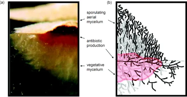
The genus Streptomyces was firstly described by Waksman and Henrici, in 1943 [1]. Streptomyces, from the Greek streptos (bent) and mukês (fungus), refers to a group of filamentous, high GC content, Gram-positive bacteria, harbouring more than 650 species [2]. The life cycle of these bacteria in solid media presents three main stages: the growth of vegetative mycelia, the formation of aerial mycelia and the differentiation of the latter into chains of spores (figure 1). Streptomyces spp. are chemoorganotrophic and are able to use a wide range of organic compounds, even from complex polymeric substrates. They are widespread in nature and can be found in a wide range of habitats (mostly in the soil and freshwaters) due to the extensive ranges of temperature and pH they tolerate. The secondary metabolism of these bacteria is amongst the most prolific sources of natural bioactive products known [3].
Figure 1. Vertical sections through a Streptomyces colony. Photograph (a) and scheme (b) of colony growing on agar. In the scheme, dead cells are represented in white and living cells in black. Adapted from [4].
Streptomyces coelicolor A3(2) was the first representative of the genus to be fully sequenced and is used in most genetic studies [5]. Its genome is constituted by a 8.7 Mbp linear chromosome, featuring 7825 coding sequences, and two circular plasmids, SCP1 and SCP2, with 356 kbp and 31 kbp, respectively [6-8]. This sequencing project shed light upon the biosynthetic potential of the genus, unveiling a high number of gene clusters dedicated for secondary metabolism. S. coelicolor M145, a S. coelicolor A3(2) derivative strain lacking the SCP1 and SCP2 plasmids, has been comprehensively
studied, namely regarding its transcriptome and proteome [9-11]. Understanding the genetic expression mechanisms in Streptomyces model organism using -omics approaches is arguably a major advantage for applied projects involving these organisms, namely in the optimization of the production titters of secondary metabolites.
Secondary metabolism
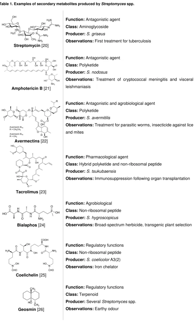
The genus Streptomyces is known to produce over 7600 natural bioactive metabolites and, remarkably, each strain has the genetic potential to produce several compounds [12]. The metabolites are grouped according their function as antagonistic agents, pharmacological agents, agro-biologicals or compounds with regulatory functions [13]. The secondary metabolites found in Streptomyces are biochemically derived from the concerted action of enzymes or enzymatic complexes generally encoded in genomic islands as clusters, some of which conserved amongst species [14]. The metabolite classes are related to the biosynthetic pathways and they include polyketides derived from polyketide synthases, peptides from non-ribosomal peptide synthetases, terpenoids, siderophores, aminoglycosides and their combinations and derivatives [14]. A representative panel of metabolites derived from Streptomyces is shown in table 1.
The number of sequencing projects of Streptomyces has been increasing at high rates since 2002, with 19 completely sequenced genomes and 125 draft genome as of May 2014 [15]. The analysis of secondary metabolites and biosynthetic gene clusters underwent great improvements thanks to the computational tools developed to identify these genomic clusters [16]. For example, antiSMASH is a web tool for genome mining of biosynthetic gene clusters that relies on gene organization and protein domain homologies [17]. The genome sequencing and annotation projects have unveil that Streptomyces devote up to 10% of their coding capacity to secondary metabolism related genes [18]. On the other hand, most of the detected biosynthetic gene clusters are not associated with previously isolated metabolites. These clusters whose product have not been detected under laboratory conditions are called silent or cryptic clusters. In S. coelicolor A3(2), from the 31 secondary metabolite gene clusters identified, only sixteen have been associated with detected metabolites [14]. The awakening of cryptic gene clusters using synthetic biology approaches has been described as a promising technique for the production of novel secondary metabolites [19]. Using the proper strategies, the vast unexplored biosynthetic libraries of Streptomyces could reveal themselves as a major source of compounds with utility to mankind.
Table 1. Examples of secondary metabolites produced by Streptomyces spp.
Streptomycin [20]
Function: Antagonistic agent
Class: Aminoglycoside
Producer: S. griseus
Observations: First treatment for tuberculosis
Amphotericin B [21]
Function: Antagonistic agent
Class: Polyketide
Producer: S. nodosus
Observations: Treatment of cryptococcal meningitis and visceral leishmaniasis
Avermectins [22]
Function: Antagonistic and agrobiological agent
Class: Polyketide
Producer: S. avermitilis
Observations: Treatment for parasitic worms, insecticide against lice and mites
Tacrolimus [23]
Function: Pharmacological agent
Class: Hybrid polyketide and non-ribosomal peptide
Producer: S. tsukubaensis
Observations: Immunosuppression following organ transplantation
Bialaphos [24]
Function: Agrobiological
Class: Non-ribosomal peptide
Producer: S. hygroscopicus
Observations: Broad-spectrum herbicide, transgenic plant selection
Coelichelin [25]
Function: Regulatory functions
Class: Non-ribosomal peptide
Producer: S. coelicolor A3(2)
Observations: Iron chelator
Geosmin [26]
Function: Regulatory functions
Class: Terpenoid
Producer: Several Streptomyces spp.
Synthetic biology in Streptomyces
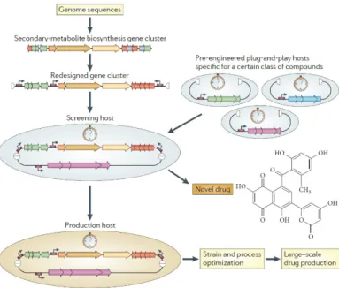
Synthetic biology has been defined as the engineering of biology through the synthesis of complex, biologically based (or inspired) systems, which display functions that do not exist in nature [27]. Synthetic biology aims to design and construct new genetic circuits and to remake natural existing ones for useful purposes. A key idea of synthetic biology approaches is the plug-and-play genetic manipulation (figure 2) [28]. The fact that secondary metabolite biosynthetic genes are already organized in clusters simplifies the application of such idea. A strategy for the activation of the cryptic clusters would consist on the complete removal of the native regulation and the substitution by a synthetic, predictable regulation. Once the strategy had been optimized, it could function as scaffold for high-throughput screening of the vast number of annotated clusters encoding for the same class of compounds [29]. Besides the awakening, the manipulation of synthetic assemblies would allow to exploit the modular nature of secondary metabolism enzymatic complexes to produce novel or optimized derivatives from a given biosynthetic pathway. Synthetic biology approaches rely on fully characterized genetic components, standardized assembly procedures and optimized hosts, also known as chassis, to achieve predictable behaviours and products in living systems. The synthetic biology toolbox for Streptomyces is based in a long tradition of genetic studies and a recent focus on strain enhancement and toolbox development per se, that will be reviewed in the following sections.
Figure 2. Plug-and-play strategy for heterologous expression of cryptic biosynthetic pathways. The redesign of gene clusters of secondary-metabolite biosynthetic proceeds from the streamlining of the coding sequences with inclusion of known transcriptional elements (represented by small arrows). Reproduced from [28].
Streptomyces-based chassis
The natural ability of Streptomyces bacteria of producing secondary metabolites fits them into the optimal choice for development of chassis for the heterologous expression of natural or engineered biosynthetic gene clusters. The strains chosen should fulfil the following characteristics: high growth rate, genomic stability, compatible molecular tools and a surplus of biosynthetic precursors. There are two main groups of Streptomyces-based chassis: wild-type strains and engineered strains. The wild-type strains used as heterologous hosts have their genome sequenced and are genetically amenable to work with. S. lividans 66 (or 1326) is an example of such wild-type strain: it was sequenced and it is naturally tolerant to exogenous methylated DNA [30]. S. albus J1074, a SalI-defective derivative of S. albus G with naturally-minimized genome [31], presented improved yields in secondary metabolite production relatively to S. lividans [32]. The engineered strains are generally derived from extensively known species by rational genome minimization, including deletion of unstable or precursor-diverting genomic regions. Several S. coelicolor A3(2) derivatives were produced by deletion of the gene clusters encoding the detectable metabolites actinorhodin, calcium-dependent antibiotic, prodiginine and yCPK and/or by modifications in the transcriptional and translational machinery [33]. Mutants of S. avermilitis with deletion of more than 1.4 Mbp that include the biosynthetic cluster for avermectins, oligomycins and filipins were also generated and characterized [34]. These optimized strains have been presented as the most adequate chassis to accommodate exogenous genetic circuits in order to achieve the expected outputs [35].
Genome editing techniques
The development of molecular tools and techniques is an important requirement for strain manipulation. The delivery of exogenous DNA to Streptomyces is mainly achieved by intergeneric conjugation [36, 37], a highly effective technique based in the horizontal gene transfer of an oriT-containing plasmid from a suitable tra-encoding E. coli host to Streptomyces. Genetic manipulation procedures in Streptomyces chromosome are based in homologous recombination events. One of the most widely used techniques is REDIRECT [38], a PCR-targeting and λ-Red mediated recombination methodology where the target sequence is replaced by cassette containing a selectable antibiotic resistance flanked by the yeast FLP-recombinase target sequences for selective marker removal. The deletion of large genomic regions has been achieved most of the times by Cre-loxP system from the P1 phage [39]. Homologous recombination is used for introduction of loxP sites flanking the target region followed by Cre expression for excision of flanked site. Recently, the Cas9/CRISPR technology,
based in a primitive bacterial immune system [40], was proven its utility in Streptomyces genome editing, including large deletion and point mutations [41, 42]. This technique is based in homologous recombination-based repair following a RNA-guided cleavage of the genomic target by Cas9 nuclease. Methodologies for introduction of exogenous DNA into Streptomyces include use of self-replicating and integrating vectors. For example, the SCP2* derivatives are self-replicating plasmids that presents a low copy number, stable inherence and being capable of carrying >30 kbp inserts [43]. The integrative vectors are based in the genomic integration systems of ΦC31 and ΦBT1 actinophages into the respective bacterial attachment sites (attB) encoded in several Streptomyces spp., including S. coelicolor and S. lividans [44, 45]. These integrative vectors contain a phage-derived integrase and the respective attachment site (attP) which allows, in theory, a single site-specific unidirectional recombination. A vast array of integrative vectors have been created, from expression [46] and reporter vectors [47, 48] to high capacity vectors such as bacterial artificial chromosomes [49] and cosmids [44].
Molecular toolbox for Streptomyces
A molecular toolbox is constituted by genetic parts that suit defined functions in transcriptional and translational control or gene encoding. The assembly of these parts allows to create molecular devices with defined purposes. The implementation of such devices in suitable chassis yields the desired output. Thus, the genetic parts, represented by promoters, ribosome binding sequences, coding sequences and transcriptional terminators represent the basis for synthetic biology approaches. The rational development of new biological devices rely on comprehensive characterization of the genomic parts employed [50].
In bacteria, transcription is driven by RNA polymerase complex, constituted by the elongation-capable core with subunits α2ββ’ω and the initiation-required sigma factor. The DNA sequences that are able to recruit a sigma-containing RNA polymerase are called promoters. The most used promoter for heterologous gene expression in Streptomyces has been ermEp*, a derivative of erythromycin resistance gene promoter of Saccharopolyspora erythraea (formerly, Streptomyces erythraeus) [51, 52]. Some promoters are under influence of transcriptional regulators. The regulation determinants are the operator, usually a conserved DNA motif in the promoter region, and the regulator, a protein that can recognize and bind the operator. A repressor is a regulator that inhibits transcription by binding its cognate operator in the absence of an inducer signal. Thereby, promoters regulated by repressors are dependent on the presence of the inducer for transcriptional activity and are known as inducible promoters. Most inducible promoters used as molecular tools for Streptomyces are listed in table 2. While
some of the reported inducible systems were based in naturally regulated promoters, others were synthetically derived from operator-free promoters merged with known operator/repressor systems.
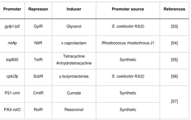
Table 2. Inducible promoters based in repressors used in Streptomyces.
Promoter Repressor Inducer Promoter source References
gylp1/p2 GylR Glycerol S. coelicolor A3(2) [53]
nitAp NitR ε-caprolactam Rhodococcus rhodochrous J1 [54]
tcp830 TetR Tetracycline
Anhydrotetracycline Synthetic [55]
cpkOp ScbR γ-butyrolactones S. coelicolor A3(2) [56]
P21-cmt CmtR Cumate Synthetic
[57]
PA3-rolO RolR Resorcinol Synthetic
Activators are regulators that positively affect the transcriptional activity of a promoter in the presence of an inducer. The tipAp is a popular activator-based inducible promoter for Streptomyces. In the presence of thiostrepton, TipA is overexpressed and recruited to tipA promoter, activating its own transcription [58-60]. Exogenous promoter/RNA polymerase systems, based on the T7 phage machinery, were adapted for Streptomyces, allowing fully orthogonal transcriptional regulation [61].
The assessment of promoter activity can be performed by cloning the query sequence upstream of reporter genes. The reporter genes generally code for enzymes which activity can be measured with high sensitivity and specificity. The specificity is a major issue in reporter gene development for Streptomyces given its natural richness in catabolic enzymes and antibiotic production (and consequently, resistance). The reporter genes described for Streptomyces are summarized in table 3.
Translation is a further layer of gene expression regulation. The 70S ribosomes are the molecular machines responsible for translation in bacteria, being constituted by two asymmetric rRNA-protein complexes, the 30S and 50S subunits. Interestingly, several antibiotics produced by Streptomyces spp. target ribosomes [62].
Table 3. Reporter genes used in Streptomyces. MIC, minimal inhibitory concentration.
Reporter
gene Protein encoded Measurements Source References
neo Aminoglycoside phosphotransferase
MIC
Enzymatic assays Tn5 transposon [63]
cat Chloramphenicol O-acetyltransferase
MIC
Enzymatic assays Tn9 transposon [64]
aacC1 Aminoglycoside O-acetyltransferase MIC Enzymatic assays Tn1696 transposon [65]
melC Tyrosinase Enzymatic assays S. glaucescens [66]
luxAB Luciferase Bioluminescence Photorhabdus
luminescens [67]
gfp Green fluorescent
protein Intrinsic fluorescence Aequorea victoria [68, 69]
xylE Catechol
2,3-dioxygenase Enzymatic assays
Pseudomonas
putida [48]
gusA β-glucuronidase Enzymatic assays E. coli [47]
bpsA Indogoidine synthase Indogoidine quantification
S. aureofaciens
S. lavendulae [70, 71]
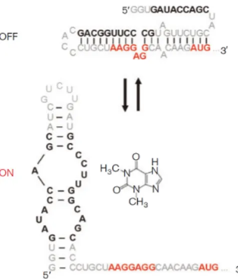
Although exciting breakthroughs in ribosome engineering have been achieved in other organisms [72], translation control in Streptomyces is mainly attained by modulating the affinity of mRNA towards ribosomes. In bacteria, the ribosome binding site (RBS, also known as Shine-Dalgarno [SD] sequence) is a sequence in the 5’ untranslated region (5’-UTR) of mRNAs that present some complementarity with the 3’ region of the 16S rRNA from 30S subunit and is involved in mRNA-ribosome recognition and positioning. The usage of RBS from genes with the desired expression level is the most common technique for tuning translational efficiency in Streptomyces [29]. Translation efficiency can also be affected by secondary structures of mRNA. Riboswitches are genetic control elements present in 5’-UTRs of mRNA that can generate alternative secondary structures through binding of a given metabolite, such as the B12 riboswitch in S. coelicolor A3(2) [73]. Synthetic riboswitches for Streptomyces have been designed using theophylline-sensitive aptamers that conditionally allow the access of ribosome to RBS and start codon of mRNAs, thereby exerting ligand-dependent translational control (figure 3) [74].
Figure 3. Alternative structures of the theophylline-sensitive E* riboswitch. The SD sequence, represented in red, is made accessible in the presence of theophylline. Reproduced from [74].
The advances in chassis, techniques and molecular tools for synthetic biology approaches in Streptomyces, although promising, are yet very modest compared with other organisms. The complex transcriptional apparatus and dynamics in Streptomyces still hampers the development of novel parts such as constitutive promoters.
Development of constitutive promoters for Streptomyces
In bacteria, transcription of DNA into mRNA by RNA polymerase is the major checkpoint for controlling gene expression. For controlling transcription, there are two main factors to be considered: the existence of regulators bound to operators in promoter regions and the sigma factors available for promoter recognition. In Streptomyces, each of these factors is represented by a massive number of variables: for the model organism S. coelicolor A3(2) a total of 499 transcriptional regulators and 64 sigma factor have been reported [75]. A successful strategy for transcriptional control in Streptomyces would need to circumvent these complex native networks. Three scenarios could be considered: (i) to implement a totally exogenous transcriptional machinery, such as the T7 RNA polymerase system [61], (ii) to rely on exogenous orthogonal regulator/operator systems [76] or (iii) to remove native regulation of stably expressed components in order to turn them into predictable parts. In the context of constitutive promoters’ development for Streptomyces, the latter strategy has been applied in a more or less declared way. Predictable promoters are indispensable tools for gene cluster refactoring. Furthermore, well-characterized promoters are building blocks for synthetic inducible systems and can be combined with riboswitches, providing a myriad of new components for synthetic biology approaches.
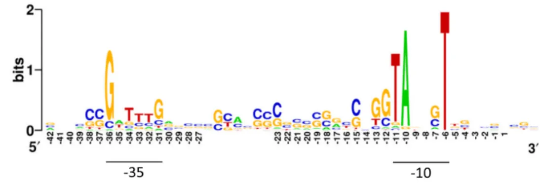
Determinants of constitutive promoters in Streptomyces
Constitutive promoters are those active at all circumstances, which, in practice, is understood as being recognized by the primary sigma factor without need of other transcriptional factors [77]. The core promoter is the minimal portion of a promoter region required to initiate transcription and it is constituted by two sigma-recognizable hexamers centred approximately 10 and 35 bp upstream of the transcription start site (TSS) and the spacing between them. The primary sigma factor in S. coelicolor, HrdB [78, 79], belongs to the sigma-70 family and recognizes the consensus sequence depicted in
figure 4 [80].
Figure 4. Representation of the sequence recognized by HrdB in Streptomyces. The Weblogo representation was derived from 29 Streptomyces’ promoters resembling sequences recognized by sigma-70 in E. coli [80].
The promoters of Streptomyces were classified based on clustered frequency and positional analysis of over-represented short sequences, including motifs predominantly centred in the -35 and -10 position relatively to the TSS [81]. This analysis revealed the prevalence of the sigma-70-compatible motifs TANNNT for -10 region and TTGAC for -35 region, but also the extended motifs TNTNNNANNT, TGNNANNNT and GTNNANNNT centred at -10 region, similar to what occurs in E. coli and Corynebacterium glutamicum [82, 83]. Recently, a RNA-seq study revealed an extended motif centred in -35 region of promoters of transcriptional machinery of S. coelicolor A3(2) (figure 5) [9].
Figure 5. Representation of conserved sequence in promoters associated with transcriptional machinery in
Strategies for design of constitutive promoters for Streptomyces
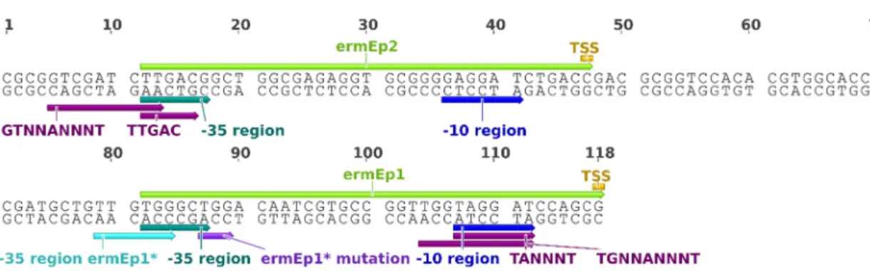
The first and most used “synthetic” constitutive promoter for Streptomyces is actually the result of a point deletion in the erythromycin resistance gene (ermE) promoter of Saccharopolyspora erythraea [52]. The genomic region upstream to ermE harbours a complex web of RNA polymerase-attracting sites, namely, two promoter in the forward strand, ermEp2 and ermEp1, and a promoter in the reverse strand [51]. Together, ermEp2 and ermEp1 constitute ermEp (figure 6). The TGG deletion from the -35 region of ermEp1 originated a 1.08-fold stronger promoter, known as ermEp* [52]. This effect was suggested to result from the more HrdB-complying -35 region. The -10 region of ermEp1 was identified as belonging to the -10 extended class, TGNNANNNT.
Figure 6. Representation of the ermE promoter. The ermEp1 and ermEp2 promoters are depicted by light green arrows. The motifs associated with core sequences of Streptomyces’ promoters [81] are depicted by dark purple arrows. The remaining annotations are derived from the original characterizations [51, 52].
In 1997, Labes et al. described SF14 [65], a promoter-containing fragment derived from the S. ghanaensis phage I19 responsible for 1.66-fold higher kanamycin resistance levels than ermEp*. SF14 is also constituted by two tandem promoters, 14-Ip and 14-IIp, but in contrast with ermEp, their -10 regions partially overlap (figure 7). One TTGAC motif and one TANNNT are present in the -35 and -10 regions of 14-IIp, respectively. Both 14-Ip and 14-IIp promoters include the HrdB-recognizable sequence and were validated by in vitro run-off transcription assays using fractions containing HrdB-enriched RNA polymerase holoenzyme.
Figure 7. Representation of the SF14 promoter. The motifs associated with core sequences of Streptomyces’ promoters [81] are depicted by dark purple arrows. The remaining annotations are from the original characterization [65].
kasOp* was generated by deleting the ScbR operators present in the inducible cpkO promoter [84]. kasOp* was recognized by HrdB in vivo and presented a constitutive pattern of transcription. Using neo as reporter gene, kasOp* presented 2-fold higher promoter activity than ermEp* in S. coelicolor A3(2). The presence of a single HrdB-binding site with 18-nt spacer was regarded as avoiding steric hindrances between RNA polymerases. The TANNNT and TTGAC motifs are present in the -10 and -35 regions of kasOp*. Besides, three additional -10 motifs are represented in its sequence (figure 8).
Figure 8. Representation of kasOp*. The motifs associated with core sequences of Streptomyces’ promoters [81] are depicted by dark purple arrows. The remaining annotations are from the original characterization [84]. The engineered
nucleotides annotation refer to the mutations in ScbR operator.
The hint for synthetic promoter libraries (SPL) construction came after the knowledge that by keeping the -35 and -10 regions and randomizing the spacer sequence one could modulate the strength of prokaryotic promoters [85]. The first attempt to produce SPL for Streptomyces spp. was based in the -35 and -10 hexamers of HrdB-recognizable sequence, using the degenerate oligonucleotide N10TTGACNN17TASVDTN5 [86]. Noteworthy that the -10 region was also partially randomized according to the consensus used, with 18 possible sequences. The strongest representative of the library, A1-14, displayed 0.92-fold activity when compared to ermEp*. A1-14 contains the TTGAC and TANNNT motifs, which were ubiquitous in the library, but also a -10 extended class motif, TGNNANNNT (figure 9).
Clustered analysis of strong versus weak synthetic promoters of the SPL showed that: (i) TAGGGT would be the typical -10 region of strong promoters, (ii) the motif RGgGn immediately upstream -10 region is an extension present only in strong promoters and (ii) that a G-rich spacer would be influential in strength of streptomycete promoters. Furthermore, it was suggested that imperfect repetitions of -10 region might help RNA polymerase positioning.
Figure 9. Representation of the A1-14 promoter, the strongest representative of a SPL based in the sequence recognized by HrdB. The motifs associated with core sequences of Streptomyces’ promoters [81] are depicted by dark purple arrows. The remaining annotations are from the original characterization [86].
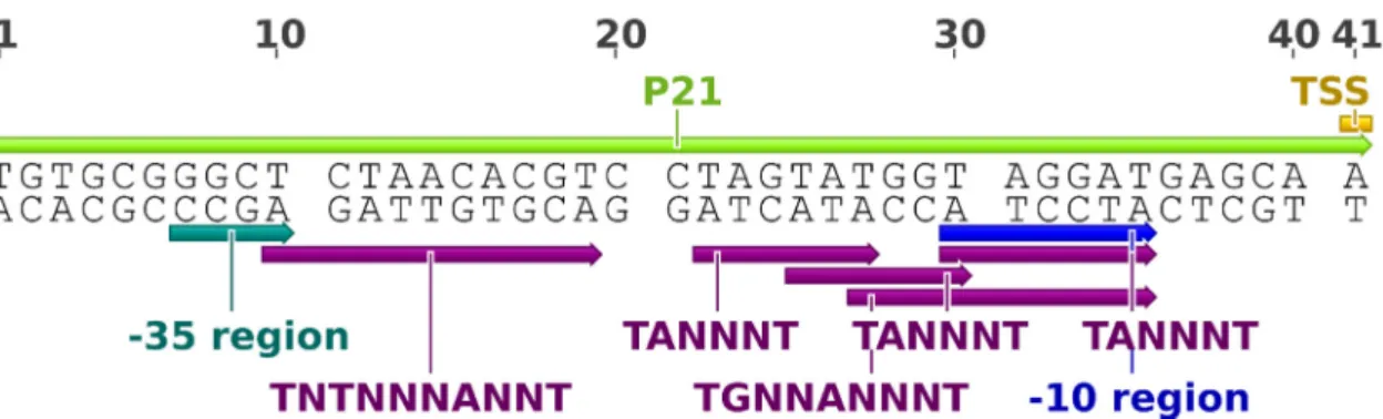
In 2013, Siegl et al. reported and characterized a new SPL based in the -10 and -35 regions of ermEp1, N6GGCTN19TAGGATN6 [87]. P21 promoter (figure 10) displayed 1.67- and 2.92- fold higher activity, in S. lividans TK24, than ermEp* and ermEp1, respectively. The promoter showed similar results in several different actinomycetes. By RNA-seq analysis, the authors showed that the TSS of P21 promoter is located 6 nucleotides downstream the -10 region.
Figure 10. Representation of the P21 promoter, the strongest representative of a SPL based in ermEp1’s -10 and -35 regions. The motifs associated with core sequences of Streptomyces’ promoters [81] are depicted by dark purple arrows. The remaining annotations are from the original characterization [87].
Several randomized nucleotides are conserved between ermEp1 and P21, including the TGG motif immediately upstream the -10 region, which classifies this region as extended (motif TGNNANNNT). Three TANNNT elements - TAGTAT, TATGGT and TAGGAT - are present in P21 promoter, surrounding the -10 region. The -35 region consensus observed in P21 does not comply with the usual TTGAC motif, albeit the G, the most conserved nucleotide of the motif, is present.
Looking for a set of constitutive promoters for gene cluster reengineering, Shao et al. [88] characterized the promoter activity of the genomic regions upstream of the coding sequences for glyceraldehyde 3-phosphate dehydrogenase (GAPDH) and 30S ribosomal protein S12 (rpS12, figure 11) of S. griseus, which they had found highly transcribed along time by RT-qPCR among 23 housekeeping genes. These genomic
regions promoted over 10-fold higher reporter activities than ermEp*, in S. lividans 1326. A whole set of 13 intergenic regions presenting promoter activity was completed from the sequences upstream gap1 and rpS12 of other actinobacteria. The cloned regions were about 300-nucleotide long and were putatively assumed to have RBSs in 6-10 bp upstream start codon, based on the presence of a AG-rich region.
Figure 11. Representation of the genomic region upstream of rpS12 in S griseus. The promoter activity of this region was assessed by Shao et al [88]. The motifs associated with core sequences of Streptomyces’ promoters [81] are depicted by dark purple arrows. The region matching the sequence recognized by HrdB with a spacer of 17 nucleotides and evidence of promoter core sequences is depicted by the green arrow.
The design of constitutive promoters for Streptomyces followed a path from the usage of naturally occurring, complex genomic regions of related organisms, to rational trimming and promoter engineering of known inducible promoters, coming to consensus-based randomization of spacers and synthetic promoter library construction. During the course of these developments, some determinants of strong constitutive promoters were added to the consensus and motifs found during the first studies in the early ‘90s. The last reported attempt to obtained strong promoters [88] reintroduced the idea of using native promoters, now selected by previous low-scale transcriptional studies. Indeed, several never-so-strong promoters were reported using this approach. However, the rising rationality in promoter choice/generation was withdrew by using full genomic regions, which could contain unexpected operators or encode 5’-UTR that acquire secondary structures that hamper translation. Hence, the combination of the increasing whole-genome transcriptomic data, mainly available for the model organism S. coelicolor A3(2), with the rational principles of trimming, operator freeing and compliance with strong promoters’ motifs, seemed as the next logical strategy to design constitutive promoters for Streptomyces.
Objectives
The current molecular toolbox for Streptomyces includes several useful representatives of constitutive promoters, such as ermEp* and SF14. However, the limited number of promoters together with the complexity and unpredictability of the existing ones still hinders genetic engineering progresses. The increasing number of whole genome transcriptome/proteomic studies and bioinformatics developments pave the way for new rational approaches to design synthetic parts. The present work aims to apply these assets to establish novel constitutive promoters for Streptomyces. The objectives of this work were:
• Selection of stable and highly expressed genes of S. coelicolor M145 using publically available whole-genome transcriptomic and proteomic data;
• In silico characterization of genomic region upstream of the selected genes regarding functional sequences, such as transcriptional terminators, core promoters and ribosome binding sites;
• Design of synthetic promoter candidates according to the described genomic regions of S. coelicolor M145;
• In vivo characterization of the synthetic promoters in the model organism S. coelicolor M145 and in a common chassis for heterologous gene expression, S. lividans 1326, using promoter probe vectors.
Material and methods
Bacterial strains, plasmids and growth conditions
All strains and plasmids used in this work are listed in table 4. Streptomyces coelicolor M145, S. lividans 1326 and their derivative mutant strains were grown at 28 ºC, at 220 rpm. For DNA extraction procedures, Streptomyces spp. were grown in YEME medium [89]. For synthetic promoter strength determination, bacteria were grown in TSB [89] or NMMP medium [89, 90]. Sporulation was achieved in MS agar medium, at 28 ºC. Spores of S. coelicolor and S. lividans were collected using a solution of 30% (w/v) glycerol and 0.021% (v/v) Triton X-100, spectrophotometrically quantified using an UVmini-1240 UV-Vis Spectrophotometer (Shimadzu) and kept at -80 ºC. For the determination of cells dry weight, 1-mL aliquots were harvested, washed once with 0.9% (w/v) NaCl solution and dried at 80 °C for at least 48 h. E. coli was routinely grown in Lysogeny broth (LB), at 37 ºC. Stock cultures of E. coli were kept in 33% (w/v) glycerol at -80 ºC. When necessary, medium was supplemented with antibiotics to final concentrations of 50 μg.mL-1 apramycin (Am), 100 μg.mL-1 ampicillin (Ap), 50 μg.mL-1 kanamycin, 25 μg.mL-1 chloramphenicol, 50 μg.mL-1 spectinomycin (Sp) and 25 μg.mL-1 nalidixic acid.
Table 4. Strain and plasmids used in this work.
Strain or plasmid Main characteristics Reference
Streptomyces
S. coelicolor M145 SCP1- SCP2- derivative from A3(2) strain [89]
S. lividans 1326 wild-type [30]
Escherichia coli
DH5α
General cloning strain [F- φ80lacZΔM15
Δ(lacZYA-argF)U169 recA1 endA1 hsdR17(rk-, mk+) phoA supE44
thi-1 gyrA96 relA1 λ-]
[91]
ET12567 [pUZ8002]
Non-methylating strain for intergeneric conjugation [dam dcm hsdM hsdS hsdR Tcr Cmr][RP4 plasmid derivative,
Kmr tra operon]
[38, 92]
Plasmids
pUC19 General cloning vector, Apr lacZα pMB1ori [93]
pGUS Promoter probe vector for Streptomyces [ΦC31-integrase
gusA Amr Spr oriT pMB1ori attB] [47]
pGUS_SP pGUS derivative with synthetic promoter fragment cloned
Selection and characterization of S. coelicolor promoters
The search for stable and highly expressed genes from S. coelicolor was based on publically available transcriptomic and proteomic data. The genes with a <5% variation of read counts per transcript were retrieved from a time-lapse whole genome transcriptome analysis study of S. coelicolor M145 [94] and ranked according to the average transcription level. The list of retrieved genes was conferred with the list of the 65 most abundant proteins retrieved from a semi-quantitative proteomics study in S. coelicolor M145 [11]. A final list of S. coelicolor genes was obtained that displayed the common entries from transcriptomic and proteomic data and was sorted according a score given by the product of read counts per transcript and the total exponentially modified protein abundance index. The transcription start site for each gene was annotated according to RNA-seq studies in S. coelicolor M145 [9, 10]. The regions upstream of the selected genes were annotated regarding ribosome binding sequences [80], promoter core motifs in Streptomyces [81], sequences recognized by HrdB [80], transcriptional terminators and regulatory motifs/operators described in the literature.
In silico
analysis of DNA sequences
The annotation of genomic sequences was performed using Geneious version 4.8.3 [95]. The intrinsic terminators of transcription in S. coelicolor were obtained from WebGesTer Database [96]. The reconstructed operators/motifs were represented using WebLogo [97]. The search of motifs in genomic sequences was done using FIMO [98].
DNA manipulation
Plasmids were isolated from 5 mL cultures of E. coli grown overnight in LB medium, at 37 ºC, using the GenElute™ Plasmid Miniprep Kit (Sigma), following the manufacturer’s instructions. Streptomyces genomic DNA (gDNA) was isolated from 1 mL aliquots of stationary phase cultures using the GeneJET Genomic DNA Purification Kit (Thermo Scientific) according to the manufacturer’s instructions. DNA digestion products and PCR products were purified using NZYGelpure Kit (NZYTech) according to the manufacturer’s instructions. DNA was eluted with ddH2O, quantified using NanoDrop™ 1000 Spectrophotometer (Thermo Scientific) and analysed by gel electrophoresis.
PCR conditions
Generic conditions of polymerase chain reactions (PCR) were as follows: 1x GoTaq® Reaction Buffer, 1.5 mM MgCl2, 0.2 mM dNTPs (Thermo Scientific), 0.2 µM forward primer, 0.2 µM reverse primer, 1.25 U GoTaq™ G2 flexi DNA Polymerase (Promega) and 5% (v/v) of dimethyl sulfoxide (DMSO). All the oligonucleotides used in this work are listed in table 5. Template DNA varied between 20 to 50 ng for plasmid DNA and 50 to 100 ng for genomic DNA. All programs included an initial denaturation step (95 ºC, 5 min), a variable number of cycles of denaturation (95 ºC, 30 s), annealing (30 s) and extension (72 ºC, 1 min/kbp) and a final elongation step (72 ºC, 7 min). The reactions were performed in a C1000™ Thermal Cycler (Bio-Rad).
Table 5. Oligonucleotides used in this work.
Name Sequence (5’-3’) Uses
bla_R TATATCTAGAGTCTGACGCTCAGTGG Synthetic promoter generation P1_F GGGGTACCTGCTGCTGACGCTACACGGCATGTCCGAGCC TCACCAGTGAGTAAGGGGTGTGCGGAACCCCTATTTG P2_F GGGGTACCCACCTCCGACCCTACCTCTCCGGGGCCTCGG GGGTGACATCGAGACGCCCCGCGGAACCCCTATTTG P3_F GGGGTACCAGGTCCGAGACTATGACTGCGATTAGCACTC GGTCAAGCGGAGGCGGAACCCCTATTTG P21_F GGGGTACCTTGCTCATCCTACCATACTAGGACGTGTTAGA GCCCGCACAGCGGAACCCCTATTTG
GUSseq_R3 GGTTTCGACGGGCCG Synthetic promoter
sequencing
GUSseq_F2 CGAAGATACCTGCAAGAA Synthetic promoter
presence confirmation pUCR ACACAGGAAACAGCTATGAC C31_int_F2 TGGGTGTCGCCGTTGGTG ΦC31 attB integration confirmation C31_int_R2 CGTCGTCGGTCGGCGGCT
Gel electrophoresis
For routine diagnosis, DNA samples were mixed with 1x loading dye and separated by gel electrophoresis (0.8 - 1.2% agarose, 0.2 µg/mL ethidium bromide, 80 - 100 V, 1x TAE [40 mM Tris, 20 mM acetic acid, 1 mM EDTA]). The gels were scanned in a Molecular Imager® GelDoc™ XR+ Imaging System (Bio-Rad). The Lambda DNA/HindIII Marker or GeneRuler™ DNA Ladder Mix (Thermo Scientific) were used as reference for DNA sizing and relative quantification.
Generation and cloning of synthetic promoters
Synthetic promoters P1, P2, P3 and P21 were generated by PCR using the oligonucleotides listed in Table. Briefly, the synthetic promoter was amplified together with the gene coding for beta-lactamase; the 1.2-kbp PCR product was cloned into pGUS plasmid digested with XbaI and KpnI (Thermo Scientific), yielding the pGUS_SP plasmids (pGUS_P1, pGUS_P2, pGUS_P3 and pGUS_P21) (figure 12). All constructions were confirmed by DNA sequencing (STAB VIDA, Portugal).
E. coli
transformation
For routine cloning procedures, E. coli DH5α chemically competent cells were prepared by the divalent cations treatment method [99]. For DNA transformation, 100 µL of cell suspension were mixed with up to 1 µg plasmid DNA and incubated on ice for 20 min. The cells were then heat-shocked at 42 ºC for 45 s and transferred to ice for 2 minutes. The cells were added 900 µL of LB medium and incubated at 37 ºC for 45 min for cell recovery prior to plating in solid medium. E. coli ET12567 carrying pUZ8002 plasmid electro-competent cells were transformed with the mobilizable plasmids for Streptomyces (figure 12) [89]. E. coli ET12567 carrying pUZ8002 plasmid was grown overnight in 5 mL LB supplemented with kanamycin and chloramphenicol. 20 mL of fresh medium supplemented with the appropriate antibiotics and 20 mM MgSO4 was inoculated with 200 µL pre-inoculum. The culture was grown at 37 ºC to an OD600nm of 0.4, washed once with 1 volume of ddH2O and washed twice with 1 volume of 10% (w/v) glycerol. The cells were resuspended in 300 µL of 10% (w/v) glycerol. 50 µL of cells were mixed with 100 ng of pGUS_SP plasmids using a Gene Pulser™ plus Pulse Controller (Bio-Rad) (0.1 cm gap width, 200 Ω, 25 µF, 2.5 kV). The cells were then let to recover in 1 mL LB at 37 ºC for one hour. The cell suspensions were plated in LB agar plates supplemented with chloramphenicol, kanamycin and apramycin and incubated overnight at 37 ºC.
Figure 12. Strategy for construction of synthetic promoters probe vectors in Streptomyces. The synthetic promoters P1, P2, P3 and P21 (generically, SP) were generated by polymerase chain reaction using SP_F/bla_R primer pairs to amplify the ampicillin resistance marker of pUC19 plasmid. The PCR products were cloned into XbaI/KpnI sites of pGUS reporter vector upstream of promoterless gusA gene, to originate pGUS_SP plasmids. pGUS and pGUS_SP plasmids were transferred to Streptomyces spp. by intergeneric conjugation using non-methylating E. coli ET12567 [pUZ8002] as plasmid donors. Integration of pGUS_SP plasmids into Streptomyces chromosome by ΦC31 integrase-mediated attB/attP recombination was confirmed by PCR. Apr, ampicillin resistance marker; Amr, apramycin resistance
Intergeneric conjugation
Foreign DNA was introduced in Streptomyces by intergeneric conjugation through cell-to-cell contact from a donor cell into a recipient cell. E. coli ET12567 [pUZ8002] carrying the pGUS_SP plasmids were used as donor cells (figure 12). 10 mL of LB medium supplemented with appropriate antibiotics and 20 mM MgSO4 was inoculated with overnight-grown E. coli and incubated at 37 ºC to an OD600nm of 0.9. Cells were washed twice with LB medium without antibiotics and resuspended in 600 μL of LB medium. 109 Streptomyces spp. spores were resuspended in 600 μL 2xYT medium [89] and incubated at 50 ºC for 10 minutes. Donors and recipients were incubated together at 28 ºC, for 20 min and then spread on MS plates supplemented with 10 mM MgCl2 and grown overnight at 28 ºC. The plates were supplemented with apramycin and nalidixic acid, air dried and incubated at 28 ºC. The presence of exconjugates was verified after 7 days. Ten colonies for each exconjugate were streaked on new MS plates supplemented with apramycin and nalidixic acid for 7 days at 28 ºC.
Glucuronidase activity assays
For in situ detection of glucuronidase activity, exconjugates were streaked on MS plates supplemented with 25 µg/mL X-Gluc (5-bromo-4-chloro-3-indolyl β-D-glucuronide sodium salt [Sigma]) and incubated at 28 ºC for 24 hours.
Promoter strength was determined by measuring the glucuronidase reporter activity in total protein extracts of Streptomyces spp. The protocol was based in a previously reported method [100], with adaptations for continuous spectrophotometric measurements [101]. For relative promoter strength assessment, protein crude extracts were obtained from 1.5 mL samples of cultures grown in TSB for 40h and 72h. For heat-shock effect assessment, cells were grown in TSB at 28 ºC. After 40h of growth cultures were divided in two flasks to grow at 28 ºC or 42 ºC. Samples were obtained after 30 and 60 minutes of growth at 28 ºC or 42 ºC. For carbon source effect assessment, the samples were obtained from cultures grown in NMMP medium supplemented with 0.5% (w/v) glucose, glycerol or mannitol for 48h. Mycelia was washed once with one volume of 50 mM NaH2PO4/Na2HPO4 buffer pH 7 and resuspended in 0.5 mL GUS assay buffer [50 mM NaH2PO4/Na2HPO4 buffer pH 7, 5 mM DTT, 10 mM Na2EDTA, 0.1% (v/v) Triton X-100] and 1 mM phenylmethylsulfonylfluoride (PMSF). Cells were disrupted by sonication (Branson Sonifier® S-250A) and the lysate was recovered after centrifugation at 4 ºC, 16000x g. Protein content of crude extracts was determined using the Bio-Rad Protein Assay Kit (Bio-Rad), following the manufacturer’s instructions and using bovine serum albumin as a standard. Crude extracts containing about 1 μg of total protein were
incubated at room temperature with 1 mM p-nitrophenyl β-D-glucuronide (PNPG) (Sigma) in 96-well plates. The conversion of PNPG into p-nitrophenol (PNP) was followed spectrophotometrically at 415 nm, for up to 20 minutes, using an iMark™ Microplate Absorbance Reader (Bio-Rad). One unit of glucuronidase (GUS unit) activity was defined as the amount of enzyme necessary to increase absorbance at 415 nm of 1 unit per minute, at room temperature. Glucuronidase specific activity was determined by calculating the PNP formation rate per minute per milligram of total protein.
Statistical analysis
All statistical analysis was performed using GraphPad Prism version 5.02.
Accession numbers
The accession numbers of sequences and studies used in this work are listed in
table 6.
Table 6. Accession numbers of sequences and studies used in this work. The accession number GSE57268 refers to an expression profiling of WT S. coelicolor M145 at 24h and 72h of culture. The accession numbers GSE46507 and GSE46232 refer to two transcription maps of WT S. coelicolor M145 produced by differential and global RNA sequencing, respectively.
Data Accession Reference
S. coelicolor A3(2) chromosome sequence NC_003888.3 [8]
S. lividans 1326 chromosome sequence NZ_CM001889.1 [30]
S. coelicolor M145 RNA-seq GSE57268 [94]
Results
Screening of constitutively expressed genes in S. coelicolor M145
In order to find suitable templates for the designing of new promoters, constitutively expressed genes in S. coelicolor were screened using genome-wide transcriptomic and proteomic data. Two criteria were established: (i) genes ought to be stable and highly transcribed along time and (ii) proteomic data should support transcriptomic evidence. All S. coelicolor M145 genes were sorted according expression level according to time-lapse genome-wide transcriptomic data [94]. Only genes with variation in expression along time lower than 5% were considered. An ordered list of 300 genes was generated. In order to provide proteomics evidence for high expression, the 300 selected genes list were verified with most expressed proteins list in S. coelicolor M145 [11]. Twelve entries were present in both lists (table 6). Results were sorted using a score defined as the product of average transcripts counts and protein abundance index. Existence of TSS upstream of the selected genes was assessed using two RNA-seq datasets [9, 10]. Only 6 of the 12 selected genes presented a TSS. The three genes with highest score, SCO4761, SCO1947 and SCO0527, had TSS in their upstream regions and were selected for further characterization.
Table 7. Ordered list of stable and highly transcribed genes in S. coelicolor M145. EmPAI refers to exponentially
modified protein abundance index, a measure of protein abundance, calculated elsewhere [11]. The score is the product of average transcript counts by the protein abundance index. TSS existence was assessed in the 500-bp region upstream each gene. Locus Average transcript counts Protein abundance index (EmPAI) Score TSS Description
SCO4761 3332 85,6 285219 Yes co-chaperonin GroES
SCO1947 5317 27,98 148770 Yes glyceraldehyde-3-phosphate dehydrogenase
SCO0527 3575,5 35,48 126859 Yes cold shock protein SCO4653 3500,5 33,5 117267 No 50S ribosomal protein L7/L12 SCO4702 3355,5 32,74 109859 No 50S ribosomal protein L3
SCO4655 3717 24,08 89505 No DNA-directed RNA polymerase subunit beta
SCO0641 593,5 87,87 52151 Yes tellurium resistance protein SCO5736 790 58,93 46555 Yes 30S ribosomal protein S15
SCO3767 614,5 60,02 36882 No hypothetical protein
SCO5776 1022 35,82 36608 No glutamate binding protein SCO1598 940,5 33,81 31798 No 50S ribosomal protein L20 SCO5595 953,5 30,14 28738 Yes 50S ribosomal protein L19
In silico
characterization of promoter regions of the selected genes
Synthetic promoter design based in upstream region of the selected genes required a prior characterization of these regions. This characterization aimed to describe conditions or mutations known to influence gene expression and to annotate functional sequences, such as sequences recognized by sigma factors, operators, transcriptional terminators and ribosome binding sites. Following this characterization, upstream regions of the selected genes were downsize to core promoter sequences following three criteria: (i) a single TSS and the associated sequence recognized by HrdB should be included, (ii) at least, one motif associated with core promoters regions in Streptomyces should be present and (iii) known and predicted operators and transcriptional terminators should be avoided.
SCO4761 - groES
S. coelicolor A3(2) groES encodes a 10 kDa chaperonin [8]. GroES protein belongs to a multimeric complex, encompassing several subunits of GroES and GroEL proteins, that provides an isolated environment that facilities correct folding and assembly of some proteins. The sequence of groES in S. coelicolor A3(2) was first published by Duchêne et al [102]. groES TSSs are located 126 bp (TSS1) and 1 bp (TSS2) upstream of start codon [9, 102]. A putative intrinsic transcriptional terminator concerning the gene upstream groES (∆G=-20.83 kcal.mol-1) and a RBS (q-value=0.05) were found 269 bp and 17 bp upstream of groES start codon, respectively. The core promoter upstream TSS1 contains a sequence recognized by HrdB and two copies of CIRCE (controlling inverted repeat of chaperone expression), a negative cis-element recognized by the heat-inducible transcriptional repressor HrcA [102, 103]. Heat shock conditions were shown to induce groES transcription in S. coelicolor A3(2) and S. lividans TK21 [104-106]. Full exclusion of CIRCE from dnaK promoter in Lactococcus lactis was shown to abolish its heat shock-dependent transcription [107]. Furthermore, introduction of point mutations in CIRCE of Bacillus subtilis dnaK promoter was shown to de-repress dnaK expression under non-heat shock conditions [108]. Hence, in order to design a heat shock-independent synthetic promoter, CIRCE motifs should to be trimmed/removed from groES promoter. In S. coelicolor A3(2), the core promoter (upstream from TSS1) of groES/groEL operon contains two CIRCE: one near TSS1 (CIRCE_B), similar to B. subtilis, and one surrounding the -35 hexamer (CIRCE_A). CIRCE_B motif would act like in B. subtilis. Nevertheless, deletion of CIRCE_B would not interfere with core promoter sequence upstream TSS1. On the other hand, -35 hexamer of TSS1 was embedded into CIRCE_A motif. Thus, CIRCE_A could not be
fully excluded from the synthetic promoter template. High pairwise identity was observed between CIRCE motifs of S. coelicolor groES and Bacillus subtilis dnaK promoter (figure 13). Hence, together with CIRCE_B full trimming, a disruption strategy similar to IR-1 mutation [108] was applied to CIRCE_A in order to remove heat-shock regulation from this synthetic promoter sequence. Other factors were shown to up-regulate groES transcription, namely, the growth in chitin-amended soil [109] and pH shock [110].
Figure 13. Alignment of CIRCE operators in promoters of S. coelicolor A3(2) groES and Bacillus subtilis dnaK .
The IR-1/2 mutations are referred in [108].
Sequences recognized by HrdB were found upstream of TSS1 and TSS2 of groES [80]. The TANNNT, CANNNT and TNTNNNANNT motifs [81] were found in the -10 region of groES TSS1. The -35 region of groES TSS1 was conform to TTGAC motif [81]. No promoter core motifs compatible with TSS2 were found. Considering the annotated elements, the 43-bp region upstream to TSS1 of S. coelicolor groES was chosen as template for synthetic promoter and it was named P3 (figure 14).
Figure 14. Annotation of the upstream region of groES in S. coelicolor A3(2). P3 promoter sequence is highlighted in pink.
SCO1947 - gap1
S. coelicolor A3(2) gap1 encodes a glyceraldehyde-3-phosphate dehydrogenase (GAPDH) [8]. GAPDH is responsible for the reversible conversion of 1,3-bisphosphoglycerate and glyceraldehyde-3-phosphate, a central step in glycolysis and gluconeogenesis. GAPDH has been classified as constitutively expressed [111]. Despite that, ∆SCO2179 mutant (lacking a leucyl aminopeptidase) was shown to down-regulate GAPDH [112], while glucose (in a glucose kinase-independent way) [113] and pH shock [114] were shown to up-regulate GAPDH expression. The TSSs of gap1 are located 200 bp (TSS1) and 67 bp (TSS2) upstream of gap1 start codon [9]. A putative intrinsic transcriptional terminator concerning the gene upstream gap1 (∆G=-24.49 kcal.mol-1) and a RBS (q-value=0.06) were found 162 bp and 14 bp upstream of gap1, respectively. TSS1 was thought to be less important for gap1 translation since it is located upstream the predicted transcriptional terminator.
The region upstream gap1 has been associated with several operators and sequences recognized by sigma factors. A sequence recognized by sigma factor B (SigB) was located 119 bp upstream of gap1 [115]. However, no TSS compatible with SigB-directed transcription of gap1 has been described. AfsQ1 operator was located 11 bp upstream of gap1, in the nontemplate strand [116]. However, gap1 transcription was not affected by inactivation of the AfsQ1/Q2 regulator [116]. No ARG box [117] was identified in gap1 upstream region of S. coelicolor A3(2) despite of gap1 down-regulation in S. coelicolor ∆argR mutant [118]. Three operators of the glycerol-inducible repressor [119] (GylR, figure 15) were found (p<0.05) in upstream region of gap1, one of them immediately upstream from TSS1.
Figure 15. GylR operator in S. coelicolor A3(2). The GylR operator motif was reconstructed from the motifs found in the promoters of gylR and glyCABX operon [119]. The logo representation was obtained using Weblogo [97].
A 3-fold down-regulation of gap1 transcription was observed in S. coelicolor ∆bldD mutant [120], despite no BldD operator has been described in the upstream region of gap1. Others factors were shown to affect gap1 transcription, namely, differentiation stage of mycelia [121] and growth in chitin-amended soil [109].
Sequences recognized by HrdB were found upstream TSS1 and TSS2 of gap1 [80]. The TANNNT motif [81] was found in the -10 region of gap1 TSS2. No promoter core motifs compatible with TSS1 were found. Considering the annotated elements, the 51-bp region upstream to TSS2 of S. coelicolor gap1 was chosen as template for synthetic promoter and it was named P2 (figure 16).
Figure 16. Annotation of the upstream region of gap1 in S. coelicolor A3(2). P2 promoter sequence is highlighted in pink.
SCO0527 - scoF
S. coelicolor A3(2) scoF encodes for a cold shock protein, containing a RNA-binding domain that functions as RNA-chaperone [8]. The ScoF homologs in Myxococcus xanthus, also a soil-dwelling high-GC bacteria, are constitutively expressed at its optimal, but also at lower and higher than optimal temperatures for growth [122]. The TSS of scoF was located 131 bp upstream the start codon [9]. No transcriptional terminator nor RBS were predicted in the upstream region of scoF. Constant transcriptional levels of scoF were described under different pH conditions and along time of culture, and, for that reason, it has being used as housekeeping gene in RT-qPCR analysis [110, 114]. S. coelicolor ∆dasR mutant grown in chitin-amended soil was shown to down-regulate scoF [109]. A DasR-responsive element (dre) has been predicted 301 bp upstream of scoF start codon [123].
Sequences recognized by HrdB were found upstream scoF TSS [80]. The GTNNANNNT and TANNNT motifs [81] were found in the -10 region of scoF TSS. Considering the annotated elements, the 52-bp region upstream TSS of S. coelicolor scoF was chosen as template for synthetic promoter and it was named P1 (figure 17).
Figure 17. Annotation of the upstream region of scoF in S. coelicolor A3(2). P1 promoter sequence is highlighted in pink.
![Figure 11. Representation of the genomic region upstream of rpS12 in S griseus. The promoter activity of this region was assessed by Shao et al [88]](https://thumb-eu.123doks.com/thumbv2/123dok_br/15679405.1063323/32.892.123.766.164.547/figure-representation-genomic-upstream-griseus-promoter-activity-assessed.webp)