UNIVERSITY OF THE AZORES Department of Agrarian Sciences
IMPLEMENTATION OF IMMATURE OOCYTES BY CRYOPRESERVATION TECHNIQUE AND SUBSEQUENT IN VITRO MATURATION AND
FERTILIZATION POST-THAWING
Master Dissertation in Engenharia Zootécnica
Sofia Margarida Pontes Teixeira
Angra do Heroísmo 2010
ii UNIVERSITY OF THE AZORES
Department of Agrarian Sciences
IMPLEMENTATION OF IMMATURE OOCYTES BY CRYOPRESERVATION TECHNIQUE AND SUBSEQUENT IN VITRO MATURATION AND
FERTILIZATION POST-THAWING
Coordinator:
Dra. Marwa Said Faheem Ali Mohamed
Master Dissertation in Engenharia Zootécnica
Sofia Margarida Pontes Teixeira
Angra do Heroísmo 2010
iii
Aos meus Pais
iv
“I am among those who think that science has great beauty. A scientist in his laboratory is not only a technician; he is also a child placed before natural phenomena that impress him like a fairy tale.” Marie Curie
v ACKNOWLEDGEMENTS
Uma vez chegada ao fim deste trabalho, gostaria de expressar o meu sincero agradecimento a todos aqueles que, de uma maneira ou de outra, contribuíram para a minha formação académica, bem como para o meu crescimento como pessoa.
Ao Prof. Doutor Joaquim Moreira da Silva por estar disponível sempre que necessário e por ser coordenador de Mestrado.
À Dr. Marwa Faheem por ter aceite ser minha orientadora da tese de mestrado. Por todos os conhecimentos partilhados, pela paciência, exigência, motivação, esclarecimentos a qualquer hora e por todo o apoio, amizade e simpatia.
Aos meus colegas de trabalho, Sara Sieuve, António Chaveiro e Isabel Carvalhais pela ajuda e boa disposição diária.
Aos meus afilhados por toda a amizade demonstrada.
Ao Pedro Nogueira, à Solange Sieuve e Susana Costa por terem sido sempre grandes amigos, como também por todo o carinho e amizade que me ofereceram. Agradeço também, o incentivo e ajuda constante nos momentos menos bons da minha vida.
À Paula Marques pela sua sincera amizade ao longo destes anos.
À Isabel Brazão, Cátia Santos e Sofia Sousa, amigas de longa data, pelo companheirismo de tantos anos, força transmitida e amizade sempre sincera.
À minha família, primos, tios e avós, um muito obrigado pelo apoio dado ao longo deste tempo e que apesar da distância, nunca me deixaram sentir só.
À minha irmã, Ana Luísa que sempre foi uma pessoa constante na minha vida
À minha mãe, Elisabete Teixeira, por todo o apoio, paciência e incentivo, não só nesta fase, bem como ao longo da minha vida.
Ao meu pai, Rui Teixeira, por tudo…..
A todos os meus amigos, companheiros dos bons e maus momentos, que de alguma forma contribuíram para que agora possa alcançar esta importante meta da minha vida.
vi E finalmente, não me sendo possível enumerar, a todas as pessoas que de alguma forma me ajudaram e apoiaram ao longo desta caminhada.
vii Abstract: “Implementation of immature oocytes by cryopreservation technique and subsequent in vitro maturation and fertilization post-thawing”
The aim of this study was to cryopreserve by vitrification by propylene glycol (PROH) and dimethylsulfoxide (DMSO) immature bovine oocytes in straws and to investigate the effects of vitrification on post-thaw oocyte maturationandfertilization
A total of 983 cumulus oocyte complexes were obtained by follicle aspiration from 263 ovaries of cows slaughtered at a local slaughterhouse. Following selection, oocytes with compacted cumulus cells and evenly granulated ooplasm were vitrified using one of the two different solutions with a non vitrified group served as control. The first step vitrification solution contained 10% PROH while the second step solution contained 20% PROH+1M sucrose in a basic media used in group of PROH. Oocytes were matured in N-2-hidroxyethyl piperazine-N-2-ethanosulfonic acid (HEPES) buffered tissue culture medium (TCM) 199 supplemented with 10% FCS, 0.02 IU/ml FSH (Sigma), 1µg/ml E2 (Sigma), 0.15mg/ml Glutamine, 22µg/ml Na-Pyruvate, 10µg/ml Gentamycin, 10µg/ml Streptomycin and 10µg/ml Nistamycin. for 24 h at 38 ◦C in a humidified atmosphere of 5% CO2 in air. Oocytes were then fertilized and cultured. The numbers of developed embryos observed were 98 (70.1%), 73 (59.9%), and 80 (67.7%) in groups Control, PROH and DMSO, respectively, without co-culture. With co-culture were observed 141 (69.5%), 54 (61.7%), and 45 (49%), also in Control, PROH and DMSO. Developed embryos rate from control, without co-culture, was not statistically different when compared with PROH and DMSO (p< 0.05), but with co-culture present statistical differences with DMSO. However, better results were obtained in DMSP group compared to PROH, without co-culture. The lowest number of
viii developed embryos was obtained in DMSO group, with co-culture. Immature bovine oocytes can be vitrified in straws, but success differs with the cryoprotectant and without co-culture.
Keywords: Cryopreservation; In vitro fertilization; Cattle; Embryo; DMSO; PROH; Vitrification.
ix CONTENTS Page Acknowledgements ... v Abstract ... vii Contents ... ix List of Figures ... xi
List of Tables ... xii
List of Abbreviation………. xiii
I INTRODUCTION ... 1
II LITERATURE REVIEW ... 4
1. Cryopreservation ... 4
1.1 Definition ... 4
1.2 History ... 4
1.3 Basics of cooling and cryopreservation ... 5
1.3.1 Principal variables ... 5 1.3.2 Cooling rates ... 6 1.3.3 Warming rates ... 7 1.3.4 Cryoprotectants ... 7 2. In vitro Fertilization ... 8 2.1 Definition ... 8 2.2 History ... 8
x
3. Oocyte collection ... 9
4. Oocyte quality ... 11
5. Oocyte maturation ... 15
5.1 Criteria of oocyte maturation ... 16
A – Expansion of COC ... 16 B – Nuclear maturation ... 16 6. Fertilization ... 18 6.1 Sperm capacitation ... 18 6.2 In vitro fertilization ... 20 6.2.1 IVF medium ... 20 6.2.2 Temperature ... 21 6.2.3 Sperm concentration ... 21
6.2.4 Sperm – Oocyte incubation time ... 22
7. Embryo culture ... 22
8. Embryo quality ... 25
III MATERIALS AND METHODS ... 30
IV RESULTS ... 40
V DISCUSSION & CONCLUSION ... 43
xi LIST OF FIGURES Page Figure 1: 2 Cell ... 26 Figure 2: 2 to 4 Cell ... 26 Figure 3: 8 to 16 Cell ... 27 Figure 4: 16 Cell ... 27 Figure 5: Morula ... 27
Figure 6: Late Morula / Early blastocyst ... 27
Figure 7: Early Blastocyst ... 28
xii LIST OF TABLES
Page Table 1 - COCs characteristics according to their morphological
Assessment………...14
Table 2 – IETS numerical codes for the stage of development of Embryos………...26
Table 3- Stocks preparation for composing media as in table 4 and 5……….35
Table 4- Recipes for preparation of TL solutions………..35
Table 5- Recipes for preparation of TALP Media………..36
Table 6 – Oocytes survival rate and their subsequent in vitro development after cryopreservation with co-culture……….40
Table 7 - Oocytes survival rate and their subsequent in vitro development after cryopreservation with co-culture………...40
xiii LIST OF ABBREVIATION:
A I – Anaphase I
BO - Bracket and Oliphant’s medium BSA – Bovine Serum Albumin
CCPE – Cumulus Cell Process Endings COC – Cumulus-oocyte complex CPA – Cryoprotectant agent DSMO – Dimethylsulfoxide E2 – Estradiol
EAA – Essencial Amino Acid FCS – Fetal calf serum
FMS – French mini straw
FSH – Follicle-stimulating Hormone GCM – Granulosa Culture Medium GVBD - Germinal Vesicle Break Down IETS – International Embryo Transfer Society
IVF – In vitro fertilization IVM – In vitro maturation
IVP – In vitro production L-15 – L-15 Leibovitz M I – Metaphase I M II – Metaphase II
MTOC – Microtubule Organizing Center
NEAA – Non Essencial Amino Acid OPU – Ovum Pick Up
PBS – Phosphate buffered saline PROH – Propylene glycol RPM – Rotations per minute SEC – Seconds
SP - Sperm
TALP – Thyroid Albumin Lactate Pyruvate
T I - Telophase I
1 I Introduction
All methods of reproduction in vitro are dependent on the oocyte development in vivo pre-ovulatory follicles, which are present in small numbers in the ovary. However, the females have hundreds or thousands of oocytes in the pre-antral that could be used to allow better utilization of genetic potential. Although the follicular population is large, while the female's reproductive life only a small portion, about 0.1% reaches the pre-ovulatory (Sato et al., 1990), and the rest is lost through atresia.
In order to avoid these losses and to make better use of oocytes, various techniques of isolation and culture of preantral follicles have been developed. Mechanical methods and / or enzymes have been optimized for greater recovery of preantral follicles for culture or cryopreservation (FIGUEIREDO et al., 1993). Several methods of enzyme isolation have been used in mice (Nayudu & Osborn, 1992), rabbits (Maresh et al., 1990), pigs (Lazzaro et al., 1992), cattle (FIGUEIREDO et al., 1993) and human (Roy & Treacy, 1993) Several procedures have been described in fetuses and adult cattle (FIGUEIREDO et al., 1993, Hulshof et al. 1994; NUTTTINCK et al. 1993; CARÁMBULA et al. 1999), goats (Lucci et al . 1999), fetal and adult pregnant and nonpregnant sheep (AMORIM et al. 2000).
Tens of thousands of preantral follicles can be retrieved from a single bovine ovary by mechanical dissociation. This would be a potencially rich source of genetic material for animal breeding to the oocytes could be preserved and then fertilized in vitro. However, besides the lack of efficient techniques of in vitro growth of these follicles, it is necessary to develop protocols for the preservation of gametes since the immediate handling of large numbers it is unenforceable. The preservation of biological material in liquid nitrogen (- 196 ° C) can be obtained for thousands of years, which is
2 an alternative to preserve the genetic material of animals of high value livestock or endangered. The cryopreservation of ovarian tissue fragments has produced encouraging results, such as the restoration of the reproductive cycle after transplantation, and the birth of viable mice after culture in vitro of these structures.
However, hasn’t been reported cryopreservation of preantral follicles in cattle, isolated using many different cryoprotectants (GLI, TSG, DMSO and PROH) and concentrations (1.5 and 3.0M).
Moreover, it is not known at what stage of the procedure of cryopreservation (period of balance and / or freezing / thawing) follicular losses occur. Once achieved this step, situations such as total sterility, resulting from the loss of ovarian function total, partial or total destruction of the population of eggs within the ovary, could be minimized.
As is known, the thawing of ovarian tissue provides options for the development of the oocyte, including auto transplants where, theoretically, the thawed tissue can be deployed in their place of origin or elsewhere in the body.
Although these techniques appear promising, the growth and maturation in vitro of primordial follicles is still a technical challenge, because the development of follicles in vitro is only fully understood when it is available a suitable system of cultivation and this is possible only after understanding the mechanisms of control of follicular growth.. The cryobiology has revolutionized studies in animal reproduction with the possibility of preserving gametes and embryos. In recent decades, cryopreservation has been widely used in the breeding of domestic animals as a procedure to preserve the genetic material come from biotech for breeding, thus allowing the formation of banks of genetic resources. In principle, banks of genetic resources offer a number of benefits in programs where the goal is to preserve genetic diversity. For animals of high value
3 livestock, conservation of genetic material offer great amenities such as their exchange between institutions in the world, its marketing and even greater control of health. This feature can also be used to wildlife in order to prevent the extinction of endangered species, and offers the added advantage of germplasm exchange among populations of animals bred in captivity and in their original habitat, thereby increasing the diversity. For female gametes, studies in cryobiology have been directed to the preservation of embryos, mature oocytes and preantral follicles.
The first positive results in cryopreservation of bovine embryos have been published in 60 years. Since then, embryo cryopreservation has become an integral part of procedures to optimize animal breeding. To get an idea of the biotech, more than 40% of bovine embryos collected in the United States, are cryopreserve. With respect to cryopreservation, the first works were published in the 50s, soon after the development of the first protocols for freezing semen. However, only in the last decade, recognizing the potential of this technique to store a large number of immature oocytes frozen, several research centers took up studies in this area. Such studies have shown that the FPA can better resist cryoinjuries than mature oocytes. Regarding the development of FPA alone, the protocols of in vitro are still inadequate to promote the complete follicular growth, especially in farm animals. However, it is believed that the challenge to optimize cryopreservation protocols and to develop cultivation techniques safe and effective and can be overcome in the coming years this technology can be fully used for genetic resource banks in the preservation of high value animals commercial or endangered species.
4 II Literature Review
1. Cryopreservation 1.1. Definition
Cryobiology is the study of the effects of low temperatures on living organisms (Read, C. 1999).
1.2. History
The history of cryobiology dates back to the late 1600’s. Henry Power froze a jar of vinegar eels in salt water after thawing, he found that they still as active as they were prior freezing. Power was the first to theorize that cold didn’t have so called “killing properties” that are possessed by heat. Another pioneer in cryobiology was Lazzaro Spallanzani who conducted extensive studies on tissues of several species and their reaction to low temperatures in the late 1700’s (Sittig, 1963).
In the late 1940’s, Christopher Polge and his colleagues at the University of Cambridge accidentally discovered the protective capabilities of glycerol when they used bottles of chemicals that had been inadvertently mislabeled. This accidental discovery enabled them to successfully cryopreserve spermatozoa of chickens and cattle (Polge et al., 1949). Discovery the ability of glycerol to protect cells against freezing damaged led to the derivation of the science of low temperature biology. In 1951, the first calf, produced by artificial insemination with frozen-thawed bovine spermatozoa, was born (Stewart, 1951).
Successful cryopreservation of mammalian cells is dependent on several variables. These variables include the type of cell itself, the solution in which the cell is suspended whether or not the solution contains a cryoprotective additive, the rate at which the cell is cooled to low subzero temperatures, the minimum subzero temperature, the rate of
5 cell warming and the conditions under which the cryoprotectant is removed from the cell.
Although the cryopreservation of cleavage-stage embryos is now a standard procedure, the mammalian oocyte has proven to be much more difficult to cryopreserve successfully. Early attempts with mouse oocytes used the conventional cryopreservation protocols for embryos, but these resulted in only 6 to 14% of these oocytes developing into fetuses of offspring after in vitro fertilization. (IVF) (Parkening et al., 1976; Whittingham, 1977; Glenister et al., 1987; Schroeder et al., 1990). Cattle oocytes cryopreserved by slow-cooling exhibited low fertilization rates after IVF and fewer than 13% developed to 2-cell embryos (Schellander et al., 1988;Lim et al., 1991). Nevertheless, offspring have been produced from oocytes that have been successfully cryopreserved in mice (Parkening et al., 1976; Whittingham, 1977), rabbits (Al-Hasani et al., 1989), cattle (Fuku et al., 1992; Otoi et al., 1992) and humans (Chen, 1988).
In recent years, cryopreservation of mammalian oocytes has become much more successful for a number of reasons. However, these latest accomplishments are most likely due to viewing the differences between oocytes and embryos, rather than their similarities. Oocytes are difficult to cryopreserve because of their large size, low surface area to volume ratio, high water content and low hydraulic conductivity (Leibo, 1980). This has led to increased investigation of the vitrification of oocytes, as an alternative to cryopreservation by slow-cooling methods.
1.3. Basics of cooling and cryopreservation 1.3.1. Principal variables
Cryopreservation involves five critical steps: (1) Exposure of cells or tissues to cryoprotectants;(2) Freezing the specimens to temperatures below 0ºC; (3) Storage at
6 the “glass” transition temperature of water below -130ºC; (4) Warming and thawing of the cells and finally (5) Removal of cryoprotectants prior to incubation (Luyet and Rapatz, 1970; Mazur, 1988, 2004; Leibo, 1986, 2004b; Lebio and Songsasen, 2002)
The causes for cellular damage and death from cryopreservation aren’t completely understood. During the cryopreservation process, cells experience several changes in their environment: water is removed from the solution in the form of ice; consequently solutes become more concentrated and can precipitate; the cell responds osmotically by losing water. These processes can also be caused by changes of temperature, except for the precipitation of solutes. Researchers have debated whether changes in temperature, several solution effects or both are the cause of cellular damage and death during cryopreservation (Mazur, 1970; Karow and Critser, 1997; Fuller et al., 2004).
1.3.2. Cooling rates
Cooling rate is one of the principal determinants of cell survival during cryopreservation. Cooling too slowly may kill cells by exposing them to concentrated solutions, whereas cooling them to quickly can cause cell death by ice crystal formation, moreover cryoprotectants permeability also changes with changes in temperature (Mazur, 1977).
When cells in suspension are cooled to subzero temperatures, ice crystals first formed in the extracellular solution and the cell cytoplasm become supercool. As the cell cytoplasm is cooled to lower temperatures (below -10ºC or -15ºC), ice crystals may form abruptly in the cytoplasm itself, a phenomenon referred to as intracellular nucleation. This is often, but not inevitably lethal to the cells. If cells that have frozen intracellularly are warmed very rapidly, the cells may be “rescued” from this damage
7 (Mazur, 1970). In contrast, when cells are cryopreserved by vitrification, they are cooled in such high concentrations of cryoprotectant agents (CPA) solution and at such high cooling rates that intracellular ice crystals do not form.
1.3.3. Warming rates
The warming rate is important for successful cryopreservation of mammalian cells, as cooling rate. The optimum warming rate for a given type of cell is highly dependent on the optimum cooling rate that preceded. Early investigators found that rapid warming of mammalian cells after cryopreservation was always better because cells had shorter times to recrystalize and were exposed for less time to CPA. However the study made by Whittingham et al (1972) showed that embryos cryopreserved by slow-cooling had greater post-thaw survival when they were warmed slowly.
The most common method for warming of oocytes after they have been vitrified is a rapid and direct method. Usually oocytes are placed into warming solutions at 20º to 37º. After warming, oocytes must rehydrate and CPAs used for vitrification must be removed.
1.3.4. Cryoprotectants
Cryoprotectants are additional chemicals used during the cryopreservation process to avoid the shock of the ice formation. They can generally be divided into two categories, permeating and nonpermeating. Permeating cryoprotectants are small molecules that readily penetrate the membranes of cells, form hydrogen bonds with intracellular water molecules, decreasing the freezing temperature of the resulting mixture and preventing ice crystallization. Propylene glycol (PROH) is the most commonly added with other permeating cryoprotectants commonly used are ethylene
8 glycol, glycerol and dimethylsulphoxide. On the other hand, nonpermeating cryoprotectants, with high molecular weight, remain extracellular, drawing free water from within the cell and causing dehydration of the intracellular space. They are used in combination with a permeating cryoprotectant, to increase the net concentration of permeating cryoprotectant inside the cell and also preventing ice-crystal formation. Freezing and thawing protocols commonly use a high concentration of nonpermeating cryoprotectants during the thawing phase (Jain and Paulson, 2006). The most commonly used nonpermeating cryoprotectant is sucrose, but other disaccharides, such as galactose and threalose and other nonpermeating agents, such as macromolecules and cytoskeletal relaxants, may also be used.
2. In vitro Fertilization 2.1. Definition
In vitro fertilization (IVF) is a technique of assisted reproduction which consists in incubating the spermatozoa with the oocytes in a media that is able to provide them with the required elements for their final maturation, their fusion and is also capable of supporting the first stages of embryonic development (Guerin et al., 1996)
2.2. History
In 1954, the first IVF was achieved by Thibault in a rabbit. The first IVF was accomplished recurring to oocytes matured in vivo and spermatozoa recovered from the uterus of female rabbits shortly after mating. In vitro semen capacitation and in vitro maturation of the oocytes opened up a new pathway for IVF.
However pregnancy and birth of calves after IVF of in vitro matured oocytes wasn’t reported until 1986 ( Critser et al., 1986b and Hanada et al., 1986). In the in vitro
9 system, fertilized bovine oocytes were cultured in vivo in sheep oviducts before being transferred to recipient cows. To avoid the need to use a inter on mediate host, a co-culture system capable of supporting in vitro development of one cell embryos to the morula/blastocyst stage was developed by Eyestone et al. (1987). The birth of calves following the transfer of bovine embryos produced exclusively by in vitro technique was independently reported by Lu et al. (1988) and Goto et al. (1988).
The fertilization is a complex process, which results from the union of two gametes, restoring the number of somatic chromosomes and the beginning of the development of a new individual. For successful IVF, is necessarily and adequate preparation of semen and oocytes, as well as the conditions of culture which should benefit the metabolic activity of male and female gametes (Xu and King, 1990).
From the set of factors that affects the IVF success, the factor bull has an important role to play (Santos et al., 2007). Another factor is the quality of COCs (Santos et al., 2008). So, it is important to standardize the preparation of oocytes to IVF, in order to obtain a homogeneous population of oocytes with a similar quality as possible prior to replication. In fact, oocytes with lower grades are more susceptible to polyspermy. Presenting, for example, an ultra-structure of the pellucid zone with fewer and larger pores, having less opportunities to develop to blastocyst stage (Santos et al., 2008).
3. Oocyte collection
The oocytes used in IVF can be obtained by two distinguished puncture techniques: Ovum Pick Up (OPU) or follicular puncture from slaughterhouse ovaries.
10 The OPU method allows the collection of intra-follicular oocytes from a live animal, with the aid of a punction needle and a probe inserted into the vagina. The follicle and the needle are visualized the screen of the echograph. The aspirated oocytes are evaluated by their morphological aspect, and those considered viable are matured, fertilized and cultured in vitro, in order to obtain transferable embryos (Nibaurt, 1995).
In bovines, the number of oocytes and viable oocytes collected per session and per unstimullated donors is 6.0 and 5.0, respectively. The number of oocytes collected from heifers is 25% less, but this percentage varies a lot. The main factors of variation are the donor herself and the operator. Females superovulated by FSH during a progesterone treatment can be collected once a month and the mean number of oocyte per session and per cow is twelve (Nibart & Marquant-LeGuienne, 1995).
Follicular punction from slaughterhouse ovaries
IVF in ovaries recovered from slaughterhouses allows the production of embryos for the study of gamete physiology and biotechnologies applied to animal reproduction (Guyader-Joli, 1994)
The ovaries contain a large pool of oocytes able to complete the procedures that lead to maturation, fertilization and embryo development (Marquant-LeGuienne et al., 2004). The punction can be performed with the aid of a syringe or with any other device that allows a constant depression. The diameter of the needle and the depression applied are crucial to the quality of the collected oocytes; a diameter to small or a depression too powerful may deteriorate the oocytes.
The aspirated follicular fluid is then examined with a binocular magnifying glass and the Cumulus-Oocyte Complexes (COC) is evaluated with basis in the integrity of the cumulus and aspect of the ooplasm. The oocytes are then washed and maturated in vitro.
11 4. Oocyte quality
Cumulus investment morphology and the microscopic aspect of the ooplasm are generally considered as two main parameters to assess the quality of the COC (De Loos et al., 1989 and Hazeleger et al., 1995). Oocyte quality is the consequence of booth oocyte capacitation during the course of folliculogenesis and oocyte maturation after the LH surge in vivo or after in vitro culture during in vitro maturation (IVM) (Mermillod, 1999). Pavlok et al. (1992) reported that cattle COCs recovered from follicles ranging from 1-2mm in diameter had a very low competence to undergo IVM and IVF and lack the capability to cleave beyond the 8-cell stage. However, no difference was found in the developmental potential of COCs recovered from 2-4mm follicles and that ranging from 4-8mm in diameter. Lonergan et al. (1992) found that a significantly higher proportion of high-grade oocytes and blastocyst were obtained when COCs were recovered from follicles larger than 6mm in diameter compared to 2-6mm follicles. Van Soom and De Kruift (1996) stated that oocytes destined for IVM were obtained from follicles larger than 2mm in diameter.
Wit and Kruip (2001) classified bovine oocyte quality into five categories based on their morphology:
COC A: compact cumulus investment and bright oocyte;
COC B1: compact cumulus investment, but darker oocyte than COC A;
COC B2: compact cumulus investment, but with the corona radiate dissociated from the rest of the cumulus investment;
COC B3: almost black oocyte and the corona radiate almost completely dissociated from the rest of the cumulus investment;
COC C: strongly expanded cumulus investment with dark spots of degenerated cells.
12 Oocyte quality is an important factor and it largely affects the success of in vitro production systems. The extent and quality of the cells that surround te oocyte has been suggested as one suitable criteria for its developmental ability (Khurana and Niemann, 2000). It was then necessary to conceive a non invasive COC classification procedure. This method of classification would end up being of extreme importance for maturation and in vitro fertilization studies (Loos et al., 1989).
The classification of bovine COCs, according to LeGuinne (1998), is based on morphological aspects, namely the number of layers of cumulus cells, and the aspect of the cytoplasm. Four degrees of classification were used, as described below:
Quality 1: Q1
Aspect of the cumulus oosphorus- all cells are present; it may be spherical or irregular;
Aspect of the cytoplasm- the oocytes cytoplasm, view through the cumulus cells, must be homogeneous and of uniform coloration.
Only the COCs that present a homogeneous cytoplasm and a dense and complete cumulus will be considered as quality 1.
Quality 2: Q2
Aspect of the cumulus oopherus – not all cumulus layers are present. Nevertheless, the number of layers observed is superior or equal to five. The visible ensemble of the cumulus is compact.
Aspect of the cytoplasm – the cytoplasm may present some pigmentation.
The presence of one of these conditions, or even both, classes these COCs in quality 2.
13 Aspect of the cumulus oosphorus – the ensemble of layers that compose the cumulus isn’t complete, but the number of layers observed is equal or superior to three. Some cells may be absent in the periphery of the oocyte. The existing cells are compact. Aspect of the cytoplasm – the oocytes cytoplasm may present some larger or smaller areas of pigmentation.
The presence of one of these conditions, or even both, classes these COCs in quality 3.
Quality 4: Q4
Aspect of the cumulus oosphorus - the cumulus may be partially or completely absent. The cumulus may also be totally present, but the cells aren’t compact (expanded cumulus) and the number of layers is inferior to three.
Aspect of the cytoplasm - the oocytes cytoplasm may present very different zones of pigmentation.
The presence of one of these conditions or even both classes these COCs in quality 4.
Loos et al., (1989), evaluated the morphology of the different quality of bovine COCs and assessed their ultrastructure, using the following criteria: the nucleus was evaluated by its localization and possible signs of GVBD; the cytoplasm, was evaluated by the distribution of the organelles and their associations; the cumulus was studied for its compactness and for the cellular processes that traverse the zona pellucida and that enter in contact with the oocyte.
The next table summarizes the most important characteristics of the four quality of oocytes.
14 Table 1- COCs characteristics according to their morphological assessment.
COC Quality
1 2 3 4
Nucleus
GV located in the periphery + + + +
Undulating Nuclear membrane +/- + + +
Mitochondria next to the nuclear membrane - - +/- +/-
Cytoplasm
Large number of lipid droplets - - + +
Lipid droplets of large diameter - - +/- +
Mitochondria clustered with lipids - - + +
Individual cortical granules - - +/- +/-
Organelle free areas - - - +
Cumulus
Degenerative expansion - - - +
CCPE* penetration + +/- +/- -
*CCPE- Cumulus Cell Process Endings
Legend: + characteristic that applies to that quality; +/- characteristic that applies to some oocytes from that quality; - characteristic
that does not apply to that quality.
Studies developed by Rizos et al (2002), indicated that existence of different species resulted in differences in oocyte quality. Moreover, they demonstrated that these differences between species, reflected in their adaptability to culture conditions, embryo morphology and cryotolerance, are related to differences in RNA messenger’s relative
15 abundance. The observed differences in quality between in vitro produced ovine and bovine blastocysts are almost certainly related to differences in gene expression in the embryos. It’s well known that the culture environment influences the expression of developmentally important genes in the embryo. Modifications made to culture media can have serious consequences for RNA messenger expression in the embryo; which in turn can be associated with alterations in embryo quality. For example, presence of serum in a standard culture media, alters the pattern of RNA messenger expression, affects cryotolerance, in which is associated with a reduction of the pregnancy rate after transfer to recipients, and may produce long term effects on postnatal development and behavior. The results highlight the usefulness of transcript analyses as a marker of embryo quality.
5. Oocyte maturation
Thibault (1987) emphasized that the concept of oocyte maturation must be widened to include all those events that permit the oocyte to express is developmental potential after fertilization and should be limited merely to nuclear events or the ability of the oocyte to be fertilized.
Loos et al. (1991) stated that it’s important to elucidate the nature of the many changes that take place in the follicle destined for ovulation during the final 24h before ovulation in cattle. More precise information on the involvement of hormones, growth factors and cytokines during in vivo maturation of the oocyte in the preovulatory follicle vital clues as to what is required in a optimal in vitro maturation medium.
Eppig (1991) defined oocyte maturation as the reinitiating and completion of the first meiotic division, subsequent progression to metaphase-II (M II), and the
16 accompanying cytoplasmic processes essential for fertilization and early embryonic development.
Wu et al. (1997) stated that oocyte maturation is multifactorial and is normally dependent on the follicular environment in which oocyte maturation takes place.
5.1. Criteria of oocyte maturation
A- Expansion of COC
Cumulus expansion occurs by synthesis of hyaluronic acid and gap junction endocytosis (Chen et al., 1990) and considered the most morphologically criteria used for evaluation of oocytes maturation.
B- Nuclear maturation
When the resumption of meiosis becomes possible, the oocyte is said to be competent.
In mice and sheep, the oocytes originated from small antrum follicles become competent once they have achieved 80% of their final size whereas in cows and pigs, only the oocytes originated from medium size follicles (>3mm) are considered competent.
The oocytes competence to resume its meiosis, results in transcription and localization, in a programmed chronological order, of the products of their genes. The growth of the cytoplasm, mitochondria differentiation, synthesis and deposit of vitellus as well as of the RNA messengers, and finally the disappearance of the centriullums must take place before the resumption of meiosis.
17 The most curious event is the dislocation of the centriullums. In the oocytes originated from primordial follicles of mice and rabbit, two pairs of centriullum are still present. They then disappear and only the pericentriullum material persists, forming a structure known as Microtubule Organizing Center (MTOC), which plays an important role in the formation of the meiotic fuses (Szollosi, 1991).
Mammalian oocytes undergo spontaneous meiotic maturation when they are removed from antral follicles and exposed to in vitro maturation medium.
Staigmiller (1988) noted that before fertilization the oocyte should be at metaphase II with the first polar body evident.
When nuclear maturation is reinitiated, the oocyte will progresses meiotically to metaphase II, and remains blocked at this stage, until activated by sperm (Downs, 1993). The defects in oocyte maturation can be possibly caused by an inadequate nuclear or cytoplasmic maturation or even by failure or both. Besides many immature oocytes are capable of completing meiosis in vitro, only a small percentage of the pool of immature oocytes is competent to continue development to blastocyst stage (Krisher and Bavister, 1999).
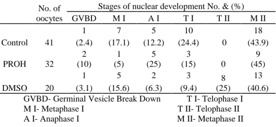
The meiotic resumption is characterized by Germinal Vesicle Break Down (GVBD), chromosomal condensation, progression to metaphase I (M I), release of the first polar body, and then arrested at M II (Motlik and Kubelka, 1990). At M II stage, oocytes achieve the competence to undergo normal fertilization and subsequent embryonic development (Sato and Koide, 1987). According to Datta and Goswami (1999) the first sign of meiotic resumption, GVBD was found to occur between 6 to 10h of culture, whereas at 16h M I, anaphase I (A I ) and telophase I (T I) were equally prevalent. M II stage started appearing in sizable percentage from 16h of culture and reached a peak value of 92.10% at 24h of in vitro culture for maturation.
18 Matsumo et al (2001) only used one step vitrification, so he had low maturation rates, so in this study, we had better results in nuclear stage MII, using 3 step vitrification.
Similarly, Dhali et al (2000) showed that low permeability in immature oocytes as compared to mature ones as a reason for low maturation ratio.
The CPAs used could change the success of maturation and when analyzing the rate of maturation it was found that the most advantageous CPA is PROH.
6. Fertilization
Fertilization is a procedure that assures the creation of a new individual from two gametes, female and male.
Fertilization is a complex event whose steps are regulated by varied cellular and molecular mechanisms. The duration of each step varies from one species to another, but is relatively long in all of them (Crozet, 1994).
The success of in vitro fertilization depends in large part on the maturation of the oocytes, as well as the ability to capacitate spermatozoa in vitro (Crozet, 1994). Once this difficulty is overcome all the steps of fertilization that occur in vivo also take place in vitro.
6.1. Sperm capacitation
In vitro capacitation procedure of semen aims at stimulating the sequence of events that normally occur in the cow’s reproductive tract. This involves the removal of seminal proteins and other substances that coat the sperm membrane of the semen (Duran, 2000).
19 For in vitro fertilization, we should assure a similar environment comparing in vivo, so that spermatozoa are able to undergo capacitation and penetrate the oocyte. Investigators first used several biological fluids, such as oviduct fluid, follicular fluid or blood serum, but these media had such a complex composition, that it was very difficult to determine exactly which components were actually involved in capacitating the spermatozoa. As the techniques evolved, we were able to formulate chemically defined mediums that provide good results. The media frequently used in IVF and in vitro capacitation are Tyrode’s Solution or Ringer-Krebs Solution, supplemented with the appropriate energy sources (glucose, lactate, Pyruvate) and albumin (Yanagimachi, 1994).
Parrish et al. (1986) stated that the major advances in controlling the IVF process have been the use of heparin (10 µg/ml) as capacitating agent and the swim-up technique to allow the use of frozen semen. Several methods for sperm selection have been described, such as: washing/centrifugation (Fukuda et al., 1990), density gradient centrifugation (Parrish et al., 1995), deferential filtration through a glass wool column (Stabbings and Wasik, 1991) and self-migration procedure (Lonergan et al., 1994).
The simple (centrifuge) sperm wash should be performed on a sample that has a decreased concentration and/or motility. A sample containing round cells and debris should not be prepared by this method. Sperm washing media is added to the specimen and centrifuged. The pellet is recovered, resuspended, and centrifuged again. The final pellet is resuspended in approximately 0.4 mls of media.
The level of pH (7.4, 7.7 and 8.0) of the sperm capacitation medium as a highly significant (P<0.01) effect on individual motility and completely acrosome reacted sperm. The individual motility declined as the pH of medium increased. However, a different trend was observed in the completely acrosome reacted sperm which increased
20 from 11.24±0.03 to 19.57±0.05% as the pH of medium increased (Brahmkshtri et al., 2000).
PH and viscosity are two factors that could have a major initial effect on sperm motility during the process of fertilization (Rutlant, J. et al., 1973). Furthermore, studies involving chemo tactic agents for sperm have yielded results suggesting evidence of follicular fluid involvement after ovulation as a sperm attractant (Eisenbech M.,1999; Mohammad S.N., 1997). Rikmenspoel and others have worked extensively on the effects of multiple variables on bovine sperm motility (Rikmenspoel, R.,1994; Rikmenspoel, R, et al., 1984) . Included in these studies are viscosity and pH. However, these variables were always tested with other confounding variables.
The pH of semen is slightly alkalotic and varies between 7.5 and 8.5. Studies quantifying physiological conditions affecting sperm motility have been done in the past (Rikmenspoel, R.,1994; Rikmenspoel, R, et al., 1984). However, research evaluating sperm motility at varied levels of pH and viscosity is lacking.
6.2.In vitro fertilization
Oocytes obtained from medium or large sized follicles possess higher developmental potential than oocytes obtained from small follicles, the treatment they received in terms of maturation medium, maturation time conditions can largely affect IVF.
6.2.1. IVF medium
Palta and Chauhan (1998) stated that the basic medium employed for IVF must be capable of providing an environment conducive to penetration of the oocyte by the sperm.
21 Tyrode’s modified (TALP) medium have been successfully used as the basic media for IVF of oocytes and a temperature of 38.5º-39ºC is optimal for performing IVF.
Bracket and Zuelke (1993) reported that insemination in TALP medium for a 24h interval led to the best results. Im et al. (1995) found that the fertilization and cleavage rates of bovine oocytes were 67.4 and 23.3% respectively in Bracket and Oliphant’s medium (BO) and 84.3 and 56.9%, respectively in TALP medium. Several components, added to the fertilization media, were tested in improving rate of fertilization. The presence of BSA as a source of protein in IVF medium was found to enhance sperm mobility and promote the acrosome reaction in capacitated spermatozoa (Fraser, 1985). Heparin in the IVF medium was reported to enhance sperm motility and fertilization rate (Parrish et al., 1985 and Saeki et al., 1994).
6.2.2. Temperature
Temperature plays an important role in capacitation. The most adequate temperature to support in vitro capacitation is 37-38ºC for most species. Nonetheless, for pigs or sheep, capacitation seems to be more effective at 39ºC. Even such a small variation can make all the difference in the physical status of the lipidic membrane (Yanagimachi, 1994).
6.2.3. Sperm concentration
Sperm concentration has been shown to be an important factor in determining the in vitro fertilization rate of antral oocyte. Ling and Lu (1990) reported that 6.4x10⁵ sperm/ml was optimal for in vitro fertilization. Nandi et al. (1998) indicated that the sperm concentration of 9 to 10 million sperm/ml, used in IVF yielded the highest
22 cleavage rates. However, an increase in sperm concentration has been reported to increased polyspermy, using 1x10⁶ sperm/ml (Totey et al., 1993a and Saeki et al., 1994). The role of cumulus cells during IVF is, however, not clear. Some investigators found that, oocytes with expanded cumuli fertilize at higher rates than those with compacted or poorly expanded cumuli. It has been postulated that cumulus expansion plays an important role in process of fertilization by enhancing sperm capacitation and increasing sperm motility.
6.2.4. Sperm – Oocyte incubation time
The choice of an optimum length of time for sperm-oocyte incubation is essential for obtaining high rates of fertilization without increasing the incidence of polyspermy (Palta and Chauhnan, 1998). The duration of incubation is dependent on the sperm concentration used for IVF. The use of low sperm concentration of 0.7 million sperm/ml requires a longer duration of 48h which could be reduced to 20-24h by increasing the sperm concentration to 1 million (Samad et al., 1998).
The optimum duration of sperm/oocyte incubation was stated to be 24h using 1x10⁶ sperm/ml as a final concentration. Dode et al. (2002) concluded that the sperm-oocyte co-incubation time affected the fertilization rate.
7. Embryo culture
The transferrable embryos obtained at the end of the culture period must be free of pathogens. Culture of embryos in simple defined media without any cell support would be the ideal solution from a sanitary point of view.
Co culture of the ova with granulosa, cumulus or uterine cells from slaughterhouse material presents a major risk of contamination. This type of co culture
23 should be used only if cells can be controlled for the presence of viroses, and the results are known before transfer of the resulting embryos into recipients. Media conditioned with granulosa, cumulus or uterinetubal cells are an easier alternative to co culture as they can be controlled in advance and kept frozen until use (IETS, 1998).
After fertilization of the oocyte, a complex program at the level of genes, directs the process of embryogenesis. The genetic program in mammalian embryos coordinates and develops series of divisions, migrations and differentiations. These process transform a single embryonic cell, the oocyte fertilized, into a complex embryo matured (Burdsal, 1998).
Some major events occur during the period of six days from the zygote until the beginning of blastocyst formation in cattle (Lonergan et al., 2003). This period includes the first division of the zygote, the period known to be critically important in the determination of subsequent ability like development of the embryo (Lonergan et al., 2003); the embryonic genome activation stage in 8-16 cells (Memli and First, 2000); the compaction of morula on day 5, which involves the establishment of first contiguous cell-to-cell contacts in the embryo (Boni et al., 1999) and the blastocyst formation on the day 6-7, involving the differentiation of two types of cells : the trophoblast and the embryonic button (Watson and Bareroft, 2001). Thus any modification of the culture environment, which may affect any or all of these processes, may have a detrimental effect on embryo quality (Lonergan et al., 2003).
Despite the conditions of culture can influence the development of the embryo at the stage of pre-implantation (Pinyopummintr and Bavister 1994; Langendonckt et al., 1997), is likely that the main factors controlling the ability of the embryo development are intrinsic to the oocyte (Lonergan et al., 2000), the sperm (Ward et al., 2001) or both (Lonergan et al., 2003).
24 The development of the blastocyst is just one step on a long road to production of a foal (Lonergan et al., 2003).
The proportion of cattle oocytes that become transferable embryos remains sub optimal, usually no more than 50% and typically between 25 to 30% (Hansen and Block, 2004). The failure to obtain higher rates of blastocyst production due a combination of factors, including the collection of oocytes with low aptitude for fertilization and subsequent development (Hansen, 2002), inadequate conditions during maturation and fertilization (Rizos et al., 2002) and embryo culture systems sub optimal process (Thompson, 2000).
As pointed out by McEvoy and his collaborators (McEvoy et al., 2000), the attainment of the state of blastocyst is more a reflection of the past than a guarantee of future success. Well beyond increasing the success rate of embryo production in vitro is essential that the embryos that actually reach this state, are of the highest quality possible, so as to ensure that a normal pregnancy after embryo transfer.
Today there is a large source of studies showing that the culture environment after fertilization has a huge effect on the expression of genes in the embryo, which in turn has serious implications in normal development of the embryo until the blastocyst stage. Historically, there was great difficulty in the embryos cultured in vitro to pass the mythical block of the 8-16 cells, an event triggered by sub optimal characteristics from culture (Eyestone and First, 1991). In vitro culture systems to the reproductive tract environment were improved, to support the development of the embryo in the pre-implantation and thereby be possible to overcome the block of 8-16 cells.
The co-culture is one of the methods to improve embryo development in vitro. At the beginning of the 80 decade, the co-culture was the only method to achieve good rates from blastocyst production of large domestic animals species. It was evident that,
25 the knowledge about the physiology of the blastocyst would come through the co-culture. Both basic knowledge (effect on maternal and paternal) and applied knowledge (how to handle a program of IVF blastocysts in large scale, such as freezing blastocysts...) was based on the co-culture. The effects of these embryotrophics mono-layers of cells are known as epithelium more than hormone-dependent (Papaioannou and Ebert, 1986), and are not specific to species (Boland, 1984) or from organs (Ménézo et al., 1990). The mechanism by which the co-culture cell embryotrophic exerts its effect, an effect that allows increasing the formation of blastocysts is still not completely known. The mechanism of action of the cells may be to remove toxins from the environment, culture, or to help dilute the effect of any potential inhibitor compound in the microenvironment of the embryo. According to several studies, it is also known, that during its development and proliferation, these cells produce somatic embryotrophics substances which are beneficial to the embryo (Ménézo et al., 1990; Desai et al., 2000).
8. Embryo quality
After oocyte fertilization, a complex program at the level of genes directs the process of embryogenesis.
The International Embryo Transfer Society (IETS) established a series of numerical codes in order to facilitate the certification and identification of the stage of development of the embryo, as well as its quality.
26 Table 2 – IETS numerical codes for the stage of development of embryos
Numerical Code Stage of development
1 Unfertilized 2 2 to 12 cell 3 Early Morula 4 Morula 5 Early Blastocyst 6 Blastocyst 7 Expanded Blastocyst 8 Hatched Blastocyst
27 Fig.1- 2 Cell Fig.2 –2 to 4 Cell
28 Fig.5- Morula Fig.6 – Late morula/ Early Blastocyst
Fig.7- Early Blastocyst Fig.8 – Blastocyst
Like oocytes, embryos can also be classified into different quality categories, according to their morphological excellence. There are also four grades of quality as follows (LeGuienne et al., 1990):
29 Excellent - ideal embryo, spherical, symmetrical, with cells of comparable texture and color; for those in morula stage-compact;
Good – embryo a little late in its development, or embryo with excellent qualities but asymmetrical, or even that shows the exclusion of any blastomer in the perivitelline space.
Quality 2
Fair – embryo one or two days late in its development with precise defaults, like: too many cells in the perivitelline space; vesicles; degenerated cells; variable sized blastomers; with a lighter or darker aspect than normal.
Quality 3
Poor – defaults like loose, degenerated or different sized cells and thick vesicles in large number, but with the presence of an homogeneous mass that looks viable.
Quality 4
30 III Materials and Methods
Ovary source
Ovaries (n=340) were obtained from cows slaughtered in a local slaughterhouse, washed and transported to the laboratory in PBS saline solution (Phosphate Buffered saline, P 0290, sigma) at 38ºC being processed within 2 hours of slaughter.
Preparation of culture medium
All media used in this study were prepared in a sterile environment through a horizontal laminar flow hood. This chamber is in a room, free of drafts, to enable a stable ambient temperature.
The media for vitrification were frozen until use and the culture media were filtered into sterile tubes with filters of 22 µm and placed in a incubator at 38.5ºC in 5% CO2 and saturated humidity, 2 hours before use.
Collection of oocytes
The ovary was dried lightly with paper towels before follicular cumulus-oocyte complex (COCs) was aspirated from 2-6 mm diameter antral follicles using an 18-gauge needle connected to a 10 ml syringe. To avoid disruption of the COCs, by applying the aspiration method, the needle and syringe were primed with approximately 0.25 – 0.5 ml of washing medium, consisted of TCM199 buffered with Hepes, supplemented with serum of cow (2%), Glutamine (0.3mg/ml), Gentamycin (50µg/ml) and Streptomycin (50µg/ml).
31 COCs and follicular fluid were slowly expelled into 10 ml tube and maintained there for at least 5 minutes for sedimentation of the COCs. The precipitate was taken into sterile petri dish for subsequent studies on COCs.
Evaluation of the quality of the COCs
With the aid of a binocular magnifying glass, the COCs were picked up and they underwent a couple of rinses in afore mentioned washing medium. They were evaluated according to morphological criteria and separated according to their quality.
Oocytes Cryopreservation
The cryoprotectors that were use in this experience was the DMSO and PROH. For their preparation, it was used L-15 (L-15 Leibovitz) instead of the TCM-199.
DMSO is used as a cryoprotectant, added to cell media to prevent cell death during the freezing process (Pegg, E. 2007). Approximately 10% may be used with a slow-freeze method, and the cells may be frozen at -80ºC or stores in liquid nitrogen safely.
Oocytes were exposed, in a way, in a two steps to balance the vitrification solution. The first one was at 10% PROH and 10% DMSO for two minutes, in a room temperature, and after those two minutes they were transfer for 20% PROH and 20% DMSO only for 30 seconds. After this, we put around 15-20 oocytes in 20µl from the last solution, and put them in a French Mini Straw (FMS), which already contain sucrose (0.5M) and after the addiction of the oocytes, more sucrose is addicted and we sealed the straw with isolant and put the straw in the liquid nitrogen. The straws are
32 thawed in a water bath at 37ºC, for 30 sec, after a minimum period of storage for a week.
.
Thawing
For thawing, the straws were thawed for 30 sec, for a minimum period of storage for a week. The straws were submitted to several concentrations of sucrose during 5 minutes, to balance the medium. The first concentration was 0.5M, the second 0.1M and the last 0M. All of these concentrations were in a plate with a drop of 200µl.
In vitro oocytes Maturation
After thawing, intact oocytes were washed two times in washing medium. Subsequent two washings were carried out in a maturation medium consisting of TCM 199-Hepes supplemented with 10% FCS, 0.02 IU/ml FSH (Sigma), 1µg/ml E2 (Sigma), 0.15mg/ml Glutamine, 22µg/ml Na-Pyruvate, 10µg/ml Gentamycin, 10µg/ml Streptomycin and 10µg/ml Nistamycin. Groups of 15-20 COCs were randomly allocated to 100µl microdrops of the maturation medium under mineral oil (Sigma) in small petri dish and incubated in a incubator at 38.5ºC in 5% CO2 and satured humidity
Nuclear Maturation
After 24h of incubation for maturation, a part of it was utilized to obtain artificially denuded oocytes from surrounding cumulus cells using a sterile glass pipette with bore small enough to remove the cumulus cells without damaging the oocytes. The oocytes were then fixed by modified whole oocytes mounting method to access the state of nuclear maturation. The denuded oocytes were taken in a petri dish containing
33 fixation solution (methanol:glacial acetic acid, 3:1) and left for 24h. After the fixation period was over, the oocytes were replaced on glass slide followed by covering them with a cover slip and stained with 1% aceto orcein for 5 minutes before examining under inverted microscope.
The different nuclear configurations observed were classified according to Datta and Goswami (1999) and Marilia (1999) into the following categories:
I – Germinal Vesicle (GV):
Oocytes nucleus stage 1 (ON1)
The oocytes having a distinct nuclear envelop, rounded GV, chromatin only around the nucleolus and not condensed thus not always visible.
Oocytes nucleus stage 2 (ON2)
The nuclear envelop still present but less distinct and with irregular form, with chromatin condensed and visible.
II – Metaphase I:
The chromosomes are arranged in the metaphase plate and the diploid sets of chromosomes (2n) are fully condensed with absence of polar body component of chromatin mass.
III – Anaphase I:
Separation of bivalent pairs of chromosomes along with their spindles, when observed laterally.
34 IV – Telophase I:
The chromatids are separated, and one set of the chromosome (n) reached one of the two poles, while the remnants of spindles are still not detached.
IV – Metaphase II:
The polar body is observed in the perivitelline space. Initially the oocytes and polar body chromatin components are observed as pairs of darkly stained masses, which subsequently started condensing to reveal two sets of condensed chromatin materials. The polar body component subsequently started degenerating. The oocytes chromatin is condensed.
Preparation of spermatozoa and in vitro fertilization of the oocytes
The spermatozoa used in this experiment came only from one donor, “Winter”. Straws were place in a water bath at 37ºC, for one minute.
Sperm were recovered from frozen semen by “Swim Up” separation in TALP medium (Thyroid Albumin Lactate Pyruvate). The contents were washed twice in SP-TALP (Tables X and Y) (4ml) by centrifugation at room temperature at 1800 rpm for 5 minutes to remove extender and cryoprotectant. The sediment of spermatozoa was resuspended in IVF-TALP (Tables X and Y) (4ml) containing 10µg/ml heparin and then centrifuged at 1800 rpm for 5 minutes. The supernatant was removed leaving 0.25-0.5 ml of IVF-TALP and sperm pellet. The concentration of sperm was adjusted by adding IVF-TALP medium to reach 1x10⁶sperm/ml as tested by a haemocytometer.
35 Table 3- Stocks preparation for composing media as in table 4 and 5
Stocks Preparation
NaCl (g) Dissolve 6.665g in 50 ml water* KCl (g) Dissolve 0.588g in 50 ml water NaHCO3 (g) Dissolve 1.052g in 50 ml water NaH2PO4 (g) Dissolve 0.235g in 50 ml water Na-lactate
CaCl2 (g)
60% syrup
Dissolve 1.470g CaCl2.2H2O in 50 ml water MgCl2 (g) Dissolve 1.017g MgCl2.6H2O in 50 ml water Na-pyruvate (g) Dissolve 0.220g in 100 ml water
Heparin Dissolve 20mg in 10 ml water
HEPES (g) 119g HEPES to 400ml water, then adjust the pH to 7.0 and bring volume up to 500ml
*Distilled or deionized water
Table 4- Recipes for preparation of TL solutions, according to Hansen (2000)
Ingredient (ml) Sp-TL HEPES-TL IVF-TL
Water (ml) 40.5 ml 30 ml 40.157 ml Stock 1: NaCl (ml) 2.220 ml 1.7 ml 2.5 ml Stock 2: KCl (ml) 1.0 ml 677 µl 1.0 ml Stock 3: NaHCO3 (ml) 5.110 ml 270 µl 5.0 ml Stock 4: NaH2PO4 (ml) 511 µl 340 µl 500 µl Stock 5: CaCl2 (ml) 511 µl 340 µl 500 µl Stock 6: MgCl2 (ml) 562 µl 170 µl 250 µl Stock 7: HEPES (ml) 511 µl 340 µl ----
36
pH 7.4 7.3 7.4
Osmolarity (mOsm) 295 – 305 275 – 285 290 – 300
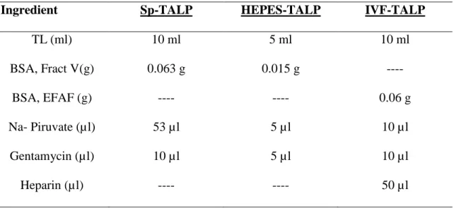
Table 5- Recipes for preparation of TALP Media according to Hansen (2000)
Ingredient Sp-TALP HEPES-TALP IVF-TALP
TL (ml) 10 ml 5 ml 10 ml BSA, Fract V(g) 0.063 g 0.015 g ---- BSA, EFAF (g) ---- ---- 0.06 g Na- Piruvate (µl) 53 µl 5 µl 10 µl Gentamycin (µl) 10 µl 5 µl 10 µl Heparin (µl) ---- ---- 50 µl
After 24h of IVM, COCs were washed two times in Hepes-TALP, followed by final washing in fertilization medium (IVF-TALP). Oocytes were allocated in small petri dish containing 50 µl droplets of fertilization medium at the rates of 10 oocytes per drop. Aliquots of the sperm suspension were added to each droplet containing matured oocytes. The oocytes and sperm were co-cultured in a incubator at 38.5ºC in 5% CO2 in air, with maximal humidity for 22-24h.
37 In vitro embryos culture
I- Culture without granulosa cells monolayer:
After 22h of co-incubation of sperm and COCs, the presumptive embryos were washed in Hepes-TALP medium and stripped of cumulus cells by repeated pipetting. The final washing was done in a culture medium consisting of: TCM-199 supplemented with 3 mg/ml BSA, 22 µg/ml Na-Pyruvate, 10 µl/ml NEAA(100x), 20 µl/ml EAA (50x) and 50 µg/ml Gentamycin. Presumptive embryos were placed, in a four well petri dish in the culture medium covered with paraffin oil during culture period in incubator. Half of the medium was changed every 48 hours.
II- Co-culture with granulosa cells monolayer:
For the culture with granulosa cells, after withdrawing the COCs of the follicular fluid with washing medium, the suspension was placed in a small falcon tube and centrifuged for ten minutes. The suspension was taken and thrown out, leaving at the bottom a follicular supernatant (white part). This supernatant was homogenized with 1ml of granulosa cells medium (GCM), composed of 9 ml TCM-199, without Hepes, 1 ml of FCS, 10µl gentamycin, 10µl penistreptomycin and 10µl nystatin. Using a syringe (1ml) and a needle (19G) we mix everything and add 1ml of GCM and mix again.
To count the cells, we put in ependorf, 20 µl of suspension consisting of granulosa cells and 20 µl tripan blue, mix and put in Newbower chamber. 5 squares were counted on diagonal. In a well box we put 50 µl of the concentration and cover with oil. After fertilization, the carpet is changed, passed 22 h.
38 Assessment of embryo quality
The successful formed embryos by fertilization were examined by inverted microscope.
Grade 1: Excellent or Good
The development stage corresponds to the expected, the embryonic mass is spherically symmetric and with individual blastomeres that are uniform in size, color and density; regular shape, the pellucid zone should not have concave or flat surface should be smooth, preferably intact, especially if the embryo is intended to export; extruded cells from the mass cells of the embryo comprise less than 15% of total cellular material.
Grade 2: Fair
The development stage corresponds to the expected; regular shape, pellucid zone non-intact, moderates irregularities in the general shape of the embryonic mass or size, color and density of individual cells; extruded cells from the cells mass of the embryo, comprise less than 15% of material from cells; at least 50% of the cells comprise a viable embryonic mass intact.
Grade 3: Poor
The developmental stage doesn’t match the expected; major irregularities in the general shape of the embryonic mass or size, color and density of individual cells; less than 75% degenerated cells; at least 50% of the cells comprise a viable embryonic mass intact.
39 Grade 4: Dead or Degenerate
The developmental stage doesn’t match to the expected, embryo degeneration; embryonic mass of less than 25% of all cellular material present in the pellucid zone; oocytes or unicellular structures are degenerated.
These are the criteria used to evaluate the quality of embryos, proposed by IETS (International Embryo Transfer Society) (1998).
Statistical Analysis
The data of post-thaw in vitro development of morula, early blastocyst, blastocyst and the rate of cleavage were analized by one way ANOVA to verify differences between the groups followed by the LSD test between group means by using computer assisted statistical software SPSS Statistics 17.0. The significance of differences between means values was determined at P < 0,05. Results were expressed as means ± SE. Pearson’s correlation coefficient was calculated.
40 IVResults
Table 6 – Oocytes survival rate and their subsequent in vitro development after cryopreservation without co-culture.
Collected
Oocyte Cleaved Morula
Early Blastocyst Blastocyst Developed Embryos * Control 187 74.3 ± 1.6 a 39.8 ± 4.5a 21.1 ± 3.1a 9.1 ± 4.9a 70.7 ± 3.3a (139) (56) (30) (12) (98) PROH 174 71.4 ± 7.1 a 26.1 ± 3b 29.3 ± 3.9a, b 5.5 ± 2.8a 59.9 ± 9.4a (123) (32) (34) (7) (73) DMSO 172 69.7 ± 2.9 a 32.4 ± 3.4a, b 34.4 ± 0.5b 0.9 ± 0.9a 67.7 ± 4.3a (119) (31) (41) (1) (80)
* This is the total of embryonic developmental stage, including Morula, Early Blastocyst and Blastocyst a,b
Numbers in the same column with different letters (a,b,c) differ significantly at P<0.05
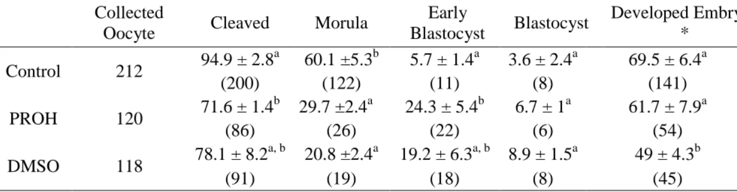
Table 7 – Oocytes survival rate and their subsequent in vitro development after cryopreservation with co-culture.
Collected
Oocyte Cleaved Morula
Early Blastocyst Blastocyst Developed Embryos * Control 212 94.9 ± 2.8 a 60.1 ±5.3b 5.7 ± 1.4a 3.6 ± 2.4a 69.5 ± 6.4a (200) (122) (11) (8) (141) PROH 120 71.6 ± 1.4 b 29.7 ±2.4a 24.3 ± 5.4b 6.7 ± 1a 61.7 ± 7.9a (86) (26) (22) (6) (54) DMSO 118 78.1 ± 8.2 a, b 20.8 ±2.4a 19.2 ± 6.3a, b 8.9 ± 1.5a 49 ± 4.3b (91) (19) (18) (8) (45)
* This is the total of embryonic developmental stage, including Morula, Early Blastocyst and Blastocyst a,b
Numbers in the same column with different letters (a,b,c) differ significantly at P<0.05
Embryo culture without co-culture system, there was no significant difference between cleaved embryos in control, PROH and DMSO groups. In morula, there was significant difference between control 56 (39.8%) and PROH 32 (26.1%), however there was no statistical difference between PROH and DMSO 31 (32.4%). Early