ESCOLA UNIVERSITÁRIA VASCO DA GAMA
MESTRADO INTEGRADO EM MEDICINA VETERINÁRIA
MAGNETIC RESONANCE IMAGING AND ITS
APPLICABILITY IN VETERINARY CARDIOLOGY
José Manuel de Seiça Ferreira
ESCOLA UNIVERSITÁRIA VASCO DA GAMA
MESTRADO INTEGRADO EM MEDICINA VETERINÁRIA
MAGNETIC RESONANCE IMAGING AND ITS
APPLICABILITY IN VETERINARY CARDIOLOGY
Coimbra, Fevereiro de 2016
José Manuel de Seiça Ferreira
Aluno do Mestrado Integrado em Medicina Veterinária
Orientador Interno
Professora Doutora Maria João Nobre de Matos Pereira Vieira
Orientador Externo
Dr. João Manuel Pimenta Ferreira de Oliveira
Médico Veterinário
Dissertação do estágio curricular dos ciclos de estudo conducentes ao Grau de Mestre em Medicina Veterinária da EUVG
iv
Abstract
Magnetic Resonance Imaging (MRI) is a technique whereby images are created by the manipulation of hydrogen atoms in magnetic fields; it is based on the principle of nuclear magnetic resonance (MR), which is non-invasive and non-ionising (Constantine, Shan, Flamm, & Sivananthan, 2004). Cardiac Magnetic Resonance Imaging (CMRI) uses the same principle: application of magnetic-field gradients that are adjusted to highlight desired tissue characteristics, producing a variety of sequences that allow detection of cardiac tissue and blood, and consequently anatomical and/or physiological abnormalities (Jeudy & White, 2008; Constantine et al., 2004). Basic pulse sequences used in CMRI are spin-echo and gradient-echo sequences, or their faster hybrids dark- or black-blood and bright-blood respectively (Constantine et al., 2004).
CMRI is rapidly developing and is now an important diagnostic tool in human clinical cardiology (Gilbert, McConnell, Holden, Sivananthan, & Dukes-McEwan, 2010). In veterinary medicine the use of CMRI is still sporadic; its limitations in this field include the need for general anaesthesia, the cost and availability of the equipment, the steep learning curve to obtain and analyse the images, and the time needed to manually trace endocardial borders if semi-automated analysis is not available (MacDonald, Kittleson, Garcia-Nolen, Larson, & Wisner, 2006). CMRI was considered to be the reference method in many veterinary studies (Eskofier, Wefstaedt, Beyerbach, Nolte, & Hungerbuhler, 2015; Fattal et al., 2015; Sargent et al., 2015).Still, not many studies have been published or made available in this field. It is therefore essential to fully ascertain the clinical applications, advantages and limitations of CMRI in veterinary medicine. The aim of this review is to identify the potential applications of CMRI from a clinical point of view and compare it with echocardiography, which is still the gold standard in veterinary cardiology. We describe the principles and technique of MRI in small animal cardiology, and the diseases in which CMRI could be an important tool for diagnosis and prognosis.
Key-words: Magnetic Resonance Imaging; Cardiac Magnetic Resonance Imaging; veterinary cardiology; dogs; cats
v
List of contents
Introduction ... 1
1. Principles and Technique of Magnetic Resonance Imaging ... 2
1.1. Magnetic Resonance Imaging physics ... 2
1.2. Sequences used in Cardiac Magnetic Resonance Imaging ... 3
1.2.1. Spin-echo pulse sequence: Black-Blood ... 4
1.2.2. Gradient-echo pulse sequence: Bright-Blood ... 4
1.3. Performance of a Cardiac Magnetic Resonance Imaging examination under general
anaesthesia ... 5
1.4. Contrast agents used in Magnetic Resonance Imaging ... 6
2. Cardiac Magnetic Resonance Imaging in veterinary medicine ... 8
2.1. Echocardiography vs Magnetic Resonance Imaging ... 8
2.2. Morphology and function... 8
2.3. Left ventricular myocardial mass, chamber volume and haemodynamic ... 12
2.4. Valvular heart disease ... 13
2.5. Cardiomyopathies ... 14
2.5.1. Hypertrophic cardiomyopathy ... 14
2.5.2. Dilated cardiomyopathy ... 15
2.5.3. Arrhythmogenic Right Ventricular Cardiomyopathy ... 15
2.5.4. Myocarditis and myocardial viability ... 16
2.6. Congenital heart disease ... 17
2.7. Ischemic disease ... 18
2.8. Pericardial disease ... 18
2.9. Other applications ... 19
3. Specific difficulties with transposition of human Cardiac Magnetic Resonance Imaging to veterinary
cardiology ... 19
4. Cardiac Magnetic Resonance Imaging studies in cats and dogs – experimental or clinical? ... 19
5. Future perspectives ... 20
Conclusion ... 21
vi
List of figures
Figure 1: Representation of the application of radiofrequency (RF) and the transversal magnetisation. 3 Figure 2: Representation of the deflected protons returning to its original position. ... 3 Figure 3: Comparison between black-blood (left) and bright-blood (right) axial images of the human hearth. Adapted from: Jeudy and White (2008). ... 5 Figure 4: Positioning of the cat for CMRI examination. Adapted from: MacDonald, Kittleson, et al. (2005). ... 6 Figure 5: Late gadolinium enhancement imaging of a rat heart with myocarditis in short-axis (left) and four-chamber (right) views. Image courtesy of Rinkevich-Shop et al. (2013). ... 7 Figure 6: Short axis view of the left ventricle: end diastolic (left) and end systolic (right). Adapted from: Drees, Johnson, Stepien, Munoz Del Rio, Saunders, et al. (2015) ... 10 Figure 7: End systolic (left) and end diastolic (right) three-chamber view. Adapted from: Drees, Johnson, Stepien, Munoz Del Rio, Saunders, et al. (2015). ... 11 Figure 8: Approximate four-chamber view (left) and transverse plan (right). Adapted from: Drees, Johnson, Stepien, Munoz Del Rio, Saunders, et al. (2015). ... 11 Figure 9: Pathological changes in boxer dogs with ARVC. Source: Basso et al. (2004). ... 16 Figure 10: Late gadolinium enhanced (LGE) distribution in left ventricle (LV) of rats with myocarditis. Image courtesy of Rinkevich-Shop et al. (2013). ... 17
vii
List of tables
Table 1: Left ventricular planar measurements generated from echocardiographic exams obtained in 10 awaken dogs, as well as CMRI with two different anaesthetic protocols. Adapted from: Drees, Johnson, Stepien, Munoz Del Rio, and Francois (2015). ... 9 Table 2: Right ventricular function variables comparing CMRI, computed tomography and three-dimensional echocardiography in dogs. Adapted from: Kim et al. (2013), Sieslack et al. (2014) and Meyer et al. (2013) ... 12
viii
List of abbreviations
3DE – three-dimensional echocardiography Ao – aorta
Ao annulus 3ch – aortic annulus measured on three-chamber view
Ao/PA ratio – aorta to pulmonary artery ratio ARVC – arrhythmogenic right ventricular cardiomyopathy
CCT – cardiac computed tomography CMRI – cardiac magnetic resonance imaging ECG – electrocardiogram
EDV – end-diastolic volume EF – ejection fraction ESV – end-systolic volume IVS – interventricular septum
IVSd – diastolic interventricular septal IVSs – systolic interventricular septal LA – left atrium
LA diam 3ch – left atrial diameter measured on three-chamber view
LA/Ao ratio – left atrium to aorta ratio LGE – late gadolinium enhanced
LPA – left pulmonary artery LV – left ventricle
LVID – left ventricular internal diameter LVIDd – diastolic left ventricular internal diameter
LVIDs – systolic left ventricular internal diameter
LVPW – left ventricular posterior wall
LVPWd – diastolic left ventricular posterior wall LVPWs – systolic left ventricular posterior wall Mitral annulus 3ch – mitral annulus measured on three-chamber view
Mitral annulus 4ch – mitral annulus measured on four-chamber view
MPA – main pulmonary artery MRI – magnetic resonance imaging
prox Ao – proximal aorta measured on transverse plane
RF – radiofrequency
RPA – right pulmonary artery RV – right ventricle
1
Introduction
MRI has become recognized as a useful referral diagnostic method in veterinary medicine, and is widely used in small animal brain and spinal diseases (Gavin, 2011; Gilbert et al., 2010), aural, nasal and orbital disorders, planning soft tissue surgery, oncology and small animal and equine orthopaedics (Gilbert et al., 2010). The use of MRI in these subjects has grown due to its unparalleled capability to image soft tissue structures (Gilbert et al., 2010) to a much better degree than other modalities (Constantine et al., 2004).
During the past decade, the application of MRI to cardiology has provided new insights in the investigation of cardiovascular diseases. With technological advancements, we can now rapidly acquire large imaging datasets that are essential for imaging the heart (Constantine et al., 2004). This has been successfully applied to human cardiology where, despite the inherent difficulties in imaging a moving and contractile structure, CMRI has become the optimal technique for the morphological assessment and quantification of ventricular function (Gilbert et al., 2010; Vallee, Ivancevic, Nguyen, Morel, & Jaconi, 2004). It is also a clinically important technique for the assessment of cardiac structure, function, perfusion, and myocardial viability (Constantine et al., 2004; Vallee et al., 2004). In veterinary medicine, CMRI is considered to be the gold standard technique for right ventricular volumetric measurement (Sieslack, Dziallas, Nolte, Wefstaedt, & Hungerbuhler, 2014) because of its high temporal and spatial resolution and its detailed soft tissue contrast, resulting in high accuracy and reproducibility of the measurements. CMRI has also been considered to be a reference method in many studies when compared with real-time two-dimensional and three-dimensional echocardiography; in dogs it was been used to the quantification of left ventricular volume (Eskofier et
al., 2015; Sieslack et al., 2014), and function in anaesthetized dogs (Eskofier et al., 2015); to quantify
right ventricular volume (Sieslack et al., 2014); to evaluate mitral regurgitation (Sargent et al., 2015); to study cardiac morphology and function in Boxer with arrhythmogenic right ventricular cardiomyopathy (ARVC) (Baumwart, Meurs, & Raman, 2009); to differentiate neoplastic and non-neoplastic pericardial effusion (Boddy et al., 2011); and in cats to study hypertrophic cardiomyopathy (MacDonald, Wisner,
et al., 2005; Sugimoto, Fujii, Sunahara, & Aoki, 2015).
CMRI is a promising diagnostic tool for the majority of cardiac pathologies. However, some studies are needed to compare the measurements and values obtained by CMRI and echocardiography to standardize the technique and establish reference values by animal breed, weight and age. It is essential to fully ascertain the clinical applications, advantages and limitations of CMRI in veterinary medicine in comparison to the more widespread and proven diagnostic tool that is echocardiography.
2
1. Principles and Technique of Magnetic Resonance Imaging
MRI is a non-invasive and non-ionising radiation technique based on the principle of nuclear magnetic resonance, and has intrinsic attributes well suited for cardiac imaging as being a tomographic technique that can acquire images in virtually any orientation (Constantine et al., 2004).
1.1. Magnetic Resonance Imaging physics
The acquisition of MRI images depends on protons; images are derived from signals produced by the protons (hydrogen nuclei), which are mainly attached to water molecules. The proton behaves like a small magnet that spins when placed in a magnetic field; this produces a small magnetic field that can align with a superconducting magnet of a larger magnetic field (Constantine et al., 2004; Jeudy & White, 2008). Protons will align with the primary magnetic field, both parallel and antiparallel to its direction; in addition, a spatially applied field generates slightly different spins along a gradient. A radiofrequency pulse can then be applied in such a way that only protons spinning at the same frequency as the radiofrequency pulse will capture the radiofrequency energy and change the direction of their magnetization (figure 1) (Jeudy & White, 2008). The magnetization of these protons is then momentarily deflected away from the direction of the vector of the superconducting magnet. After the radiofrequency pulse ceases, these deflected protons relax back towards their original position (relaxation) releasing energy in the form of a radiofrequency signal (figure 2). This signal is captured by a radiofrequency receiver coil and forms the basis for images obtained on MRI (Jeudy & White, 2008). The return of the net magnetisation vector to equilibrium has two components: the vector component parallel to the main field returns to equilibrium by interacting with surrounding molecules which is a relatively slow process and is known as T1 relaxation (longitudinal). The vector component transverse to the field is more rapid to decline, resulting from the interaction between individual spins, and is termed T2 relaxation (transverse) (Constantine et al., 2004). A series of radiofrequency pulses of varying duration or strength and application of magnetic-field gradients that are adjusted to highlight desired tissue characteristics produce a variety of sequences that are applicable to CMRI (Constantine et al., 2004; Jeudy & White, 2008).
Cardiac and respiratory motion artifacts can be overcome in human awake subjects by electrocardiography-gating (using electrocardiogram (ECG) signal to synchronizing data acquisition with specific phases of the cardiac cycle to eliminate artifacts due to cardiac motion (Gilbert et al., 2010; Mai, Badea, Wheeler, Hedlund, & Johnson, 2005)) and breath-hold navigation; this allowed for real-time techniques to be developed and refined to synchronise data acquisition and keep artifacts to a minimum or eliminate them, thus allowing for clear and precise images of morphology and function. However when using ECG-gating, cardiac dysrhythmias can significantly interfere with synchronised data acquisition (Constantine, Shan et al. 2004).
3
Figure 1: Representation of the application of radiofrequency (RF) and the transversal magnetisation. The
application of a RF pulse in such a way that only protons spinning at the same frequency as the RF pulse will capture the radiofrequency energy and change the direction of their magnetization.
Source: https://upload.wikimedia.org/wikibooks/en/9/97/InvRecov3.jpg; acessed on 13.02.2016
Figure 2: Representation of the deflected protons returning to its original position.
Source: https://upload.wikimedia.org/wikibooks/en/2/27/InvRecov6_1.jpg; acessed on 13.02.2016
1.2. Sequences used in Cardiac Magnetic Resonance Imaging
Most common pulse sequences used in CMRI are spin-echo and gradient-echo, or their faster hybrids dark- or black-blood and bright-blood respectively (Constantine et al., 2004). Spin-echo sequences are chosen for the assessment of morphology, and flowing blood appears black (figure 3); on the other hand gradient-echo sequences are used in the assessment of valvular lesions, shunts, great vessels, ventricular function and wall-motion characteristics (Constantine et al., 2004; Gilbert et al., 2010). Gradient-echo sequences have fairly low soft-tissue contrast compared with spin-echo sequences, in gradient-echo the flowing blood is represented by high signal intensity and turbulence as areas of signal void (figure 3) (Constantine et al., 2004).
4 1.2.1. Spin-echo pulse sequence: Black-Blood
Black-blood imaging was conventionally done with multi-slice T1-weighted spin-echo sequences; this sequence frequently provides excellent high quality anatomic images of cardiac chambers, great vessels and pericardium, and is relatively unaffected by metallic artifacts. These sequences are of uttermost importance when precise anatomic detail is necessary (Jeudy & White, 2008) such as in the assessment of scar tissue formed in chronic infarct (Kali et al., 2014; Thajudeen et al., 2015).
Recently, T1-weighted spin-echo imaging sequences have largely been supplanted by echo-train imaging with double inversion sequences because of its rapidity, this approach can be applied in a single slice per breath-hold fashion or images of the entire heart can be obtained in a single breath hold using a single-shot strategy (Jeudy & White, 2008), on the other hand fast T2-weighted images are used occasionally, especially for patients with cardiac masses or pericardial disease (Boddy et al., 2011; Jeudy & White, 2008). The major disadvantage of this kind of sequences is the long imaging time of several minutes and the associated motion artifact that may occur (Jeudy & White, 2008).
1.2.2. Gradient-echo pulse sequence: Bright-Blood
Gradient-echo techniques are turbulence sensitive, and thus can be employed to detect areas of turbulent flow due to stenosis or regurgitation of blood flow around the lesions, as well as to explore the pattern of flow in shunts and vessels, and in the assessment of ventricular wall-motion (Constantine et al., 2004; Jeudy & White, 2008); turbulent blood appears black in these techniques, while laminar flow appears bright (Gilbert et al., 2010). The extent of stenosis or regurgitation can be quantified using velocity-encoded sequences (Jeudy & White, 2008). Using phase encoding velocity maps, flow volumes, and flow velocity, may then be quantified across valves or shunts (Constantine et
al., 2004; Gilbert et al., 2010).Typically bright-blood images are placed in a cine-loop; this format
allows good visualization of cardiac motion at a single level in any plane during the cardiac cycle, and is essential to determine cardiac ejection fraction and to assess wall motion (Jeudy & White, 2008). Important disadvantages of gradient-echo techniques include high sensitivity to inhomogeneity in the magnetic field, images’ propensity to artifacts from metal (Jeudy & White, 2008) and less contrast between blood and soft tissue being provide by these sequences (Constantine et al., 2004).
Bright-blood sequences have increasingly become the backbone of human CMRI. Initially, bright-blood imaging was performed with a spoiled gradient-echo sequence and a single view was obtained per cardiac cycle, requiring an acquisition time of several minutes; later on, further development of gradient-echo techniques allowed for the acquisition in a single breath-hold. Spoiled gradient techniques are also limited by relatively poor endomyocardial detail (Jeudy & White, 2008). Using bright-blood sequences allows for global and regional function to be evaluated, and parameters like ejection fraction or wall thickness being reproducibly extracted based on the high contrast between blood and the myocardium (Vallee et al., 2004). The current bright-blood sequence benchmark is steady-state free precession, which allows more reliable distinction of the endomyocardial border,
5 better contrast, and less sensitivity to slow flow. When compared with earlier techniques this new method employs a very short reaction time and is thus substantially faster than spoiled gradient echo techniques. The similarity between spoiled gradient sequences and steady-state free precession sequences is that they can be performed in a single breath-hold but with improved temporal resolution; such fast imaging is critical to obtain physiologic information regarding myocardial function (Jeudy & White, 2008). Steady-state free precession sequences can also be used to obtain non-gated free-breathing images, which may be advantageous in patients who have difficulty breath-holding or in individuals with arrhythmias that make gating difficult (Jeudy & White, 2008; Vallee et al., 2004).
Figure 3: Comparison between black-blood (left) and bright-blood (right) axial images of the human
hearth. In black-blood (left) the flowing blood appears black and in bright-blood (right) the flowing blood is represented by high signal intensity and turbulence as areas of signal void. Adapted from: Jeudy and White (2008).
1.3. Performance of a Cardiac Magnetic Resonance Imaging examination under general anaesthesia
In human subjects, there is no need for general anaesthesia for CMRI examinations as humans remain calm within the noisy and confined MRI environment, and can be instructed to hold their breath (Gilbert et al., 2010). This is not the case for infants and small children, who are routinely imaged under general anaesthesia or sedation (Jeudy & White, 2008), nor it is for animals, for whom general anaesthesia or sedation is always required (Gilbert et al., 2010; Sharpley et al., 2009). To perform the examination, the animal is intubated with a cuffed endotracheal tube, placed on dorsal recumbence with the hind limbs extended (Drees, Johnson, Stepien, Munoz Del Rio, Saunders, et al., 2015), electrodes are placed (MRI-compatible electrocardiography) and the appropriate surface coil is positioned on the thorax at the level of the heart (Gilbert et al., 2010) (figure 4). The animal kept anaesthetised with controlled rate infusion of propofol (MacDonald, Kittleson, et al., 2005) or on volatile anaesthesia and 100% oxygen with positive pressure ventilation (Drees, Johnson, Stepien,
6 Munoz Del Rio, Saunders, et al., 2015; MacDonald et al., 2006; Sharpley et al., 2009). The imaging can then be carried out during apnoea following hyperventilation (Gilbert et al., 2010).
General anaesthesia in CMRI cases is often complicated by the presence of cardiac disease, and many animals will be geriatric, so the anaesthetic complexity is higher. In addition, the CMRI room environment imposes restrictions on the anaesthetic and monitoring equipment which must be safe for use near the magnet (i.e. must not contain ferrous metals) (Gilbert et al., 2010). In a retrospective study of infant and small children, Odegard et al. (2004), listed the difficulties in monitoring, and enumerated the anaesthetic protocols to be adopted. They concluded that CMRI could be performed safely under general anaesthesia in children with congenital cardiac disease despite the complexity and pathophysiology of many of the defects, the frequent breath-holding for image acquisition and the CMRI environment. Another important fact of this study was the fact that one child had moderate to severe left ventricular dysfunction and another child had a body mass of 1,3 kg, similarly to cats or small dogs (Gilbert et al., 2010).
Figure 4: Positioning of the cat for CMRI examination. Adapted from:
MacDonald, Kittleson, et al. (2005).
1.4. Contrast agents used in Magnetic Resonance Imaging
Gadolinium-based MRI contrast agents are by far the most commonly used; there are some gadolinium-based contrast agents commercialized for clinical use, primarily indicated for central nervous system, vasculature, and whole body exams. Typically, gadolinium-based contrast agents lower the T1 in vivo to create a higher signal in T1-weighted MRI scans (Hao et al., 2012). Administration of gadolinium contrast is also useful for tissue characterization and improved mass delineation, by means of differential enhancement due to variation in tumour vascularity and altered
7 capillary permeability at both dynamic and delayed imaging (Sparrow, Kurian, Jones, & Sivananthan, 2005). These agents for use in CMRI are safer than the iodinated ones used in radiography: the majority of gadolinium agents are not nephrotoxic and have a lower rate of serious adverse events compared to iodinated contrast agents used on radiography contrast. Even so, they still present some risk, like nephrogenic toxicity and fibrosis (Constantine et al., 2004; Hao et al., 2012).
In humans histological studies comparing late gadolinium images, macroscopic slices, and histologic samples showed concordance between the regions of enhancement and collagen (Moon et al., 2004). Rinkevich-Shop et al. (2013) did the same comparison in rats in a non-invasive assessment of experimental autoimmune myocarditis, also showing concordance. These results support the idea that late gadolinium enhancement in CMRI provides a means of quantifying fibrosis in vivo (figure 5). In a study by Moon et al. (2004) to identify the histological basis of late gadolinium enhancement CMRI in hypertrophic cardiomyopathy, a dose of 0.1 mmol/kg of gadolinium-diethylenetriamine pentaacetic acid as used, and the images acquired after 10 minutes, whereas in a case report of patent ductus arteriosus in a dog, done by Louvet, Duconseille, and Lazard (2010), a dose of 0,2 mmol/kg was used.
Figure 5: Late gadolinium enhancement imaging of a rat heart with myocarditis in short-axis (left) and
four-chamber (right) views1. Image courtesy of Rinkevich-Shop et al. (2013).
1
This figure shows typical epicardial late gadolinium enhancement (hyper-intense strip, yellow arrows), while sparing the sub-endocardium. Pericardial effusion is marked by red arrows.
8
2. Cardiac Magnetic Resonance Imaging in veterinary medicine
2.1. Echocardiography vs Magnetic Resonance Imaging
Non-invasive imaging methods like echocardiography and MRI are very valuable in longitudinal follow-up studies of cardiac function in small animals (Amundsen et al., 2011; Jeudy & White, 2008) and pericardial morphology, and is an excellent technique to use in left ventricle functional variables assessment (Jeudy & White, 2008). Regarding the evaluation of cardiomyopathies, CMRI is a reference standard imaging technique due to the accurate measurement of cardiac volumes and mass (Puntmann et al., 2013). In clinical routine, however, echocardiography is the standard first-line technique mainly due to its convenience and straightforwardness (Asferg, Usinger, Kristensen, & Abdulla, 2012; Puntmann et al., 2013).
CMRI is more accurate than echocardiography in providing measurements of cardiac chamber volumes (Rinkevich-Shop et al., 2013), dimensions and wall thickness (Puntmann et al., 2013), regional and global function, perfusion, and tissue characterization, with high reproducibility (MacDonald, Kittleson, et al., 2005; Rinkevich-Shop et al., 2013). CMRI can also quantify functional abnormalities and characterize morphological changes such as fatty infiltration of the right ventricle and an excellent tool to demonstrate complex vascular anatomy, something that is limited in echocardiography because of its narrow acoustic window (Baumwart et al., 2009; Lopez-Alvarez et al., 2011; MacDonald, Kittleson, et al., 2005). It is also more accurate and reliable in the measurement of the right heart, as shown by Shors and colleagues (2004) with a close agreement between right ventricular mass measurements at MRI and those observed at necropsy.
Echocardiography is consistently used to assess mitral regurgitation severity but measures of mitral regurgitation in dogs have not been compared with other quantitative methods (Sargent et al., 2015). However, Kim et al. (2013) suggest that it is feasible to use contrast echocardiography in dogs in order to obtain left ventricular volume measurements in agreement with CMRI values, suggesting that echocardiography can still be used instead of CMRI as a diagnostic tool for certain pathologies. Still, three-dimensional echocardiography underestimates true left ventricles volumes and ejection fraction and has a substantial degree of variance, especially in patients with poor acoustic windows or large ventricles (Dorosz, Lezotte, Weitzenkamp, Allen, & Salcedo, 2012), even so this still the most widespread diagnosis tool in cardiology.
2.2. Morphology and function
A variety of canine congenital and acquired heart diseases may disturb hemodynamic balance and lead to volume overload and regurgitation with possible effects such as pulmonary oedema, ascites, or sudden death from heart failure (Meyer et al., 2013). High-frequency echocardiography tends to under-estimate left ventricular diameters in end-diastole, but not in end-systole, when compared to CMRI (Amundsen et al., 2011; Mor-Avi et al., 2008); in end-systole high-frequency echocardiography
9 also under-estimates wall thickness in the posterior wall, while there seems to be no difference in the anterior wall (Amundsen et al., 2011) (table 1).
Table 1: Left ventricular planar measurements generated from echocardiographic exams
obtained in 10 awaken dogs, as well as CMRI with two different anaesthetic protocols2. Adapted from: Drees, Johnson, Stepien, Munoz Del Rio, and Francois (2015).
Variable Echocardiography Magnetic Resonance Imaging Protocol A Protocol B IVSd (cm) 0.76 (0.66-0.88) 0.79 (0.66-1.02) 0.77 (0.59-0.98) IVSs (cm) 1.06 (0.95-1.2) 0.94 (0.66-1.47) 0.77 (0.44-1.04) LVIDd (cm) 3.25 (2.79-3.63) 3.19 (2.97-3.45) 3.41 (2.84-3.63) LVIDs (cm) 2.15 (1.67-2.45) 2.6 (2.04-2.96) 2.67 (2.23-3.07) LVPWd (cm) 0.68 (0.53-0.79) 0.76 (0.52-0.94) 0.73 (0.7-0.94) LVPWs (cm) 1.03 (0.85-1.17) 1.04 (0.46-1.25) 0.92 (0.65-1.22) LA diam 3ch (cm) 2.21 (1.92-2.49) 2.8 (2.10-2.92) 2.72 (2.412.99) Mitral annulus 3ch (cm) - 2.02 (1.71-2.34) 1.94 (1.83-2.20) Ao annulus 3ch (%) 1.71 (1.55-1.97) 1.17 (0.94-1347) 1.18 (1.04-1.34) LA/Ao ratio 1.27 (1.18-1.49) 2.41 (1.93-3.04) 2.35 (1.8-2.64) Mitral annulus 4ch (cm) - 2.13 (1.69-2.23) 2.04 (1.8-2.34) prox Ao (cm) - 1.28 (1.07-1.6) 1.32 (1.18-1.56) MPA (cm) 1.6 (1.5-1.8) 1.23 (1.11-1.24) 1.3 (1.12-1.56) Ao/PA ratio (0.86-1.31) 0.99 (0.96-1.22) 1.03 (0.98-1.19) 1.09 2
All standard echocardiography studies were performed in awaken dogs. For CMRI all dogs were induced with propofol (2-6 mg/kg, intravenous) and maintained with isoflurane-oxygen mixture with an end-tidal concentration of 1-2%. Protocol A used fentanyl 5 μg/kg bolus for premedication followed by 10 μg/kg/h continuous rate infusion and midazolam 0.2 mg/kg bolus followed by 0.2 mg/kg/h constant rate infusion. Protocol B used dexmedetomedine 1–2 μg/kg bolus for premedication and 1–2 μg/kg/h constant rate infusion.
Legend: IVSd, diastolic interventricular septal thickness; IVSs, systolic interventricular septal thickness; LVIDd,
diastolic left ventricular internal diameter, measured just proximal to the papillary muscles; LVIDs, systolic left ventricular internal diameter, measured just proximal to the papillary muscles; LVPWd, diastolic left ventricular posterior wall thickness; LVPWs, systolic left ventricular posterior wall thickness; LA diam 3ch, left atrial diameter measured on three-chamber view; Mitral annulus 3ch, mitral annulus measured on approximated three-chamber view; Ao annulus 3ch, aortic annulus measured on three-chamber view; LA/Ao ratio, left atrium to aorta ratio; Mitral annulus 4ch, mitral annulus measured on four-chamber view; prox Ao, proximal aorta measured on transverse plane; MPA, main pulmonary artery transverse plane; Ao/PA ratio, aorta to pulmonary artery ratio.
10 Some of the observed discrepancies (table 1) could have been caused by different visualization of the endocardial trabeculations in the two methods. In echocardiography, the trabeculae are well visualized because of the difference in their echogenicity in relation to blood. CMRI using a bright-blood sequence likely leads to the disappearance of the trabeculae in end-diastole when the trabecular layer is filled with blood. In end-systole, the trabeculae will be compressed and squeeze out the blood, explaining why there was no difference in end-systole (Mor-Avi et al., 2008).
In veterinary cardiology, low-field magnetic resonance imaging of the heart can be useful, complementing the diagnostic information obtained by echocardiography (Garcia-Rodriguez et al., 2009). Nevertheless, in human medicine cross-sectional imaging of the heart using MRI has been shown to be superior for the evaluation of cardiac morphology and systolic function, when compared to echocardiography (Drees, Johnson, Stepien, Munoz Del Rio, Saunders, et al., 2015). The clinical importance of CMRI in veterinary medicine is well established with regard to morphological assessment of cardiac anomalies and great vessels. This role is partly attributed to the large field of view of CMRI, its unlimited scanning planes, and good contrast between tissue and blood. In addition, CMRI is a precise and highly reproducible method for measurement of right and left ventricular mass (Constantine et al., 2004), function, and monitor pulmonary haemodynamic in patients with pulmonary hypertension (Roldan-Alzate et al., 2014) (figures 6 to 8).
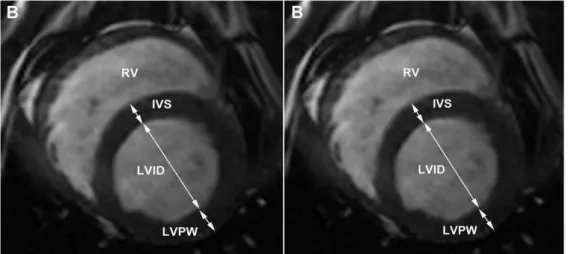
Figure 6: Short axis view of the left ventricle: end diastolic (left) and end systolic (right)3. Adapted from: Drees, Johnson, Stepien, Munoz Del Rio, Saunders, et al. (2015)
3
This figure shows measurements of interventricular septum (IVS) and left ventricular posterior wall (LVPW) thickness and left ventricular internal diameter (LVID).
11
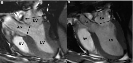
Figure 7: End systolic (left) and end diastolic (right) three-chamber view4. Adapted from: Drees, Johnson, Stepien, Munoz Del Rio, Saunders, et al. (2015).
Figure 8: Approximate four-chamber view (left) and transverse plan (right)5. Adapted from: Drees, Johnson, Stepien, Munoz Del Rio, Saunders, et al. (2015).
In veterinary medicine the use of CMRI has been limited, most likely because of the long examination times, the motion artifacts caused by heart activity and breathing, the need for anaesthesia and the high cost of the examination (Garcia-Rodriguez et al., 2009), these being its major disadvantages. Recently in veterinary medicine, problems due to cardiac and respiratory motion have been overcome through cardiac gating ECG, respiratory navigation and encoding strategies that speed up acquisition times to values compatible with in vivo imaging (Gilbert et al., 2010). The major advantage of using
4
This figure shows measurement of the diameter of the left atrial (right, dark arrow) and aortic annulus (left smallest dark arrow).
5
These figures show measurement of the mitral annulus diameter (dark arrow) at end diastole (left) and transverse plane view for measurement of the diameter of the proximal aorta (Ao) and the main pulmonary artery (MPA) (right).
Legend: LA, left atrium; LV, left ventricle; RV, right ventricle; Ao, aorta; RPA, right pulmonary artery; LPA, left
12 both high-frequency echocardiography and self-gated CMRI is the reduction of the examination time (Amundsen et al., 2011): this reduction of the anaesthetic time it’s very important in geriatric animals and animals with cardiac disease.
2.3. Left ventricular myocardial mass, chamber volume and haemodynamic
CMRI is the non-invasive method of reference for the determination of cardiac mass. Echocardiographic determination of myocardial mass has been based on measurements from two-dimension standard views with extension to three-two-dimension on the basis of a geometrical model, while contrast MRI allows direct determination of cardiac mass from three-dimension data, providing more accurate mass and volume estimation without relying on any geometric assumptions (Gilbert et
al., 2010) (table 2).
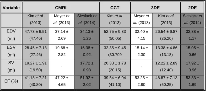
Table 2: Right ventricular function variables comparing CMRI, computed tomography and three-dimensional
echocardiography in dogs6. Adapted from: Kim et al. (2013), Sieslack et al. (2014) and Meyer et al. (2013)
Variable CMRI CCT 3DE 2DE
Kim et al. (2013) Meyer et al. (2013) Sieslack et al. (2014) Kim et al. (2013) Meyer et al. (2013) Kim et al. (2013) Sieslack et al. (2014) EDV (ml) 47.73 ± 6.51 (47.46) 37.14 ± 2.69 34.13 ± 1.26 52.75 ± 9.83 (50.05) 32.40 ± 4.15 26.54 ± 6.87 (26.20) 32.88 ± 1.17 ESV (ml) 28.45 ± 7.13 (27.46) 19.68 ± 2.82 16.38 ± 0.92 32.35 ± 9.45 (30.709 15.14 ± 2.30 13.38 ± 4.86 (13.18) 15.05 ± 0.66 SV (ml) 19.27 ± 1.91 (19.50) - 17.72 ± 0.98 20.38 ± 1.78 (20.15) - 12.22 ± 2.89 (12.40) 17.92 ± 0.96 EF (%) 41.13 ± 7.21 (40.80) 47.22 ± 4.65 51.92 ± 2.02 39.54 ± 6.04 (41.10) 53.25 ± 2.80 48.87 ± 7.13 (50.25) 53.33 ± 1.69
In a study by MacDonald, Kittleson, et al. (2005) on seven domestic cats they observed that the determination of left ventricular mass obtained by CMRI in end-systole was more accurate than echocardiography, with a significantly smaller difference between the measures obtain by CMRI and the measures obtained by echocardiography and true mass of the left ventricle. CMRI derived left ventricular mass closely approximated true left ventricular mass, but echocardiography underestimated true left ventricular mass in six of seven cats of the study, and echocardiography
6
Legend: Mean ± standard deviation values for right ventricular function variables (end-diastolic volume = EDV,
end-systolic volume = ESV, stroke volume = SV, ejection fraction = EF) obtained with cardiac magnetic resonance imaging (CMRI), cardiac computed tomography (CCT) and three-dimensional echocardiography (3DE) in 10 healthy anaesthetised beagles (Kim et al., 2013), 6 healthy anaesthetised beagles (Sieslack et al., 2014) and 10 healthy anaesthetised beagles (Meyer et al., 2013).
13 underestimated left ventricular mass quantified by CMRI in seven of seven cats. They conclude that CMRI using Simpson’s rule accurately quantifies left ventricular mass in normal cats and cats with mild left ventricular hypertrophy, and is more accurate and reproducible than the echocardiographic estimate of the same indices. Another recent study by Eskofier et al. (2015) shows the same: real-time three-dimensional echocardiography underestimates left ventricular volume in comparison with the gold standard CMRI.
Using steady-state free precession sequences, left ventricular ejection fraction can be calculated; end-diastolic and end-systolic volumes are determined for each section, allowing determination of a global left ventricular ejection fraction using a modification of Simpson’s rule. Wall motion can be assessed on short- and long-axis images either qualitatively or quantitatively, focal wall motion abnormalities suggest possible ischemia, whereas global hypokinesis may occur with a nonischemic cardiomyopathy (Jeudy & White, 2008). Roldan-Alzate et al. (2014), using the three-directional velocity information, found a strong correlation between: the ratio of the peak tricuspid regurgitation velocity to the flow through the pulmonary arteries, and pulmonary vascular resistance determined at right heart catheterization, in a canine model of acute thromboembolic pulmonary hypertension.
2.4. Valvular heart disease
As mentioned before, echocardiography is the primary non-invasive tool for initial evaluation and longitudinal monitoring of patients with significant valvular heart disease, however echocardiography can be impaired by poor acoustic windows, and is dependent on the skill and experience of the technician (Looi, Kerr, & Gabriel, 2015).
CMRI can provide a comprehensive non-invasive assessment of valvular morphology, quantification of the severity of valvular dysfunction, determination of its aetiology, assessment of the consequences for the heart from the valve lesion including measurement of ventricular volumes and function, evaluation of haemodynamic abnormalities (Looi et al., 2015), measure pulmonary artery flow, tricuspid valve regurgitation velocity, and thereby estimate pulmonary vascular resistance (Abbas et
al., 2003). This is a useful adjunct, but is unlikely to replace echocardiography as the first line
diagnostic tool for valvular disease in animals with adequate acoustic access (Gilbert et al., 2010). CMRI is reliable for the assessment of regurgitant blood volume and semi-quantitative grading of regurgitated jets and hence for the diagnosis of aortic and mitral regurgitation (Asferg et al., 2012; Gilbert et al., 2010). However, subjective visual assessment of jet properties is of limited value as their characteristics vary depending on CMRI sequences and scanning variables; pressure gradients can be determined and correlate well with echocardiography and catheterisation (Gilbert et al., 2010). Aortic valve disease can manifest as aortic regurgitation, aortic stenosis or a mixture of both. Structural abnormalities of the valve, congenital or acquired, or disease of the aorta can cause aortic valve disease (Looi et al., 2015). In some diseases, like myxomatous mitral valve disease the advances in CMRI have enabled researcher and clinicians to determine the earliest remodelling
14 changes that occur (Burchell & Schoeman, 2014). Recently Sargent et al. (2015) concluded that, although technically challenging, CMRI is a feasible method to quantify mitral regurgitant fraction in small dogs. To the best of our knowledge there are no reports describing the assessment of valvular disease by CMRI in small animals. The techniques (quantitative flow) should be applicable to clinical veterinary medicine, but may be more challenging in cats due to small cardiac size and faster heart rates. Additionally, the effects of anaesthetic protocols on systolic function may affect valve dynamics (MacDonald et al., 2006).
2.5. Cardiomyopathies
CMRI is well established in the early diagnosis and follow-up of human cardiomyopathies, where it is valuable for both tissue characterisation and assessment of function. Accurate measurement of myocardial mass and function can be used to differentiate physiological and pathological hypertrophy or dilatation (Gilbert et al., 2010). In small animals, one of the strengths of CMRI are the accuracy when measuring myocardial function (Vallee et al., 2004) and tissue morphology (Fattal et al., 2015).
2.5.1. Hypertrophic cardiomyopathy
CMRI has proved to be a useful tool for assessing a hypertrophic heart, especially when echocardiography is inconclusive or suboptimal. This technique allows for a better and more precise characterization of heart volumes and function, tissue morphology (Fattal et al., 2015) and distribution of myocardial hypertrophy (Chun et al., 2010; Fattal et al., 2015), particularly in apical hypertrophic cardiomyopathy (Wigle, 2001). CMRI either makes it possible to exclude other diseases’ aetiology that could perhaps mimic hypertrophic cardiomyopathy when assessed by other imaging modalities; CMRI can also detect features related to sudden death, apical aneurysms, and clots, and provides new information for prognosis stratification which helps in treatment planning (Chun et al., 2010).
Currently, it appears that gradient echo CMRI is not useful for measuring diastolic function in cats, the easier and faster method of tissue doppler imaging seems to be a more accurate, less expensive, and also a safer, non-invasive assessment method of diastolic function in cats (MacDonald et al., 2006). In a study by MacDonald, Wisner, et al. (2005) using a family of Main Coon cats with familial hypertrophic cardiomyopathy, contrast enhancement CMRI was not useful for quantification of myocardial fibrosis.
In animals, CMRI was established as an accurate method to quantify the left ventricular mass in cats, and a difference in left ventricular mass can be observed between cats with hypertrophic cardiomyopathy and normal cats (Fattal et al., 2015; MacDonald et al., 2006).
15 2.5.2. Dilated cardiomyopathy
To the best of our knowledge there are no published studies on dilated cardiomiopathy with CMRI, though based on echocardiographic findings, some diagnosis criteria can be used; CMRI has yet to show advantages over echocardiography in dogs.
2.5.3. Arrhythmogenic Right Ventricular Cardiomyopathy
ARVC is a myocardial disease characterized by fibrofatty replacement of the right ventricular myocardium and ventricular tachyarrhythmia being reported most commonly in the Boxer dog. Although ARVC is characterized as a myocardial disease and the impact of the disease on the function of the right ventricle has not been well studied (Baumwart et al., 2009). A spin echo technique allows for the identification of the characteristic high signal intensity associated with fat infiltration, diffuse and focal thinning (figure 9), focal aneurysms and systolic and diastolic dysfunction. Fat suppression techniques can be used in order to diminish the high fat signal and confirm fatty infiltration (Constantine et al., 2004).
Baumwart et al. (2009) found an abnormal global right ventricular systolic function and little to no morphological changes in dogs with ARVC compared with healthy control dogs. The observation of abnormal right ventricular function is consistent with the presence of a myocardial disease but has not been previously demonstrated in Boxers with ARVC because of the difficulty in imaging and assessing the right ventricle. The findings observed in that study are similar to human CMRI studies of young individuals with mild ARVC. Supporting the close resemblance of the canine disease and human ARVC, both diseases share a profile of sudden death, ventricular arrhythmias of probable right ventricular origin, syncope, right ventricular chamber enlargement, aneurysm, right ventricular myocyte loss, fatty replacement, myocarditis and apoptosis, Basso et al. (2004) proposed Boxer ARVC as a new animal model for human ARVC. These authors concluded that in vivo CMRI should allow for the diagnosis of the condition and the assessment of its progression. In the same study, all 14 hearts with ARVC displayed high transmural signal intensity in the anterolateral and/or infundibular regions of the right, corresponding anatomically to those areas of right ventricle where fat was identified by histopathology.
16
Figure 9: Pathological changes in boxer dogs with ARVC7. Source: Basso et al. (2004).
2.5.4. Myocarditis and myocardial viability
Currently, CMRI is the preferred non-invasive imaging tool in the workup of myocarditis in humans (Rinkevich-Shop et al., 2013), CMRI is being used in ischemic and nonischemic myocardial disease to assess myocardial perfusion, viability (Jeudy & White, 2008) and infarct size (Amado et al., 2004). Rinkevich-Shop et al. (2013) found that CMRI accurately identified the location and the extent of myocardial damage, which corresponded with the histopathology of myocarditis and detected regional and global left ventricular dysfunction and an increase in wall thickness that most likely reflects inflammation and oedema (figure 10).
7
A, B, and C are from a boxer dog with ventricular tachycardia and sudden death during physical activity. A, Gross heart specimen cut in cross section. B, T1-weighted post-mortem MRI corresponding to same cross-sectional plane as shown in A, presenting dilatation of right ventricle cavity however the wall thickness is normal. C, histopathological section of right ventricle wall from region of bright MRI signals (delineated by the box in B); this histological preparation shows marked transmural fatty replacement. D and E are from other boxer dog with ventricular tachycardia and congestive heart failure. D, Cross-sectional MRI image shows bright, high-intensity signal in right ventricle infundibulum. E, Panoramic histopathological section from region of bright MRI signals demonstrating massive, diffuse fatty replacement of atrophic myocardium. Inset shows small islands of a few surviving myocytes surrounded by fat. F and G are from a normal control dog. F, Cross-sectional MRI showing absence of bright MRI signals in RV wall. G, Panoramic histopathological section demonstrates normal right ventricle myocardial architecture, from a healthy dog.
17
Figure 10: Late gadolinium enhanced (LGE) distribution in left ventricle (LV) of rats with myocarditis8. Image courtesy of Rinkevich-Shop et al. (2013).
2.6. Congenital heart disease
Diagnosis of extracardiac intrathoracic vascular anomalies is of clinical importance but remains challenging. Traditional imaging modalities, such as radiography, echocardiography, and angiography, are inherently limited by the difficulties of a two-dimension approach to a three-dimensio object (Henjes, Nolte, & Wefstaedt, 2011). Recently, MRI has been introduced as a cardiac diagnostic tool and has gained increasing importance in the non-invasive examination of congenital heart defects in humans (Garcia-Rodriguez et al., 2009; Sieslack et al., 2014), playing a key role in the diagnostic of infantile congenital heart disease, even in complicated cases (Garcia-Rodriguez et al., 2009; Gilbert et
al., 2010).
Another study by Lopez-Alvarez et al. (2011) showed that echocardiography did not identify an aberrant vessel entering the caudal right atrium, apart from the coronary sinus. Also, the echocontrast study was negative (as no contrast was detected in the caudal right atrium). However, CMRI showed that the coronary sinus and caudal vena cava converged into the caudal right atrium. This negative echocontrast study may be the consequence of insufficient contrast administered, but was most likely due to the large derivation of the flow to the azygous vein, with contrast arriving in the cranial right atrium, indicating disruption of caudal venous caval flow to the heart.
Ranjan et al. (2014) used MRI and magnetic resonance angiography to further characterize a rare venous anomaly in a goat, the persistent left cranial vena cava. This is the first report including MRI and magnetic resonance angiography characterization of persistent left cranial vena cava and prominent coronary sinus with successful cardiac pacemaker implantation using the persistent left cranial vena cava. Contrast-enhanced magnetic resonance angiography offers an additional non-invasive method of patent ductus arteriosus examination especially when transthoracic
8
(B) The short-axis view of representative late gadolinium enhanced images of a rat heart with typical epicardial
late gadolinium enhanced (white strip, yellow arrows). (C) Equivalent heart section shows a high correlation in the location of fibrosis between histopathology and CMRI.
18 echocardiography is unable to provide precise measurements of the duct and most importantly from the moment that an interventional procedure is anticipated. The wide field of view, multiplanar imaging and the ability to reconstruct complicated anatomical information in three-dimensional are all valuable attributes of magnetic resonance angiography (Louvet et al., 2010).
2.7. Ischemic disease
Imaging of human ischaemic heart disease including the coronary arteries, coronary flow and myocardial viability is of increasing importance in human medicine, but is unlikely to be important in clinical veterinary medicine (Gilbert et al., 2010).
In animal experiments, excellent agreement between the extent of hyperenhancement on contrast-enhanced MRI and the histologically determined infarct size was demonstrated. The major advantage of contrast-enhanced MRI is superior spatial resolution, which allows differentiation between transmural necrosis and subendocardial necrosis (Schinkel, Poldermans, Elhendy, & Bax, 2007), as was confirmed by Rinkevich-Shop et al. (2013). A paramagnetic contrast agent, gadolinium meglumine, is administered intravenously and becomes distributed in the extracellular space before reabsorption by the myocardial capillary bed (and final excretion via the kidneys). In abnormal myocardial fibrosis, the extracellular space is expanded resulting in pooling of the contrast agent with slow washout. This appears as hyperintense bright areas on T1-weighted gradient echo images – a phenomenon known as ‘delayed enhancement’(Gilbert et al., 2010).
Thajudeen et al. (2015) demonstrated that the high-resolution mapping system and the multielectrode catheter accurately localize ventricular scar and abnormal myocardial tissue in an experimental canine infarct model. The system helps to rapidly map and identify abnormal electrocardiograms and delineates the presence, location and timing of isolated late potentials. Using the high-resolution bipolar and unipolar voltage maps may allow to differentiate between scar types and degree of transmurality.
2.8. Pericardial disease
CMRI is widely applied in human medicine in the imaging of pericardial disease, and this is likely to be in the same way applicable to dogs and cats (Gilbert et al., 2010). CMRI also has the potential to yield clinically relevant information in many cases of canine pleural and pericardial effusion (Boddy et al., 2011).
In a recent study to differentiate benign and neoplastic causes of pleural effusion, performed by Boddy
et al. (2011) CMRI did not substantially improve diagnosis of cardiac tumours compared with
transthoracic echocardiography in the cases on the study, but it yielded useful descriptive information regarding extent, anatomic location, and potential tumour type and confirmed that CMRI requires extensive additional training for tumour identification.
19 2.9. Other applications
A current development in human clinical practice is interventional CMRI – the application of CMRI as an imaging modality in interventional cardiology (Gilbert et al., 2010) as an alternative real-time technique to conventional fluoroscopy (Constantine et al., 2004). CMRI has inherent advantages over fluoroscopy in interventional imaging: it is three-dimensional, has better soft tissue contrast, provides pathological information, and does not expose the operator and subject to ionising radiation (Gilbert et
al., 2010). In 2014 Markovic et al. (2014) showed the utility of multiple imaging modalities for
assessment of anomalies of the ascending aorta, main pulmonary artery, and dissecting main pulmonary artery aneurysms. This information is particularly useful in planning surgical treatment of these disorders. Even though CMRI has shown to be reliable and accurate in human cardiology diagnosis, the need for general anaesthesia, CMRI compatible monitoring equipment and the time required for a CMRI study means it is not likely to be a first line tool in small animal the near future (Lopez-Alvarez et al., 2011; MacDonald, Kittleson, et al., 2005).
3. Specific difficulties with transposition of human Cardiac Magnetic Resonance
Imaging to veterinary cardiology
The vast majority of the existing studies are human clinical studies and experimental small rodent studies, hence the need for more research in veterinary medicine.
4. Cardiac Magnetic Resonance Imaging studies in cats and dogs
– experimental or
clinical?
MRI was adopted slowly by veterinary schools because of the expense and uncertainty about its real life applications in animals (Constantine et al., 2004; Gilbert et al., 2010). Initially, MRI was considered mainly as a research tool, and relatively few clinical studies were performed. However, with the increasing numbers of veterinary specialists and referral practices, greater public awareness of MRI, decreasing operating costs of MRI units, and – in some countries – increased popularity of veterinary health insurances, veterinary MRI has become increasingly available. The growing tendency is for veterinary schools and referral practices to install MRI systems for their exclusive use (Constantine et
20
5. Future perspectives
Future use and clinical utility of CMRI in the evaluation of small animal patients with congenital or acquired cardiac disease has being assessed in recent studies (Drees, Johnson, Stepien, Munoz Del Rio, & Francois, 2015). Other emerging techniques based on MRI, is cardiac magnetic resonance elastography (this is a novel imaging technique to noninvasively quantify myocardial stiffness) (da Silveira et al., 2014), and magnetic resonance angiography (Contreras et al., 2008).
A methodology for the simulation of heart function that combines an MRI-based model of cardiac electromechanics with a Navier–Stokes-based hemodynamic model is in development. This technique accurately describes ventricular geometry and fiber orientation (Choi, Constantino, Vedula, Trayanova, & Mittal, 2015).
21
Conclusion
CMRI has a strong potential for cardiac imaging in small animals (Vallee et al., 2004). MRI technology will surely continue to develop and magnetic resonance scanners will become more readily available to the veterinary curricula, requiring more teaching and training on this subject in veterinary curricula and at postgraduate level; veterinary radiologists must develop and participate in educational programs in MRI, in order to optimally support colleagues in first opinion and referral practices who need guidance selecting patients for MRI, and help interpreting magnetic resonance scans (Gavin, 2011). MRI is an alternative to many existing imaging modalities and veterinarians must take decisions concerning its benefits on their patients. It is important that these choices are made according to evidence acquired from well-designed studies of diagnostic performance and clinical impact (Gavin, 2011).
CMRI is still an emerging technique and has become an important diagnostic methodology in human clinical cardiology. Echocardiography will not be replaced by CMRI but this offers distinct advantages in imaging when three-dimensional organisation of the vasculature is required, when precise volume measurements are needed or when myocardial characterisation is indicated (Gilbert et al., 2010). The main generic advantages of CMRI include a wide field-of-view, absence of ionizing radiation, a variety of imaging sequences to optimize contrast and highlight specific structures, and for some applications, avoidance of intravenous contrast medium (Constantine et al., 2004; Jeudy & White, 2008). One of the major disadvantages of CMRI and other magnetic resonance techniques has been the need to anaesthetize the animal for the examinations (Sieslack, Dziallas, Nolte, & Wefstaedt, 2013). In comparison with echocardiography, CMRI provides a better understanding of the anatomy of intra- or para-cardiac abnormalities. A recent study in dogs suggest that echocardiography is a good technique for detecting heart base tumours, but CMRI provides a better assessment of the exact relationship of the tumour with the surrounding great vessels and general hemodynamic consequences, such as alternate venous return through the azygos vein (Mai, Weisse, & Sleeper, 2010). CMRI also provides improved resolution and better soft tissue contrast (Sparrow et al., 2005).
Advanced techniques like perfusion imaging, delayed enhancement or tag imaging look promising, but an effort is still needed to standardize the acquisition protocols and data analysis (Vallee et al., 2004). More recently, veterinary clinical studies have validated left ventricular mass determination by CMRI as an effective diagnostic and clinical research tool, and ‘delayed enhancement’ techniques have shown promising results. Diastolic function assessment by CMRI has been proven to be useful in the cat thus far, but this should be investigated in further studies (Gilbert et al., 2010).
In conclusion, we can affirm that CMRI is a well developed and useful diagnostic tool in human cardiology. In small animal cardiology it poses some additional obstacles that impair its feasibility, mainly the need for general anaesthesia of the patient and relative cost of the technique for the owner. Also, it is a technique with a relatively difficult learning curve and maintenance costs. We hope that in the near future these obstacles can be overcome and this technique can be made widely available for veterinary patients worldwide.
22