U
NIVERSIDADE DEL
ISBOAF
ACULDADE DEC
IÊNCIASD
EPARTAMENTO DEB
IOLOGIAA
NIMALSMALL MAMMALS AS BIOINDICATORS IN THE
ASSESSMENT OF TOXICOLOGICAL EFFECTS RESULTING
FROM THE EXPOSURE TO HEAVY METALS
C
ARLA
C
RISTINA
A
NTUNES
M
ARQUES
DOUTORAMENTO EM BIOLOGIA (ECOFISIOLOGIA)
U
NIVERSIDADE DEL
ISBOAF
ACULDADE DEC
IÊNCIASD
EPARTAMENTO DEB
IOLOGIAA
NIMALSMALL MAMMALS AS BIOINDICATORS IN THE
ASSESSMENT OF TOXICOLOGICAL EFFECTS RESULTING
FROM THE EXPOSURE TO HEAVY METALS
C
ARLA
C
RISTINA
A
NTUNES
M
ARQUES
(FCT/SFRH/BD/5018/2001)
Tese orientada por:
Professora Doutora Maria da Luz Mathias
Investigadora Doutora Ana Maria Viegas-Crespo
DOUTORAMENTO EM BIOLOGIA (ECOFISIOLOGIA)
N
OTAP
RÉVIAA presente Tese Doctoral foi financiada pelas seguintes intituições: Fundação para a Ciência e a Tecnologia (Projecto: POCTI/BSE/39917/2001; Bolsa de Doutoramento: FCT/SFRH/BD/5018/2001) e pelo Conselho de Reitores das Universidades Portuguesas (Projecto CRUP: Acção Integrada Luso-Espanhola E-1505).
Na elaboração desta dissertação, e nos termos do n.º 1 do Artigo 40, Capítulo V, do Regulamento de Estudos Pós-Graduados da Universidade de Lisboa, publicado no Diário da República – II Série N.º 153, de 5 de Julho de 2003, foi efectuado o aproveitamento total de resultados de trabalhos já publicados, submetidos ou a submeter para publicação. Tendo em conta que os referidos trabalhos foram realizados em colaboração com várias entidades científicas, a candidata esclarece que em todos eles participou na sua concepção, na obtenção, análise e discussão dos resultados, bem como na redacção dos artigos científicos.
Devido ao facto de esta tese integrar vários artigos, o padrão de redacção apresentado é diverso, variando de acordo com as normas de cada revista científica em que os artigos se encontram publicados, submetidos ou em preparação para publicação.
Lisboa, Setembro de 2008
T
ABLEOFC
ONTENTSA
CKNOWLEDGMENTS/A
GRADECIMENTOS………... iA
BSTRACT………... viiR
ESUMO………... ixL
IST OFA
BBREVIATIONS ANDS
YMBOLS………... xv1.
G
ENERALI
NTRODUCTION……….. 11.1. Abandoned mining areas in Portugal: present situation, environmental impacts and some examples………... 3
1.2. Metals toxicity ……… 7
1.3. Biological markers for metal toxicity……… 12
1.3.1. Enzymatic antioxidant defenses……….. 13
1.3.2. Metallothioneins………. 15
1.3.3. Genotoxic biomarkers………. 17
1.3.4. Morphological, histological and physiological biomarkers……… 19
1.4. Small mammals as bioindicators of metal pollution………..……….. 20
1.5. Thesis context and objectives ……… 23
1.6. Thesis structure………... 25
1.7. References………... 27
2.
I
NS
ITUM
ONITORING –T
HES
MALLM
AMMALS ASB
IOINDICATORS OFM
ININGP
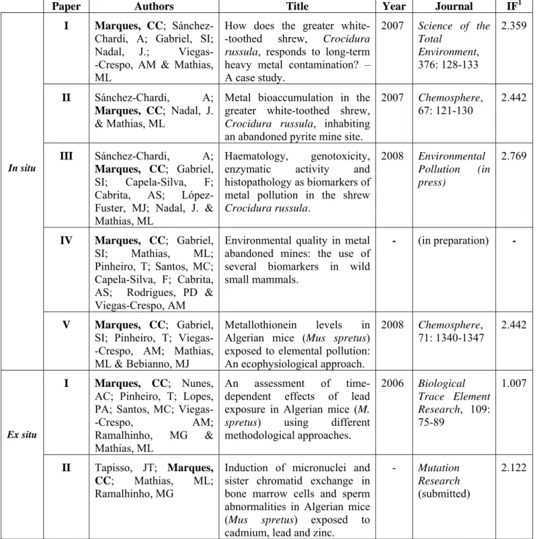
OLLUTION………... 41Paper I. How does the greater white-toothed shrew, Crocidura russula, responds to a long term heavy metal contamination? – A case study. Science of the Total Environment 2007, 376: 128-133………... 43
3 Paper II. Metal bioaccumulation in the greater white-toothed shrew, Crocidura russula, inhabiting near an abandoned pyrite mine site. Chemosphere 2007, 67: 121-130... 51
Paper III. Haematology, genotoxicity, enzymatic activity and histopathology as biomarkers of metal pollution in the shrew Crocidura russula. Environmental Pollution (in press).………... 63
Paper IV. Environmental quality in metal abandoned mines: the use of several
biomarkers in wild small mammals (in preparation)………... 73
Paper V. Metallothionein levels in Algerian mice (Mus spretus) exposed to elemental pollution: An ecophysiological approach. Chemosphere 2008, 71: 1340-1347………... 105
3.
EX
SITU
T
OXICITYT
ESTS -D
OSE-R
ESPONSEA
SSESSMENT:C
ONTROLLEDL
ABORATORYC
ONDITIONS……… 115Paper I. An assessment of time-dependent effects of lead exposure in Algerian mice (Mus spretus) using different approaches. Biological Trace Element Research 2006, 109: 75-89... 117
Paper II. Induction of micronuclei and sister chromatid exchange in bone marrow cells and sperm abnormalities in Algerian mice (Mus spretus) exposed to cadmium, lead and zinc (submitted to Mutation Research)………. 135
4.
G
ENERALD
ISCUSSION AND CONCLUDING REMARKS... 1574.1. Metal abandoned mines as an environmental problem………... 159
4.2. Uptake of elements by small mammals………. 161
4.3. Toxicity biomarkers during chronic elemental exposure……… 163
4.3.1. In Situ monitoring……… 163 4.3.2. Ex Situ monitoring………... 166 4.4. Final remarks……….. 168 4.5. Future perspectives………. 169 4.6. References………... 171
A
CKNOWLEDGMENTS/
A
GRADECIMENTOSAo olhar para o volume de papel em que resultou o presente trabalho ficamos na dúvida a pensar como pode só isto resultar de longos anos de trabalho. No entanto, o curioso é que mais uns tantos anos ainda seriam necessários sem a preciosa ajuda de todos aqueles que em diferentes fases contribuíram para a concretização deste estudo. Como tal, gostaria de expressar a minha sincera gratidão a:
▫ Fundação para a Ciência e a Tecnologia (Projecto: POCTI/BSE/39917/2001 e Bolsa de Doutoramento: FCT/SFRH/BD/5018/2001) e Conselho de Reitores das Universidades Portuguesas (Projecto CRUP: Acção Integrada Luso-Espanhola E-1505), pelo financiamento deste trabalho.
▫ Professora Doutora Maria da Luz Mathias e Doutora Ana Maria Viegas-Crespo. Às minhas duas orientadoras agradeço por me terem iniciado nesta área de investigação interessante que é a Ecotoxicologia, pelos diversos conhecimentos científicos que me transmitiram e pelos meios técnicos e financeiros que me disponibizaram. Não poderei nunca esquecer também, em fases mais complicadas deste percurso, as palavras de encorajamento, as mensagens a perguntar se tudo estava bem e a incentivar um último esforço. Obrigada pela vossa Amizade. ▫ Sofia, como Bolseira do Projecto de Ecotoxicologia. Para capturar os ratinhos apanhámos calor, frio, chuva e carraças até (mas agora já sei que o azeite é o melhor para as tirar...). No laboratório tinhamos quase como principal actividade a limpeza e tratamento dos animais. Em vários fins-de-semana lá estávamos nós a ouvir aquela impressora, ttrrr...ttrrr... Fomos a congressos levando posters que tu fazias questão de lhe dar o melhor aspecto estético possível. Mais para final fizeste revisões de alguns textos e de artigos. Que posso dizer mais..., foste imprescindível na concretização desta minha tese. Boa Sorte para o teu percurso científico é o que eu mais desejo.
▫ Doutora Teresa Pinheiro do Instituto Tecnológico e Nuclear que me acompanhou sempre fosse dia de semana ou fim-de-semana nas análises elementares através de PIXE. Que esteve sempre presente e disponibilizando-se para me apoiar em todos os domínios, quer ao nível laboratorial quer já na fase final na revisão de textos e artigos. Muito Obrigada.
▫ Alejandro Sánchez-Chardi da Faculdade de Biologia da Universidade de Barcelona, pela colaboração científica que estabelecemos e que nos permitiu partilhar conhecimentos, experiências e no final publicar vários artigos. Obrigada Alejandro e também Professor Jacint Nadal pelo apoio que me deram quando me desloquei a Barcelona (belíssima cidade).
▫ Doutor Fernando Capela-Silva, do Departamento de Biologia da Universidade de Évora, pelas análises histológicas, pela bibliografia disponibilizada ao longo deste trabalho, pelas respostas rápidas quando eu tinha dúvidas sobre algum assunto e pela colaboração na redacção de artigos.
▫ Professora Doutora Maria Cristina Santos do Departmento de Química e Bioquímica pelo apoio científico na determinação das actividades de enzimas antioxidantes e pelas palavras de encorajamento.
▫ Professora Doutora Maria João Bebianno da Universidade do Algarve, por me ter recebido, mesmo não me conhecendo, no seu laboratório para a determinação das metalotioninas, pelo apoio científico dado e palavras de encorajamento para continuar a avançar.
▫ Joaquim, Bolseiro de Mestrado do Projecto de Ecotoxicologia, pela ajuda na implementação da experiência laboratorial, pela tua determinação e excelentes ideias no decorrer do trabalho. Acho que nunca mais vamos esquecer o trabalhão que dava manter todos aqueles animais, mas conseguimos. Boa Sorte para o teu projecto de Doutoramento.
▫ Doutora Graça Ramalhinho do Museu Bocage, pela preciosa ajuda durante a experiência laboratorial e pelas palavras de incentivo sempre que nos encontramos.
▫ Doutor Vitor Oliveira do Instituto Geológico e Mineiro de Beja e Doutor Luís Martins do Centro de Dados Geológico-Mineiros de Lisboa pela documentação valiosa sobre minas abandonadas.
▫ Centro de Biologia Ambiental, pelo determinante apoio financeiro e logístico para a realização desta tese.
▫ Doutora Fátima Araújo e Doutor Pedro Valério do Instituto Tecnológico e Nuclear, pelo apoio técnico e científico durante análises elementares de várias amostras de solo.
▫ Anabela Cruces do Departamento de Geologia pela preciosa ajuda na determinação da matéria orgânica nos solos e pela tua simpatia.
▫ Doutor António Mira da Universidade de Évora, por no início do Projecto ter estabelecido a ligação com o Doutor Capela-Silva e pelas sempre palavras de apoio.
▫ Rute Pinheiro do Instituto Tecnológico e Nuclear, por toda a ajuda laboratorial, disponiblidade e conhecimentos que me deste.
▫ Engenheira Maria Teresa Pereira do Departamento de Química e Bioquímica, pelo apoio laboratorial.
▫ Professor Doutor Pedro Duarte Rodrigues pelo apoio estatístico dado num dos artigos científicos.
▫ Professor Eduardo Crespo obrigada pela revisão do resumo e pela sua opinião já na fase mais que final da tese.
▫ Professor Jean Dubois, Pierre e Gilles da Universidade de Bruxelas, por me aceitarem nas vossas instalações para um pequeno estágio sobre a Técnica ‘Comet’. Por todos os conhecimentos que foram muito úteis e valiosos.
▫ Martine e Martina, que conheci em Bruxelas, muito obrigada pela vossa ajuda no laboratório, companhia e simpatia.
▫ Inês Sousa por me ter acompanhado no terreno durante a selecção das áreas de estudo. ▫ Joana pela tua ajuda numa das muitas idas ao campo para apanhar os ratinhos.
▫ Aos vários colegas que conheci na Universidade do Algarve, Denise, Luísa, Ângela e Rui pelo apoio no laboratório e convites para almoçar e jantar.
▫ Ana Reis, pela revisão do inglês que fez num dos artigos.
▫ Cristina, já nos conhecemos desde os tempos de Faculdade e sempre foste uma amiga com quem eu muitas vezes partilhava a minha antiga viagem de barco. Muito obrigada pela força, e mesmo, mesmo no final de tudo por me ajudares a formatar a tese em pdf.
▫ Rita, pelos teus preciosos conhecimentos ao nível informático que em algumas alturas me foram muito úteis.
▫ Paula Lopes, conheço-te desde o primeiro projecto de toxicologia. Sempre te admirei pela tua determinação. Foste uma ajuda essencial ao longo do meu trabalho, quer através dos teus conhecimentos de laboratório quer ao nível científico. Também nunca esquecerei aquele dia (...) em que com as tuas palavras me fizeste sentir melhor.
▫ Inês e Sara, pelas nossas grandes conversas sobre as dificuldades de um Doutoramento, pelas idas à macrobiótica que nós adoramos (que pena agora que termino já não poderei lá ir...). Obrigada Sara pelo mapa que me fizeste e Inês por te teres lembrado de mim, quando eu já não tinha Bolsa, para participar num trabalho com Cabrera. Obrigada pela vossa Amizade e muita força para os vossos Doutoramentos.
▫ Patrícia, pela tua paciência em ouvir os meus desabafos, pela tua disponibilidade para me ajudares em tudo o que fosse preciso e pelas palavras de incentivo. Foste importante, por isso agradeço a tua Amizade.
▫ D. Branca, que eu conheço à muitos anos, desde os tempos do meu estágio, e que está lá sempre para nos ajudar.
▫ D. Irene, pela sua constante e valiosa presença na resolução dos mais diversos problemas que surjem no dia a dia de um laboratório. Pelas nossas conversas sobre os mais diversos assuntos. Pelos bolos deliciosos. Pela força que me deu para não desistir quando estamos em ‘dia não’. E já agora lhe digo que as dores de costas resultantes das ‘mil e uma mudanças’ de instalações que efectuámos foram menos dolorosas devido à sua sempre alegre companhia.
▫ Fundação da Faculdade de Ciências Universidade de Lisboa, que através da Doutora Teresa, Professora Margarida, Isabel, Ana, Miguel, Gabriela, Lívia, Paula, Elisabete, Sandra, Natália, Carlos, André, Rafael, Dália e Teresa sempre me apoioaram e me incentivaram a avançar com a Tese.
▫ Amigos de Almada, Patrícia, Paula, Teresas, Pedros, Zé, Vasco, Rute, João, Carrão e Luís, gosto da vossa simpatia e foi importante conhecer-vos. Desculpem muitas vezes dizer que não poderia estar convosco, já que tinha sempre a tese para terminar.
▫ Gemas, só vocês percebem este nome, não é ? Obrigada Amigos por existirem enquanto grupo e por naquele Fevereiro me terem quase obrigado a ir convosco para o Gerês. Nunca tinha tocado em tanta neve... Expresso também o meu agradecimento a cada um de vós individualmente:
Alexandra, a minha Amiga que faz anos quase no mesmo dia que eu, obrigada pela tua maneira positiva de viver a vida e alegria que me transmites.
Ana, as tuas gargalhadas, que nos deixam sempre um pouco embaraçados... são fundamentais. Obrigada Amiga por me empurrares para a frente.
Paulo, nos momentos mais difícies foram muito importantes aqueles convites para ir a Castro Verde, não esquecerei. Obrigada Amigo por tudo.
Claudia, de facto isto foi demasiado pesado para nós. Mas fiquei extraordinariamente feliz quando terminaste a tua Tese. Afinal era possível... Deste-me força para eu continuar a avançar. Obrigada também pelas revisões de textos e por seres minha Amiga.
Bruno, obrigada por seres de uma ilha que eu adoro, pela tua boa disposição e como já te disse temos de voltar a fazer um novo passeio de canoa.
▫ Patrícia, Maria Amélia e Henrique, pela sempre vossa boa disposição, por me fazerem rir... Não esqueço o dia em que me tentaram arrancar de casa para me levarem à Lagoa de Albufeira, dizendo que traziam uma toalha de praia a contar comigo.
▫ À Lídia, Francisco e Telmo, a minha segunda família. Obrigada pela partilha de momentos deliciosos, pela vossa dedicação, preocupação, Amizade, pelas palavras constantes de incentivo e pelos almoços e jantares sempre apetitosos e que eu tanto aprecio. Obrigada Amigos.
▫ Avós António e Adelaide, que nunca perceberam muito bem que trabalho era este com ratos, mas que foram sempre muito importantes para mim. Tenho muitas saudades vossas... mas as minhas memórias continuam cá todas.
▫ À família CM, HP e CPM: irmão, Carlitos, mesmo um dia quando fores muito velhinho serás sempre o meu irmão Carlitos. Obrigada pela tua constante presença em todas as fases da minha vida. Obrigada também Lena pela tua simpatia e pelos óptimos cozinhados naqueles dias de mini-maratona. À Catarina, a minha pequerucha sobrinha, obrigada por me puxares para brincar contigo e pelos teus enormes risos que são fundamentais para mim.
▫ Tiago, My Prince... Este dia foi muito esperado por ambos. O teu constante carinho, compreensão, paciência e disponibilidade foram essenciais. Pelas tuas palavras de incentivo e pelos teus ‘puxões de orelhas’ para avançar e não desistir. Pelos livros e música que me ajudaram a pensar noutras coisas. Pelos nossos passeios magníficos, em que me empurras para ver tudo e mais alguma coisa. Pelas revisões rápidas e constantes dos textos. Pela tua garra perante a Vida. Obrigada por seres a pessoa extraordinária que és. Adoro-te.
▫ Aos meus Grandes Pais, por todo o amor e carinho que sempre me deram. Pela alegria e conforto que me transmitem e por me terem dito um dia ‘agora não é hora de desistir’. Adoro-vos...
MUITO OBRIGADA A TODOS
‘Pense nos seus problemas como potenciais ensinamentos’
A
BSTRACTAbandoned metal mines are among the most severe environmental problems in Portugal, particularly taking into account the fact that in many cases the closing of mines are not followed by environmental requalification plans. As a consequence tones of metal residues are still circulating in their surroundings, with unpredictable effects to living organisms. However, in selected animal models, in spite of the potential environmental risk that these polluted areas may represent, few studies have investigated the environmental health impacts of long-term exposure to mining residues.
The main objective of the present PhD project was the evaluation of the environmental impact of metal abandoned mines located in southern Portugal (the Preguiça mine deactivated since 1964, and the Aljustrel mine deactivated since 1996), using two small mammals species, the white-toothed shrew (Crocidura russula) and the Algerian mouse (Mus spretus), as sentinels.
The main results can be summarized, as follows: 1) environmental levels of manganese, iron, copper, zinc, lead and arsenic are still circulating in the surroundings of both mines; 2) high hepatic contents of cadmium and nickel were detected in C. russula collected in the Preguiça area, while in shrews from Aljustrel besides cadmium and nickel also an increased accumulation of iron, lead, mercury and molybdenum in liver and/or kidney was found; 3) shrews from Preguiça revealed no significant alterations on haematological parameters and antioxidant enzyme activities, while Aljustrel shrews presented a hepatic decrease in glutathione S-transferase (GST) activity, an increase in relative liver mass and in hepatic histological alterations, and also an increase in micronucleus frequency in peripheral blood; 4) findings in M. spretus showed an increased of selenium in Preguiça individuals and increased hepatic iron and selenium in Aljustrel area; 5) in Algerian mice, hepatic glutathione peroxidase (GPx) activity increase, especially in dry season, and also hepatic histological alterations in both mining areas were detected; 6) high induction of hepatic metallothioneins was found in winter in Algerian mice from Aljustrel mine; 7) season seemed to be an important factor regulating the several biomarkers variation, whereas sex and age were less relevant factors; 8) in experimental conditions using the M. spretus as a model, the toxicity of lead seemed to be dependent of the duration of exposure and originate alterations in body and spleen mass, and in haematological and cytogenetical parameters; 9) also in controlled conditions, contamination by cadmium, lead and zinc induced micronucleus, sister chromatid exchange and sperm abnormalities in Algerian mice and the mutagenic potential of these elements could be dependent of the time of exposure and of the interaction between elements.
These results confirm the potential environmental impact of abandoned mines, and strongly support the need of requalification plans in old mines. This study also confirms the relevance of small mammals as bioindicators of environmental pollution and the usefulness of biomarkers in identifying toxicological effects in living organisms.
KEY-WORDS: Abandoned mines, pollution, metals, biomarkers, Mus spretus, Crocidura
russula
R
ESUMOA actividade mineira tem sido considerada uma das maiores fontes de contaminação ambiental por metais pesados. Em Portugal, a exploração mineira iniciou-se há vários séculos, desde os períodos Pré-Romano e Romano, tendo dado uma importante contribuição para a economia portuguesa nos séculos XIX e XX, embora nas últimas décadas a sua relevância tenha vindo a diminuir. Consequentemente, várias explorações mineiras consideradas economicamente não viáveis foram deixadas ao abandono sem que medidas de recuperação ambiental tenham sido simultaneamente implementadas. A acumulação incontrolada de quantidades significativas de metais potencialmente tóxicos, a contaminação de águas superficiais e subterrâneas, assim como da atmosfera, do solo ou da vegetação e a degradação da paisagem, constituem alguns dos mais importantes impactos ambientais destas minas que se podem traduzir em graves consequências a nível biológico e ecológico.
O presente estudo foi realizado em duas áreas mineiras abandonadas (mina da Preguiça e mina de Aljustrel) que constituem bons modelos para a investigação dos problemas ambientais anteriormente referidos. Ambas localizadas na região do Baixo Alentejo, a Mina da Preguiça manteve-se em laboração entre 1911 e 1964, período durante o qual foram extraídos óxidos e carbonatos de zinco e de chumbo, enquanto que em Aljustrel foram explorados minérios contendo cobre, zinco, chumbo e prata, entre 1867 e 1996.
Em termos de toxicidade é relativamente bem conhecido o efeito nefasto que os metais pesados possuem sobre os seres vivos, nomeadamente quando existem em grandes concentrações e a sua biodisponibilidade é elevada como acontece neste tipo de minas abandonadas, não estando no entanto os mecanismos de acção envolvidos nestes processos ainda bem esclarecidos.
Com efeito, nos organismos de metabolismo essencialmente aeróbico com o são, na generalidade os metazoários, os metais pesados, entre outras acções específicas de cada elemento, podem provocar o aumento da produção de formas reactivas de oxigénio. A actividade pro-oxidante exercida por estas formas, a nível molecular, pode originar situações de stress oxidativo conduzindo, por exemplo, à modificação oxidativa de proteínas, lípidos e DNA, com consequentes modificações ao nível da estrutura dos tecidos e alterações cromossómicas, entre outros aspectos deletérios. Existem contudo mecanismos que, em certa medida, são capazes de prevenir ou neutralizar estes efeitos pro-oxidantes – os chamados sistemas de defesa antioxidante (enzimáticos e não enzimáticos) – que medeiam um conjunto de processos metabólicos responsáveis pela manutenção da integridade das estruturas
biomoleculares. No entanto, uma exposição prolongada a metais poderá originar um aumento exacerbado da produção de formas reactivas de oxigénio, ocasionando danos irreparáveis nas referidas moléculas por uma não compensadora eficiência das defesas antioxidantes. A acção lesiva nas proteínas poderá originar alterações da membrana celular e de receptores pondo em causa o normal funcionamento dos mecanimos de transdução de sinal, quer de hormonas quer de factores de crescimento e outros, com repercussões a nível de praticamente todos os processos fisiológicos. Por outro lado, lesões ao nível do genoma poderão ocasionar mutações e a consequente ocorrência de doenças degenerativas, processos carcinogénicos e outros, nefastos para a fitness das populações naturais.
Apesar das análises químicas do solo, plantas e água fornecerem informações acerca da concentração dos elementos no ambiente, estas tornam-se, por sí só, inadequadas na avaliação da disponiblidade biológica dos contaminantes e da sua toxicidade para os organismos vivos. Os animais presentes em ambientes sujeitos à acção destes poluentes, assimilam-nos espacialmente e temporalmente de modo diferente, sendo de esperar portanto que apresentem elevadas amplitudes nos níveis de acumulação e consequentemente também alargado leque de respostas biológicas. Diversos estudos sobre a circulação e a distribuição de poluentes no ambiente e a identificação dos seus efeitos adversos para os seres vivos, incluindo o Homem, baseiam-se na utilização in situ de pequenos mamíferos como bioindicadores,.
Neste contexto, e considerando a reduzida informação existente acerca do impacto ambiental das minas abandonadas, o principal objectivo deste projecto de tese foi o de avaliar, nas duas minas abandonadas que foram selecionadas, o estado de saúde de duas espécies de pequenos mamíferos ali residentes e relativamente abundantes (Crocidura russula e Mus spretus), previamente reconhecidas como boas indicadoras de poluição por metais pesados, recorrendo à análise de vários tipos de marcadores biológicos (morfológicos, histológicos, bioquímicos e genéticos). Esta abordagem permitiu avaliar o impacto ambiental destas minas desactivadas numa perspectiva de conservação da vida selvagem.
Os resultados obtidos são apresentados e discutidos em dois capítulos. O primeiro diz respeito à monitorização ambiental in situ, usando como bioindicadores C. russula e M. spretus integrando cinco artigos científicos sobre esta temática. O segundo capítulo inclui dois artigos científicos que se reportam a experimentações ex situ no modelo M. spretus.
No primeiro artigo do primeiro capítulo foram determinados os níveis de acumulação de metais no fígado e avaliados os efeitos da poluição ao nível de parâmetros hematológicos e
apresentaram concentrações significativamente superiores de cádmio e de níquel, no fígado. A ausência de outras alterações significativas ao nível dos biomarcadores analisados, parecem indicar haver uma reduzida biodisponibilidade ambiental dos metais, aliada possivelmente ao facto da espécie C. russula apresentar grande tolerância face à poluição.
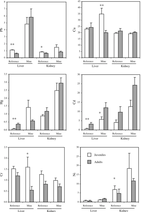
O segundo artigo deste capítulo teve por objectivo determinar as concentrações de vários metais no fígado e no rim de C. russula da área da mina de Aljustrel. Para além disso, foram avaliadas possíveis variações de vários parâmetros morfológicos (peso corporal, peso relativo de diversos órgãos e índice de condição física). Os indivíduos de C. russula da área mineira apresentaram nos tecidos uma significativa acumulação de ferro, chumbo, mercúrio, cádmio, molibedénio e níquel em relação aos valores de referência. Em termos dos metais acumulados, foram registadas, ao nível hepático e renal, variações significativas em função da estação do ano e do sexo. A idade revelou ser o factor de variação da acumulação tecidual menos relevante. Provavelmente relacionado com a toxicidade houve um aumento do peso do fígado nos animais de Aljustrel. Esta mina parece representar do ponto de vista ambiental uma situação mais preocupante do que a mina da Preguiça. As diferenças do ponto de vista ambiental entre as duas minas poderão estar relacionadas com o tipo e processos de exploração realizados e os tipos de minérios extraídos em cada uma delas, assim como com a duração do período de exploração e o período de tempo decorrente desde o fim da sua exploração.
O terceiro artigo reporta-se ao estudo dos efeitos hematológicos, genotóxicos, enzimáticos e histológicos resultantes da bioacumulação de metais nas populações de C. russula da mina de Aljustrel. Os indivíduos expostos à contaminação apresentaram alterações biológicas significativas quando comparadas com os animais de referência. No fígado foram significativas: a diminuição da actividade enzimática do glutationo S-transferase (GST), o aumento do número de micronúcleos e algumas alterações histológicas. A idade revelou ser um factor importante nas variações registadas, enquanto que o sexo apresentou-se como um factor menos relevante. Alguns dos biomarcadores analisados mostraram-se úteis para avaliação do estado de saúde de C. russula, comprovando-se igualmente a perigosidade que constitui a mina de Aljustrel para as populações selvagens de pequenos mamíferos.
O quarto artigo refere-se a um estudo que teve por alvo os indivíduos da espécie M. spretus residentes nas duas áreas mineiras. Foi realizada a monitorização sazonal de vários parâmetros (morfológicos, histológicos, fisiológicos e bioquímicos). Os animais de Aljustrel apresentaram concentrações significativamente elevadas de ferro e de selénio e um decréscimo significativo de cobre no fígado, enquanto que os da Preguiça apenas
verificou-se haver um aumento estatisticamente significativo da actividade do enzima glutationo peroxidase (GPx), especialmente durante a estação seca. Para além disso, foram também observadas alterações histológicas no fígado dos animais das duas áreas mineiras. Este estudo confirma o perigo ambiental que constituem as duas minas abandonadas para as populações selvagens de pequenos mamíferos, embora relativamente, Aljustrel pareça apresentar maior perigosidade. O significativo aumento da actividade do GPx aliado ao aumento moderado da concentração de selénio no fígado dos animais poderá, talvez, explicar a existência de um certo grau de tolerância observado para nesta espécie, tal como também, aliás, para C. russula.
No quinto e último artigo deste capítulo foram estudadas pela primeira vez as variações das concentrações de metalotioninas (MTs) no fígado e no rim de M. spretus, através de uma abordagem sazonal, comparando-se a área de referência com a área mineira de Aljustrel. Os níveis de MTs embora não significativamente diferentes foram aparentemente superiores na área mineira. O Inverno foi a estação do ano em que se verificaram níveis hepáticos significativamente superiores de MTs nos animais de Aljustrel, sugerindo que a além da poluição por metais, a indução da produção destas proteínas poderá ser influenciada por outros factores para além da poluição (e.g. temperatura, fotoperíodo, actividade sexual). Os valores de concentração de MTs obtidos pela primeira vez para esta espécie constituem, dada a inexistência de dados anteriores, uma importante referência para trabalhos futuros.
Com o objectivo de confrontar e analisar diferentes metodologias e resultados neste projecto para além da monitorização ambiental in situ foram também desenvolvidos alguns testes laboratoriais.
O primeiro artigo do segundo capítulo reporta-se a uma experiência laboratorial em que foi administrado em M.spretus e através da água de beber, uma dose fixa de acetato de chumbo durante vários períodos (15, 45 e 90 dias). Comprovando a elevada toxicidade do chumbo nesta espécie, foram observadas alterações significativas do peso corporal e do baço e igualmente ao nível da frequência de micronúcleos na medula óssea. As alterações dos parâmetros hematológicos, devem-se muito provavelmente à já conhecida interferência do chumbo com enzimas intervenientes na síntese da hemoglobina. Os efeitos do chumbo parecem estar dependentes da duração do período de exposição. Consequentemente a exposição prolongada dos animais a este metal, na natureza, poderá resultar num decréscimo da sua esperança de vida, aumentando a sua vulnerabilidade aos predadores e originando disfunções reprodutoras, com graves consequências na fitness populacional.
irmãos) e anomalias dos espermatozóides, em M. spretus quando exposto individualmente a cádmio, a chumbo e a zinco ou a combinações selecionadas destes elementos. O estudo demonstrou que todos os metais poderão ter potencial mutagénico, o qual está dependente do tempo de exposição e das interacções que possam ocorrer entre estes elementos, confirmando assim a perigosidade ambiental que as áreas mineiras, onde circulam misturas de elementos que apresentam grande biodisponiblidade e que interactuam entre si, podem ter sobre a saúde da vida selvagem e/ou humana, muito em particular ao nível reprodutor.
Os resultados do presente estudo, vêm confirmar o impacto ambiental negativo das minas abandonadas e a necessidade da existência de planos de requalificação ambiental como forma de minimizar os efeitos adversos quer para o ambiente quer para populações animais aí residentes e em última análise, para o próprio Homem. O estudo veio ainda confirmar a importância dos pequenos mamíferos como bioindicadores de poluição ambiental e a utilidade da integração de vários biomarcadores na avaliação dos efeitos toxicológicos dos metais associados aos minérios explorados nestas minas.
PALAVRAS-CHAVE: minas abandonadas, poluição, metais, biomarcadores, Crocidura russula,
L
IST OFA
BBREVIATIONS ANDS
YMBOLSALT – alanine animotransferase AMD – acid mine drainage AST – aspartate aminotransferase BL – body length BM – body mass BW – body weight CAT - catalase Cd – cadmium CDNB – 1-chloro-2,4-dinitrobenzene Cr – chromium Cu – copper
CuZnSOD – copper and zinc containing superoxide dismutase DNA – deoxyribonucleic acid
DPP – differential pulse polarography DW – dry weight
EDXRF – energy-dispersive x-ray fluorescence spectrometry Fe – iron
FeCl3 – iron chloride FeS2 – pyrite GPx – glutathione peroxidase GR – glutathione reductase GSH – reduced glutathione GSSG – oxidized glutathione GST – glutathione S-transferase H2O – water H2O2 – hydrogen peroxide H&E – hematoxylin and eosin HCT – haematocrit
Hg – mercury HGB – haemoglobin HNO3 – nitric acid
ICP-MS – Inductive coupled plasma mass spectrometry
ICP-OES – Inductive coupled plasma optical emission spectrometry IPB – iberian pyrite belt
KCL – potassium chloride LD50 – median lethal dose
MCH – mean corpuscular haemoglobin
MCHC – mean corpuscular haemoglobin concentration MCV – mean corpuscular volume
Mg – magnesium Mn – Manganese
MNCE – micronuclei normochromatic erythrocytes MnSOD – manganese containing superoxide dismutase MNT – micronucleus test
Mo – molybdenum
MPCE – micronuclei polychromatic erythrocytes MT – metallothionein
MV – million volts
NADP+ – nicotinamide adenine dinucleotide phosphate, oxidized form NADPH – nicotinamide adenine dinucleotide phosphate, reduced form Ni – nickel NOEL – no-observable-adverse-effects-level O2 – singlet oxygen O2-• – superoxide radical OH• – hydroxyl radical OM – organic matter PAS – periodic acid schiff Pb – lead
PIXE – particle induced x-ray emission RBC – red blood cells
RI – residual index
ROOH – organic peroxides ROS – reactive oxygen species SCE – sister chromatid exchange Se – selenium
Se-GPx – selenium dependent glutathione peroxidase SOD – superoxide dismutase
WBC – white blood cells Y – yttrium
1.
G
ENERALI
NTRODUCTION1.1. ABANDONED MINING AREAS IN PORTUGAL: PRESENT SITUATION, ENVIRONMENTAL
IMPACTS AND SOME EXAMPLES
Since the late 1970s there has been a general increase of concern for the environment, mainly due to evidence that human actions are causing visible and significant environmental changes.
Among the anthropogenic activities that may raise environmental issues the mining industry is probably the one that produces the deepest impacts on the environment. Moreover, deactivated mines are broadly considered a relevant environmental problem, as they represent uncontrolled sources of release of metals into the environment (Luoma, 2000).
In Portugal, several historical records point out for the existence of mining activities since the Pre-Roman and Roman ages (Schermerhorn et al., 1987). During the first decades of the last century, about 240 mines were known to be in full activity, but fifty years after (in 1989) this number decreased to about 65 mines (Barbosa and Dray, 1992). At the present (2006), only 5 active mines of extraction of metallic minerals were reported in Portugal (Direccção Geral de Energia e Geologia, 2008). This significant decline of mining activity is related to the loss of economical and technological viability of these structures, resulting in a great number of abandoned mining areas, mostly without considering any sort of environmental recovery plan. So, due to long time of permanence of metals, in those areas, high elemental levels may still be circulate in environment with unpredictable consequences for living organisms, including man (e.g. Walker et al., 1997).
Portugal is recently starting to developed specific procedures and methodologies for the assessment of polluted mining areas. The increase of environmental and governmental pressures in the European Union, which are also related to some environmental mining disasters, like those that occurred in Aznalcóllar (Spain, 1998) e Baia Mare (Romania, 2000), resulted, in 1990, in the setup of more restrictive European and Portuguese rules for the exploitation of geological resources. Those include a description of the measures to be adopted to prevent environmental pollution and to ensure the restoration of the exploited area after mine shutdown. It was also established that labouring mines involving areas bigger than 5ha or an annual production above 150,000 ton, are compiled to assess potential impacts on human health and on the environment (soil, water, air, landscape) (Rodrigues, 1998).
Notwithstanding the current legislation aimed at preventing and reducing the environmental impacts resultant from mining industry, large volumes of mine wastes occurs in the south of Portugal as a result of ore extraction from several metalliferous labouring mines.
At most mines, some of the metals existing in the mined materials cannot be recovered during mill operations and are discharged into a tailing disposal facility. These mining wastes containing high metal concentrations are released continuously in the environment, from erosion and leaching of the mine tailings, shafts and open pits, being responsible for chemical contamination of waters, soils and sediments and overall for the landscape degradation.
Mining drainage waters in contact for a long time with mineralized veins, often presents high levels of dissolved metals. For instance, the sulphide minerals, frequent in several mines, are quite vulnerable and susceptible to be oxidized by the oxygen present in pluvial waters (Santos Oliveira et al., 2002). Through chemical, electrochemical, and biological reactions, the oxidation of sulphides originate ferric hydroxides and sulphuric acid producing acid mine drainage (AMD) which is characterized by high metal content at low pH. AMD is one of the biggest environmental problems caused by the sulphide mining deposits (Akcil and Koldas, 2006), since under acidic conditions, the mobility of elements, such as iron, cooper, zinc, selenium, arsenic, cadmium and mercury, tends to increase in solution phase. Consequently, metals can be transported from the mine site to a receiving stream or water body, or can leach into the groundwater (Förstner and Wittmann, 1983; Simón et al., 2005). In addition, the effluents contaminated with heavy metals (e.g. spilling waters from the debris and/or mine drainage waters), which flow freely into the water streams, penetrate the subterranean aquifers through infiltration and percolation processes, becoming an important source of anthropogenic pollution of the hydric resources in mining surrounding areas (Santos Oliveira et al., 2002). These processes go on, not only during the mine’s life cycle, but also for many decades and even centuries after the close of mining exploitation.
Besides the knowledge of the total heavy metal content as well as the main chemical, physical and mineralogical characteristics of the mining tailings, it is in fact essential to have information on the mobility and bioavailability of the heavy metals in order to estimate the real environmental impact of abandoned mines (Gäbler, 1997). Several factors control the processes of mobility and availability of elements, and in general they are of geochemical, climate and biological origin. Some of the most important variables of soils which control element availability are the pH, redox potential, organic matter, mineral composition,
including humans, or the potential of living organisms to take up chemicals from food or abiotic environment and to incorporate them into their metabolism (National Research Council, 2003). In this context, the biological effects of heavy metals pollution are complex and strongly dependent of local geographic and climatic factors, the mixture of chemical constituents, and the vulnerability of the affected organisms.
From a geological point of view, Portugal is a considerably heterogeneous and complex country, sub-divided into various geotectonic units (Ribeiro et al., 1979; Quesada, 1992) (Fig. 1). The main metal resources are located in two areas in the south, in the Ossa-Morena zone and in the South-Portuguese zone. Two examples of abandoned mining areas here located are the Preguiça mine and the Aljustrel mine.
Figure 1. The geotectonic units in Iberian Peninsula (adapted from Instituto Geológico e Mineiro, 1998).
The Preguiça mine is situated in the Ossa-Morena zone, more precisely in the Magnetite-Zinc Belt, located in the NE part of the ‘Serra da Preguiça’, about 20 km southeast from the city Moura (Fig. 2). This open pit and underground mine began labouring in 1911 and closed in 1964, because the deposits were small and economically not viable. The deposit mine comprises domains particularly enriched in zinc and lead where the stratiform sulphide mineralizations are associated to dolomitic marbles of the Lower Cambrian age (Vairinho and Fonseca, 1989). After a decade no actions of environmental requalification were taken and the area once occupied by the mine was naturally covered by vegetation, hiding all the tailings and scoria produced and accumulated in the soil (Fig. 3a)
Figure 2. Geographic location of the major abandoned mining areas in the south of Portugal (Preguiça mine ; Aljustrel mine )
Which concerns the Aljustrel mine, this is located in the unit South-Portuguese zone in the most important metallogenetic province in Portugal, the Iberian Pyrite Belt (IPB), in the district of Beja near the small village of Aljustrel (Fig. 2). The IPB corresponds to an area of Devonian-Carboniferous volcanic and sedimentary rocks containing massive polymetallic sulphide deposits, which forms an arcuate belt, of about 250 km long and up to 60 km wide, trending westwards from near Seville in Spain to west-northwest in south Portugal (Fig. 4). The IPB massive sulphide deposits have been known and exploited from around the year 2000 BC and in Phoenician and Roman times, mainly for extraction of gold, silver and copper ore (Barriga et al., 1997).
The Aljustrel mine operated from 1867 to 1996 and shut down its operation after the mining activity became unproductive. The massive ore body of this mine comprises a mineral composed by iron and sulphur (pyrite - FeS2) with variable amounts of chalcopyrite, galene,
and blend (Barriga et al., 1997). Aljustrel landscape reveals high degradation, since old mine structures with a high level of corrosion, ruins of industrial buildings, dams, channelling streams with reddish-yellow banks, and tons of mining tails were left in the surroundings of this mine (Fig. 3b).
Figure 4. Geographic localization of the Iberian Pyrite Belt (adapted from Instituto Geológico e Mineiro, 1999).
1.2.METALS TOXICITY
Metals have been used by humans for thousands of years and are natural constituents found in rocks. Although several adverse health effects of metals have been known for a long time, exposure to heavy metals continues, and has even increasing in some parts of the world, in particular in less developed countries (Jarup, 2003).
Generally, metals are categorized as being essential or non-essential. Essential elements (e.g. manganese, iron, zinc, copper and selenium) are physiologically present in the living organisms, since they are important in many molecular and cellular functions, and are thus often regulated by efficient homeostatic mechanisms (Hoffman et al., 2001). Even though essential elements are involved in different multiple functions and structures (enzymes, transport proteins, hormone specific receptor sites and transcription factors), it
Iberian Peninsula
30% of enzymes (Apostoli, 2002). In organisms, the dose-response relationship for essential elements reflect the fact that at very low intakes of the metal, biological effects may appear due to deficiencies, whereas at high intake, effects may be due to an overdosage (Fairbrother et al., 2007). Other metals, however, such as lead, cadmium, mercury and arsenic, which have no known function in living organisms, are toxic even at very low doses and may displace or substitute for essential metals and interfere with proper functioning of enzymes and associated cofactors (Hoffman et al., 2001).
While each metal may have its own mechanisms of action, the generation of reactive oxygen species (ROS) by metals and the resulting effects on cell signalling appear to result from a common mechanism. Reactive oxygen species is a collective term which includes both oxygen radicals and certain non-radicals entities that are oxidizing agents and/or easily converted into radicals (Evans and Halliwell, 2001). These free radicals can be defined as molecules or molecular fragments containing one or more unpaired electrons being continually formed in small amounts by normal metabolism processes (Halliwell and Gutteridge, 1999). Radicals derived from oxygen represent the most important class of radical species generated in living systems. One of the members of ROS family is the superoxide anion radical (O2-•), which can be dismutated to form hydrogen peroxide (H2O2) and the
highly reactive hydroxyl radical (OH•) in the presence of certain transition metal ions
(reviewed by Leonard et al., 2004).
One of the most important mechanisms of metal-mediated free radical generation is via a Fenton-type reaction (Stohs and Bagchi, 1995). In this reaction a transition metal ion interacts with H2O2 to generate OH• and an oxidized metal ion (equation 1)
metaln + H2O2 metaln+1 + OH•+ OH- (1)
The Haber-Weiss-type reactions are also important for metal-induced free radical generation (reviewed by Leonard et al., 2004). In these reactions, an oxidized metal ion is reduced by superoxide radical (O2-•) and then reacts with H2O2 to generate OH• radical
(equations 2 and 3).
metaln+1 + O2 -• metaln + O2 (2)
metaln + H
In addition to the Fenton- and Haber-Weiss-type mechanisms, certain metal ions can react directly with cellular molecules to generate free radicals or induce cell signalling pathways.
ROS are well recognized for playing a dual role as both deleterious and beneficial species, since they can be either harmful or beneficial to living organisms (reviewed by Valko et al., 2007). Beneficial effects of ROS occur at low/moderate concentrations and involve physiological roles in cellular responses to noxia, as for example in defence against infectious agents and in the function of a number of cellular signalling systems.
The harmful effect of free radicals causing potential biological damage is termed oxidative stress. This occurs in biological systems when there is an overproduction of ROS on one side and a deficiency of enzymatic and non enzymatic antioxidants on the other. So, the oxidative stress results from imbalance between the generation and the neutralization of ROS by antioxidant mechanisms in living organisms (reviewed by Valko et al., 2007). As a result, through ROS-mediated reactions, metals can damage cellular lipids, proteins or DNA inhibiting their normal function and may contribute to metals toxicity and carcinogenesis (Fig. 5).
Figure 5. Role of free radicals in the mechanism of metal-induced carcinogenesis (adapted from Shi et al., 2004).
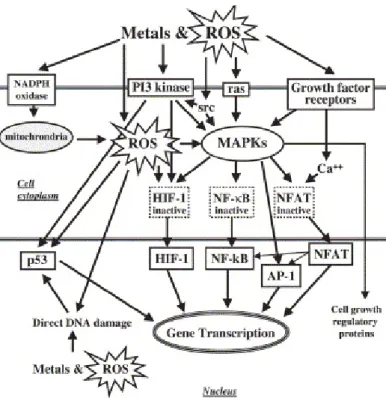
Cells communicate with each other and respond to extracellular stimuli through biological mechanisms called cell signalling or signal transduction (Poli et al., 2004). Signal transduction is a process enabling information to be transmitted from the outside of a cell to various functional elements inside the cell. Signals sent to transcription machinery responsible for the expression of certain genes are normally transmitted to the cell nucleus by a class of proteins called transcription factors. However, metals can affect the gene transcription, expression, and activation of numerous signalling proteins including growth factor receptors, G-proteins such as ras, tyrosine kinases, such as c-src, MAPK proteins, and nuclear transcription factors such as NF-kB, NFAT, AP-1, p53 and HIF-1. These effects may involve either activation or inactivation. Effects may be direct and through the interaction of metals with proteins, or indirect and through the formation of metal-induced ROS (reviewed by Leonard et al., 2004) (Fig. 6).
Figure 6. Interactions between metal-induced proteins and ROS (adapted from Leonard et al., 2004).
The receptors and genes affected by metals and metal-induced ROS interacting via signal transduction pathways can cause major cellular events including changes in cell cycle and apoptosis. This later process is an important part of normal cell development and function of organism. Properly well-functioning apoptotic mechanisms are crucial for the removal of damaged cells and the prevention of cancer development (reviewed by Leonard et al., 2004)
Aerobic organisms have developed through evolutionary processes antioxidant mechanisms designed to prevent cellular damage from ROS. Antioxidants are substances that have the ability to inhibit free radical generation, scavenge free radicals, and or reduce the oxidation and damage cause by these radicals (Shi et al., 2004). Living organisms have the ability to synthesize and control specific enzymatic systems which can be used for repair and removal of damaged proteins, lipids and DNA (Fenech and Ferguson, 2001). Also, since oxidative stress levels may vary from time to time, organisms are able to adapt to such fluctuating stresses by inducing the additional synthesis of antioxidant enzymes to regulate oxidative stress (Martins et al., 1991) (see more details in next sub-chapter ‘Biological Markers for Metal Toxicity’).
Different toxicological effects for single metals have been extensively described in literature. However, less is known about the effects related to exposure to mixtures of metals, which is the common situation in nature. Mixtures of metals can influence expected adverse health effects because their components can individually act upon the same target organs or, together, overwhelming a particular mechanism the body uses to defend itself against toxic substances. Thus, metal mixtures can interact in the body in such a way that the combined toxicity is more serious than the individual toxicity of each metal alone. In this way, low doses of a certain element that might not individually cause health effects, in combination with other elements may become a public health issue (Calderón et al., 2003). In fact, metals act additively when they are present together, others act independently of each other, and still others are antagonistic or synergistic. Interactions among metals within organisms may occur when they compete for biding locations on specific enzymes or cellular receptors during the process of absorption, excretion, or sequestration at the target site (Fairbrother et al., 2007).
Additionally, metal bioavailability greatly determines the behaviour and the toxicity of metals in environment and consequently in living organisms. Effects of metals on organisms must be considered within a context of physical and chemical influences affecting transport and fate, as well as vulnerabilities that are unique to individuals, species, populations, and communities (Peakall and Burger, 2003; Fairbrother et al., 2007). For example the forms of the metal (chemical species, particle size) can influence the metal bioavailability, fate and effects. On the other hand, the form of the metal is influenced by environmental characteristics (e.g. pH, particle size, organic matter, cation exchange capacity) (Fairbrother et al., 2007). Moreover, many other factors may affect the bioavailability of metals, including features of host organisms. A host factor can be defined as any attribute of an individual, group of individuals, or species that influences the amount and degree of metal exposure,
gender, size and weight, nutritional status, genetics (due to genetic polymorphisms), and some behaviours which influence exposure (Lopes et al., 2002; Burger et al., 2003; Fairbrother et al., 2007).
All these processes are often highly dynamic and dependent on the metal, of its form, and of organism’s ability to regulate and/or stored the metal. Many organisms have developed also physiological or anatomical abilities for regulating and/or storing metals up to certain exposure levels such that metals may not be present in organisms in a concentration, form, or place that can result in a toxic effect (Fairbrother et al., 2007). The best-known detoxification molecules, which serve as storage place as well as transportation molecules in cells, are metallothioneins (MTs) and glutathione (GSH) (Chan and Cherian, 1992; Klaassen and Liu, 1998). The both molecules are responsible for the intracellular fate of essential and non-essential metal ions (Eaton et al., 1980; Foulkes, 1993). Production of cysteine-rich metallothionein and/or binding of free metal ions to glutathione have been suggested to play a cooperative protective role against metal toxicity and in this way to prevent any detrimental metabolic reaction in cells (Chan and Cherian, 1992). The MTs structure and function will be described further on, since they were one of the subjects in the present study.
Taking in account the toxicity mechanisms discussed in this chapter, it is reasonable to say that when high concentrations of heavy metals are released into the environment they could, if are available to organisms, damage several biological macromolecules. Information about these biological effects in wildlife is generally very limited, but may be relevant to predict environmental risks in highly polluted mining areas.
1.3.BIOLOGICAL MARKERS FOR METAL TOXICITY
The presence of metals in the environment can always represents a risk for living organisms. Assessing the risk of pollutant exposure in wildlife or human populations involves the measurement of specific chemical residues in soil/water/air or in tissues, which apart from being time consuming is not per si a good indicator of the bioavailability of a chemical (Kakkar and Jaffery, 2005). One of the methods to quantify the interaction with metals and its potential impact on living organisms is the monitoring by the use of the so-called biomarkers or biological makers. According to Peakall and McBee (2001) a biomarker is ‘any biological response to an environmental chemical at the individual level or below demonstrating a
The use of several biomarkers is recognized as an important approach for the assessment of pollution, as chemical analysis of environmental samples alone does not provide evidence of the impacts in biota. Laboratory studies can help establishing cause and effect relationships that can be used to verify and predict pollution effects on natural populations in the field.
1.3.1. Enzymatic antioxidant defenses
Biochemical mechanisms involved in the cellular detoxification are particularly relevant in order to understand the deleterious effects of several metals (Viegas-Crespo et al., 2003; Bonilla-Valverde et al., 2004; Świergosz-Kowalewska et al., 2006). The antioxidant defense systems of living organisms are mainly composed by nonoenzymatic antioxidants (e.g. glutathione, ascorbic acid, α-tocopherol and β-carotene) and specific antioxidant enzymes. As pointed out in several studies, antioxidant enzymes can constitute good molecular markers for oxidative stress as well as indicate the magnitude of response in populations chronically exposed to metals (Lopes et al., 2001; Viegas-Crespo et al., 2003; Berglund et al., 2007). However, enzymes responses can vary for different chemicals pollutants and species and also in relation to environmental factors and the duration of exposure (Tsangaris et al., 2007).
The major antioxidant enzymes are superoxide dismutases (SOD), glutathione peroxidases (GPx) and catalase (CAT) that are located in different cellular compartments in virtually all tissues of vertebrates, although showing in general, higher activities in liver (Cnubben et al., 2001). These enzymes require micronutrients as cofactors such as copper, zinc, manganese, iron and selenium for optimum catalytic activity and effective antioxidant defense mechanisms (Halliwel and Gutteridge, 1995). They are also capable of removing, neutralizing, or scavenging ROS, and are strategically compartmentalized in subcellular organelles to provide maximum protection (Yu, 1994). For instance, SOD and GPx are not only distributed in the cytosol, but are also localized in mitochondria, where most of the intracellular free radicals are produced (reviewed by Yu, 1994). Additionally, other enzymes such as glutathione reductase (GR) and glutathione S-transferases (GST) are secondary help in the detoxification of ROS by decreasing peroxide levels or maintaining a steady supply of compounds, like glutathione (GSH) necessary for optimum functioning of the primary antioxidant enzymes (Habig et al., 1974; Vendemiale et al., 1999; Singh et al., 2004).
2000). It exists in virtually all O2- respiring organisms, and its major function is to catalyze
the dismutation of O2-• to H
2O2 and O2, according to equation 4 (Misra and Fridovich, 1972).
SOD
2 H+ + 2 O2-• H2O2 + O2 (4)
In mammalian tissues, SOD is classified into three distinct classes depending on the metal ion content: cytosolic CuZnSOD, mitochondrial MnSOD and extracellular EcSOD (Matés, 2000). CuZnSOD is believed to play a major role in the first line of antioxidant defense by catalysing the dismutation of superoxide anion radicals to form hydrogen peroxide and molecular oxygen. CuZnSOD induction appears as a very important enzyme for the prevention of aging and mutation by oxidative stress and hazardous effects from environmental factors (reviewed by Matés, 2000). The activity of SOD varies among the tissues. The highest levels are found in the liver, adrenal gland, kidney, and spleen (Yu, 1994). In general, an increase of SOD activity represents an adaptive response to higher superoxide ions production and disequilibrium between SOD and GPx activity could represent a marker of oxidative damage in cells (Gaeta et al., 2002).
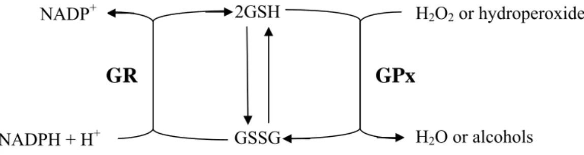
Glutathione peroxidases have two different forms, one of which is selenium-dependent (GPX, EC 1.11.1.9) while other is selenium-inselenium-dependent (glutathione S-transferase, GST, EC 2.5.1.18) (Matés et al., 1999). These two enzymes differ in the number of subunits, the bonding nature of the selenium at the active centre and their catalytic mechanisms. The selenium-dependent peroxidases (Se-GPx) are able to reduce H2O2 and
organic hydroperoxides (ROOH), while the selenium-independent peroxidases, formerly called glutathione S-transferases, only catalyze reduction of organic hydroperoxides (Cnubben et al., 2001) (Fig. 7).
Another enzyme, glutathione reductase (GR), regenerates GSH by catalysing the oxidation of GSSG to GSH (Paglia and Valentine, 1967; Pinto and Bartley, 1969) using NADPH as the reductor (Fig. 7).
2GSH
GSSG
Figure 7. Schematic representation of the glutathione redox cycle.
The capacity of glutathione to regenerate the most important antioxidants is linked with the redox state of the glutathione disulphide–glutathione couple (GSSG/2GSH) (Valko et al., 2007) (Fig. 7).
The enzymes, GPx and GR, are closely involved in glutathione enzymatic change. The form of glutathione dominant in the cell depends on the activity of those enzymes. Ikediobi et al. (2004) showed that radical oxygen appearance in cells following heavy metals intoxication may results in interruption of GSH/GSSG ratio, which then may affect intracellular GPx and GR activity. The main role in all conjugation processes is played by glutahione S-transferases (GST) (Deneke and Fanburg, 1989).
GSTs are a multigene family of isoenzymes that catalyze the conjugation of electrophilic compounds to glutathione (GSH). GSTs can also express through some of their isoenzymes glutathione peroxidase activity towards lipid hydroperoxides generated by pollutants, such as heavy metals or organic pollutants (Iscan et al., 1995; Tjaalkens et al., 1998). The diversity of substrates accommodated by GST is a result of the relatively non-specific nature of the binding site for the hydrophobic substrate and also the existence of numerous isoforms of GST. GST metabolize carcinogens, environmental pollutants, drugs and a broad spectrum of other xenobiotics (Cnubben et al., 2001).
1.3.2. Metallothioneins
Metallothioneins (MTs) are a superfamily of ubiquitous low molecular weight proteins (6000-7000 Da, 61 amino acid residues in mammal MTs), that are sulphydryl-rich proteins (up to 30% cysteine in mammal MTs), with peculiar amino acid sequence (characteristic distribution of cysteinyl residues such as Cys-x-Cys, Cys-Cys, Cys-xy-Cys, where xy are amino acids different from cysteine) (Kägi and Nordberg, 1979; Kägi and Kojima, 1987; Kägi and Shaffer, 1988; Milles et al., 2000) (Fig. 8). They were first recognized by Margoshes and
NADP+
NADPH + H+
GR
GPx
H2O2 or hydroperoxides
Vallee (1957) in equine kidney in their search for macromolecule accounting for the natural accumulation of cadmium in this organ.
Figure 8. Schematic representation of the amino acid sequence of mammalian MT (adapted from Kojima and Kägi, 1978).
MTs have a high heavy metal affinity and binding capacity (7-9 g atom mol-1 thionein) and are able to chelate both essential (e.g. zinc and copper) and non-essential metals (e.g. cadmium and mercury) by cysteine tetrathiolate clusters. The metal-thiolate clusters form in two well-defined domains, named β (N-terminal) and α (C-terminal), with stoichiometries of M3Cys9 and M4Cys11, respectively, for divalent metal ions (Vašák, 2005) (Fig. 9).
Figure 9. Schematic representation of the β and α metal-sulfur clusters with the divalent metal ions tetrahedrally arranged to bridging and terminal sulfurs (adapted from Hansen, 2002).
The metallotetrathiolate clusters provide the protein with a highly stable tertiary structure that renders it stability to heat (Kägi and Kojima, 1987). The metal-thiolate clusters within the MT molecules allow rapid exchanges of metallic ions between clusters and with other molecules. These characteristics of binding and transference of metals appear to be unique to MT and fundamental to their biological role (Viarengo et al., 2000).
Biological functions of MT include homeostasis of physiological important metals
CYS CYS CYS CYS CYS CYS
CYS
CYS CYS CYS CYS CYS
CYS CYS CYS CYS CYS CYS
CYS CYS
CYS CYS CYS CYS CYS CYS
CYS
CYS CYS CYS CYS CYS
CYS CYS CYS CYS CYS CYS
CYS CYS
cellular defence against oxidative stress (Kiningham and Kasarskis, 1998; Viarengo et al., 2000). However, it should be noted that MT could protect the cells from oxidative stress not only acting as oxyradical scavenger, but through metal binding/release dynamics (Viarengo et al., 2000).
Mammalian species have multiple MT genes coding for a family of isoforms. In fact, distinct MT isoforms designated MT-1 through MT-4 have been found (Vašák and Hasler, 2000; Milles et al., 2000). MT-1 and MT-2 isoforms are present in all organs being the best studied. MT-3 is expressed mainly in brain and MT-4 is most abundant in certain stratified tissues. The expression of MT-1 and MT-2 is regulated at the transcriptional level. Metals, glucocorticoids, cytokines and a variety of chemical and physical stress conditions co-ordinately induce these genes, whereas the other two MT genes are relatively unresponsive to the inducers. The regulation of MT genes by heavy metals appears to be mediated by a zinc-sensitive inhibitor that interacts with a constitutively active transcription factor (Palmiter, 1994). In many species (bivalves, fish, and mammals) induction of MT synthesis by metal contaminants (e.g. copper, cadmium and mercury) has been demonstrated (Rotchell et al., 2001; Bebianno et al., 1993; Świergosz-Kowalewska et al., 2007) suggesting the potential use of MTs concentrations in organisms as biomarkers of metal exposure.
1.3.3. Genotoxic biomarkers
As referred in previous sub-chapter metals through ROS-mediated reactions can cause DNA damage. Consequently, environmental genotoxicity of heavy metals have been investigated several times in small mammal species (e.g. Ieradi et al., 1998; Tanzarella et al., 2001; Topashka-Ancheva et al., 2003). Nevertheless, this type of assessment is a complex issue because of the referred diversity of potential mixtures that are often found in miming areas (Calderón et al., 2003).
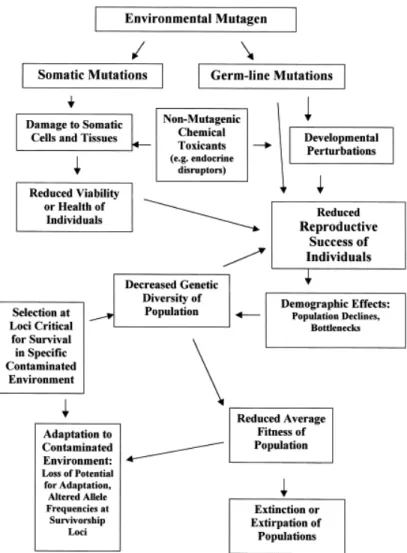
Environmental contaminants can affect genetic systems at a variety of levels of organization (Fig. 10). Beginning at the molecular level, mutagens metals interact with DNA to form lesions (somatic effects) that can cause cell or tissue damage leading to adverse health effects, or stress at individual level. This in turn can lead to reproductive impairment or result in high mortality rates. This be able to cause population bottlenecks (an ecological effect), which probably result in reduction of genetic variability in populations (population genetic effects). Shifts in allele frequencies might result from selection at loci important for survival
(evolutionary effects). All of these might ultimately lead to a reduction in the average fitness of the populations and eventually to extinction or extirpation of the population. Therefore, pollutants might show their toxic effects at the molecular level, but also initiate a cascade of responses at higher levels including tissue, reproduction, population demographics, population genetics, and finally, evolutionary processes including extinction (Bickham et al., 2000).
Figure 10. Model to illustrate the interrelationship among factors related to chemical pollution of the environment and the different levels of organization (adapted from Bickham et al., 2000).
So, biomarkers of DNA damage are valuable tools to assess effects of acute and chronic exposure of living organisms to genotoxic substances. Moreover, as genotoxins may induce changes in DNA that are passed on to future generations, this kind of biomarker can be used in a predictive way, avoiding irreversible ecological consequences. A variety of biomarkers is available to examine for possible damage to, or changes in genetic material
loci between chromatids at the four-strand stage during replication of chromosomal DNA. Chromosomes that have undergone SCE should not be regarded as damaged in the conventional sense as they are morphological intact. Nevertheless, SCE occurs at site of mutational events including chromatid breakage (Peakall and McBee, 2001). Alterations at chromosomal level can be also assessed by micronucleus test. The micronucleus test is a simple, cytogenetic test that has been used in laboratory test of clastogenicity. Micronuclei are thought to be the result of chromosome breakage (or other anomalies occurring during mitosis) that result in the retention of small fragments of chromatin or whole chromosomes in polychromatic erythrocytes after expulsion of the nucleus in the processes of red blood cell maturation (Peakall and McBee, 2001). Another simple test widely used is the analysis of sperm alterations (Wyrobek and Bruce, 1975). Although the mechanism of induction of anomalies in spermatozoa is not completely clear some advantages in the use of this marker is the reproducibility of the results and, overall to the possibility to get information on the transmission of genetic damages of generation to generation. In accordance to some authors (e.g. Hedge and Sujatha, 1995) the alterations in sperm occur as a consequence of small mutations in the animals DNA and endocrine dysfunction.
1.3.4. Morphological, Histological and Physiological Biomarkers
Morphological measures such as body and internal organs masses are commonly used to assess the health status of wild populations of small mammals exposed to some kind of pollution (e.g. Ma, 1996; Ma and Talmage, 2001; Nunes et al., 2001a; Sánchez-Chardi et al., 2007). For beyond the morphological aspects caused by pollution, histological alterations can also occur. Disturbance of living processes at the molecular and sub-cellular levels of biological organization by metals frequently leads to cell injury resulting in degenerative and neoplasic diseases in target organs. Therefore, histological biomarkers provide important qualitative and quantitative information about acute and chronic effects of toxic compounds, sometimes not so finely predicted by other parameters (e.g. Reynolds et al., 2006; Thijssen et al., 2007).
Assessment of haematological parameters also provides important information on health and physiological status. Several studies have reported alterations of haematological parameters in small mammals (Nunes et al., 2001a; Tersago et al., 2004; Topashka-Ancheva et al., 2003). Some reports have indicate that toxic metal exposure may cause a significant