UNIVERSIDADE DE LISBOA
FACULDADE DE FARMÁCIA
Targeting Membrane Proteins of Latently
Infected CD4+ T Cells Providing a Starting
Point to the Eradication of HIV1
Vasco Gonçalves Dissertação de Mestrado MESTRADO EM CIÊNCIAS BIOFARMACÊUTICAS Microbiologia 2013
UNIVERSIDADE DE LISBOA
FACULDADE DE FARMÁCIA
Targeting Membrane Proteins of Latently
Infected CD4+ T Cells Providing a Starting
Point to the Eradication of HIV1
Vasco Gonçalves Dissertação orientada pelo Profº Doutor João Gonçalves MESTRADO EM CIÊNCIAS BIOFARMACÊUTICAS 2013“The greatest ideas are the simplest.” William Golding, Lord of the Flies, 1974
ABSTRACT
AIDS is characterized by an immunodeficiency developed due to an infectious disease caused by a world spread virus, the HIV‐1. Infection with this virus culminates invariably in the death of the infected individual due to opportunistic infections that find a way into the organism. Today, HIV‐1 is fought against with therapies based in antiretroviral drugs that, although efficient and able to stop or difficult the viral infectious cycle, do not provide a real cure for AIDS, this is, HIV‐1 infection is still considered a chronic and fatal condition. The incapacity of such therapies to eradicate HIV‐1 infection is in part due to the existence of viral reservoirs like the CD4+ T cells population which are responsible for maintaining the HIV‐1 in a latent state inside the cell, protecting it from the host immune system and administered drugs, waiting for some trigger capable to reactivate it. This project was born from the possibility to achieve a sterilizing cure upon finding that specific trigger, enabling the depletion of such reservoirs and subjugating the reactivated HIV‐1 to the effects of the antiretroviral drugs. For this, we traced and objective in selecting antibodies through a run of the mill technology, the phage display technique, against unknown membrane determinants of the latently infected cells searching for agonist antibodies capable of reactivating the latent HIV‐1 and possibly, bringing us one step closer to a cure for AIDS. scFv of human origin were selected against J‐Lat 10.6 and theiragonist capacity in causing a phenotypic modification was evaluated resorting to flow cytometry.
The work here presented permitted us to identify four promising scFv with agonist HIV‐1 latent reactivation capacities against J‐Lat 10.6 cells and, additionally demonstrated the possibility for our simple approach to be an alternative methodology to the lentiviral agonist selection method developed by Hongkai Zhang and associates. Key‐Words: HIV‐1 Latency, AIDS sterilizing cure, Agonist antibodies, scFv, Phage display. vi
RESUMO
A SIDA, ou síndrome da imunodeficiência humana adquirida desenvolve‐se devido a uma doença infeciosa de incidência mundial causada pelo vírus da imunodeficiência humana, o VIH tipo‐1. Este vírus tem um tropismo específico para células do sistema imunitário, o que leva à depleção desta população celular inevitavelmente debilitando as defesas naturais do organismo. Tal estado imunodeprimido contribui para que infeções oportunistas se instalem, causando complicações adicionais o que resulta invariavelmente na morte do individuo seropositivo. Até à data os doentes com VIH‐1 são sujeitos a terapias que se baseiam em combinações de fármacos antirretrovirais e que, apesar de eficientes bloqueando ou dificultando o ciclo infecioso do vírus em algum momento, não constituem uma verdadeira cura para a SIDA. A infeção com VIH‐1 continua a ser considerada uma condição crónica e fatal. A dificuldade que tais terapias têm em erradicar a infeção causada pelo VIH‐1 é em parte devida à existência de reservatórios virais, como a população de células T CD4+. Esta células inadvertidamente mantêm o VIH‐1 num estado latente no interior da célula, protegido do sistema imunitário do hospedeiro e de qualquer terapêutica farmacológica, aguardando por algum evento desencadeador do processo de reativação. É derivado desta observação fisiológica que este projeto nasce. A partir da possibilidade de se obter uma cura esterilizante ao se encontrar um gatilho capaz de reactivar o VIH‐1 latente e que permita então a depleção
dos reservatórios de latência viral e submeta o HIV‐1 à mercê dos fármacos antirretrovirais. Para tal traçamos um objetivo baseado numa técnica laboratorial corriqueira, o phage display, para se selecionar anticorpos contra determinantes membranares apresentados pelas células latentemente infetadas. A identificação de anticorpos agonistas capazes de reativar o HIV‐1 latente num modelo celular laboratorial, as J‐Lat 10.6, aproxima‐nos da possibilidade de encontrar uma cura para a SIDA. Tal identificação foi facilitada recorrendo à técnica de citometria de fluxo que permitiu avaliar a capacidade dos anticorpos selecionados desencadearem a alteração fenotípica desejada.
O trabalho aqui apresentado permitiu‐nos identificar quatro scFv agonistas com capacidade de reativar o HIV‐1 latente em células J‐Lat 10.6 e, adicionalmente, demonstrou que pode ser utilizado como uma alternativa à metodologia utilizada por Hongkai Zhang na sua seleção de anticorpos agonistas através do uso de partículas lentivirais. Palavras‐Chave: Latência viral do HIV‐1, Cura esterilizante da SIDA, Anticorpos agonistas, Phage display. viii
AKNOWLEDGMENTS
“Tell me and I forget, teach me and I may remember, involve me and I learn.” Unknown Uma viagem. É assim que, ao olhar para trás, reconheço este último ano. Não uma viagem que começou e acabou no espaço de um ano agora passado, mas uma viagem que se insinuava na eminencia desde a primeira vez que abri os olhos e vi com vontade de conhecer. Esta dissertação representa, para mim, não apenas um único período, o finalizar estéril de uma caminho, mas um quarto de século, uma vida, uma fértil viagem ainda a decorrer.Mas a verdade é que este caminho estaria toldado na escuridão se o percorresse sozinho, condenado a andar em círculos ou a parar nas depressões dos trilhos não calcetados. Por isso, agradeço a todas as caras que me sorriram, mesmo quando, por questões próprias, não conseguia retribuir esse sorriso de volta. Agradeço a todos aqueles que da sua forma pessoal e única tocaram e iluminaram o meu trajeto, mostrando‐me que a estrada continua para além da ocasional escuridão.
Agradeço ao Professor Doutor João Gonçalves, meu orientador, pela oportunidade que me deu e pelo seu voto de confiança. Por me ter aceite no mundo da ciência e da investigação. Por ix
me ter encontrado um lugar no seu laboratório na Faculdade de Farmácia da Universidade de Lisboa e por me ter incutido o bichinho pelos anticorpos e as suas potencialidades.
À Doutora Mariana Santa Marta e à Doutora Paula Brito o meu obrigado. Obrigado por me mostrarem como a vida continua após o doutoramento. Obrigado pelas informações partilhadas, obrigado pelos conhecimentos que tiveram disponibilidade para me oferecer e obrigado pelos momentos de debate proporcionados.
Acima de tudo quero agradecer aos meus Pais. Pelo carinho que me deram, pelos valores que me imprimiram. Agradeço‐vos por me terem ensinado a querer ser eu próprio e não uma sombra replicativa de um padrão. Agradeço também pelo esforço e pelo tempo que venderam para me verem a seguir o meu caminho. Tenho que agradecer em particular à minha Mãe pelas suas perguntas insuportavelmente insignificantes que se multiplicavam com o aproximar do prazo para a entrega da tese. Obrigado Mãe, mas está descansada, continuo a gostar muito de ti e nada alguma vez pode alterar isso. Obrigado Pai, por me dares a liberdade para seguir o meu caminho apesar que de quando a quando te preocupes demais com o destino final. Adoro‐vos.
Agradeço à família com que nasci, e à família que escolhi.
A todos os meus amigos, novos ou velhos, antigos ou recentes. À Patx pela sua constante presença, pela sua durável voz, eletrónica ou biológica, mas definitivamente incalável. À Madalena, pela sua constante companhia, mesmo depois de anos fisicamente separados por quatro fronteiras. Ao Roby que me traz a beleza do mundo das artes e me mostra interesse do mundo físico e da matéria. À Fran que me mostra a beleza do mundo interno, com quem partilho uma ligação que vai para além do mundo físico e material. À Di com quem partilho o mesmo desejo pelo conhecimento e gosto pela ciência. À Nesa com quem partilho as mesmas frustrações profissionais. Ao Nelson por partilhar comigo quão bem lhe corre a vida lá em cima nas nuvens e por gozar comigo, obrigado pela força. Ao Tiago e à Elle (aka Mónica) pelo carinho nunca esquecido que trazem das terras da Rainha. Ao Aníbal (aka Nibbler), ao Nuno e ao Avelar por me arrastarem para fora de casa (ou eu vos arrastar a vocês) e me forçarem copos sempre cheios para a mão (ou eu força‐los para as vossas mãos). Obrigado.
À Cátia pela constante companhia e ajuda (não me esqueci da nossa ida ao Lux), à Catarina pela incansável ajuda e orientação na bancada, ao Pedro, pelas horas no GUAVA, à Soraia pela x
estrutura e consistência científica, à Ana pelas Heineken escondidas na ‐4, ao Luis e à ACS pelos risos, à Sofia e à Margarida por todas as partilhas. À Technophage pelo tempo e recursos disponibilizados.
A todos os meus colegas de licenciatura e mestrado por serem sempre uma motivação tão presente na pluralidade do mundo científico (tomem, inchem!). À faculdade e a todos nela empregados por todos os protocolos, regulamentos e informações sempre tão exatas e a tempo certo.
Ao pavilhão F, que se despede de mim com uma vénia.
E por ultimo, agradeço e sorrio de volta a todas as pessoas que por minha falha escaparam a esta enumeração mas que contribuíram para colorir de infinitos pontos incandescentes o caminho que percorro. “Can it be that I have not lived as one ought?" suddenly came into his head. "But how not so, when I've done everything as it should be done?” Leo Tolstoy, The Death of Ivan Ilych, 1886 xi
ABBREVIATIONS
Ab Abtibody Abs Absorbance ADCC Antibody Dependent Cell Mediated Cytotoxicity Ag Antigen AIDS Acquired Immunodeficiency Syndrome Amp AmpicillinAPOBEC3G Apolipoprotein B mRNA-editing Enzyme-Catalytic Polypeptide-like 3G
BSA Bovine Serum Albumin CA Capsid cART Combination antiretroviral therapy CDR Complementary Determining Regions CH Constant Heavy Chain CL Constant Light Chain dAb Single Domain Antibody DNA Deoxyribonucleic acid xiii
DR5 Death Receptor 5 E. coli Escherichia coli Env Envelope Fab Fragment Antigen Binding FACS Flourescence Activated Cell Sorting FBS Fetal Bovine Serum Fc Fragment Crystallizable FcR Fragment Crystallizable Receptor FR Framework Region Gag Group Specific Antigen GFP Green Fluorescent Protein Glu Glutamic acid Gly Glycine HAART Highly Active Antiretroviral Therapy HAMA Human Anti‐Mouse Antobody HIV Human Immunodeficiency Virus HIV‐1 Human Immunodeficiency Virus type 1 HPV Human Papilloma Virus Ig Immunoglobulin IgA Immunoglobulin A IgD Immunoglobulin D IgE Immunoglobulin E IgG Immunoglobulin G IgM Immunoglobulin M IgSF Immunoglobulin SuperFamily IN Integrase IPTG Isopropyl‐beta‐D‐Thiogalactopyranoside Kan Kanamycin kDa Kilodalton LTR Long Terminal Repeat Lys Lysine xiv
MA Matrix MHC Major Histocompatibility Complex mRNA Messenger Ribonucleic Acid NC Nucleocapsid NF‐κB Nuclear Factor kappa B NK Natural Killers OD Optical density ORF Open Reading Frame PBS Phosphate Buffered Saline PCR Polymerase Chain Reaction PEG Polyethylene Glycol PKC Protein Kinase C Pol Polymerase PR Protease RNA Ribonucleic Acid RPMI Roswell Park Memorial Institute Medium RT Reverse Transcriptase scFv Single‐Chain Variable Fragment Ser Serine SU Surface glycoprotein Tet Tetracycline TM Transmembrane glycoprotein TNF‐α Tumor Necrosis Factor alpha TRAIL Tumor Necrosis Factor Related Apoptosis Inducing Ligand TRAIL‐R2 Tumor Necrosis Factor Related Apoptosis Inducing Ligand Receptor 2 TSA Trichostatin A U Deoxyuridine VH Variable Heavy Chain VL Variable Light Chain μg Microgram μl Microlitre xv
TABLE OF CONTENTS
ABSTRACT v RESUMO vii AKNOWLEDGMENTS ix ABBREVIATIONS xiii TABLE OF CONTENTS xvii 1. GENERAL INTRODUCTION 1 1.1 The Antibody 2 1.1.1 Antibody structure 2 1.1.2 Genetic organization of immunity 4 1.1.3 Antibodies and its effector functions 5 1.1.4 Genetic engineering and the recombinant antibody 6 1.1.4.1 New antibody formats: the advent of the single chain variable fragments 7 xvii1.2 New antibody selection techniques 9 1.2.1 Phage display technology 10 1.2.2 Lentiviral selection method 14 1.3 The HIV problem 14 1.3.1 Characterization of HIV‐1 16 1.3.2 HIV‐1 and viral latency 18 1.3.2.1 Therapeutic strategies against viral latency 19 2. Vas AIMS OF THE PRESENT WORK 21 3. METHODS 23 3.1 Recombinant phage library reamplification and recombinant phage purification 23 3.2 Recombinant phage panning against eukaryotic whole cells in suspension 24 3.3 Recombinant phage J‐Lat reactivation assay and flow cytometry analysis 24 3.3.1 Preliminary selected recombinant phage library J‐Lat reactivation assay 25 3.3.2 Single Clone recombinant phage J‐Lat reactivation screening assay 25 3.4 Sorting of GFP+ J‐Lat cells and single clone individualization 25 3.4.1 Recombinant phage recuperation and individualization 25 3.5 Sequencing of the selected scFv clones 26 4. RESULTS 27 4.1 Recombinant phage panning against eukaryotic whole cells in suspension 27 4.2 Preliminary selected recombinant phage library J‐Lat reactivation assay 29 4.2.1 Sorting of GFP+ J‐Lat cells and single clone individualization 31 4.3 Single clone recombinant phage J‐Lat reactivation assay 31 xviii
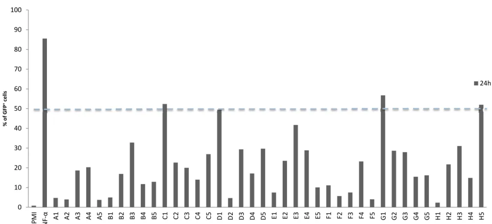
4.3.1 Single clone recombinant phage screening reactivation assay against multiple J‐Lat lineages 34 4.4 Sequencing results 35 DISCUSSION 37 CONCLUDING REMARKS AND FUTURE WORK 43 REFERENCES
Figures
47 Figure 1.1 Typical immunoglobulin G format 3 Figure 1.2 Representation of the natural IgG and some of its derived new formats 8 Figure 1.3 Generic structure of a recombinant phage 11 Figure 1.4 Detail of a phagemid 12 Figure 1.5 The phage display cycle 13 Figure 1.6 The HIV‐1 genome 16 Figure 1.7 HIV‐1 Structure 17 Figure 4.1 Human scFv recombinant phage library panning against eukaryotic cells in suspension 28 Figure 4.2 Rabbit scFv recombinant phage library panning against eukaryotic cells in suspension 28 Figure 4.3 Preliminary selected recombinant phages library J‐Lat reactivation assay 29 Figure 4.4 Flow cytometry preliminary evaluation of negative controls 30 Figure 4.5 Preliminary single clone recombinant phage J‐Lat reactivation screening assay 32 Figure 4.6 Single clone recombinant phage J‐Lat reactivation screening assay 33 Figure 4.7 Single clone recombinant phage screening assay against multiple J‐Lat lineages 34 xixxx
GENERAL INTRODUCTION
In 1901, Emil von Behring got his work recognized with a Nobel Prize in Physiology or Medicine. Such achievement was attributed to the effort he had done in developing the then known as serum therapy and its application against diphtheria. As it was stated, his work “opened a new road in the domain of medical science and thereby placed in the hands of the physician a victorious weapon against illness and deaths” (“The Nobel Prize in Physiology or Medicine”, 1901). Being this weapon what we now know as antibodies. Since then, and until 1975, six more Nobel Prizes were attributed for advances related to such important proteins when Kohler and Milstein revolutionized biological research (Köhler & Milstein, 1975). They created a key tool for research, diagnosis and clinical applications that granted antibodies the place as the most important biomolecule in modern times. Understanding the structure and the genetics of such molecules granted two more Nobel Prizes to Baruj Benacerraf, Jean Dausset and George Snell (1980) and to Susumu Tonegawa (1987) and paved way to the development of important new technologies that enabled in vitro selection and the production of large quantities of new antibodies in new formats (Miersch & Sidhu, 2012). The great promise of antibodies led pharmaceutical companies everywhere to compile and exploit the knowledge acquired by all these years of research in the hope of creating new drugs or to generate new forms of diagnosis capable of fighting old diseases. This thesis started on this 1
idea, in the importance of antibodies, the importance of the new technologies used for selecting them and the possibility to use these molecules in innovative ways to fight diseases such like acquired immunodeficiency syndrome (AIDS).
1.1 The Antibody
In the microscopic biological realm entity recognition, binding and/or adhesion is imperial for the precise functioning of either a single cell or a complex organism (Kindt, Goldsby, & Osborne, 2007a). Such processes are made possible, in part, due to the existence of a super family of proteins, soluble or membrane‐bound, called the immunoglobulin superfamily (IgSF). One of its integrants, the immunoglobulins (Igs), or simply putting it: the antibodies; are the main effectors for the natural process of adaptive immunity in complex organisms such as the human species. Antibodies by themselves form a family of Y shaped glycosylated proteins, produced by a specific class of lymphocytes, the B cells, and are characterized by their capability of binding to specific immunologic active regions of molecules. These molecules are given the designation of antigens and present one or more specific sites or antigenic determinants, the epitopes, to which the antibody’s paratopes bind to in a non covalent way. When membrane‐bound, upon contact with the immunogen, antibodies act as triggers for B cell clone proliferation and differentiation into memory B cells and plasma cells, and when secreted in its soluble form, antibodies travel through the circulatory system acting as the effectors of humoral immunity. In normal conditions, they are responsible for identifying, binding, neutralizing or starting a cascade of events that leads to the neutralization and/or elimination of foreign particles that have found a way into our bodies. Deregulations of antibody activity and immune responses and their intervenients are in the base for the emergence of genetic or acquired immune deficiencies. Antibodies are therefore an important and powerful component of the immune system defending our organism from foreign menaces (Kindt, Goldsby, & Osborne, 2007b).
1.1.1 Antibody structure
Antibodies are not all the same and therefore form a rather significant family composed by similar but yet different members. One can say that the immunoglobulin family is complex and presents different classes that can be grouped and distinguished by their ability to perform several unique binding and effector functions directly related to their individual structures and formats. Although there are five major classes worth mentioning: IgG, IgA, IgM, IgE, IgD; thiswork will only focus on the IgG class, as it is the most abundant in serum samples, representing about 80% of total immunoglobulins present in the serum.
In nature, human IgG is a protein of 150 kDa formed by four individual amino acid sequences illustrated in figure 1.1. Those are organized in a series of antiparallel β sheets that fold themselves into 110 amino acid long globular domains due to the existence of a conserved inter‐domain disulfide bonds. They consist of two γ heavy chains (55 kDa) and two kappa (κ) or lambda (λ) light chains (22 kDa) arranged into two heterodimers connected by an intra‐domain disulfide bond. The quaternary structure of the well known Y shape is given by two intra‐ domain disulfide bridges that connect the hinge regions of the two adjacent heavy chains and thus, dimerizes both heterodimers. Consequently, being a homodimer of heterodimers, an antibody can be represented in its molecular formula as γ2κ2 or γ2λ2 (Kindt, Goldsby, &
Osborne, 2007c).
Figure 1.1 Typical immunoglobulin G format: this class of antibodies is composed by four chains grouped in
identical pairs, two Heavy and two Light chains that maintain their quaternary structure due to disulfide bridges. Each chain is composed by globular domains named according to their sequence identity between different antigen binding antibodies. Heavy chains present three constant regions (CH3 to CH1) and one
variable (VH) while the light chains present one constant (CL) and one variable region (VL). Variable regions
have peptide loops that extend outside of its frame work called CDRs and are responsible for forming the paratope.
On each monomer it is possible to identify several regions according to the globular domains. In the heavy chain four globular domains define three constant regions (CH3 to CH1 starting
from the carboxyl‐terminal region) and one variable region (VH – amino‐terminal), on the other
hand, each light chain consists only in two regions, one constant (CL) and one variable (VL – also
at the amino‐terminal region). Constant regions acquired such name logically, as when compared with the same regions on other antibodies few differences in sequence are spotted. Variable regions on the other hand differ between antibodies with different antigenic determinants. Certain areas of these variable regions, called the complementary‐determining regions (CDRs), exhibit a higher variability in amino acid sequences and are in fact responsible for forming the antigen binding site. Each variable domain is composed by three loops that correspond to three CDRs (CDR1 to CDR3) which in turn are responsible for connecting the conserved β strands that compose the so called framework regions (FRs). FRs are then responsible for maintaining a universal structure for variable regions acting as a scaffold upon which the six CDR loops are supported permitting them to undertake their hypervariable conformation unique to each monoclonal antibody (Kindt et al., 2007c).
Enzymatic digestion with the proteolytic enzyme papain cleaves the immunoglobulin at the hinge region producing three fragments. Two of those fragments (45 kDa each) presented antigen‐binding activity, and thus were named Fragment antigen binding (Fab). The other one was named Fragment crystallisable (Fc) (55 kDa) as it was found to crystallize during cold storage. Fc corresponds to the portions CH3 and CH2 of both heavy chains explaining why it
does not present antigen‐binding activity. Functions for these regions will be analyzed further down the line (Kindt et al., 2007c).
1.1.2 Genetic organization of immunity
Considering the outside of our individual systems, the number of particles capable of being considered foreign is enormous. Therefore, in order to interact with as many foreign molecules as possible, there is need for an enormous number of antibodies as well. At this point it is important to mention that although response to an antigen can mobilize different B cell clones, one antigenic determinant mobilizes only one B cell clone and thus, a monoclonal antibody. It is estimated that the human organism is capable of, during its lifetime, producing between 10 to 300 million different antibodies. Such variability would mean a copious amount of genes. And looking at the size of our genome, one understands that all possible antibodies cannot be coded in individual genes. Actually, variability is assured by somatic recombination,P and N nucleotide adition and hypermutagenesis of a small set of genes that culminates in a pool of circulating polyclonal B cells (Kindt et al., 2007a).
Ig genes are organized in three separate families: Heavy chains are coded from the IgH locus in chromosome 14 while light chains are coded either in the Ig or Igλ locus, in chromosome 2 or 22 respectively. As said before, human light chains can be either κ or λ, being κ more frequent than λ (60% to 40%), while heavy chains can be of five isotypes depending on the Ig class: γ (IgG); μ (IgM); α (IgA); δ (IgD) and ε (IgE). Germ‐line DNA sequences are actually different for mature sequences and are composed of different segments, coding and non‐coding alike. While the portions of these genes that correspond to the constant regions of Igs are called constant (C) segments, variable regions are coded by two or three segments, depending if they belong to light our heavy chains respectively. A variable (V) segment fallowed by a joining (J) segment for both regions and the addition of a diversity (D) segment in between both of them for the variable regions of the heavy chains. During lymphocyte maturation V(D)J segments are randomly combined in an event called somatic recombination which adds another layer for antibody diversity from the already so many possible sequences existent for antibody generation. DNA repairing enzymes active during somatic recombination are responsible for generating even more diversity by adding nucleotides during the joining of the coding sequences. If palindromic junctions are generated P nucleotides were added, otherwise nontemplated stretches with new nucleotides are generated (N nucleotides).
After lymphocyte activation upon contact with the antigen, two more diversity generating processes occur: hypermutagenesis and Ig class switch recombination. Somatic hypermutation or hypermutagenesis is responsible for random insertions of point mutations in V segments that leads to the selection of the lineage with the best affinity for the specific antigen – affinity maturation; and Ig class switch recombination, which only occurs in the C segments, is responsible for changing isotypes and therefore effector functions as well.
1.1.3 Antibodies and its effector functions
Besides antigen‐binding activity, which by itself does not neutralizes or removes the foreign particle from our body, immunoglobulins play a determining role in humoral response. They are able to promote biological responses that lead to the effective removal or neutralization of the said foreign particle, this is, they have effector functions (Kindt, Goldsby, & Osborne, 2007d). Such functions are assured by interactions between the constant regions of the heavy chain and other soluble or membrane bound proteins and therefore are dependent upondifferent immunoglobulin classes. One of those effector functions is opsonization, one of the most important defenses against bacterial invasions. It is characterized by antibody triggered phagocytosis of antigens by macrophages and neutrophils. Activation of this event is dependent on cross‐linking of Fc receptors (FcR) presented on macrophages membranes upon binding to Fc regions of antibody‐antigen complexes, setting off a signal‐transduction pathway responsible for starting phagocytosis. Destruction of the infectious agent is carried out inside the phagocyte. Complement activation and antibody‐dependent cell‐mediated cytotoxicity (ADCC) are other examples of antibody effector functions. The complement system is composed by a group of soluble proteins present in the serum capable of many actions such as the formation of pores in cellular membranes leading to their destruction or phagocytosis induction. Antibodies can act as cellular receptors as well, binding to target cells and exposing their Fc region to effector cells like natural killer (NK) cells activating a cytotoxic reaction – ADCC.
But for medical uses the most important feature of the antibodies is their antigen‐binding activity. Antibodies can be used as a means of diagnosis passively marking target molecules or as therapeutic agents binding to certain molecules in the human system, sequestering them, or to their respective receptors, blocking them, and therefore preventing their natural interaction antagonizing the receptor. But they can also bind to a specific cellular receptors and act as an agonist triggering some form of cellular response. Present day research is avidly turning its efforts into the search for antibodies capable of causing an action in the cell whose receptor they bind to. Conatumumab (AMG 655) developed by Amgen Inc is an example of an agonist monoclonal antibody designed for the treatment of solid tumors of hematopoietic origin (Rosevear, Lightfoot, & Griffith, 2010). It targets the tumor necrosis factor‐related apoptosis‐inducing ligand receptor 2 (TRAIL‐R2 or death receptor 5 – DR5) mimicking its natural ligand and starting a caspase cascade that leads to the apoptosis of the tumor cell. Agonist antibodies can be studied and developed for use in other diseases rather than just neoplasic conditions. Syndromes and diseases caused by virus that do not have yet a cure, like the human immunodeficiency virus (HIV) or even the human papilloma virus (HPV), can possibly represent new target afflictions for agonist modulation.
1.1.4 Genetic engineering and the recombinant antibody
In the living organism, as it has been stated before, contact with an antigen causes proliferation and differentiation of a pool of B cell clones rather than just one specific clone with affinity for one singular epitope. This results in a mixture of antibodies directed for 6different epitopes of the same antigen, this is, a polyclonal antibody mixture. Although advantageous for a living organism, for research or medical purposes this represents a difficulty. Thus monoclonal antibodies, with specificity to a single epitope, are preferred. Purification of a monoclonal antibody from a polyclonal mixture was not feasible until 1975 when Kohler and Milstein described a method that permitted monoclonal antibody production (Kindt, Goldsby, & Osborne, 2007e). Fusing a B‐cell recovered from an immunized donor, like a mouse, with an immortal myeloma cell they created a hybrid cell, the hybridoma, capable of antibody production in large quantities with immortal properties and therefore with the possibility to be cultured indefinitely (Köhler & Milstein, 1975). Although this technology revolutionized the field of antibody research and application several difficulties were associated with it. B‐cells used for generation of hybridomas were of murine origin and thus when used for medical purposes in humans created immunologic and hypersensitive reactions with human anti‐mouse antibodies (HAMAs) limiting their use (Jaffers et al., 1986). Still, associated with their animal of origin, there was the problem of affinity. Different animals have different innate libraries of possible antibodies, which mean that high‐affinity antibodies for a specific antigen are not always provided from mouse immunization. Other difficulties resided in the fact that the antibodies obtained, when used for medical purposes, did not stay in circulation for long periods of time. Also this technology is very laborious, time consuming and expensive (Jaffers et al., 1986). Cellular stability and storage costs, impossibility to immunize against toxic targets and difficulty in direct access to antibody genes are examples of some other problems for hybridoma technology.
1.1.4.1 New antibody formats: the advent of the single chain variable
fragments
With recombinant DNA technology on the rise, new approaches to antibody production started to appear. Antibodies began to be designed rather than just produced. This genetic antibody manipulation contributed to furthering the knowledge about structure and functional organization of immunoglobulins which by itself permitted the emergence of new engineered antibodies for research, diagnosis, and therapeutic applications with new specificities beyond conventional antibody technologies. Coding sequences from Igs of different species started to be used to create chimeric or humanized antibodies trying to surpass some problems of hybridoma technology (Morrison, Johnson, Herzenberg, & Oi, 1984; Jones, Dear, Foote, Neuberger, & Winter, 1986). And not only molecules of different origins were added to the natural backbone of antibodies, but fragmentation also started to occur. Antibodies began to be divided into smaller fragments and their sizes were reduced considerably. Changes were 7
made in order to achieve specific goals such as increased protein expression, better biodistribution and longer circulation half‐life, lower immunogenicity or even better tissue penetration. New formats like Fragment antigen binding (Fab) single chain variable fragments (scFvs) or single domain antibodies (dAbs) started to appear (figure 1.2).
The scFv format is a fusion protein of approximately 30kDa. It consists of a VH and a VL region
connected by a flexible peptide linker of about 15 amino acids (Bird et al., 1988). Such linker contributes to protein stability allowing the variable regions to acquire their correct conformation and also facilitates expression in E. coli systems. Most common linkers are composed by stretches of Gly and Ser and/or Glu and Lys residues for flexibility or solubility respectively (Ahmad et al., 2012). scFv are one of the smallest portions of the immunoglobulin still retaining its antigen‐binding activity and thanks to this characteristic associated with the easiness of expression in bacterial systems scFv have undertaken a central role in new antibody selection techniques like phage display. Also, scFvs can easily be transformed in bivalent or trivalent scFvs be linking this proteins together creating multimers. Such fusions can be between equal or different scFvs creating bi‐specific diabodies.
Figure 1.2 Representation of the natural occurring IgG and some of its derived new formats: scFv
(30kDa) can be fused together to create diabodies (50kDa), triabodies (75kDa) and so forth. They can also be conjugated with other proteins of the same origin but with different affinity (bi‐specific antibodies) or even chemical compounds.
Yet, the smallest possible antibody format is the dAb with 15kDa (Putnam, Liu, & Low, 1979). They are formed by a sole variable domain either from the heavy or light chain of immunoglobulins and thus only present three CDRs against one specific epitope (Ward, Güssow, Griffiths, Jones, & Winter, 1989). Like scFvs, their small sizes give them special attributes like higher tissue penetration and lesser immunogenic activity, besides the facility of expression in bacterial systems.
1.2 New antibody selection techniques
As we have seen before, antibody diversity in higher organisms is assured by combinatorial recombination of germ‐line antibody genes associated with somatic hypermutation. This means that each individual organism caries a library of all its possible antibodies with the majority of them presented in its circulating B cells. Hybridoma technology was based on this natural occurring library using it as a form of in vivo selection. Since then, antibody generating technology evolved due to impending medical necessity and new forms of in vitro antibody selection arouse such like phage, microbial or cell display or even cell free technologies like ribosome, DNA or mRNA display (Oliveira, 2013). Such technologies rely in the possibility to present a protein conjugated to a particle able to genetically identify it. Ribosome, mRNA and DNA display require for in vitro transcription and translation and are by themselves variants of each other. Translation can be stopped by the addition of antibiotics like rifampicin which permits the generation of protein‐ribosome‐mRNA complexes presenting the basis for ribosome display. If mRNAs are treated to present at their 3’ end a puromycin oligonucleotid which is incorporated at the end of the translation process, a protein‐mRNA complex is formed and mRNA display is made possible. DNA display was developed to circumvent the fast degrading mRNA usage and uses a mixture of reversed micelles where individualized DNA fragments, marked for example with biotin, are transcribed and translated to streptavidin‐ fused proteins. Cell dependent displays work in a similar way. Cells provide the machinery necessary for our protein production and present them is their surface therefore forming a protein‐cell‐DNA conjugate. Phage display will be further explained below as it is the mains focus in this work.
But for in vitro selection techniques to work the huge diversity of antibodies observed in humans had to be duplicated in manmade libraries. Presently it is possible to create several different types of libraries that differ in their origin depending on the selection technique and purpose of the intended result. Obviously each immune library has its own limitations that
have to be equated depending on the type of antigen or the affinity and quantity of antibodies expected from the selection procedure.
scFv libraries can be divided into three categories, immune, naïve and synthetic (Ahmad et al., 2012; Oliveira, 2013), explained hereafter. Immune libraries derive from the variable domains of antibody genes collected from enriched pools of activated B cells of immunized animals like mice (Xu, Jin, & Fan, 2003) or camels (Rahbarizadeh, Rasaee, Moghadam, Allameh, & Sadroddiny, 2004). Meaning that previous animal injection with the desired antigen is required in order to construct such libraries. Due to the previous in vivo affinity maturation, in vitro selection is biased. Selection procedures result in greater pools of specific binders against the pre‐immunized antigen and selected antibodies usually present a wider range of higher binding affinities. Immune libraries are constructed for specific uses and therefore, when considering another antigen, a new library needs to be made. Naïve libraries also derive from live donors, but lack the pre‐immunization procedure and therefore are not biased against any particular antigen. They are constructed from samples of non activated B cells (Vaughan, Williams, & Pritchard, 1996). This feature allows them to be used in various selection procedures against non related antigens without the need to construct a new library and most importantly, they overcome the inability to select antibodies against toxic, self or non‐ immunogenic antigens. One important aspect of such libraries is their size and its relation to selection success. Libraries with larger sizes, usually maxing at 109‐1010 individual scFv, present better chances to select more antibodies with higher affinities than smaller libraries (Griffiths et al., 1994; Vaughan et al., 1996; Winter, Griffiths, Hawkins, & Hoogenboom, 1994). All these characteristics contributed for the choice to work with this kind of libraries in this experimental work. The last category is the synthetic libraries. These libraries are also not biased against any particular antigen as they derive from non‐immunized samples prepared resorting to manmade combination of germ‐line gene sequences and randomized CDRs (Hoogenboom & Winter, 1992). Being CDR3 the CDR with the most diversity and the one that is essentially responsible for antigen binding it is understandable that most synthetic libraries focus on randomizing this particular CDR and therefore are created specifically to yield high affinity antibodies.
1.2.1 Phage display technology
Derived from the work developed by George Smith in 1985, phage display technology is based in the possibility to present a determined protein on the surface of a filamentous bacteriophage (Smith, 1985). Smith demonstrated that DNA from foreign origin could be fused 10together with the gene for a coat protein of a non‐lytic filamentous phage and thus, expressed and displayed at its surface as a fusion protein without affecting the normal infectious cycle of the bacteriophage. In 1990 and with the help of recombinant DNA technology, McCafferty created a recombinant phage that successfully displayed a functional scFv (McCafferty, 1990). He demonstrated that the fusion scFv maintained the same antigen‐binding activity as the original scFv and that, even diluted in a mixture with 106 different phages, it was possible to re‐ isolate it. Additionally, access to the gene sequence of the fusion protein was made possible due to phenotype genotype conjugation, this is, scFv displaying particles have associated to them the DNA for the respective fusion scFv displayed. This became one of the most important characteristics of phage display technology permitting quick and easy access to the gene sequence of any selected scFv amongst an enriched and diverse library (Griffiths et al., 1994). This technology was further perfected in the following years (Barbas, 1991; Clackson, 1991; Winter, 1994) and now it is a widely used procedure presenting proteins other than scFv. Phage display technology was then born and permitted the fast and efficient selection of antibodies isolated from large populations of recombinant phages based on their affinity against a specific target.
Figure 1.3 Generic structure of a recombinant phage: A phagemid coding for a fusion protein
between pIII and a scFv (or any other displayable protein) is the basis of phage display.
In more detail, the phage display technique relays in filamentous bacteriophage biology. Phages like M13 and its relatives are non‐lytic bacteriophages that present a rod‐shaped capsid composed mostly by the major coat protein, pVIII, with approximately 2700 copies, that encapsulates its single strand circular DNA genome. On one tip of the phage particle there are approximately five copies of each minor coat protein pVII and pIX and on the other tip also around five copies of each minor coat protein, pVI and pIII, being the last responsible for the infection of the host bacteria by binding its n‐terminal region to the F pilus (Smith & Petrenko, 1997). Infection ends with the viral genome being injected unto the bacterial cytoplasm where it remains as a separate entity from the bacterial genome being transcribed and replicated. But, in order to create
recombinant phages expressing or protein of interest, one has to first clone it in a phasmid (Figure 1.3). Phasmids are vectors derived from small coding plasmids with a phage origin of replication which code for only one phage coat protein preceded by a multiple cloning site. Therefore, a phasmid by itself is not capable of producing phages (Garrard, Yang, O’Connell, Kelley, & Henner, 1991) and only when complemented with helper phages like VCSM13, with a complete viral genome, recombinant phage production is possible (Vieira & Messing, 1897). Proteins of interest cloned in such vectors, like the pCOMB vector family (Barbas, Burton, Scott, & Silverman, 2001), can be fused to any desired coat protein (Barbas et al., 1991) but most common practices clone the protein of interest in fusion with pIII or pIX. Such choices depend on the desired final valence. Phasmid may also present leader sequences enabling export of the synthesized protein to the oxidative environment of the periplasm and an amber stop codon separating the cloned protein and the phage coat protein (Figure 1.4). The ambar stop codon, glnV, is especially important for the production of the cloned protein in its soluble form. This is made possible resorting to an ambar non‐suppressor strain like E.coli TOP10F’ where glnV is read as a stop codon (Hoogenboom et al., 1991) supplemented with IPTG. Figure 1.4 Detail of a phagemid: pCGMT is a phagemid derived from pCOMB that codes for a FLAG epitope before the ambar stop codon facilitating the screening protocols of the purified scFv. A lacZ promoter makes scFv production inducible by the presence of IPTG when in ambar non‐suppressor strains like TOP10F’. For recombinant phage scFv selection to be achieved, inquiry of a built library against a target antigen of interest is necessary (figure 1.5). Multiple rounds of panning of the phage library against the antigen are performed with multiple washing steps in order to remove unbound recombinant phages. Due to the washing steps antigens need to be immobilized in a solid support like small peptide high binding microtiter plates, or streptavidin coated beads (for biotinylated antigens) per example. At the end of each panning specifically bound phages are eluted and reamplified allowing to continue the selection process serving as the input of the next panning. Normally three rounds of pannings are performed, allowing specifically bound phage enrichment (Smith & Petrenko, 1997), ending with the infection of an ambar non suppressor E. coli strain allowing the production of soluble proteins for further characterization. When performing this technique it is expected to select a high affinity clone against the chosen antigen from a large number of nonspecific recombinant phage clones.
Figure 1.5 The phage display cycle: After phage library amplification and recombinant phage production the phage pool is incubated against the immobilized antigen for a determined period of time before any washing steps, responsible for removing unbound recombinant phages, and posteriorly eluted to recover the binding phages. Eluted phages are in turn used for creating a new library biased against the antigen by infecting an E. coli strain and are then reamplified to produce a new pool of recombinant phages, which can be the starting point of a new selection round, or directed for screening purposes.
For the presentation of scFvs this technique exhibits several advantages over hybridoma technology. First of all, animal immunization is easily avoided, and if necessary to library creation, no hybridomas need to be produced. Selection of antibodies against difficult or toxic antigens becomes more accessible and feasible. Additionally, phages are more stable and therefore can be stored at 4ºC for long periods of time and if needed are also easily, rapidly and inexpensively produced. And finally, scFv genes are easily obtainable and thus more easily manipulated by mutagenesis as affinity or specificity capacities of the selected scFv, although normally sufficient for research usage, might be unsatisfactory for medical applications.
1.2.2 Lentiviral selection method
Although the vast amount of selection technologies at hand, new purposes and intentions pave the way for new selection techniques to be imagined and developed every day. The lentiviral selection method developed by Hongkai Zhang and its associates was designed to answer impending new needs (H. Zhang et al., 2013; H. Zhang, Wilson, & Lerner, 2012). As we mentioned before, great efforts are being made in order to discover antibodies that present agonist capacities, but selection techniques are mainly focused in binding rather than function. This group states that the generation of antibodies capable of perturbing cellular functions is hampered by a secondary screening against antigens in their natural environment presented by eukaryotic cells while the first screening was performed under different conditions against a highly purified antigen in vitro, and for a different function – binding. Also, they state that the success of the first screen is of upmost importance affecting the complexity and success of the second. Therefore they designed a method where scFv can be produced by the eukaryotic model cells themselves bypassing the first screening and selecting for function right from the start. For this scFv libraries are constructed in an eukaryotic infectious agent, the lentivirus, which is actually a problem for this method as lentiviral libraries reach at maximum 107 elements against the 1010 in phage libraries (H. Zhang et al., 2012). But on the other hand, one advantage of this method is the possibility for multiple lentiviral infection of one cell creating a possibility to study combinatorial synergy maximizing the size of the library. After insertion of the foreign scFv DNA in the cellular genome due to lentiviral infection cells need to be incubated with a methylcellulose based medium in order to confine the secreted scFv to the vicinity of the cell producing it permitting the phenotype genotype conjugation. After a pre‐ determined incubation period necessary for the agonist scFv to act cellular samples are prepared and sorted resorting to flow cytometry in order to individualize them for screening and sequencing purposes.1.3 The HIV problem
Since the very beginning, the human race has always fought against all adversities. Predators, self‐imposed wars, lack of food or plagues. And, since that very beginning, and always with costs associated, the human race recovered, learned and prevailed. One of our most notable achievements against the natural predators of our own body, the infectious microorganisms, was accomplished during the twentieth century. This was due to the discovery and commercialization of natural antibiotics. The war against infectious diseases had therefore officially begun. During most part of the previous century we considered ourselves on the 14
upper hand. Eradication of infectious agents was soon to be seen (Fauci, 2001; Pier, 2008; Spellberg, 2008). But our foresight was blinded by success. In 1981, the syndrome we now know as acquired immunodeficiency syndrome was for the first time described (Gottlieb, 1981; Masur et al., 1981; Siegal et al., 1981). It is characterized by a severe trauma to the patient immune system making him prone to opportunistic fatal illnesses. Some years later, in 1983, AIDS was associated with an infectious agent, a virus – the human immunodeficiency virus (Barré‐Sinoussi et al., 1983). This date marked and scared our society as we witnessed our own limitations. In 2010, and according to data from UNAIDS, it was estimated that 2,6 million new infected individuals arise each year and another 1,8 million die due to the infection. Thirty years later the identification of HIV, the true cure for AIDS still elope us. HAART, or highly active antirretroviral therapy (also known as cART ‐ combination antiretroviral therapy) was proposed in 1996 during the XI International AIDS Conference in Vancouver. This therapy encompasses a cocktail combination of three or more antiretroviral drugs that target different essential stages of the HIV‐1 lifecycle (Richman et al., 2009), reaching more than 5 million infected individuals (WHO, UNAIDS, & UNICEF, 2010). Although science has managed to greatly reduce morbidity and mortality rates for those with access to care (Braitstein et al., 2006; Mocroft et al., 2003; Palella et al., 1998), viremia does not disappear during treatment. Plasma viral RNA levels are only dropped below 20 copies per mL (Dornadula et al., 1999; Geeraert, Kraus, & Pomerantz, 2008). As of the moment therapy is discontinued, viremia rebounds to measurable levels in about two weeks (Harrigan, Whaley, & Montaner, 1999). This means that, in the present day, even though forty six compounds were developed and at some time and place commercialized (data compiled from www.fds.org and www.ema.europa.eu/ema/), HIV infection passed only from fatal illness to chronic disease (Pomerantz & Horn, 2003; Richman et al., 2009). Also, patient adherence to this therapeutic strategy is mined due to the costs related to a lifelong medical treatment, long‐term side effects, social stigma, and more importantly due to the fact that HAART is not sufficient to eradicate HIV‐1 from circulation. Reasons for this inefficiency possibly relay on a cryptic ongoing replication, poor drug penetration (L. Zhang, Ramratnam, & Tenner‐Racz, 1999) or, most importantly, on the persistency of the virus in cellular reservoirs due to viral latency. So, paraphrasing, no efficient way to eradicate this virus from the human body as yet been found and new ways to arrest infection and to stop viremia rebound are needed.
1.3.1 Characterization of HIV1
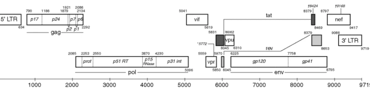
Although the HIV family is divided into two sub types both infectious the human species, HIV type 1 (Barré‐Sinoussi et al., 1983) and type2 (Clavel et al., 1986), focus is taken mostly on HIV‐ 1 as it is responsible for a more severe syndrome associated with higher infectious rates that resulted in the AIDS pandemic. Therefore, during this thesis, focus will be on HIV‐1 only. HIV‐1 belongs to the Lentivirus genus of the Retroviridae family, meaning that it is an RNA virus, and one of the most complex of its family (Wagner, Hewlett, Bloom, & Camerini, 2008a). Its genome (figure 1.6) is composed of two positive single strain RNA molecules of about 9kb that code for 9 open reading frames (ORFs): gag, pol, vif, vpr, tat, rev, vpu, env and nef; flanked by long‐terminal repeats (LTRs). The promoter for viral transcription is found on the 5’ LTR. As in other retrovirus proteins are transcribed in polyproteins that are subsequently proteolyzed into their individual components (Wagner, Hewlett, Bloom, & Camerini, 2008b) (figure 1.7 for HIV‐1 structure). The Gag (group specific antigen) polyprotein is cleaved into four other proteins: matrix (MA), capsid (CA), nucleocapsid (NC), and p6; while Env (envelope) is cleaved into two: glycoprotein gp120 (surface or SU) and glycoprotein gp41 (transmembrane or TM) (Allan et al., 1985; Veronese et al., 1985). These six proteins are structural components that build the core of the virion and outer membrane envelope. Pol (polymerase) polyprotein is proteolyzed into three proteins – protease (PR), reverse transcriptase (RT) and integrase (IN). These are responsible for providing the essential enzymatic functions for infection and are also encapsulated within the viral particle. The remaining proteins are called accessory proteins. One is responsible for indirectly assisting in the assembly of the virion – Vpu; while two of them are essential to gene regulation – Tat, a transcription transactivator and Rev, a protein responsible for the regulation of viral protein production. The remaining proteins Vif, Vpr, and Nef are the only accessory proteins encapsulated and are responsible for modulating cellular events like blocking APOBEC3G action, arresting the cell cycle and downregulating CD4 and the major histocompatibility complex (MHC) expression respectively (Wagner et al., 2008a). 16 Figure 1.6 The HIV1 genome: the HIV‐1 genome is composed by three structural genes: gag, pol and env; three regulator genes: tat, rev, nef; and three accessory genes: vif, vpr, vpu; all flanked by LTRs coded in one single strand of RNA. Proteolyses of the polyproteins is necessary for individual proteins to perform their action. (adapted from http://www.hiv.lanl.gov/content/sequence/HIV/MAP/landmark.html)
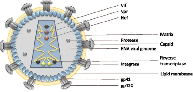
Figure 1.7 HIV1 structure: This virus is composed by two positive RNA single strands coated by a
nucleocapsid (not present in the figure) encapsulated together with Vif, Vpr, Nef, Integrase and Reverse transcriptase in a particle of close to 100nm in diameter. Right below the lipid membrane there is the matrix that surrounds the viral capsid.
HIV infects CD4 positive cells like CD4+ T cells, macrophages and dendritic cells, by fusing with their membrane. This is due to the viral proteins gp120 and gp41 presented in the outer membrane envelope responsible for the targeting and fusion process. gp120 initially binds to the cellular receptor CD4 and subsequently to a group of CC and CXC chemokine receptors like CCR5 and CXCR4, a coreceptor. At this point gp41 undergoes conformational changes in order to expose a fusion peptide that triggers the membrane fusion process. Once in the cytoplasm, the viral core suffers an uncoating process by dissociation of the CA important for RT to copy the RNA genome into a double stranded linear DNA genome which in turn is delivered to the nucleus and intregrated onto the cellular genome with the help of IN. The provirus is transcribed by the host RNA polymerase II into spliced and unspliced mRNA transcripts. In an early phase Tat, Rev and Nef are translated from small multiple spliced mRNAs. While Tat enhances viral mRNA transcription by binding to the trans‐activating response element (TAR) in association with other host proteins, Rev is responsible for halting viral mRNA cleavage allowing the multiplication of the viral genome and for Gag and Pol polyproteins to by synthesized. The Env polyprotein is produced in the endoplasmatic reticulum and cleaved and glycosylated into gp120 and gp41 (McCune et al., 1988) and transported to the plasma membrane where in association with Gag and Pol polyproteins, protease and genomic RNA form the immature virions. Upon release by gemulation the viral protease triggers the maturation of the viral particles with a drastic reorganization of the core and gain of infectious capacity.