Universidade de Lisboa
Faculdade de Ciências
Departamento de Biologia Vegetal
Study of a new pathway involved in
electron bifurcation in anaerobic
bacteria
Gonçalo Pizarro Madureira Salgado de Oliveira
Dissertação
MESTRADO EM MICROBIOLOGIA APLICADA
Universidade de Lisboa
Faculdade de Ciências
Departamento de Biologia Vegetal
Study of a new pathway involved in
electron bifurcation in anaerobic
bacteria
Gonçalo Pizarro Madureira Salgado de Oliveira
Dissertação
MESTRADO EM MICROBIOLOGIA APLICADA
Dissertação orientada por
Doutora Inês Cardoso Pereira
Doutora Sofia Venceslau
Professora Doutora Ana Tenreiro
Study of a new pathway involved in
electron bifurcation in anaerobic
bacteria
Gonçalo Pizarro Madureira Salgado de Oliveira
2014
This thesis was fully performed at Instituto de Tecnologia Química
e
Biológica
(ITQB)
under
the
direct
supervision
of
Dr. Inês Cardoso Pereira and co-supervision of Dr. Sofia
Venceslau. Dr. Ana Tenreiro was the internal supervisor in the
scope of the Master in Applied Microbiology of the Faculty of
Sciences of the University of Lisbon (FCUL).
Acknowledgements
First of all I would like to thank my supervisor Dr. Inês Cardoso Pereira for giving me the opportunity to work and learn in the Bacterial Energy Metabolism Lab and off course for all the guidance, expertise and knowledge provided during this work.
I also thank Dr. Sofia Venceslau who helped me since the very beginning of the experimental work and who taught me most of the things I know today with infinite patience.
I thank Dr. Raquel Ramos for allowing me to collaborate with her in the Hdr-Flox project from which an award-winning poster and a paper soon to be published emerged.
I also thank my internal supervisor Dr. Ana Tenreiro for promptly analyzing and correcting my thesis and also for the useful advices in the last phase of this work.
I thank Cátia Santos from the Genomics and Stress Lab (ITQB) for all the support during the RT-qPCR experiments.
I would like to thank all my colleagues at Bacterial Energy Metabolism Lab (Américo, André Santos, Cláudia, Isabel, Marta, Mónica, Sónia and more recently André Preto and Irene) for all their help during this work and for making ITQB a better place to be.
A special thanks to my girlfriend Marta Lourenço for all the patience and support throughout this work and to my parents for always providing me the best conditions to work and be fulfilled.
I thank Fundação para a Ciência e Tecnlogia for financial support of my research fellowship under the scope of the project “Energy conservation by a novel NADH dehydrogenase family widespread in Bacteria” (PTDC/BBB-BQB/0684/2012).
Abstract
Bioenergetics of chemotrophic bacteria is based on substrate-level phosphorylation and electron transfer phosphorylation for energy conservation. Recently, a third mechanism of energy coupling named flavin-based electron bifurcation (FBEB) was proposed for anaerobic bacteria. Sulfate-reducing organisms (SRO) are a polyphyletic group of anaerobic microorganisms, which perform dissimilatory sulfate reduction. Interestingly, studies concerning SRO identified protein homologies to enzymes engaged in FBEB in methanogens, suggesting the presence of FBEB in sulfate reducers. Here we studied: i) the physiological role of a protein complex (HdrABC-FloxABCD) possibly involved in FBEB; and ii) the function of DsrD, a protein potentially involved in sulfite reduction, the last step of the dissimilatory sulfate reduction.
Firstly, phenotypic characterization of hdrC and floxA mutants in Desulfovibrio
vulgaris Hildenborough with ethanol as electron donor revealed no cell growth, while a
complemented strain of the floxA mutant grew similarly to the wild-type (WT). Then, under pyruvate fermentation conditions, both mutants produced low levels of ethanol comparing to the WT and complemented strain. Gene and protein expression analysis in WT strains cultured with different electron donors/acceptors showed upregulation of FloxA and HdrA when ethanol is the electron donor. Moreover, an alcohol dehydrogenase (adh1) gene present upstream of the hdr-flox cluster is also upregulated in the medium containing ethanol. Altogether, these results show that the HdrABC-FloxABCD complex is involved in the ethanol metabolism of D. vulgaris.
Secondly, the dsrD gene from D. vulgaris was cloned, overexpressed and the protein purified. Sulfite reduction activity and protein-protein interaction studies showed no direct biochemical role of DsrD in sulfite reduction. Then a dsrD deletion mutant was generated showing a long lag phase under sulfate reduction conditions when compared to the WT. This mutant did not grow with sulfite as electron acceptor, revealing the importance of dsrD in sulfite reduction, most likely at a regulatory level.
Overall, this work allowed a better understanding of energy conservation mechanisms in SRO, proposing a new mechanism for ethanol metabolism played by the FloxABCD-HdrABC complex and producing new insights into the function of DsrD in the dissimilatory sulfate reduction.
Keywords: Bioenergetics; energy conservation; flavin-based electron bifurcation; sulfate-reducing organisms; gene expression studies, phenotype characterization; protein purification.
Resumo
Os sistemas bioenergéticos de bactérias quimiotróficas baseiam-se em dois mecanismos de conservação de energia: a fosforilação ao nível do substrato (FNS) e a fosforilação oxidativa (FO). A FO é também conhecida como respiração e envolve acoplamento quimiosmótico. Recentemente, um outro mecanismo de acoplamento energético, chamado bifurcação electrónica baseada em flavinas (BEBF), foi proposto para bactérias anaeróbias quimiotróficas. Este mecanismo é caracterizado por acoplar uma reacção termodinamicamente não favorável a uma reacção favorável e foi já experimentalmente demonstrado em organismos fermentativos, árqueas metanogénicas e bactérias acetogénicas. Os complexos proteicos envolvidos na BEBF são citoplasmáticos, contêm flavinas como cofactores (FMN ou FAD) e um dos aceitadores/dadores de electrões é normalmente uma ferredoxina (Fd).
Os organismos redutores de sulfato (ORS) são um grupo polifilético de microrganismos existentes em ambientes anaeróbios, tendo um metabolismo versátil. Os sulfato-redutores têm a capacidade de realizar a redução dissimilativa do sulfato, i.e., de reduzir grandes quantidades de sulfato como mecanismo de conservação de energia produzindo sulfureto, um produto tóxico do seu metabolismo. Deste modo estes organismos contribuem para o ciclo biogeoquímico do enxofre usando o sulfato como aceitador de electrões durante a degradação de matéria orgânica, produzindo sulfureto que pode ser oxidado por outros microrganismos. Pela capacidade que têm de utilizar diversos dadores e aceitadores de electrões, os ORS são usados em bio-remediação de metais tóxicos e compostos orgânicos como hidrocarbonetos; todavia estes organismos comportam igualmente um impacto negativo, nomeadamente contribuindo para a bio-corrosão de metais ferrosos e de betão e contribuindo ainda para a acidificação de reservas de petróleo. Apesar da importância ambiental destes microrganismos, os mecanismos de conservação de energia neste grupo permanecem por conhecer com clareza. Para estudos fisiológicos, genéticos e bioquímicos é normalmente utilizado o organismo modelo Desulfovibrio vulgaris Hildenborough, uma vez que é relativamente fácil e rápido de cultivar em laboratório, tendo sido o primeiro ORS a ter o seu genoma sequenciado. Estudos genómicos identificaram proteínas semelhantes à heterodilsulfureto redutases (Hdr) em ORS, especialmente proteínas homólogas à HdrA, a subunidade proteica que contém uma flavina como co-factor e que foi proposta ser responsável pela BEBF em árqueas metanogénicas. Este estudo sugere desta forma que o mecanismo de BEBF pode estar presente em procariotas sulfato-redutores.
Assim sendo, o objectivo deste trabalho centra-se em contribuir para um maior e mais profundo conhecimento dos mecanismos de conservação de energia em sulfato-redutores, estudando o papel fisiológico do complexo citoplasmático heterodisulfureto redutase-oxidase de flavina (HdrABC-FloxABCD), que se pensa estar envolvido em BEBF, e investigando a potencial função da proteína redutase dissimilativa de sulfito (DsrD) na via da redução dissimilativa do sulfato.
Na primeira parte do trabalho experimental, estudou-se o papel fisiológico dos genes
flox-hdr em D. vulgaris Hildenborough, usando duas estirpes mutantes: IPFG01, com uma
cassete de resistência a canamicina inserida no gene hdrC, e IPFG02, uma estirpe em que o gene floxA foi substituído por uma cassete de resistência a canamicina por recombinação homóloga. A caracterização fenotípica das estirpes mutantes revelou que ambos os mutantes são incapazes de crescer quando o etanol é usado como dador de electrões para reduzir sulfato. Por outro lado uma estirpe mutante complementada com o gene floxA (IPFG03) cresceu de forma semelhante ao WT. Durante o crescimento fermentativo em piruvato, realizou-se a quantificação da concentração de etanol no meio extracelular através de um método enzimático. Este ensaio experimental revelou uma produção muito baixa de etanol por parte das estirpes mutantes IPFG01 e IPFG02, comparativamente às estirpes WT e IPFG03, indicando deste modo que em condições fermentativas de crescimento, as proteínas FloxABCD estão envolvidas na redução de NAD+ para produção de etanol. Foram adicionalmente realizados estudos de expressão génica ao nível do gene (por PCR quantitativo) e da proteína (por “Western blotting”) na estirpe selvagem (WT) em condições de cultura contendo diferentes dadores e aceitadores de electrões. Estes revelaram que os genes floxA e hdrA são sobre-expressos quando é usado etanol como dador de electrões, e o mesmo foi observado ao nível da expressão proteica. A montante dos genes hdr-flox está situado um gene que codifica para uma álcool desidrogenase (adh1) revelando igualmente uma sobre-expressão quando o etanol é usado como dador de electrões. Contudo a expressão do gene adh1 é consideravelmente superior à dos genes anteriormente referidos, indicando que os genes adh1 e hdr-flox não se encontram no mesmo operão. Esta diferença observada ao nível da expressão génica reflectiu-se na purificação das proteínas: enquanto que a proteína Adh1 foi facilmente purificada, o mesmo sucesso não foi obtido na purificação das proteínas Flox e Hdr devido à sua baixa expressão. Os resultados obtidos considerando esta parte do trabalho demonstram que as proteínas FloxABCD estão envolvidas no metabolismo do etanol em D. vulgaris. Propomos então que o complexo FloxABC-HdrABCD seja capaz de realizar BEBF acoplando a redução de Fd com NADH à redução da proteína DsrCigualmente com NADH.
Na segunda parte deste trabalho, o gene dsrD foi clonado, sobre-expresso em
Escherichia coli e a proteína subsequentemente purificada por técnicas cromatográficas. O
gene dsrD encontra-se situado imediatamente a jusante do gene dsrAB, fazendo ambos parte do mesmo operão. A redutase dissimilativa de sulfito (DsrAB) é, juntamente com a proteína DsrC, responsável pelo último passo da redução dissimilativa do sulfato. Tendo em conta a potencial importância da proteína DsrD na redução do sulfato, foram realizadas ensaios espectrofotométricos de actividade enzimática de redução do sulfito e testes de interação proteína-proteína (por Biacore) e proteína-ligando (por Calorimetria de Titulação Isotérmica) de modo a detectar interações entre a DsrD e DsrAB, DsrC e/ou sulfito. Apesar das diversas tentativas, não foram obtidos resultados positivos, levando-nos a crer que a proteína DsrD desempenha um papel regulatório em vez de um papel funcional directo na redução do sulfato. Um vez que a estrutura tridimensional da proteína foi já obtida e inclui um
domínio de ligação ao DNA, considera-se que a proteína DsrD desempenhe um papel regulatório in vivo. Durante este trabalho experimental foi construído um mutante de deleção por troca da sequência codificante para o gene dsrD com uma cassete de resistência a canamicina através de recombinação homóloga. Os estudos fenotípicos com esta nova estirpe revelaram uma longa fase de adaptação na curva de crescimento em meio de cultura contendo sulfato como aceitador de electrões, comparativamente com a estirpe WT. Pensamos que após a longa fase de adaptação as células adquirem espontaneamente mutações que lhes permitem adaptar-se ao meio contendo sulfato, demonstrando que o gene dsrD pode estar a regular os genes dsrAB afectando deste modo a via respiratória. Adicionalmente, a estirpe mutante não foi capaz de crescer em meio com sulfito como aceitador de electrões, o que revela que este gene é essencial para a redução do sulfito.
De uma forma geral, este trabalho contribuiu para uma melhor e mais profunda compreensão do modo como os organismos sulfato-redutores desempenham a conservação de energia, com especial interesse na nova via de conservação de energia, BEBF, que aparenta estar disseminada pelas bactérias quimiotróficas anaeróbias. Em perspectivas futuras seria ideal conseguir expressar e purificar o complexo FloxABCD-HdrABC de modo a analisar a BEBF in vitro. Adicionalmente, novas indicações foram obtidas no que toca a compreender qual a função ao nível fisiológico da proteína DsrD, envolvida na redução dissimilativa do sulfato. Em continuação deste projecto, seria interessante determinar a capacidade de interação da proteína DsrD com o DNA in vitro, analisar que genes substituem a função do gene dsrD, permitindo a adaptação da estirpe mutante ao meio de cultura contendo sulfato como aceitador de electrões, e ainda qual a contribuição desses mesmos genes para a via da redução dissimitativa do sulfato.
Palavras-chave: Bioenergética; conservação de energia; bifurcação electrónica baseada em flavinas; organismos redutores de sulfato; estudos de expressão génica; caracterização fenotípica; purificação de proteínas.
Index
LIST OF ABBREVIATIONS XI
1 – INTRODUCTION XII
1.1 – Energy conservation in anaerobic bacteria 1
1.1.1 – Bioenergetic origins 2
1.1.2 – Flavin-based electron bifurcation – a new pathway for energy coupling 3
1.1.3 – FBEB examples 4
1.2 – Sulfate-Reducing Organisms (SRO) 6
1.2.1 – Sulfur cycle 7
1.2.2 – Physiology and Biochemistry 7
1.2.3 – Taxonomy 8
1.2.4 – Environmental impact 10
1.3 – The Desulfovibrio genus 10
1.3.1 – Morphology, biochemistry, and genome features 11
1.3.2 – Taxonomy 11
1.3.3 – Dissimilatory sulfate reduction 11
2 – OBJECTIVES 14
3 – MATERIALS AND METHODS 15
3.1 – The hdr-flox gene cluster 15
3.1.1 – Strains and media 15
3.1.2 – Growth curves 15
3.1.3 – Ethanol quantification 15
3.1.4 – Adh1 purification and activity measurements 17
3.1.5 – Reverse Transcriptase quantitative PCR (RT-qPCR) 17 3.1.6 – Western blot analysis of FloxA, HdrA, and Adh1 expression 18
3.2 – Studies with dsrD 19
3.2.1 – dsrD heterologous expression 19
4 – RESULTS 22
4.1 – The hdr-flox gene cluster 22
4.1.1 – Growth curves 22
4.1.2 – Adh1 purification and activity measurements 24
4.1.3 – Reverse Transcriptase quantitative (RT-qPCR) 25
4.1.4 – Western blot analysis of FloxA, HdrA, and Adh1 expression 25
4.2 – Studies with dsrD 26
4.2.1 – DsrD activity and interaction assays 26
4.2.2 – Growth curves 27
5 – DISCUSSION 28
5.1 – The hdr-flox gene cluster 28
5.2 – dsrD 32
List of abbreviations
Ack – Acetate kinase
Adh – Alcohol dehydrogenase ADP – Adenosine diphosphate Al-dh – Aldehyde dehydrogenase AMP – Adenosine monophosphate
Aor – Aldehyde:ferredoxin oxidoreductase. Apr – Adenosine-5’-phosphosulfate reductase APS – Adenosine-5’-phosphosulfate
ATP – Adenosine triphosphate BCA – Bicinchoninic acid assay
Bcd/Etf – Butyryl-CoA dehydrogenase/electron transfer complex BCIP – 5-bromo-4-chloro-3-indolyl phosphate
CoA – Coenzyme A
CoB-SH – Coenzyme B, N-7-mercaptoheptanoyl-L-threonine phosphate CoM-SH – Coenzyme M, 2-mercaptoethanesolfonate
CoM-S-S-CoB – Heterodisulfide of CoM-SH and CoB-SH dCTP – Deoxycytidine triphosphate
DMRB – Dissimilatory metal-reducing bacteria Dsr – Dissimilatory sulfite reductase
E0’ – Standard electron potential
EDTA – Ethylenediaminetetraacetic acid EtOH – Ethanol
ETP – Electron transport phosphorylation FAD – Flavin adenine dinucleotide FBEB – Flavin-based electron bifurcation Fd – Ferredoxin
Flox – Flavin oxidoreductase FMN – Flavin mononucleotide Hase – Hydrogenase
Hdr – Heterodisulfide reductase Hyd – [Fe]-only hydrogenase
ITC – Isothermal Titration Calorimetry Km – Kanamycin
LA – Luria agar LB – Lysogeny broth
Ldh – Lactate dehydrogenase
MOY – MO basal medium with yeast extract MQ – Menaquinone
MQH2 – Menaquinol
Mvh – F420 non-reducing hydrogenase
NAD+ – Nicotinamide adenine dinucleotide
NADH – Nicotinamide adenine dinucleotide reduced form NBT – Nitro-blue tetrazolium chloride
Nfn – NADH-dependent reduced ferredoxin:NADP+
oxidoreductase OD – Optical density
PCR – Polymerase chain reaction Pi – Inorganic phosphate
Por – Pyruvate:ferredoxin oxidoreductase PPi – Inorganic pyrophosphate
Pta – Phosphate acetyltransferase PVDF – Polyvinylidene difluoride
Qmo – Quinone-interacting membrane-bound oxidoreductase Rnf – Rhodobacter nitrogen fixation
RT-qPCR – Reverse Transcriptase quantitative Polymerase Chain Reaction SLIC – Sequence ligation independent cloning
SLP – Substrate-level phosphorylation SPR – Surface Plasmon Resonance SRO – Sulfate-reducing organisms TBS – Tris-buffered saline
TBST – Tris-buffered saline Tween 20 TpIc3 – Type I cytochrome c3
Tris – Tris(hydroxymethyl)aminomethane UV – Ultraviolet
Vho – Methanophenazine-reducing [NiFe] hydrogenase
1 – Introduction
1.1 – Energy conservation in anaerobic bacteria
Life requires energy. One of the features that distinguish living beings from inanimate objects is their metabolism. Metabolism balances energy through a set of chemical transformations occurring in two forms: anabolism, biosynthetic reactions requiring energy input; and catabolism, molecular breakdown reactions that allow to restore the cellular energy budget. The universal molecular currency of energy adopted by living beings is proton motive force, which is consumed for cellular processes and produced by energy conservation mechanisms. Additionally, there are two environmental sources for energy conservation: in chemotrophic organisms, energy derives from the oxidation of environmental electron donors, while in phototrophic organisms energy comes from sunlight (Figure 1.1). Chemotrophic organisms exist virtually since the dawn of life on Earth and have evolved to very distinct metabolic systems ever since.
Figure 1.1 – Energy transformations via the proton motive force system. Adapted from (1).
HEAT
Energy input
Energy output
1. Chemical Energy
(Chemotrophy)
2. Light Energy
(Phototrophy)
1. Chemical Work
(Biosynthesis)
2. Osmotic Work
(Active Transport)
3. Mechanical Work
(Active Movement)
Anabolism
Catabolism
Proton Motive Force
+ + + + + + +
− − − − − − −
1.1.1 – Bioenergetic origins
At the time life is estimated to have emerged on Earth (about 4.6 billion years ago) our planet had quite different geological and atmospheric characteristics from the ones observed today. The prebiotic atmosphere was mainly composed of hydrogen, ammonia, methane, carbon dioxide, and water (2). In the 1920’s Aleksandr Oparin and John Haldane hypothesized that these compounds, combined with the strong UV radiation present at that time, led to the emergence of the first biomolecules in ocean waters. The combination of these molecules, along with the action of Natural Selection during millions and millions of years, formed the complex life forms we know of today.
Since the primordial atmosphere had no oxygen, the first living beings had to replicate and generate their own energy from an anaerobic environment. Energy was stored in the form of ATP, an energy-rich compound required for anabolic reactions and cell function; this biochemical signature has been conserved by nature thereafter and is still present among all living forms. In this regard, the first metabolism capable of sustaining life would use the compounds available in the primitive environment in order to generate energy for cellular biochemical reactions.
During the evolution of life, new metabolic machineries started to emerge. Despite the diversity of mechanisms, energy conservation in chemotrophs is thought to exist only via two different ways: substrate-level phosphorylation (SLP) and electron-transfer phosphorylation (ETP), also known as oxidative phosphorylation (1). In the first mechanism, energy-rich compounds form ATP by transferring a phosphoryl group to ADP in anaerobic conditions. The second mechanism (ETP) involves the formation of an electrochemical gradient (∆pH for protons or ∆pNa for sodium ions) across the cytoplasmic membrane generated by an electron transfer chain that reduces a terminal acceptor (3). The electrochemical gradient formed drives a cationic flow through ATP synthase, which causes subunits of this enzyme to rotate, leading to conformational changes in the active site, which culminates in the formation of ATP from ADP and Pi (4). Two major types of oxidative
phosphorylation, also called respiration, emerged differing in the electron acceptors used. These respiratory organisms used both SLP and ETP pathways using a diverse range of electron donors/acceptors for energy conservation (Table 1.1). Anaerobic respiration emerged first, using reduced compounds such as sulfur or iron as electron acceptors. After the rise of atmospheric oxygen concentration caused by the metabolism of photosynthetic organisms, aerobic respiration emerged using oxygen as terminal electron acceptor. Since oxygen has a higher redox potential (E0’) than the electron acceptors used in anaerobic respiration it permits the release of more energy per oxidized molecule.
Although it is classically considered that fermentative organisms do not perform respiration, recent studies indicate that their energy conservation mechanisms may be more complex. There are several examples of an electrochemical gradient necessary for ATP synthesis being formed in the membrane of fermentative organisms, such as by electrogenic transport in lactic acid bacteria (5), electron transfer through energy-conserving
hydrogenases in hyperthermophiles (6), and also by sodium-translocating NADH dehydrogenases in glutamate fermenting bacteria (7).
Table 1.1 – Redox potential of electron donors/acceptors involved in electron transport
phosphorylation. Adapted from (1).
Redox compound E0’ (mV) Redox compound E0’ (mV)
SO4
2-/HSO3- –516 Flavodoxin ox/red (E0’2) –115
CO2/formate –432 HSO3 -/HS- –116 H+/H2 –414 Menaquinone ox/red (MK) –74 S2O3 2-/HS-+ HSO3 - –402 APS/AMP + HSO3 - –60
Flavodoxin ox/red (E0’1) –371 Rubredoxin ox/red –57
Ferredoxin ox/red (E0’1) –398 Acrylyl-CoA/propionyl CoA –15
NAD+/NADH –320 Glycine/acetate- + NH4
+ –10
Cytochrome c3 ox/red –290 2-Demethylvitamin K12 ox/red +25
CO2/acetate - –290 S4O6 2 / S2O3 2-+24 S0/HS- –270 Fumarate/succinate +33 CO2/CH4 –244 Ubiquinone ox/red +113 FAD/FADH2 –220 S3O62-/S2O32- + HSO3- +225 Acetaldehyde/ethanol –197 NO2 -/NO +350 Pyruvate-/lactate- –190 NO3 -/NO2 - +433 FMN/FMNH2 –190 Fe 3+ /Fe2+ +772 Dihydroxyacetone phosphate/glycerol-phosphate –190 O2/H2O +818 HSO3 -/S3O6 2- –173 NO/N2O +1175
Oxaloacetate2-/malate2- –172 N2O/N2 +1355
1.1.2 – Flavin-based electron bifurcation – a new pathway for energy
coupling
In recent years, a new mechanism of energy coupling named flavin-based electron bifurcation (FBEB) has been proposed (8, 9). This mechanism couples a thermodynamically unfavorable reaction (often the reduction of ferredoxin) to a favorable reaction. Reduced ferredoxin (Fd) then functions as an electron and energy carrier and its oxidation can be linked to energy conservation. At the base of FBEB is a stepwise electron transfer from a reduced flavin co-factor (FADH2) in which the low-potential electron is used for reduction of a
high potential electron acceptor, while the second electron reduces Fd (Figure 1.2) (9). The reduction of low potential Fd is only possible since a lower redox potential flavin semiquinone (“hot flavosemiquinone”) is formed after the first electron is transferred to the high potential electron acceptor (10). The logic behind FBEB is similar to the Q-cycle proposed by Peter Mitchell, involving a quinone electron bifurcation at the bc1 complex (11). Energy conservation
via reduced Fd occurs by either proton reduction to H2, increasing SLP in the oxidative branch
of fermentation, or by generation of an electrochemical gradient via the Rnf membrane complex (8). The Rnf complex (for Rhodobacter nitrogen fixation) is a H+/Na+-pumping Fd:NAD+ oxidoreductase found in many anaerobes (12). Since low redox potential Fd is required by autotrophs to reduce CO2, FBEB might have been important, if not essential,
during early life evolution (13).
1.1.3 – FBEB examples
The FBEB mechanism was first observed in the Gram-positive bacterium Clostridium
kluyveri (9), which is able to ferment ethanol and acetate to butyrate, caproate, and H2, a
unique feature among clostridia (14). H2 formation was shown to be Fd-dependent, requiring
acetyl-CoA for reduction of crotonyl-CoA to butyryl-CoA. Also, H2 was formed by an
endergonic reaction during fermentation from NADH. The work of Li et al. revealed that the cytoplasmic butyryl-CoA dehydrogenase/electron transfer complex (BcdA/EtfBC complex) was responsible for coupling Fd reduction with NADH to the reduction of crotonyl-CoA to butyryl-CoA with NADH (Figure 1.2) (9). Since this complex contains four FAD cofactors with no additional prosthetic groups this new energy conservation mechanism was named flavin-based electron bifurcation. Other examples of FBEB enzymes are the NfnAB complex present in C. kluyveri, which couples the reduction of Fd with NADPH to the reduction of NAD+ (15) and the reverse electron bifurcation (confurcation) Hase (HydABC) present in
Thermotoga maritima, which couples the oxidation of Fd and NADH to generate H2 (16).
Figure 1.2 – Flavin-based electron bifurcation mechanism by Bcd/EtfCB from Clostridium kluyveri.
The endergonic reduction of Fd by NADH is coupled to the exergonic reduction of crotonyl-CoA with NADH. Adapted from (9).
2 NADH
2 FADH
22 FADH
2 FAD
Crotonyl-CoA
4 e
-+ 4 H
+2 e
-+ 2 H
+Fd
red+ 2 H
+Fd
oxE
o’= – 410 mV
E
o’= – 10 mV
FAD
FAD
FAD
2 NADH
2 NAD
++ 2 H
+Fd
oxCrotonyl-CoA
Butyryl-CoA
EtfC
Etf
B
Bcd
Bcd
E
o’= – 320 mV
Fd
red 2-In methanogens, the formation of methane from CO2 reduction with H2 is coupled to the
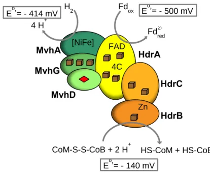
formation of a heterodisulfide (CoM-S-S-CoB). The mechanism for heterodisulfide reduction is distinct in methanogens with cytochromes from methanogens without cytochromes (17). In methanogens with cytochromes VhoACG hydrogenase/HdrDE heterodisulfide reductase membrane complex couples H2 oxidation to heterodisulfide reduction and proton pumping
(18). In methanogens without cytochromes the MvhADG hydrogenase/HdrABC heterodisulfide reductase cytoplasmic complex couples the reduction of Fd (unfavorable step) with H2 to the reduction of the heterodisulfide by H2 (favorable step) in a FBEB mechanism
(Figure 1.3) (19). Interestingly, we can find HdrABC related proteins in non-methanogenic organisms in which the function of these proteins remains unsolved. The most evident of these Hdr-containing organisms are the sulfate-reducing organisms (SRO) group, whose energy metabolism remains incompletely understood. Surprisingly, these organisms contain several proteins similar to HdrA and HdrB/D (20, 21).
Figure 1.3 – Schematic representation of HdrABC/MvhADG complex from methanogens without
cytochromes. The complex is responsible for the endergonic reduction of Fd with H2 coupled to the
exergonic heterodisulfide reduction with H2. Adapted from (17). C – cysteine; Cube – [4Fe-4S];
diamond – [2Fe-2S].
In the sulfate reducer Desulfovibrio vulgaris Hildenborough, a novel gene cluster includes the floxABCD (flavin oxidoreductase) genes, which code for a new NADH dehydrogenase and a set of hdrABC genes (Figure 1.4). The flox/hdr cluster is predicted to form an operon possibly involved in electron bifurcation from NADH. Additionally, this cluster is found in a great diversity of Bacteria, including members of different phyla such as
Chlorobi, Proteobacteria, Firmicutes, Bacteroidetes, Spirochaetes, and Actinobacteria (20). It
is proposed that the Flox proteins oxidize NADH via FloxA that will then transfer electrons to
FAD
4C
Zn
[NiFe]
Fd
oxFd
redH
24 H
+CoM-S-S-CoB + 2 H
+HS-CoM + HS-CoB
E
o’= - 500 mV
E
o’= - 140 mV
MvhA
MvhD
MvhG
HdrA
HdrB
HdrC
E
o’= - 414 mV
HdrABC (Figure 1.5). In D. vulgaris Hildenborough, the flox/hdr genes are flanked by two alcohol dehydrogenase genes one of which (adh1) is one of the most highly expressed genes in D. vulgaris cells, revealing an important function in the energy metabolism (22).
Figure 1.4 – Gene locus of D. vulgaris Hildenborough containing the floxABCD, hdrABC, and adh
genes (in this organism the floxC and floxD genes are fused).
Figure 1.5
– Schematic representation of the D. vulgaris Hildenborough Flox and Hdr proteinswith corresponding cofactors. NADH may be formed upon oxidation of ethanol by the Adh1 encoded next to the flox and hdr genes. Cube – [4Fe-4S]; diamond – [2Fe-2S]; Fd – ferredoxin.
1.2 – Sulfate-Reducing Organisms (SRO)
There is geological evidence for the activity of SRO by 3.5 billion years ago (23). Since then, this polyphyletic group of microorganisms has been playing a critical role in the biogeochemical sulfur and carbon cycles. SRO live in anaerobic conditions such as marine sediments, fresh waters, soil and also in the mouth and gut of some animals, including humans. Due to the high concentration of sulfate in sea water (~28 mM) they are more abundant in marine environments and are responsible for 50 % of total carbon mineralization in marine sediments (24). This group of organisms is capable of anaerobic respiration, performing energy conservation through reduction of sulfate to sulfide. Also, since these organisms are able to use a vast variety of compounds as electron donors and electron acceptors, they have a great potential for biotechnological applications, such as bioremediation. Furthermore it was reported that sulfate-reducing bacteria can cause inflammatory bowel diseases in animals and humans (25).
adh1 floxC floxB floxA adh2
D hdrB hdrA hdrC
FAD
FAD
NADH
NAD
+Fd
oxFd
redFloxA
FloxCD
FloxB
HdrA
HdrC
HdrB
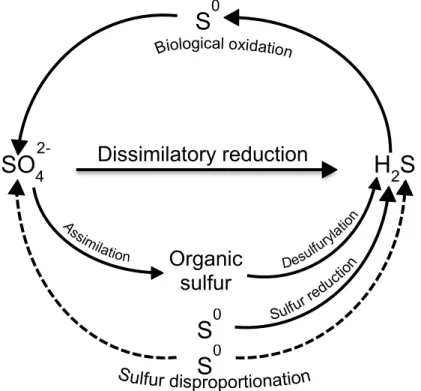
2-1.2.1 – Sulfur cycle
Sulfur is one of the most abundant elements on Earth and it can be found in different oxidation states, being sulfate (SO4
2-; oxidation state +6), elemental sulfur (S0; oxidation state 0), and sulfide (S2-; oxidation state -2) the most significant in nature (26, 27). The sulfur biogeochemical cycle is not only important in geology since many minerals are composed of sulfur but is also essential in biology since amino acids (cysteine and methionine) and many other biomolecules contain sulfur atoms. SRO play a critical role in the sulfur cycle since they use sulfate as terminal electron acceptor in the degradation of organic matter to produce sulfide (Figure 1.6). Both these sulfur forms are used in the metabolism of other organisms generating the microbial sulfur cycle (26). Stable isotope fractionation studies show that sulfur has been reduced biologically for over 3.5 billion years (28).
Figure 1.6 – Microbial sulfur cycle. SRO reduce sulfate (SO4
2-) to sulfide (H2S). Sulfide can then
be oxidized aerobically by chemolithotrophic sulfur-oxidizing bacteria (e.g. Thiobacillus or
Beggiatoa spp.) or anaerobically by phototrophic sulfur bacteria (e.g. Chlorobium spp.) to
elemental sulfur (S0) and sulfate. Other transformations, which are carried out by specialized groups of microorganisms, result in sulfur reduction (e.g. Desulfuromonas spp.) and sulfur disproportionation (e.g. Desulfovibrio sulfodismutans). Adapted from (24).
1.2.2 – Physiology and Biochemistry
SRO are chemoheterotrophs since they generate their energy through chemical reactions and require organic carbon for cell growth. They are also capable of either lithotrophic or organotrophic growth since they can use a great diversity of electron donors such as hydrogen, fatty acids, aromatic compounds, sugars, monocarboxylic acids, and dicarboxylic acids (29). Sulfate is not the sole terminal electron acceptor in SRO, as other
S
0SO
4H
2S
S
0S
0Organic
sulfur
Dissimilatory reduction
2-sulfur compounds can also be used such as sulfite (SO3
2-), thiosulfate (S2O3
2-), elemental sulfur (S0), and organosulfonates. Moreover, some species of SRO can use other electron acceptors as nitrate and nitrite, which are reduced to ammonium and also metal ion oxides including iron (FeIII), uranyl (UVI), selenite (SeVI), chromate (CrVI), and arsenate (AsVI) therefore revealing a great potential in bioremediation of toxic metals (24).
Many organisms including bacteria, fungi, algae, and plants are capable of assimilatory sulfate reduction in which small amounts of sulfide are produced and incorporated into sulfur-containing amino acids, vitamins and cofactors (30). SRO perform dissimilatory sulfate reduction, i.e. the reduction of sulfate for energy conservation while producing large amounts of sulfide, a toxic gas in the protonated form. Moreover, some sulfate reducers are able to survive in low concentrations of oxygen, showing that these organisms are not strict anaerobes (31).
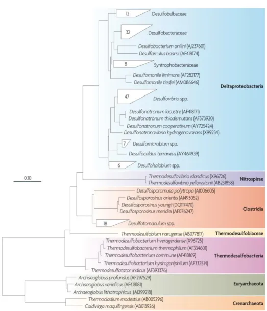
1.2.3 – Taxonomy
The first SRO to be isolated and described was named Spirillum desulfuricans due to its morphology and ability to produce sulfide from sulfate (32). This discovery dates from 1895 and was attributed to the Dutch microbiologist and botanist Martinus Beijerinck; the bacterium was later reclassified as Desulfovibrio desulfuricans (Figure 1.7). Classification of these organisms was based on phenotypic characteristics, which limited the number of species until the 1980’s. With the advent of Molecular Biology new methods based on the rRNA 16S gene, PCR, and genome sequencing started to emerge and were used to discover numerous novel species of SRO (24).
Figure 1.7
– The figure is a reproduction of Vibrio desulfuricans, which was painted by HenriëtteBeijerinck, the sister of Martinus Beijerinck, and is reproduced courtesy of the Beijerinck Museum (24).
There are more than 220 species of 60 genera of SRO described, either from the Bacteria or Archaea Domain. Within the Bacteria Domain, these organisms belong to five divisions: Deltaproteobacteria class (including Desulfovibrio and 11 other genus), Nitrospira phylum (containing the genus Thermodesulfovibrio), Clostridia class (containing the Gram-positive, spore-forming genus Desulfotomaculum, Desulfosporomusa, and
Desulfosporosinus), Thermodesulfobiaceae family, and Thermodesulfobacteria phylum.
Within the Archaea Domain, two phyla are distinguished: Euryarchaeota (containing the
Archaeoglobus genus) and Crenarchaeota (containing the genus Thermocladium and Caldivirga) (Figure 1.8) (24, 29). From these organisms, 72 genome sequences are already
available according to the Integrated Microbial Genomes (IMG) website.
Figure 1.8 – Phylogenetic tree of the SRO described, based on 16S ribossomal RNA (rRNA)
sequences. It is possible to observe five phylogenetic lineages of Bacteria Domain organisms and two of Archaea Domain organisms. The 0.10 scale bar indicates 10 % of sequence difference. From (24).
1.2.4 – Environmental impact
SRO have an important environmental impact since they intervene in biocorrosion of ferrous metals and also corrosion of concrete and stonework (29). These microorganisms also have a negative impact on the petroleum industry since they can contribute to a phenomenon called “oil souring” by sulfide production in oil fields and pipelines.
Since SRO have a very plastic metabolism they have a great biotechnological potential. These microorganisms are capable of bioremediation of several compounds by oxidation, such as organic compounds, monoaromatic hydrocarbons present in contamination due to petroleum spills; reduction of halogenated compounds, which contaminate waters and sediment, and are used as terminal electron acceptor (dehalorespiration); nitroaromatic respiration, such as TNT (trinitrotoluene) which contaminates soils and ground water near places where explosives are manufactured (29). Also, SRO are capable of immobilization of hazardous and toxic metals released by metallurgic plants, nuclear plants or oil refining industry via precipitation as metal sulfides. A restricted group of SRO called DMRB (dissimilatory metal-reducing bacteria) is capable of toxic metal reduction (such as uranium) posing an important contribution for toxic environment bioremediation. Reduction of azo dyes, which contain an azo bond (N=N) and are abundantly produced in the textile industry, is also performed by this group of organisms; finally, SRO are capable of biorecovery of precious metals such as gold, platinum, or palladium from industrial waste streams (33).
Concerning the high potential of SRO in biotechnology, the study of the physiology and full comprehension of the metabolism of these organisms is imperative to further develop new and more powerful biotechnological tools.
1.3 – The Desulfovibrio genus
Desulfovibrio is one of many genus of sulfate-reducing bacteria belonging to the Deltaproteobacteria class. Desulfovibrio species exist in marine environents, but also in hot
springs, geothermal vents, the human digestive tract, and in soil (34). These bacteria are not strict anaerobes as previously considered, since they tolerate oxygen concentration but their growth is limited in aerobic environments. The optimal growth temperature for the mesophilic
Desulfovibrio bacteria ranges from 25 – 40 ºC while the optimal pH ranges between 6.6 – 7.5.
Since D. vulgaris Hildenborough was the first SRO to have its genome sequenced and due to its easy and rapid growth it has been used as a model for studying the physiology, genetics, and biochemistry of these organisms (35).
1.3.1 – Morphology, biochemistry, and genome features
Desulfovibrio species are non-sporulating curved rod-shaped cells with sizes
between 0.5 – 1.3 x 0.8
–
5 μm (36). These cells are motile with a single polar flagellum and stain Gram negative. These microbes use hydrogen, organic acids, or alcohols as electron donors for sulfate reduction. Lactate is preferentially used for carbon metabolism but pyruvate, formate, and certain primary alcohols also function as carbon sources. The model organism D. vulgaris Hildenborough was discovered and isolated in 1946 in clay soil near Hildenborough, Kent, England (35). The genome contains two replicons: one chromosome containing 3.57 Mbp and one mega plasmid containing 202 kbp. The GC percentage of the replicons is 63.2 and 65.7, respectively (35).1.3.2 – Taxonomy
D. vulgaris Hildenborough belongs to the phylum Proteobacteria and class Deltaproteobacteria. The order of these bacteria is Desulfovibrionales, the family is Desulfovibrionacea and the genus is Desulfovibrio. In the last decade several genomes of
SRO were sequenced and deposited in online databases such as MicrobesOnline, Integrated Microbial Genomes (IMG), and National Centre for Biotechnology Information (NCBI). There are over 60 known species of the Desulfovibrio genus, from which 43 genomes are available according to the IMG website. Very recently, the genome of Desulfovibrio gigas was sequenced through a collaboration between a group at ITQB and Brazilian researchers (37).
1.3.3 – Dissimilatory sulfate reduction
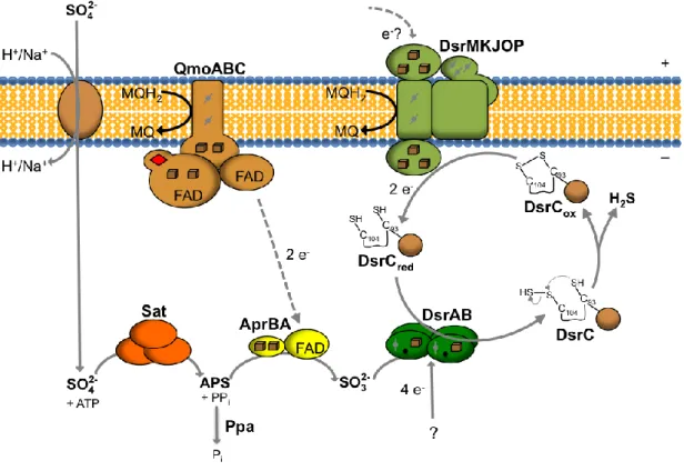
Dissimilatory sulfate reduction occurs in the cytoplasm of SRO and requires the presence of four soluble enzymes. In order to fully reduce sulfate to sulfide, eight electrons are necessary and approximately two ATP molecules are used for sulfate activation (Figure
1.9). Sulfate is transported inside the cell by symport with Na+ or H+, depending on the marine or fresh-water environment, respectively (38). Since sulfate reduction to sulfite is chemically unfavorable [Eo, (SO4
2-/SO3
2-) = – 516 mV] sulfate requires activation to a more favorable compound. In the presence of the ATP sulfurylase or adenylyltransferase (Sat), sulfate and ATP are converted to adenosine-5’-phosphosulfate (APS generating pyrophosphate (PPi).
Some SRO have membrane associated pyrophosphatases that couple the hydrolysis of PPi
to proton translocation across the membrane minimizing the energy costs of sulfate reduction (20). APS [Eo, (APS/SO3
2-) = – 60 mV] accepts two electrons being reduced to sulfite and AMP by the APS reductase enzyme (AprBA). The physiological electron donor to AprBA is still unknown but several lines of evidence point to the Quinone-interacting membrane-bound oxidoreductase (QmoABC) membrane complex (39, 40). After sulfite formation, the final reduction of sulfite to sulfide [Eo, (SO3
2-/H2S) = – 116 mV] requires 6 electrons and two
mechanisms were proposed to explain this reduction: the trithionate pathway, in which sulfite is reduced to sulfide in three steps, by the intermediates trithionate (S3O6
2-) and thiosulfate (S2O3
step, catalyzed by the dissimilatory sulfite reductase (DsrAB or dSir) without formation of any intermediates (41). There is evidence that a third protein, named DsrC but not encoded in the same operon of DsrA and DsrB, is involved in the final reduction of sulfite to sulfide (41). DsrC functions not as a subunit of DsrAB but as an interacting protein by entering the cleft between DsrA and DsrB, as the crystal structure of DsrAB-DsrC complex from D. vulgaris revealed (41). DsrK (a subunit from the DsrMKJOP membrane complex) functions as the potential physiological electron donor to DsrC (42). Since the dsrMKJOP gene cluster is in close proximity to a gene encoding a Fd, there might be an involvement of a Fd in sulfite reduction and other soluble electron transfer pathways (20).
Figure 1.9 – Schematic representation of the proposed model of dissimilatory sulfate reduction
pathway. Adapted from (41). Cube – [4Fe-4S]; diamond - [2Fe-2S]; – heme b; – heme c; – sirohydrochlorine.
Immediately downstream from the dsrAB genes a small gene named dsrD which codes for a 9 kDa protein is present (43). This gene is specific of SRO, although dsrD is absent in some thiosulfate reducers (e.g. Thermosinus carboxydivorans and
Thermanaeromonas toyohensis) and is also not present in sulfur-oxidizing organisms (44).
Furthermore, in the anaerobic taurine-degrading gut bacterium Bilophila wadsworthia RZATAU the dsrD gene is fused to the dsrB gene, forming a DsrB-DsrD fusion protein (45). Interestingly it has also been shown that dsrD is strongly downregulated in the presence of sulfide (46). These results suggest a possible involvement of DsrD in the dissimilatory sulfite
reduction. However, DsrD was not found associated with DsrAB during the purification methods and in vitro studies revealed that this protein binds sulfate, sulfite, and sulfide with low affinity (47). Since the structure of DsrD includes a winged-helix motif, a B- and Z-DNA binding motif, it has been hypothesized that DsrD is associated to sulfate reduction not in a direct biochemical association but via regulation of sulfite reduction genes (Figure 1.10) (48). Despite some structural, biophysical, and biochemical studies performed with this protein, the physiological function of DsrD remains to be solved.
Figure 1.10 – (A) Stereo view of DsrD from D. vulgaris Hildenborough at 1.2 Å resolution. DsrD
has a winged-helix motif composed of three α–helixes (H1, H2, H3) and three β-sheets (s1, s2, s3). DsrD has an additional α-helix (H4) in the C-terminal region (B) Sequence alignment of DsrD from D. vulgaris Hildenborough, D. salexigens, D.desulfuricans, D. piger, D. alaskensis, the DsrD fragment (residues 405-483 of DsrBD) from Bilophila wadsworthia, DsrD from Archaeoglobus
profundus, and Archaeoglobus fulgidus. Box – highly conserved residues in the DsrD family; red –
highly conserved hydrophobic residues in the winged-helix motif. Adapted from (48).
2 – Objectives
This thesis focuses on two main objectives.
The first objective is to study the hdr-flox gene cluster possibly involved in an electron bifurcation pathway present in the model organism D. vulgaris Hildenborough. It was ultimately desired to elucidate and better understand the metabolic importance of this gene cluster and to analyze in which conditions these genes were more expressed both at a gene and protein level.
The second part of this thesis focuses on the study of dsrD, a potentially relevant gene involved in the dissimilatory sulfate reduction whose function in the physiology of SRO remains a mystery. The main objective of this study is to provide new hints about the function of this protein both with in vitro and in vivo assays, while contributing to a better understanding of the dissimilatory sulfate reduction pathway, in particular in the sulfite reduction.
3 – Materials and Methods
3.1 – hdr-flox gene cluster
3.1.1 – Strains and media
The strains used in this work were previously constructed by Fabian Grein (49) and are listed in Table 3.1. All D. vulgaris Hildenborough strains were grown in anaerobic conditions, in MOY basal medium (39). MOY medium contains 8 mM MgCl2, 20 mM NH4Cl,
0.6 mM CaCl2, 2 mM K2HPO4-NaH2PO4, 0.06 mM FeCl, 0.12 mM EDTA, 30 mM Tris-HCl pH
7.4, 1 g/L of yeast extract, 6 mL of trace element solution (39), and 1 mL of Thauers vitamins solution (50) per liter. Thauer’s vitamin solution contains 82 μM biotin, 45 μM folic acid, 468 μM pyridoxine hydrochloride, 148 μM thiamine hydrochloride, 133 μM riboflavin, 406 μM nicotinic acid, 210 μM DL-panthotenic acid, 365 μM p-aminobenzoic acid, 242 μM lipoic acid, 14 mM choline chloride, and 7.4 μM vitamin B12. Additionally, 1.2 mM thioglycolate was used as a reducing agent and 640 nM resazurin solution was used as a redox potential indicator (which becomes pink when the potential exceeds 110 mV). The pH was adjusted with 1 M HCl to the final value of 7.2. MOY was supplemented with different electron donors-acceptors in five different culture conditions: 60 mM lactate-30 mM sulfate (LS4); 30 mM lactate-20 mM sulfite (LS3); 60 mM pyruvate-2 mM sulfate (P); 40 mM ethanol-20 mM sulfate (ES4); and finally 1 bar hydrogen (H2)-30 mM sulfate supplemented with 10 mM acetate (HS4). The
cultures containing hydrogen as electron donor were gassed at 1 atm pressure with 80% H2
(v/v) and 20 % CO2 and incubated at 37 °C in a horizontal position in order to increase the
gas-liquid surface area.
3.1.2 – Growth curves
D. vulgaris Hildenborough WT and mutant strain cultures were grown anaerobically at
37 °C in 100 mL flasks containing a final volume of 50 mL. All flasks were inoculated with 2 % (v/v) of fresh precultured cells grown on lactate-sulfate medium except for the pyruvate growth (P), in which cells were precultured in pyruvate medium (P), and for the media with hydrogen, in which 10 % (v/v) of pre-culture cells was used. The optical density (OD at 600 nm) was monitored at various time points with a spectrophotometer Shimadzu UV-1603. All reported optical density measurements are the mean of four biologically independent experiments.
3.1.3 – Ethanol quantification
Ethanol accumulation in the growth medium was determined with an enzymatic kit from NZYTech. This method is based on quantifying NADH formed from ethanol through the combined action of Alcohol dehydrogenase (Adh) and aldehyde dehydrogenase (Al-dh).
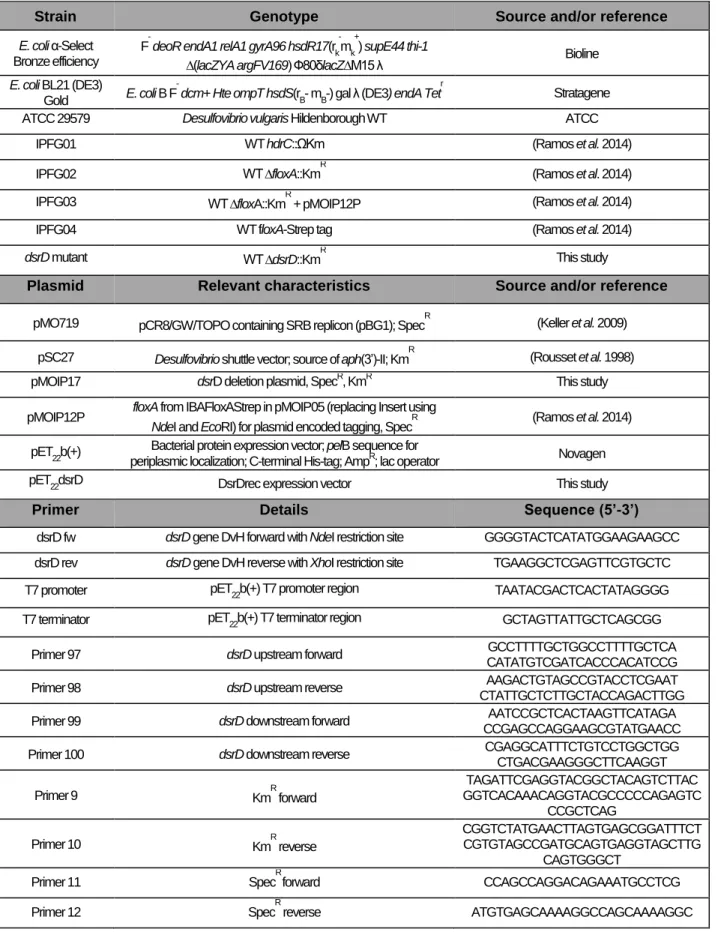
Table 3.1 – List of strains, plasmids, and primers used in this work.
Strain Genotype Source and/or reference
E. coli α-Select Bronze efficiency
F- deoR endA1 relA1 gyrA96 hsdR17(rk-mk+) supE44 thi-1
∆(lacZYA argFV169) Ф80δlacZ∆M15 λ Bioline
E. coli BL21 (DE3)
Gold E. coli B F
dcm+ Hte ompT hsdS(rB- mB-) gal λ (DE3) endA Tetr Stratagene
ATCC 29579 Desulfovibrio vulgaris Hildenborough WT ATCC
IPFG01 WT hdrC::ΩKm (Ramos et al. 2014)
IPFG02 WT ∆floxA::KmR (Ramos et al. 2014)
IPFG03 WT ∆floxA::KmR + pMOIP12P (Ramos et al. 2014)
IPFG04 WT floxA-Strep tag (Ramos et al. 2014)
dsrD mutant WT ∆dsrD::KmR This study
Plasmid Relevant characteristics Source and/or reference pMO719 pCR8/GW/TOPO containing SRB replicon (pBG1); SpecR (Keller et al. 2009)
pSC27 Desulfovibrio shuttle vector; source of aph(3’)-II; KmR (Rousset et al. 1998)
pMOIP17 dsrD deletion plasmid, SpecR, KmR This study
pMOIP12P floxA from IBAFloxAStrep in pMOIP05 (replacing Insert using
NdeI and EcoRI) for plasmid encoded tagging, SpecR (Ramos et al. 2014)
pET
22b(+)
Bacterial protein expression vector; pelB sequence for
periplasmic localization; C-terminal His-tag; AmpR; lac operator Novagen
pET22dsrD DsrDrec expression vector This study
Primer Details Sequence (5’-3’)
dsrD fw dsrD gene DvH forward with NdeI restriction site GGGGTACTCATATGGAAGAAGCC dsrD rev dsrD gene DvH reverse with XhoI restriction site TGAAGGCTCGAGTTCGTGCTC
T7 promoter pET
22b(+) T7 promoter region TAATACGACTCACTATAGGGG
T7 terminator pET
22b(+) T7 terminator region GCTAGTTATTGCTCAGCGG
Primer 97 dsrD upstream forward GCCTTTTGCTGGCCTTTTGCTCA
CATATGTCGATCACCCACATCCG
Primer 98 dsrD upstream reverse AAGACTGTAGCCGTACCTCGAAT
CTATTGCTCTTGCTACCAGACTTGG
Primer 99 dsrD downstream forward AATCCGCTCACTAAGTTCATAGA
CCGAGCCAGGAAGCGTATGAACC
Primer 100 dsrD downstream reverse CGAGGCATTTCTGTCCTGGCTGG
CTGACGAAGGGCTTCAAGGT Primer 9 KmR forward TAGATTCGAGGTACGGCTACAGTCTTAC GGTCACAAACAGGTACGCCCCCAGAGTC CCGCTCAG Primer 10 KmR reverse CGGTCTATGAACTTAGTGAGCGGATTTCT CGTGTAGCCGATGCAGTGAGGTAGCTTG CAGTGGGCT
Primer 11 SpecR forward CCAGCCAGGACAGAAATGCCTCG
3.1.4 – Adh1 purification and activity measurements
Adh1 was purified inside a Coy anaerobic chamber [95 %/ 5 % (v/v) N2/H2] using an
AKTATM Prime plusTM system from the soluble fraction of IPFG04 cells grown in ethanol, using first a Q-Sepharose HP according to (51). NaCl was added to the fraction with Adh activity, to a concentration of 1 M. The fraction was then purified in a HiTrapTM Phenyl HP (2 mL column volume, CV) equilibrated with 50 mM Tris-HCl (pH 7.6) and 1 M NaCl. The protein elution was performed with a linear gradient of 1 to 0 M NaCl (20 CV). Fractions with highest Adh activity eluted between 1 and 0.7 M NaCl. The identity of the enzyme purified was confirmed as Adh1 by Mass spectrometry. Adh activity was determined by NADH formation at 340 nm (εNADH = 6.22 mM
-1
cm-1) in 50 mM Tris-HCl (pH 9), 5 mM NAD+, and 20 mM ethanol, at room temperature (51) with a Shimadzu UV-1800 spectrophotometer in a stirred cuvette (Hellma). Adh1 antibody was produced from pure protein injected in rabbits, at Davids Biotechnologie GmbH®.
3.1.5 – Reverse Transcriptase quantitative PCR (RT-qPCR)
Reverse Transcriptase quantitative PCR (RT-qPCR) was used in D. vulgaris WT to analyze gene expression of adh1, floxA, and hdrA in cells grown in LS4, LS3, P, ES4, and HS4. Cells from three independent experiments were collected at the mid-exponential phase, centrifuged for 12 min at 3,000 g, washed with ice-cold sterile Mili-Q water, and frozen for later RNA extraction. Total RNA was extracted as described in (52). DNase treatment was performed with Turbo DNase (Ambion) in order to avoid genomic DNA contamination in the RNA extracts and was also followed by a RNA clean-up kit (Qiagen). cDNA synthesis from each RNA sample (1 μg) was performed using Transcriptor Reverse Transcriptase (Roche Diagnostics). Primers were designed to amplify a region of about 100 bp of adh1, floxA, hdrA, and the 16S rRNA gene was used as an internal reference gene for each analyzed sample (Table 3.2). Reverse Transcriptase quantitative PCR reactions were performed in a Light Cycler 480 Real-Time PCR System (Roche), with Light Cycler 480 SYBR Green Master I (Roche). Relative standard curves and gene expression were calculated by the relative quantification method with efficiency correction, using the LightCycler Software 4.1, using 16S rRNA gene as a reference (53). For the final results three biological replicates and two technical replicates were used for each condition. The unpaired (two-sample equal variance with two-tailed distribution) t-test was used to determine the significance of the differences in gene expression between cells grown in LS4 and cells grown in the other culture conditions, with a significance defined as p-value < 0.10 and p-value < 0.05.
Table 3.2 – Primers used in qPCR to study the expression of adh1, floxA, and hdrA.
Target Primer sequence (5’ 3’) Amplicon size (bp)
adh1 Forward: ACCAAGAACGCGCAGAA Reverse: CGGTTCTGTCTGTACTCCTTAC 111
floxA Forward: ACCAAGTACGTGTGTGTCG Reverse: CTGCATCGCGGCTACAA 83
hdrA Forward: CATTCCCAAGAAGGCGATCA
Reverse: CGACAATCTCATCCTCCATGTC 125
16S rRNA gene Forward: CCTATTGCCAGTTGCTACC Reverse: AAGGGCCATGATGACTTGAC 100
3.1.6 – Western blot analysis of FloxA, HdrA, and Adh1 expression
D. vulgaris WT cells grown in lactate-sulfate (LS4), lactate-sulfite (LS3), pyruvate (P),
ethanol-sulfate (ES4), and hydrogen-sulfate (HS4), were collected at two different time points, mid-exponential and stationary phase, and centrifuged for 12 min at 3,000 g. Cells were then disrupted by adding 1 mL of BugBuster® Protein Extraction Reagent (Novagen®) and 10 μL of lysonase per g of cells, followed by 20 min incubation with slow stirring at room temperature. The soluble crude extract was obtained by centrifugation at 16,000 g and 4 °C for 20 min. Protein concentration was determined by the Bradford method (Bio-Rad) with bovine serum albumin as standard (NZYTech).
Protein samples of 25 μg were run in a SDS-PAGE gel [12 % acrylamide, (v/v)] and transferred to 0.45 μm polyvinylidene difluoride (PVDF) membranes (Roche) for 30 minutes at 100 V and 350 mA in a Mini Trans-Blot® electrophoretic transfer cell (Bio-Rad) containing Transfer Buffer (48 mM Tris pH 9.2, 39 mM glycine). The membranes were treated with Blocking Buffer [20 mM Tris-HCl pH 7.5, 150 mM NaCl, 0.05 % Tween 20 (v/v), and 5 % non-fat milk (w/v)], overnight at room temperature. In the following day, after three washing steps with TBST [20 mM Tris-HCl pH 7.5, 150 mM NaCl, 0.05 % Tween 20 (v/v)], anti-FloxA at 1 : 1000, anti-HdrA 1 : 500, and anti-Adh1 at 1 : 5000 dillution in TBST were incubated with the membrane for 1 h at room temperature; after two washing steps with TBST, membranes were incubated with anti-rabbit IgG antibody linked to alkaline phosphatase conjugate (Sigma-Aldrich®) at 1 : 15 000 dilltuion in TBST for 45 minutes. After three washing steps with TBS (20 mM Tris-HCl pH 7.5, 150 mM NaCl) protein detection was performed with Alkaline Phosphatase Buffer (100 mM Tris-HCl pH 9.5, 100 mM NaCl, and 5 mM MgCl2) and NBT
(nitro-blue tetrazolium chloride)/BCIP (5-bromo-4-chloro-3-indolyl phosphate) (Carl Roth®). The antibodies used against FloxA and HdrA subunits were produced in rabbits with synthesized peptides and Adh1 antibody was produced from pure protein, as previously referred.
3.2 – Studies with dsrD
3.2.1
– dsrD heterologous expression
3.2.1.1
– dsrD gene cloning and protein expression
The dsrD gene of Desulfovibrio vulgaris Hildenborough was amplified by PCR using genomic DNA as template and the dsrD fw and dsrD rev primers with NdeI and XhoI restriction sites, respectively (Table 3.1). The PCR product was cloned into a pET22b(+)
vector (Novagen®), allowing the insertion of a 6-His tag at the C-terminus of DsrD protein. The recombinant plasmid (pET22dsrD) was heat-shock transformed into E. coli BL21 (DE3) Gold (Stratagene®) strains which were grown at 37 °C in Minimal Medium with ampicillin (100 µg/mL) until an optical density of 0.4. Then 100 µM IPTG (isopropyl β-D-1-thiogalactopyranoside) was added and cells were permitted to grow for 4 additional hours.
3.2.1.2 – DsrD purification
The cells were harvested by centrifugation, washed with Buffer A (25 mM potassium phosphate, 300 mM NaCl, 30 mM imidazole pH 7.2) and frozen to – 20 ºC. After thawing and ressuspending with Buffer A the cells were disrupted using a French Press cell in the presence of DNase. Afterwards, the cell lysate was centrifuged for 15 minutes at 20,000 g in order to remove cell debris, and then ultra centrifuged for 2 h at 140,000 g to separate the membrane from the soluble fraction. The supernatant was injected into a HiTrap Chelating HP column (GEHealthcare) charged with NiCl2 and equilibrated with Buffer A. The protein
was eluted with Buffer B (Buffer A with 100 mM imidazole) and dialyzed to 25 mM potassium phosphate pH 7. The protein purity was analyzed by SDS-PAGE stained with Coomassie Blue and the concentration was determined at 280 nm using the absorption coefficient of 10 mM-1cm-1 and with the BCA (bicinchoninic acid assay) kit by Pierce®.
3.2.1.3 – DsrD activity and interaction assays
DsrABC was tested for sulfite reduction activity in the presence of DsrD in order to analyze if there was any interaction between these molecules capable of modifying the reaction rate. The enzymatic activities were performed in a Coy anaerobic chamber [95 %/ 5 % (v/v) N2/H2] with a Shimadzu UV-1800 spectrophotometer in a stirred cuvette (Hellma),
monitoring the oxidation of methyl viologen (an electron acceptor reduced by Zn2+) mediated by DsrABC, which transfers electrons to sulfite. The assays were developed in Wash Buffer (25 mM KPi pH7) at room temperature, measuring the variation of Abs732 nm through time.
BIAcore experiments were performed with DsrD and DsrABC. BIAcore is a technology based on the Surface Plasmon Resonance (SPR) principle, which permits the determination of affinity constants between two interacting proteins. DsrD was chemically immobilized in a CM5 sensor chip (GE® Healthcare) to which DsrABC was injected and interaction of these two proteins was measured. The SPR experiments were performed at 25 °C on a BIAcore 2000 instrument (Biacore Inc., GE® HealthCare).
Additionally, Isothermal Titration Calorimetry (ITC) technique was also performed to measure interactions between DsrD and DsrABC or sulfite. ITC is a biophysical method that allows the measurement of thermodynamics and binding association of molecules in solution by detecting the heat change in molecular reactions. The ITC experiments were performed at 25 °C on a MicroCalTM (GE® Healthcare).
3.2.2 – dsrD deletion
3.2.2.1 – Strains and media
The strains used in this work are listed in Table 3.1. Escherichia coli α-Select strain was cultured in LB medium (per liter of medium: 10 g tryptone, 10 g sodium chloride, and 5 g yeast extract). Where indicated, kanamycin or spectinomycin was added to LB medium to a final concentration of 50 µg/mL and 100 µg/mL, respectively. All D. vulgaris strains were grown at 37 °C in MOY medium according to (39). Sodium lactate (60 mM) or sodium pyruvate (30 mM) were added as electron donors and sodium sulfate (30 mM for lactate and 3 mM for pyruvate) or sodium sulfite (20 mM for lactate) were added as terminal electron acceptors Antibiotics were added to the MOY medium as follows: G418 (geneticin) at 400 µg/mL or spectinomycin at 100 µg/mL. G418 was used in place of kanamycin as described by Zane et al. 2010. For solidified MOY medium, 15 g agar per liter was added.
3.2.2.2 – Plasmid and strain construction
The pMOIP17 plasmid for insertion of ΩKm cassette in dsrD was constructed by SLIC (sequence ligation independent cloning). The upstream and downstream regions flanking
dsrD were amplified using the primers 97, 98, 99, and 100, respectively, (Table 3.1) and
chromosomal DNA of D. vulgaris was used as template. Also, a spectinomycin resistance cassette and the pMO719 template containing pUC ori and Kanamycin resistance cassette were all amplified by PCR, using pSC27 and pMO719, respectively, as templates and primers 9, 10, 11, and 12. The four PCR products were added to a reaction mix with T4 ligase incubated for 30 min at room temperature. 1 mM dCTP (deoxycytidine triphosphate) was then added the DNA was heat-shock transformed into E. coli α-Select competent cells and plated into kanamycin and spectinomycin containing LA plates. The assembled plasmid (pMOIP17) was extracted, sequenced, and electroporated into D. vulgaris cells according to Keller et al. (2011) from which double recombinants were selected in MOY solid medium with 30 mM of sodium pyruvate by secondary antibiotic screening as described in (54). G418 was used in place of kanamycin as described by Zane et al. (2010). G418 resistant but spectinomycin sensitive colonies were selected and allowed to grow in G418 containing growth medium. The absence of the dsrD gene in the dsrD mutant strain was verified by PCR using the dsrD fw and dsrD rev primers with the chromosomal DNA extracted from the screened cells.
3.2.2.3 – Growth curves
D. vulgaris Hildenborough WT and dsrD mutant strains were grown anaerobically at
37 °C in 100 mL flasks with 50 mL of MOY basal medium (39). MOY was supplemented with different electron donors-acceptors in three different culture conditions: 60 mM lactate-30 mM sulfate (LS4); 30 mM lactate-20 mM sulfite (LS3); 30 mM pyruvate-3 mM sulfate (Pyr). All media were inoculated with 2 % (v/v) fresh precultured cells grown in pyruvate-sulfate medium (Pyr). The OD of the cultures was monitored at various time points with a spectrophotometer Shimadzu UV-1603. All reported optical density measurements are the mean of three biologically independent experiments.