Universidade de Lisboa
Faculdade de Farmácia
Study of neuroinflammation markers in the
hippocampus of adolescent rats after
binge-like ethanol exposure
Alexandra Antunes Ângelo
Mestrado Integrado em Ciências Farmacêuticas
Universidade de Lisboa
Faculdade de Farmácia
Study of neuroinflammation markers in the
hippocampus of adolescent rats after
binge-like ethanol exposure
Alexandra Antunes Ângelo
Trabalho de Campo de Mestrado Integrado em Ciências Farmacêuticas apresentado à Faculdade de Farmácia da Universidade de Lisboa
Orientador: Dr. Catherine Vilpoux
Co-orientador: Dr. Manuel Caneira, Professor Convidado
~
The work accomplished on this monograph was possible thanks to
the University of Picardie Jules Verne (UPJV) and to the GRAP
6
RESUMO
O início do consumo de álcool ocorre tipicamente durante a adolescência e os modos de consumo produzem efeitos visíveis a nível social e de saúde. A adolescência é um período de vida durante o qual ocorrem importantes processos do desenvolvimento cerebral, tendo sido previamente demonstrado que o cérebro adolescente é mais susceptível ao comprometimento da memória e danos cerebrais induzidos pelo etanol, pelo que o consumo excessivo de álcool pode gerar níveis neurotóxicos de intoxicação e ao desenvolvimento de efeitos profundos no sistema nervoso central.
Estudos previamente realizados no laboratório GRAP demonstraram que uma exposição ao álcool do tipo binge-drinking através da administração de duas injecções consecutivas de etanol (3g/kg i.p.) em ratos foi suficiente para originar a abolição da plasticidade sináptica na região CA1 do hipocampo, bem como perdas nas habilidades de aprendizagem. Dada a relação recentemente descoberta entre a plasticidade sináptica e neuroinflamação, pretende-se agora investigar se este protocolo de exposição do tipo binge-drinking será capaz de provocar eventos neuroinflamatórios nas subregiões CA1 e Dentate Gyrus do hipocampo, utilizando dois marcadores de neuroinflamação, HMGB1 (High-mobility group box 1 protein) e TLR4 (Toll-like receptor).
Os resultados do protocolo de imunohistoquímica realizado para HMGB1 revelaram que não existe diferenças estatisticamente significativas da expressão de HMGB1 entre o grupo de ratos submetidos à exposição do tipo binge-drinking e o grupo de controlo, o que sugere que a exposição excessiva ao etanol não altera a expressão de HMGB1, nem induz a ocorrência de fenómenos inflamatórios nas células neuronais, em nenhuma das subregiões do hipocampo em estudo. O estudo de imunofluorescência realizado revelou a ocorrência de um aumento da expressão de TLR4 nas duas subregiões do hipocampo em estudo dos ratos sujeitos à administração de álcool. Estes resultados sugerem que este protocolo do tipo binge-drinking parece ser suficiente para induzir fenómenos de neuroinflamação no hipocampo dos ratos.
7
ABSTRACT
The initiation of alcohol consumption typically occurs during adolescence and the patterns of drinking have an effect on health and social outcomes. The adolescence is a period of life, during which strong processes of brain development take place, having been previously demonstrated that adolescent brain is more susceptible to ethanol-induced memory impairment or brain damage, whereby excessive alcohol consumption may drive to neurotoxic levels of intoxication and to the development of profound effects on the central nervous system.
Previous studies performed in GRAP laboratory demonstrated that binge-like ethanol exposure with two close injections of ethanol (3g/kg i.p.) in rat was enough to produce synaptic plasticity abolition in the CA1 of hippocampus, together with losses in learning abilities. Given the recently discovered relation between synaptic plasticity and neuroinflammation, we intended in this work to investigate whether this binge-like exposure protocol would be able to produce neuroinflammatory events in the CA1 field and Dentate Gyrus of the hippocampus, using two neuroinflammation markers, HMGB1 (High-mobility group box 1 protein) and TLR4 (Toll-like receptor).
The results of the immunohistochemistry experiment performed for HMGB1 revealed that there are no statistically significant differences between the HMGB1 expression in group of rats exposed to the binge-drinking and the control group, which suggest that the binge-like ethanol exposure doesn't alter the HMGB1 expression and it doesn’t induce inflammatory phenomena in neurons, neither in CA1 nor Dentate Gyrus. The immunofluorescent study performed revealed an increase of TLR4 expression in both hippocampus subregions of binged rats, which suggests that this binge-like ethanol exposure protocol seems to be sufficient to induce neuroinflammation in the hippocampus of rats.
Keywords: HMGB1; TLR4; neuroinflammation; binge-drinking; alcohol.
8
CONTENTS
Chapters Pages FIGURES ... 10 TABLES ... 10 ABBREVIATIONS ... 11 1. INTRODUCTION ... 121.1 Aims of the research project ... 12
2. LITERATURE REVIEW ... 13
2.1 Adolescence and the binge drinking problem ... 13
2.1.1 Alcohol and alcohol addiction ... 13
2.2 Pattern of alcohol consumption in Europe, Portugal and France ... 14
2.2.1 Alcohol and harmful pattern of consumption: the “binge drinking” phenomena in adolescence ... 17
2.3 Hippocampus ... 18
2.4 HMGB1 ... 20
2.5 Toll-like receptor 4 (TLR4) ... 21
3. MATERIALS AND METHODS ... 23
Animal model ... 23
Treatment: Ethanol exposure ... 23
Preparation of slices ... 24 3.3.1 Sacrifice of Rats ... 24 3.3.2 Brain Sectioning ... 24 3.4 HMGB1 Immunohistochemistry staining ... 24 3.4.1 Fundaments ... 24 3.4.2 Protocols ... 27 3.4.2.1 HMGB1 Immunohistochemical Staining ... 27
3.4.2.2 Microscopic observation, acquisition and analysis of images ... 27
3.5 Immunohistofluorescence staining ... 28
3.5.1 Fundaments ... 28
3.5.2 Protocols ... 30
3.5.2.1 Co-labelling of TLR4 and NeuN immunofluorescence ... 30
3.5.2.2 Microscopic observation, acquisition and analysis of images ... 31
3.6 Statistical analysis ... 31
9
4.1 HMGB1 Immunohistochemical Staining ... 32
4.2 Co-labelling of TLR4 and NeuN immunofluorescence... 34
4.2.1 Co-labelling of TLR4 and NeuN immunofluorescence in CA1 ... 34
4.2.2 Co-labelling of TLR4 and NeuN immunofluorescence in DG ... 37
5. DISCUSSION ... 39 6. CONCLUSION ... 42 7. REFERENCES ... 43 APPENDIX 1 ... 50 APPENDIX 2 ... 53 ACKNOWLEDGMENTS ... 55
10
FIGURES
Figure 1 - Life-time risk of death from alcohol-related injuries . ... 15
Figure 2 - Schematic representation of a cross-section of the hippocampus ... 20
Figure 3 - Schematic representation of ABC method . ... 26
Figure 4 - Structural formula of 3-3'-diaminobenzidine tetrahydrochloride (DAB). ... 26
Figure 5 - Schematic representation of immunofluorescence methods. ... 28
Figure 6 - Principle of fluorescence . ... 29
Figure 7 - Representative photomicrographs of HMGB1 expression in the CA1 and DG ... 32
Figure 8 - Number of HMGB1 positive cells per 100 000 µm2 ... 33
Figure 9 - Mean optical density of HMGB1 labelling in CA1 and DG ... 33
Figure 10 - Representative photomicrographs of TLR4, NeuN and merge in the CA1 ... 35
Figure 11 - Expression levels of TLR4 and NeuN in CA1 field ... 35
Figure 12 - Magnifying of CA1 region showing NeuN labelling, TLR4 labelling and merge ... 36
Figure 13 - Optical density ratio for TLR4/NeuN labelling in CA1 field ... 36
Figure 14 - Representative photomicrographs of TLR4, NeuN and merge in the DG ... 37
Figure 15 - Expression levels of TLR4 and NeuN in DG field ... 38
Figure 16 - Optical density ratio for TLR4/NeuN labelling in the DG field ... 38
TABLES
Table 1 - Diagnostic and Statistical Manual of Mental Disorders [4]. ... 1411
ABBREVIATIONS
AUD - Alcohol use disorder CA - Ammon’s HornCNS - Central nervous system
DAMPs – Danger-associated molecular patterns DG – Dentate gyrus
EC – Entorhinal Cortex
ECATD - Estudo sobre o consumo de álcool, tabaco e droga
ESPAD - European School Survey Project on Alcohol and Other Drugs EU – European Union
GCL – Granular cell layer
GRAP - Research Group on Alcohol and Pharmacodependences HMGB1 – High-mobility group box 1 protein
IF - Immunofluorescence IHC - Immunohistochemistry
INME - Inquérito Nacional em Meio Escolar
INSERM - Institut National de la Santé et de la Recherche Médicale LTD – Long-term synaptic depression
NIAAA - National Institute on Alcohol Abuse and Alcoholism NPCs – Neural progenitor cells
PAMP – Pathogen-associated molecular pattern PBS - Phosphate buffered saline
PFA – Paraformaldehyde PFC - Prefrontal cortex
PRR - Pattern recognition receptor SGZ – Subgranular zone
12
1.
INTRODUCTION
1.1
Aims of the research project
GRAP team has been studying the impact of alcohol binges in rats. In a previous study, the GRAP team showed that this binge-like pattern produced modification on memory process and electrophysiological properties in the hippocampus.
Taking into account these findings, we intend to analyse if other cellular damages, such as neuroinflammation, happen in the hippocampal structure, following a binge-like alcohol exposure. A previous labelling of a neuroinflammation marker, High-mobility group box 1 protein (HMGB1), was done by GRAP team following the 2 binges. I started by analysing this labelling and based on these results, we decided to move on the study of the receptor of HMGB1, the Toll-like receptor 4 (TLR4).
Therefore, this study aims to evaluate the expression and signalling of these two neuroinflammation markers, HMGB1 and TLR4, in ethanol-induced brain alterations, using a binge-like alcohol exposure in vivo through the administration of two alcohol injections in adolescent rats (3g ethanol /kg i.p.) at an 8-hour interval.
We intend to perform an immunohistochemistry study to analyse the expression of HMGB1 in hippocampus neurons, following these two massive alcohol injections in the rats. We also intend to analyse the activation of TLR4 and consequent neuroinflammation in hippocampus neurons, following this double binge-like alcohol exposure, through an immunofluorescence study, performing a co-labelling of TLR4 and a marker of neurons (NeuN). These techniques will be performed on rat brain slices, more specifically in the CA1 field and in the Dentate Gyrus of the hippocampus.
13
2.
LITERATURE REVIEW
2.1
Adolescence and the binge drinking problem
2.1.1 Alcohol and alcohol addiction
Alcohol, or ethanolic alcohol, is one of the most widely used and abused substances in the world (1). Initiation of alcohol use typically occurs during adolescence (2) and its excessive consumption represents a serious health challenge (3). Since May 2013, the American Psychiatric Association has published the fifth edition of the "Diagnostic and Statistical Manual of Mental Disorders" or DSM-5, which presents the addictions in the chapter "Addiction and Related disorders" (4). The psychiatric disease resulting from alcohol addiction is named Alcohol Use Disorders (AUD) by the DSM-5 (See table 1 for diagnosis criteria).
The etiology and pathophysiology of alcohol abuse is influenced by numerous and environmental factors (5), which affect the magnitude and patterns of consumption, and increase the risk of developing AUD (6).
Alcohol Use Disorder (AUD) (7,8), is a complex multifactorial disease influenced by genetical and environmental factors (9), and psychiatric disorders. Some studies with twins and adoption families reveal an alcoholism heritability of ~50%, with identification of many genetic variants that influence the risk of alcoholism (5,10). These genetic variants may modify an individual’s susceptibility because it alters the tolerance, acute sensitivity to intoxication, dependence and craving (5). Also, the environmental factors such culture, economic development and availability of alcohol can explain the vulnerability between societies and trend of alcohol consumption. Although there is no single risk factor that is dominant, the literature suggests that the more vulnerabilities a person has, the more likely the person is to develop alcohol-related problems (6).
14
Table 1 - Diagnostic and Statistical Manual of Mental Disorders (4).
2.2
Pattern of alcohol consumption in Europe, Portugal and France
Alcohol consumption and patterns of drinking have an effect on health and social outcomes. Alcohol abuse is an important public health problem that is related with premature male deaths (11) both in Portugal, France and in Europe as a whole. Alcohol is responsible for 1 in 7 male deaths and 1 in 13 female deaths among people aged between 15 and 64 years old. This means almost about 120000 deaths each year in EU for attributable causes (12). The lifetime risk of dying from an alcohol-related injury across the total population aged over 15 years increases exponentially with a daily alcohol consumption beyond 10 g of alcohol per day (Figure 1). The risks are higher for men than for women, for any dose of alcohol consumption. (12). It is estimated that in 2004, more than 4 million disability-adjusted life-years (DALYs) were caused by alcohol consumption in EU, corresponding to 15% of all DALYs in men and 4% of all DALYs in women (12). These outcomes makes alcohol the third highest of twenty-six risk factors for ill-health in the EU, behind tobacco and high blood pressure (13).
15
Figure 1 - Life-time risk of death from alcohol-related injuries (12).
An analysis between the various countries of Europe reveals a substantial variation in prevalence of alcohol consumption. However, the majority of people in all countries claimed to have consumed alcoholic drinks in the past 12 months (14). In this study, 76% of European citizens said to have consumed alcoholic drinks in the past year. The percentage of people who had consumed alcoholic drinks in the past year is lowest in Portugal (58%) than the European average (76%) and is higher in France (83%) (14), which shows the trend to a higher alcohol consumption in France.
Out of those people who said they had consumed alcoholic beverages in the last 12 months, about 91% both in Portugal and in France affirmed had consumed alcoholic drinks in the last 30 days. Among these, about 1 in 3 European citizens reports a binge drinking intake at least once a week, mainly among the youngest age groups (15-24 years old) (14). As more extensively described below, binge-drinking represents a specific pattern of high ethanol intake. In general, men are far more likely to have consumed alcoholic drinks than women, but this difference is almost nil in the youngest age groups (14).
ESPAD (European School Survey Project on Alcohol and Other Drugs) is a European project which is occurring since 1995. Its aim is to study the evolution of prevalence of alcohol and other drugs consumption, among students with 16 years old in about 40 European countries (15, 16). In 2011, ESPAD showed that 74% of Portuguese students of this age group had consumed alcohol in the last 12 months and, about 52% in the last 30 days (16, 17). In 2015, 20 years after the first ESPAD study, ESPAD demonstrated that the alcohol consumption among adolescents with 16 years
16
old remains high all over Europe but with a positive development trend – lifetime alcohol consumption decreased from 89% (in 1995) to 81% (in 2015) and last-30-day consumption from 56% to 47% (18, 19). However, the prevalence of heavy episodic drinking / binge drinking has remained unchanged over the 20 years, with values in 2015 similar to those in 1995, with still 35% of student reporting heavy episodic drinking in the past month in the latest survey in 2015 (18, 19, 20).
Since 2003, the Portuguese ESPAD group of researches opted to extend this study, in Portugal, to students not only 16 years old but also in the age groups between 13 and 18 years, giving origin to the ECATD project (16). The last ECATD study, conducted in 2015, reveals that prevalence of alcohol consumption varies in direct ratio to the student’s ages. The prevalence of recent consumption (last 12 months) was about 20% in the 13 years old students and 86% in the 18 years old students, and the prevalence of current consumption (last 30 days) was about 10% in students with 13 years old and 68% in students with 18 years old. These data evidence a slight decrease of alcohol consumption , four years after the last study (15, 16).
INME studies were done, also in Portugal, since 2001 with middle and high school students, aiming at characterizing both psychoactive substances consumption and their consumers, allowing to identify and analyze the needs of preventive intervention. In 2011, INME results showed that the majority of middle and high school students had a recent alcohol consumption in the last 12 months (respectively 55% and 87%) and half of the high school students reports a binge drinking consumption also in the last year (16, 21).
These studies revealed a substantial variation in prevalence of alcohol consumption, but also reveal an elevated consumption especially in adolescents and young adults all over Europe. This alcohol consumption pattern consists, mainly, in a heavy episodic drinking, also called “binge drinking”, which has become very common in this age groups, and whose effects on the nervous system are still under study.
17
2.2.1 Alcohol and harmful pattern of consumption: the “binge drinking” phenomena in adolescence
The harmful effects of alcohol consumption on health vary according to extension and habits of usage, like dose, duration and pattern of intake (22), and depend on numerous environmental and individual factors (23).
Binge drinking or heavy episodic drinking is one of the most common and harmful pattern of alcohol consumption in adolescents and young adults (24, 25). It is defined as the consumption of large amounts of alcohol within a limited period (26).
According to theNational Institute on Alcohol Abuse and Alcoholism (NIAAA), binge
drinking is characterized as a drinking pattern that corresponds to consuming 5 or more drinks (for males) or 4 or more drinks (for females) on a single occasion in about 2 hours, which brings blood alcohol concentration to levels ≥ 0,8 g/L.
The adolescence is a period of life, during which strong processes of brain development take place, for example, extensive synaptic remodeling (27, 28) which could explain the differences of sensitivity between adolescents to the rewarding and aversive effects of alcohol (29). Because of this, people who initiate alcohol consumption during adolescence are more likely to develop an Alcohol Use Disorder (AUD). Some studies showed that an early beginning of alcohol consumption seems to seriously increase the risk of developing AUDs later in life (7, 30, 31).
Due to its developmental state, the adolescent brain is more resistant to the motor impairing and sedative effects of alcohol than the adult brain but is more sensitive to its rewarding and reinforcing properties (8, 32, 33) and more susceptible to
ethanol-induced memory impairment or brain damage (34, 35). For this reason,
excessive alcohol consumption may drive to neurotoxic levels of intoxication(8) and to
the development of profound effects on the central nervous system (36, 37) . However,
the impact of only a few alcohol intoxications on learning and memory sometimes appears underestimated or even neglected, revealing the need for a better understanding of both the short-term and long-term effects of a few binges on cognitive functions (34). Previously, several studies have been done at the prefrontal cortex, striatum, amygdala and hippocampus: These brain regions belong to the reward system, the brain
18
regions system that is strongly dysfunctional in all addictions, including alcohol addiction (38). Many of these studies demonstrated that the excessive consumption of alcohol increases the risk of developing brain damages or dementia (1), lack of control of compulsivity (39), and cognitive deficits in learning, memory and visuospatial capabilities (1, 34). This, because alcohol acts on many molecular targets, and affects many different cellular processes, related with neurotransmission. Understanding these mechanisms associated with the neurotoxic action of alcohol abuse is fundamental to identify new therapeutic targets and develop treatment strategies (5).
2.3
Hippocampus
Hippocampus is a bilateral cerebral structure located in temporal lobe, which belongs to the limbic system (40). This region has a major role in several cognitive functions and processes, including working and spatial memory, regulation of emotions, learning and process of adapting an individual to his environment. Hippocampus also belongs to the reward system, a set of brain regions managing the processes of addiction (41).
On the neuro-anatomical plane, the hippocampus is composed by two main structures, Dentate Gyrus (DG) and the Ammon’s Horn (CA), itself subdivided into 3 regions: CA1, CA2 and CA3 (41).
In the GRAP laboratory, a protocol was used to model “binge drinking” consumption in rats: it consisted in the intraperitoneal injection of two successive massive alcohol dose of 3g ethanol/kg, one in the morning and one 8 hours after. In GRAP laboratory, Pr Pierrefiche et al. studied synaptic plasticity in the hippocampus. He and his collaborators showed an abolition of the long-term synaptic depression (LTD) in CA1 field of the hippocampus of adolescent rats 48 hours after submitting them to this binge-like exposure. LTD is a mechanism which contributes to synaptic plasticity and, therefore, allows the learning and memory processes. That way, the learning deficit exhibited by the rats in the acquisition phase may be explained by that lack of LTD (34). Other studies have shown that DG displays neuronal renewal stem cells, in SGZ, which could be linked to neuronal losses induced by the alcohol exposure (42).
19
Considering these findings, in this study we are concerned about the region CA1 and the region DG, both with a glutamatergic nature. CA1 field displays an area, called pyramidal stratum, which contains the cell bodies of pyramidal neurons (43). In respect to DG, it displays a granular cell layer (GCL) with high body cells density, a molecular layer and a hilus (44). Beyond that, DG presents a subgranular zone (SGZ) delineated, which is a narrow band of tissue of DG between the granule cell layer and the hilus (44, 45). Previous animal research studies have shown that SGZ contains the microenvironment that is permissive for neuronal development to occur and which it is called the neurogenic “niche” that comprises the precursor cells themselves, their immediate progeny and immature neurons, other glial cells and endothelia, very likely immune cells, microglia and macrophages and an extracellular matrix (45). This microenvironment allows constant generation of new granule cells in the SGZ, from neural progenitor cells (NPCs) (44, 45, 46).
Hippocampal neurogenesis occurs, then, through the NPCs proliferation in SGZ layer of DG, and comprises a series of sequential developmental events that are all necessary for the generation of new neurons (45, 47, 48).
In the hippocampus, the flow of information is mainly unidirectional. The DG is an input region, which receives input from the axons of pyramidal cells of the entorhinal cortex (EC) layer II (first synapse); the granule cells of DG (called mossy fibers) pass on the information to the dendrites of CA3 pyramidal cells (second synapse) and then, the CA3 axons (called Schaffer collaterals) project the information to CA1 pyramidal cells, which are responsible to project back to the EC, finishing the Trisynaptic Circuit. However, some of the axons of EC project the information directly to CA3 and CA1 fields (Figure 2) (49, 50).
20
Figure 2 - Schematic representation of a cross-section of the hippocampus, illustrating
the classical view of the Trisynaptic Circuit (40)
2.4
HMGB1
High-mobility group box-1 (HMGB1) is a protein that executes two different functions: inside the cell, HMGB1 is a non-histone nuclear protein that binds DNA, regulating transcription and determining chromosomal architecture and, outside the cell, HMGB1 activates the innate immune system and mediates a wide range of physiological and pathological responses (51), working as an endogenous danger signal (alarmin) (52).
HMGB1 has been implicated in the pathophysiology of many neuroinflammatory conditions like traumatic brain injury, seizure, ischemia and chronic ethanol use (52). Just like other DAMPs, HMGB1 can induce a pro-inflammatory response through receptors for advanced glycation end products (RAGE), C-X-C chemokine receptor 4 (CXCR4) (53, 54), but mainly through TLRs (52). HMGB1 also shows several molecular features which distinguish it from other alarmins, like a redox-sensitive capacity to rapidly shift between functional states and the capacity to bind and amplify the pro-inflammatory effects of cytokines and PAMPs (52).
In the brain, HMGB1 has been localized to microglia and astrocytes and is also expressed at high levels in neurons. Therefore, this protein has been implicated in the pathophysiology of several neurodegenerative diseases (52, 55). Neuronal HMGB1 has
21
an important role as an initiator and amplifier of neuroinflammation and in neuronal excitation (52).
Considering these findings, the GRAP team was working on a project which aimed to evaluate if cellular damages like neuroinflammation occurs in the hippocampus, through the analysis of possible changes in the expression of this neuroinflammation marker, HMGB1, following a binge-like alcohol exposure. That way, I started to delineate my project by analyzing the labeling obtained by the GRAP team in that experiment, which didn’t reveal any differences in HMGB1 expression between the rats exposed to the binge-like alcohol exposure and the control rats. Thus, we chose to move on with the study about neuroinflammation associated with alcohol exposure, through the study of TLR4, a receptor of HMGB1.
2.5
Toll-like receptor 4 (TLR4)
Toll-like receptor 4 (TLR4) is a type I transmembrane protein which belongs to the family of toll-like receptors (TLRs), a member of the Pattern Recognition Receptor (PRRs) family (56, 57, 58).
Despite the wide range of ligands recognized by TLRs, all the paralogs share a common architecture of three domains: a N-terminal ligand extracellular recognition domain, a single transmembrane helix, and a C-terminal cytoplasmic signaling domain (58, 59). Upon ligand binding, two extracellular domains form an “m”-shaped dimer sandwiching the ligand molecule, bringing the transmembrane and cytoplasmic domains in close proximity and triggering a downstream signaling cascade (58).
During infections, these receptors recognize various conserved structural motifs in the extracellular environment, named pathogen-associated molecular patterns (PAMPs) (60), that are expressed on infectious agents. TLR4 responds specifically to LPS (58). Their activation triggers an intracellular signaling pathway NF-κB and the induction of genes that encode cytokines and molecules inflammation-associated, which leads to an activation of the innate immune system (56, 61). However, TLRs not only play a role in the innate immunity in response to infection, but they can also be activated by
22
endogenous danger signals called danger-associated molecular patterns (DAMPs), which are proteins released from injured or stressed cells under situation of sterile inflammation or ischemia (60, 62), as a result of cell damage and inflammatory processes. That way, TLRs also participates in CNS neurodegeneration and neural injury, wherein the activation of TLR4 contributes to neuroinflammation (56, 63). The majority of TLRs are expressed in the glial cells of the central nervous system (56, 64), but also in the neurons (65).
Silvia Alfonso-Loeches, María Pascual and their remaining team have shown in previous studies that, unlike what happens in WT mice, in TLR4-KO mice alcohol doesn’t induce any activation of kinases, cytokines and another inflammatory mediators, which means that the elimination of these receptors in mice protects against ethanol-induced glial activation, induction of inflammatory mediators and apoptosis and, consequently, protects against ischemic brain damage and injury and behavioral damages (56, 66).
Moreover, Jorge Montesinos and his collaborators have also demonstrated that, by activating TLR4, adolescent intermittent ethanol exposure causes neuroinflammation, myelin damage and behavioral dysfunctions. Intermittent ethanol treatment on TLR4-KO adolescent mice neither induced the activation of the different kinases or NF-KB nor the production of cytokines and inflammatory mediators in the PFC; this activation and mediators production only occurred on WT mice. These results suggest that mice deficient in the TLR4 function are protected against ethanol-induced neuroinflammation and another derangements induced by ethanol in adolescence, which emphasizes the role of TLR4 in signaling and neuroinflammation in ethanol-induced brain alterations (54). In a more recent study, Montesinos and his team have shown that Nalmefene (NF), which is a derivative of Naloxone and an opioid antagonist, together with a treatment against alcohol addiction, prevents the up-regulation of cytokines, chemokines and pro-inflammatory mediators, myelin damages and apoptosis, inhibiting the TLR4 signaling, which confirms that NF prevents neuroinflammation and brain damages by blocking the TLR4 response (67). These previous studies have shown that ethanol seems to be capable of activating glial TLR4 receptors and inducing neuroinflammation and brain damage in mice.
23
3.
MATERIALS AND METHODS
Animal model
Twenty-two male rats (Charles River Laboratories, France) were individually housed and kept under standard conditions with free access to food and water during their accommodation. In rats, adolescence corresponds to the period from the 30th to the 60th day of life (D30 to D60) among which specific response to alcohol exposure could be observed between early adolescence (D30-D45) and late adolescence (D45-D60) (29, 68).
The rats were sacrificed at 42 days of age, during their adolescent period.
All experiments were carried out in conformity with the European Community guiding principles for the care and use of animals (2010/63/UE, CE Off. J. 20 October 2010), the French decree n° 2013-118 (French Republic Off. J., 2013) and the local ethics committee rules (CREMEAP, University of Picardie Jules Verne).
Treatment: Ethanol exposure
In order to simulate the high amount of alcohol in a short time, characteristic of binge drinking consumption, the rats were submitted to intraperitoneal injections of a high dose of alcohol, as previously described (34).
Two groups of rats were formed: a group of rats exposed to a binge-like ethanol exposure (Binge) and control group (Control). The Binge group was formed by 12 rats that received an intraperitoneal injection of ethanol (3.0g/kg) two consecutive times 8 hours apart, using a 15% (v/v) diluted ethanol solution (VWR International (France)). The Control group was formed by 10 rats that received the same volume of a 0.9% saline intraperitoneal injections following the same schedule. The rats were sacrificed 48h after the first injection.
24
Preparation of slices
3.3.1 Sacrifice of Rats
Rats were deeply anesthetized with pentobarbital (60mg/kg, intraperitoneal, 0.5mL ± 0.2mL) and received a heparin injection (12500 UI/rat, intraperitoneal) to avoid formation of clots. Then, they were perfused by transcardiac path via the ascending aorta with 75mµ of heparinized 0.9% saline solution and then, with 200 mL of 4% iced PFA (4% paraformaldehyde, phosphate buffer PB 0.1M, pH 7.4) in order to fix the brain.
3.3.2 Brain Sectioning
Brains were removed, postfixed in 4% PFA at 4ºC for the night. They were rinsed with PBS 1x three times x20min at 4ºC and placed into a crescent concentration of saccharose cryoprotection solution (15% of saccharose and 30% of saccharose in PBS 1x) for a week.
Using the Bregma coordinates as defined into “The Rat Brain in Stereotaxic Coordinates” (69), we sliced the brain in the hippocampus region, from Bregma -2,30mm to Bregma -4,80mm. We focused on the hippocampus level chosen for electrophysiologic studies, meaning the anterior hippocampus. Therefore, 50 μm frozen coronal sections corresponding to that specific area were prepared, at the Bregma -2,5mm. These sections were sliced at -17ºC with a cryostat, put in cryoprotective solution (Ethylene glycol 30%; Sucrose 30%, NaCl 0.9%, in PBS 1X) and stored at -20°C.
3.4
HMGB1 Immunohistochemistry staining
3.4.1 Fundaments
Immunohistochemistry (IHC) is a laboratory method, which combines anatomical, histological, immunological and biochemical techniques, for detection and identification of specific components in cells of a tissue section, by the interaction of a target antigen with a specifically labelled antibody with a visible marker. This way, the
25
technique makes it possible to determine the distribution and localization of these specific cellular components, within a cell or tissue, as well as to identify cellular events. IHC presents a great specificity in diagnosis of degenerative brain diseases and muscular dystrophies (70, 71). IHC can also be used to evaluate the impact of medication action, or, as done here, the impact of alcohol administration.
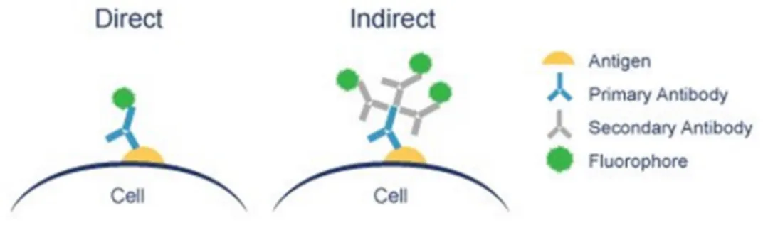
Direct IHC method is done in one step which involves a single labelled antibody to detect the target molecule within the tissue, while the indirect method involves an unlabelled primary antibody to detect the target molecule of interest and a secondary labelled antibody, which binds to the primary antibody. The direct technique is straightforward and less time-consuming, it is less sensitive and lack the ability to amplify weak signals. On the other hand, the indirect method, whilst being more time-consuming, amplifies relatively weak antigen signals in tissue as many secondary antibodies can bind to different antigenic sites of the primary antibody, which makes it a more sensitive technique (72).
Given its greater sensitivity, the indirect method of IHC was used in this protocol. The antibody-antigen binding can be visualized in different manners and for this, the secondary antibody is labelled with a visible marker, which could be a fluorescent dye, a enzymatic marker, a radioactive element, colloidal metal or hapten (73). In this protocol, an enzymatic marker was used: Avidin-Biotin-Peroxidase Complex (ABC) method for HMGB1 labelling.
The ABC method involves three main layers. The first layer is the unlabelled primary antibody; the second layer is the secondary antibody labelled with biotin molecules, which will recognize the constant part (Fc) of the primary antibody, and binds to it; the third layer is a complex of avidin-biotin-peroxidase (Figure 3) (74).The avidin of this complex is a basic glycoprotein with four binding sites for biotin. Due to the high affinity to this vitamin, avidin will bind to the biotin molecules that are labelling the secondary antibody (70). The four binding sites for biotin make lattice complexes possible, where the avidins are linked together via the enzyme peroxidase. It is just necessary that the enzyme has at least two biotin molecules attached so that it can function as a link between the avidins (75). That way, the biotinylated secondary antibodies function as links between tissue-bound primary antibodies and an avidin-biotin-peroxidase complex (71).
26
Figure 3 - Schematic representation of ABC method (71).
Then, avid-biotin-peroxidase complexes can be detected through the use of a chromogenic substrate. In this protocol, we chose the substrate 3,3’-diaminobenzidin tetrahydrochloride (DAB) (Figure 4). DAB is added in its reduced and colorless form. In the presence of peroxidase enzyme, DAB is oxidized, resulting in a deposition of a brown alcohol-insoluble precipitate at the site of enzymatic activity, by the multiple peroxidase enzyme molecules attached at the site of the target antigen (72). That way, it becomes possible to detect the ABC complex that corresponds to the epitope of interest/ antigen, by counting the brown stains. (76)
27
3.4.2 Protocols
3.4.2.1 HMGB1 Immunohistochemical Staining
Brain sections were washed 3 times x5min with PBS 1x-T for permeabilization, and treated 30min in methanol with 3% H2O2, to inactivate endogenous peroxidase activity. Sections were rinsed 2 times x10 min with PBS 1x-T and placed in goat blocking serum for 30min at room temperature. Then, sections were incubated at 4ºC for 1 night with primary antibody rabbit anti-HMGB1 (Ab18256, Abcam, France) diluted 1/100 in 500 µl of goat blocking serum (2% blocking goat + horse serum, 1% BSA, 0.2% Triton x100, PB 0.1M).
Afterwards, sections were rinsed for 3 times x5 min with PBS 1x-T and were incubated for 1h at room temperature with a solution composed of secondary biotinylated universal antibody diluted in 500 µL of PBS 1x (Vectastain® Elite® ABC Kit (Universal) PK-6200), according to supplier instructions.
Brain sections were rinsed for 5 min with PBS 1x and they were incubated for 1h with 500 µL of Avidin-Biotin-Peroxidase Complex (Vectastain® Elite® ABC Kit (Universal) PK-6200) in PBS 1x at room temperature. Then, they were rinsed for 5 min with PBS 1x and incubated for 3 min with DAB, nickel solution and hydrogen peroxide (Vector® DAB Peroxidase Substrate Kit SK-4100) at room temperature.
After 3 washes for 5 min with PBS 1x, sections were mounted in gelatinised microscope slides and dried for the night. In the next day, were performed 2 successive baths for 2min in ethanol for dehydrate slices (50% ethanol, 70% ethanol, 80% ethanol, 100% ethanol), followed by xylene bath for 1 min for delipidation of slices. Sections were mounted with Entellan resin and covered with coverslips.
3.4.2.2 Microscopic observation, acquisition and analysis of images
Immunohistochemical staining observations were performed in an optical microscope, after 1 day minimum to allow Entellan resin polymerization. Photographs were taken with a Leica® Infinity Camera 2 connected to a Leica DM 4000 microscope. The staining was examined using a Mercator® (Version 1.73; ExploraNova, La Rochelle, France), an image processing acquisition software. The cells located in the
28
granular layer and CA1 field of dorsal hippocampal sections (x10) were counted and presented as mean numbers of immunolabelled cells per 100 000 μm2. Moreover, we
also counted the Optical Density of each labelled cell and expressed the result as mean Optical Density for Control and Binge rats.
3.5
Immunohistofluorescence staining
3.5.1 Fundaments
Immunohistofluorescence is a laboratory microscope-based technique, with the aim of detecting and determining the location of antigen or antibody in a tissue by the pattern of fluorescence, when the tissue is exposed to the specific fluorescent-labeled antibody or antigen (77, 78). This technique allows to generate high-resolution images for protein localization studies and to quantify the fluorescent signal (79). This labeling of the antibodies consists in a chemical conjugation of the antibodies to fluorescent dyes (also called fluorophores or fluorochromes).
These labeled antibodies can bind cellular antigens through two main ways: directly or indirectly (Figure 5) (79). Direct immunofluorescence uses a single antibody that is chemically linked to a fluorescent dye. The antibody recognizes the target molecule and binds to it. The fluorescent dye it carries, can be detected on the microscope (77, 80). Indirect immunofluorescence requires two antibodies: a primary antibody that is unlabeled and which specifically recognizes the target molecule and a secondary antibody which recognizes the primary antibody and binds to it, and that carries the immunofluorescent dye (77, 80).
29
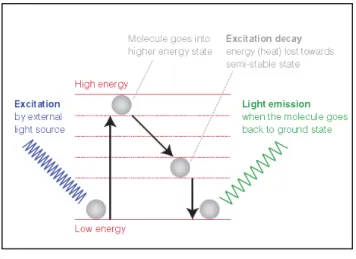
The fluorescent dye allows visualization of the target distribution in the sample under a fluorescent microscope (79). It is subjected to an excitation light with a short-wavelength and high energy, which is absorbed. Then, this molecule with luminescent properties becomes excited and jumps to a higher and less stable energy level to an excited state that doesn’t last long. The fluorescent dye loses a small amount of energy as heat and the remaining energy is given off in the form of a photon. Therefore, it emits a fluorescent light of a longer wavelength and lower energy than the absorbed light (Figure 6). In other words, the fluorescent dye appears bright when submitted to the ultraviolet light, because it can convert the UV into visible light. The fluorescence can be visualized and quantified using, for example, fluorescence or confocal microscopy, that allows a visualization of the presence and distribution of target molecules in a sample (77, 79).
Figure 6 - Principle of fluorescence (78).
The direct immunofluorescent method is very specific and, due the direct conjugation, reduces the number of steps in the staining procedure, making the process faster than the indirect method and can reduce the background signal by avoiding some issues with antibody cross-reactivity or non-specificity. However, this technique is less sensitive than indirect immunofluorescence due the limited number of fluorescent molecules that can be bound to the primary antibody. (78)
Regarding the indirect immunofluorescence method, although more complex and time consuming than the direct immunofluorescence, this technique is more sensitive and allows more flexibility in the experimental design because multiple secondary antibodies and detection techniques can be used to bind to each primary antibody, which amplifies the fluorescence signal (77). Because of this, in the protocols used in my work
30
here, we used only indirect immunofluorescence, with one primary antibody associated with a fluorochrome-carrying secondary antibody. Besides, we did double immunofluorescence labelling against TLR4, the neuroinflammation marker studied, and NeuN, specifically labelling neurons, in order to evaluate TLR4 labelling inside neurons.
3.5.2 Protocols
3.5.2.1 Co-labelling of TLR4 and NeuN immunofluorescence
For immunolabelling, brain sections were washed 3 times x5min with PBS 1x-T (PB 0.1M 0.9%, 0.2% Triton x100, pH 7.5), treated 30min in methanol with 3% H2O2, washed 2 times x10min in PBS 1x-T and placed in goat blocking serum (PBS 0.1M; 0.1% BSA; 0.2% Triton X-100; 2% Goat serum, Bio West, Cat nº S2000-100) for 30min at room temperature. Then, sections were incubated at 4ºC for 48 hours with primary antibodies rabbit anti-TLR4 (Ab13556, Abcam, France) and mouse anti-NeuN (MAB 377 Merck Millipore), diluted 1: 50 and 1:100 respectively in goat blocking serum.
Afterwards, sections were washed 3 times x5min with PBS 1x-T and were incubated for 1 hour at room temperature in a solution composed of Alexa 594 goat anti-rabbit IgG secondary antibody (111-585-003, Jackson ImmunoResearch Laboratories) and Alexa Fluor 488 goat anti-mouse IgG secondary antibody (115-545-003, Jackson ImmunoResearch Laboratories), both diluted 1:250 in a solution of blocking serum of goat (Bio West, Cat nºS2000-100) and horse (Vectastain ABC Kit (Universal) Pk-6200).
Following four washes of 5 min with PBS 1x sections were mounted on gelatinized microscope slides and lamellae with DAPI (4',6'-diamidino-2-phenylindole) containing mounting solution (Life Technologies Prolong Gold P36931). DAPI is a nucleic acid stainer that is commonly used in multicolour fluorescent techniques, since it stains specifically the nucleus in blue, when bound to AT regions of DNA. The specificity of the two antibodies used was tested by omitting the primary antibody from an additional well of free-floating samples. In such conditions, no immunoreactive cells were detected.
31
3.5.2.2 Microscopic observation, acquisition and analysis of images
Immunohistofluorescence observations were performed in a confocal microscope (Zeiss LSM170) and photographs were taken with an AxioCam camera. A z-stack of seven 2.42 µm adjacent layers with the highest fluorescence intensity was recorded for each CA1 and Dentate Gyrus field, and pooled. Equally sized images were recorded from each tissue section with identical settings for excitation and detection, objective lens, aperture, laser power and photomultiplier gain/offset.
All image analyse was performed using ImageJ software. We acquired the fluorescence intensity of NeuN labelling of 10 neurons chosen by chance on each Control and Binge slices in CA1, and in DG. Then the same drawing was placed over the same region for TLR4 labelling, and for each of these 10 neurons, we acquired the fluorescence intensity of TRL4 labelling. The fluorescent intensity was acquired as arbitrary unit (AU). Then we calculate the ratio of TLR4/NeuN labelling, for each of the 10 neurons of each slices, and then for n=9 control and n=10 Binge rats. For each slice, the average intensity of NeuN, TRLR4 and ratio TLR4/NeuN labelling was calculated.
3.6
Statistical analysis
All data is expressed as mean ± standard error of the mean (SEM), which was calculated by dividing the value of the standard derivation divided by the square root of the number of total values used to determine the mean (1). Statistical analyses were conducted using Student’s T-tests, which were performed using Microsoft Office Excel (Redmond, USA) between Binge and Control groups for each marker. The results for which p ≤ 0.05 were considered significant.
32
4.
RESULTS
4.1
HMGB1 Immunohistochemical Staining
With the aim to study if binge ethanol exposure in adolescence upregulates proinflammatory mediator HMGB1, we carried out an immunohistochemistry study in the hippocampus of adolescent rats. The hippocampus sections were subjected to an immunohistochemistry labelling for this protein HMGB1 in two subregions of hippocampus, CA1 field and Dentate gyrus (for the experiment protocol, see appendix 1).
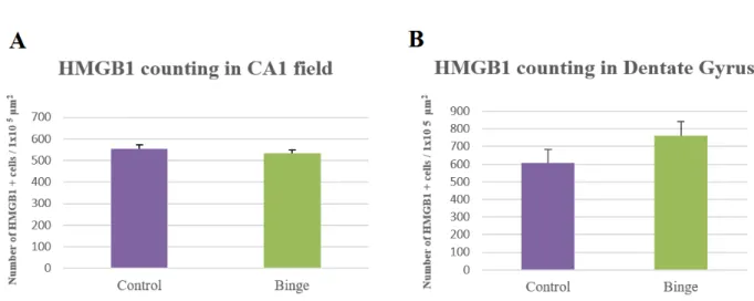
As presented in Figure 7, the number of HMGB1 positive cells seems to be similar between the binge group (A and C) and the control group (B and D). The statistical analysis showed that, in fact, there was no statistically significant difference between the alcohol-treated rats and rats of the control group for CA1 field (Figure 8-A) nor the Dentate Gyrus (Figure 8-B) (p>0,05). Moreover, there were no statistically significant changes in the mean optical density between both groups, which is directly proportional to labelling intensity, in both subregions(Figure 9).
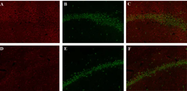
Figure 7 - Representative photomicrographs of HMGB1 expression shown in the CA1
field and Dentate Gyrus of ratsexposed to two binges of ethanol administration and allowed 48h of recovery (A-C) or to control (B-D). Images were taken with a 10x objective.
33
Figure 8 - Number of HMGB1 positive cells per 100 000 µm2in the CA1 field (A) and
in the Dentate Gyrus (B) of binge rats and control group, after 48H of recovery (p>0,05).
Figure 9 - Mean optical density of HMGB1 labelling in the CA1 field and in the DGin
34
4.2 Co-labelling of TLR4 and NeuN immunofluorescence
In order to verify if binge ethanol exposure in adolescence upregulates the TLR4 receptors (the receptor for the proinflammatory mediator HMGB1), we carried out an immunofluorescence study in two subregions of the hippocampus of adolescent rats, CA1 field and Dentate Gyrus (for the experiment protocol, see appendix 2).
TLR4 receptors are expressed both in glia cells and neurons, so here we produced a double labelling of TLR4 and NeuN, a marker of neuronal cell. The merger of both labelling allows to look at TLR4 expression inside neurons.
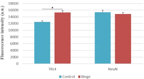
4.2.1 Co-labelling of TLR4 and NeuN immunofluorescence in CA1
The immunofluorescence labelling suggests that exists a slight increase in TLR4 fluorescence intensity in CA1 of binge rats, compared to the control group (Figure 10) and the statistical analysis shows an alteration in TLR4 fluorescence intensity in the CA1, which increased 19.97% in binge treated rats, indicating a significant increase of
TLR4 expression (Figure 11).This could reveal the presence of proinflammatory events
in these subregion, induced by the treatment of 2 binges of ethanol. As contrats the value for NeuN labelling is unchanged, as expected here: no changes was expecteed in the level of intensity of the markers NeuN, used here as a “reference” labelling to produce a ratio of TLR4 on NeuN.
There is also a statistically significant increase of ratio of TLR4/neuN in rats after the administration of 2 binges of ethanol and 48h of recovery, compared to the control group (Figure 13). This indicate that neuronal TLR4 is induced in the CA1 by the double binge exposure.
35
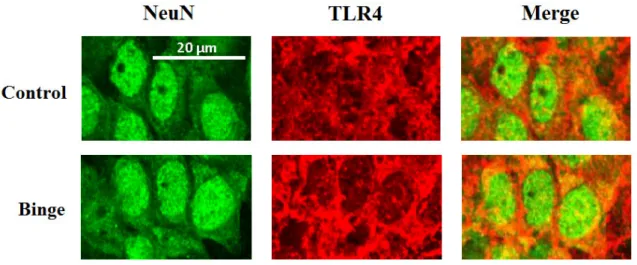
Figure 10 - Representative photomicrographs of TLR4 (red), NeuN (green) and
TLR4+NeuN (merger), shown in the CA1 fieldof binge rats (A-C) and control rats (D-F). Images were taken with a 80x objective with a confocal microscope.
Figure 11 - Expression levels of TLR4 and NeuN in CA1 field of both binge and control groups. The immunolabelling shows a significant increase of 19,97% of TLR4 fluorescence intensity in binged rats, which could mean a possible proinflammatory event in the CA1 field, induced by the alcohol treatment (*p<0,001). Values represent mean ± SEM, n=10 rats/group.
36
Figure 12 - Magnifying of CA1 region showing NeuN labelling, TLR4 labelling and
Merger of both signals, in Control (above) and Binge rats (below). Note that the TLR4 labelling reveals a more enhanced intensity of fluorescence in Binge rats than in the control group.
Figure 13 - Optical density ratio for TLR4/NeuN labelling in CA1 fieldin Control
37
4.2.2 Co-labelling of TLR4 and NeuN immunofluorescence in DG
In the DG, the immunofluorescence labelling shows an evident alteration in TLR4 fluorescence intensity between the Binge group and the control group (Figure 14), which increased 22.7% in binge treated rats (Figure 15), indicating a significant increase of TLR4 expression, which could mean the presence of proinflammatory events in these subregion, induced by the treatment of 2 binges of ethanol. As contrats the value for NeuN labelling was unchanged, as expected here.
The statistically analysis also reveals a significant increase of ratio of TLR4/neuN in rats after the administration of 2 binges of ethanol and 48h of recovery, compared to the
control group (Figure 16).This indicate that neuronal TLR4 is induced in the DG by the
double binge exposure.
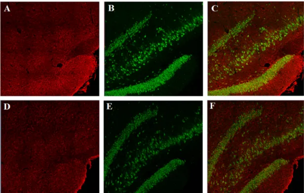
Figure 14 - Representative photomicrographs of TLR4 (red), NeuN (green) and
TLR4+NeuN (merger), shown in the dentate gyrus (DG) of Binge rats (A-C) and Control rats (D-F). Images were taken with a 80x objective with a confocal microscope. It is possible to verify that TLR4 labelling presents a more enhanced intensity of fluorescence in Binge rats than in the control group.
38
Figure 15 - Expression levels of TLR4 and NeuN in DG field,of both binge and control
groups. The immunolabelling shows a significant increase of 22,7% of TLR4 fluorescence intensity in binge rats, which could mean a possible proinflammatory event in the DG, induce by the alcohol treatment (*p<0,001). Values represent mean ± SEM, n= 8 rats/group.
Figure 16 - Optical density ratio for TLR4/NeuN labelling in the DG fieldin Control
(0,917 ± 0,045) and Binge (1,075±0,041) groups.
There is a statistically significant increase of ratio of TLR4/NeuN in rats after the administration of 2 binges of ethanol and 48h of recovery, compared to the control group. (**p<0,01). This indicate that neuronal TLR4 seems to be induced also in the DG by the double binge exposure.
39
5.
DISCUSSION
In the Grap laboratory, it was previously shown that a binge-like exposure with two close injections of ethanol 3g/kg in rat was enough to produce synaptic plasticity abolition in the CA1 of the hippocampus, along with losses in learning abilities, observed in a behavioural task.
In this work, given the recently discovered relationship between synaptic plasticity and neuroinflammation, we aimed at investigating whether this binge-like exposure protocol was able to produce neuroinflammatory events in the hippocampus. Our results
showed that HMGB1 expression was not altered neither in CA1 nor in dentate gyrus,
while the expression of neuronal TLR4 was induced in both the CA1 and dentate gyrus. Those two markers are linked, as HMGB1 is the ligand for TLR4 receptors. In basal metabolism, HMGB1 is present in the intracellular compartment and interacts with transcription factors and nucleosome formation. During neuroinflammation processes, HMGB1 is released into the extracellular space, inducing the innate immune response, hence called an alarmin. Upon being released by the cells into the extracellular medium, this protein will bind and activate TLR4 receptors, which are present on the surface of neuronal cells and microglia. Its activation by HMGB1 initiates the immune response
by the production of cytokines andpro-inflammatory mediators, such as TNF-α, IL-1β,
iNOS, MCP-1, among others.
A recent study by Zou et Crews (2014) showed through a western blot analysis of a primary culture of neuronal cells of the hippocampus, that a previous treatment with alcohol leads to an increase of HMGB1 expression after 24 and 72 hours (81). In a
post-mortem analysis it was shown that the chronic administration of alcohol during 10 days
in mice leads to the increase of HMGB1 expression in the entorhinal-hippocampal cortex and in the orbitofrontal cortex in alcoholic patients (82). Vetreno et al. (2012) demonstrated that the intermittent ethanol exposure during adolescence leads to the increase of HMGB1/TLR danger signal and HMGB1 expression in the prefrontal cortex, which persists into adulthood (2). This suggests that ethanol can contribute to positive loops of neuroimmune activation (83). These previous studies have suggested the hypothesis that the alcohol consumption induces neuronal inflammation, which results in an increase in the HMGB1 expression.
40
However, it was found that there are not significant differences in the mean optical density (Figure 9) nor the number of labelled cells for HMGB1 (Figure 8) between the binge group and the control group, for both subregions of hippocampus, as
suggested in the representative photomicrographs in the Figure 7. Taken together,these
results suggest that the double binge drinking didn’t induce any changes in HMGB1 expression in the hippocampus of rats after 48 hours, which can be explained by several reasons.
We hypothesize that, as HMGB1 is released into the extracellular space during neuroinflammation, our method using immunohistochemistry would have “washed away” the released HMGB1, so that way we were unable to see any change in HMGB1 expressed on the tissue.
Another possible explanation for these results may be the fact HMGB1 is a neuroinflammation mediator that presents a low specificity, for being a ubiquitous nuclear protein and because of this, it is involved in other processes, like nucleosome formation and gene transcription, processes which can change its expression levels (84).
When compared to the Vetreno’s study, whose exposure of rats to alcohol lasted one month (2), the protocol used in this study describes a duration of treatment of just two binges 8 hours apart, which may have been not enough to induce any changes in HMGB1 expression. On the other hand, analysing the effects of the two binges after 48 hours of recovery, lead us to also consider that there was no increase in HMGB1 expression in the binge rats, because 48h after binge could be too late to detect these possible changes, which can occur earlier.
Based on these results, which didn’t show a consistent relation between the expressed levels of HMGB1 and the alcohol-induced effects, we decided to move on with the study, through a receptor of HMGB1, the toll-like receptor 4 (TLR4), taking into account the findings from other authors.
Vetreno and Crews (2012) have demonstrated that HMGB1 expression in the adult PFC was significantly correlated with TLR4 and TLR3 expression in the same brain region and have shown that HMGB1/TLR danger signalling contributes to positive loops of neuroimmune activation. A more recent study from Montesinos (2015) and his team demonstrated that ethanol, by activating innate immune receptors TLR4, induces
41
neuroinflammation and brain damage in the PFC of adult mice. That way, we intended to show if TLR4, as a receptor of the cytokine and neuroinflammation marker HMGB1, would display changes in its expression in the hippocampus of binged rats.
TLR4 is expressed both in neurons and in glial cells. In order to analyse the TLR4 expression inside neurons, we performed a double immunofluorescence study labelling of TLR4 and NeuN. NeuN is a neuronal nuclear antigen, commonly used as a biomarker for neurons.
The results obtained showed a statistically significant increase of TLR4 expression in the hippocampus of binged rats: about 19,97% in the CA1 (Figure 11) and 22,7% in the DG (Figure 15). NeuN presents a similiar fluorescence intensity in both binge and control groups, as expected, given that the NeuN labelling is used here as an internal control that allows us to calculate a ratio between TLR4 and NeuN, that will reflect the changes in TLR4 expression. Also, the ratio between the optical density of both (TLR4/NeuN) is significantly increased in the binged rats for CA1 (Figure 13) and DG (Figure 16). Therefore, there is a greater expression of the TLR4 in the CA1 and DG of the binged group, which may reveal the occurrence of proinflammatory events and neuroinflammation processes in these subregions, induced by the alcohol exposure. These findings support the idea that TLR4 expression is increased in the hippocampus of rats submitted to a binge-like exposure. This protocol seems to be sufficient to induce neuroinflammation.
42
6.
CONCLUSION
The purpose of the current study was to determine if the binge-like exposure protocol was able to produce neuroinflammatory events in the hippocampus of adolescent rats. In order to investigate if neuroinflammations events occurred in rats exposed to a double binge of alcohol, we performed two main experiments.
The immunohistochemistry study has shown that there are not significant differences in the mean optical density nor the number of labelled cells for HMGB1 between the binge group and the control group, for both subregions of hippocampus, CA1 and DG. Although there are several possible hypotheses that may explain these results, the findings suggest that the double binge drinking was not enough to induce changes in HMGB1 expression on the hippocampus of rats, 48 hours later.
The results of immunofluorescence study demonstrated that there is a significant increase of TLR4 expression in the CA1 and in the DG of binged rats, which suggests that just a double binge-like ethanol exposure 8 hours apart is sufficient to induce neuroinflammation processes in the hippocampus of rats. This result supports the role of the neuroimmune response and TLR4 signalling in the neurotoxic effects of ethanol in adolescence.
A limitation of this study is that it makes possible we study the expression of these markers only in the cell body of neurons, and not on the dendrites. Another important limitation is time point of used in the protocol (48 hours of recovery, after the first administration of ethanol), which could be too late to detect any changes in HMGB1 expression, for example.
Further studies are needed to assess the precise impact of our protocol of binge drinking on neuroinflammation. A future study could be needed to demonstrate that HMGB1, as a neuroinflammation marker, may present an expression changed by the ethanol exposure, possibly, a IHC study with another antibody for HMGB1 would be a new challenge. It would also be interesting to analyse if TLR4 expression is also increased in the microglial cells, performing a co-labelling of TLR4 and M1 and/or M2, which are different phenotypes of microglia.
43
7.
REFERENCES
1. Hayes DM, Deeny MA, Shaner CA, Nixon K. Determining the threshold for alcohol-induced brain damage: new evidence with gliosis markers. Alcohol Clin Exp Res. 2013;37(3):425-34.
2. Vetreno RP, Crews FT. Adolescent binge drinking increases expression of the danger signal receptor agonist HMGB1 and Toll-like receptors in the adult prefrontal cortex. Neuroscience. 2012;226:475-88.
3. Marshall EJ. Adolescent alcohol use: risks and consequences. Alcohol Alcohol. 2014;49(2):160-4.
4. American Psychiatric Association, Diagnostic and statistical manual of mentaldisorders: DSM-5. 5th ed. Arlington, VA: American Psychiatric Publishing; 2013.
5. Janeczek P, MacKay RK, Lea RA, Dodd PR, Lewohl JM. Reduced expression of α-synuclein in alcoholic brain: influence of SNCA-Rep1 genotype. Addict Biol. 2014;19(3):509-15. 6. World Health Organization. Global status report on alcohol and health 2014. Geneva,
Switzerland: World Health Organization; 2014. xiv, 376 pages p.
7. Grant BF, Dawson DA. Age at onset of alcohol use and its association with DSM-IV alcohol abuse and dependence: results from the National Longitudinal Alcohol Epidemiologic Survey. J Subst Abuse. 1997;9:103-10.
8. McClain JA, Hayes DM, Morris SA, Nixon K. Adolescent binge alcohol exposure alters hippocampal progenitor cell proliferation in rats: effects on cell cycle kinetics. J Comp Neurol. 2011;519(13):2697-710.
9. Enoch MA. The influence of gene-environment interactions on the development of alcoholism and drug dependence. Curr Psychiatry Rep. 2012;14(2):150-8.
10. Ducci F, Goldman D. Genetic approaches to addiction: genes and alcohol. Addiction. 2008;103(9):1414-28.
11. (Inserm) Indlsedlrm. Alcohol: Social damages, Abuse and dependance. English Short version ed. Paris2003. p. 60.
12. Moeller L, Galea G, Organization WH. Alcohol in the European Union: consumption, harm and policy approaches. Copenhagen: WHO Regional Office for Europe; 2012.
44 14. Eurobarometer 72.3 - EU citizens attitudes towards alcohol. 2010.
15. Feijão F. Estudo sobre os Consumos de Álcool, Tabaco, Drogas e outros Comportamentos Aditivos e Dependências (ECATD-CAD) - Sumário Executivo. 2015. p. 24.
16. (SICAD) SdInCAenD. A Situação do País em Matéria de Álcool - Relatório Anual 2014. Lisboa2015. p. 180.
17. Hibell B. Summary 2011 ESPAD report : substance use among students in 36 Europeans countries. Luxembourg: Publications Office of the European Union; 2012.
18. Group E. ESPAD Report 2015 - Results from the European School Survey Project on Alcohol and Other Drugs. Luxembourg: Publications Office of the European Union2016. p. 104.
19. (SICAD SdInCAenD. A Situação do País em Matéria de Álcool - Relatório Anual 2015 Lisboa2016. p. 216.
20. European School Survey Project on Alcohol and other D, Hibell Br. The 1995 ESPAD report : alcohol and other drug use among students in 26 European countries. Stockholm: Swedish Council for Information on Alcohol and Other Drugs, CAN ; [Great Britain] : Council of Europe, Co-operation Group to Combat Drug Abuse and Illicit Trafficing in Drug (Pompidou Group); 1997.
21. Feijão F. Inquérito Nacional em Meio Escolar (INME), 2011 - Secundário. Consumo de drogas e outras substâncias psicoativas: uma abordagem integrada. 2011.
22. Rehm J, Room R, Graham K, Monteiro M, Gmel G, Sempos CT. The relationship of average volume of alcohol consumption and patterns of drinking to burden of disease: an overview. Addiction. 2003;98(9):1209-28.
23. (Inserm) Indlsedlrm. Alcohol: Health effects English Short version ed. Paris2001. p. 43. 24. Carbia C, Cadaveira F, Caamaño-Isorna F, Rodríguez-Holguín S, Corral M. Binge drinking
during adolescence and young adulthood is associated with deficits in verbal episodic memory. PLoS One. 2017;12(2):e0171393.
25. Wechsler H, Davenport A, Dowdall G, Moeykens B, Castillo S. Health and behavioral consequences of binge drinking in college. A national survey of students at 140 campuses. JAMA. 1994;272(21):1672-7.
26. Townshend JM, Duka T. Binge drinking, cognitive performance and mood in a population of young social drinkers. Alcohol Clin Exp Res. 2005;29(3):317-25.