Joana Carolina
Quintela Carrola
Avaliação do impacto das nanopartículas de prata
no metabolismo celular: um estudo in vitro por
metabolómica de RMN
Assessing the impact of silver nanoparticles on cell
metabolism: an in vitro NMR metabolomics study
Joana Carolina
Quintela Carrola
Avaliação do impacto das nanopartículas de prata
no metabolismo celular: um estudo in vitro por
metabolómica de RMN
Assessing the impact of silver nanoparticles on cell
metabolism: an in vitro NMR metabolomics study
Tese apresentada à Universidade de Aveiro para cumprimento dos requisitos necessários à obtenção do grau de Doutor em Nanociências e Nanotecnologia, realizada sob a orientação científica da Doutora Iola Melissa Fernandes Duarte, Investigadora Principal do CICECO – Instituto de Materiais de Aveiro,
Departamento de Química da Universidade de Aveiro e da Doutora Helena Cristina Correia de Oliveira, Investigadora em Pós-Doutoramento do CESAM e Departamento de Biologia da Universidade de Aveiro
Apoio financeiro da Fundação para a Ciência e a Tecnologia (FCT) - bolsa de investigação FCT SFRH/BD/79494/2011 no âmbito do Programa Operacional Capital Humano (POCH), comparticipado pelo Fundo Social Europeu e por fundos nacionais do Ministério da Ciência, Tecnologia e Ensino Superior, e projeto FCOMP-01-0124-FEDER-021456 (Refª. PTDC/SAU-TOX/120953/2010) financiado pelo Fundo Europeu de Desenvolvimento Regional (FEDER) através do Programa Operacional Fatores de Competitividade (COMPETE); da
Universidade de Aveiro - CICECO-Instituto de Materiais de Aveiro, POCI-01-0145-FEDER-007679 (FCT UID/CTM /50011/2013) e CESAM (FCT
UID/AMB/50017/2013). Agradece-se ainda à Rede Nacional de RMN (RNRMN), apoiada por fundos da FCT, e à empresa Bruker BioSpin GmbH.
o júri presidente
orientadora
Professor Doutor Rui Luís Andrade Aguiar
Professor Catedrático, Departamento de Eletrónica, Telecomunicações e Informática, Universidade de Aveiro
Professora Doutora Eduarda das Graças Rodrigues Fernandes
Professora Associada com Agregação, Departamento de Ciências Químicas, Faculdade de Farmácia, Universidade do Porto
Professor Doutor Carlos Manuel Marques Palmeira
Professor Catedrático, Departamento Ciências da Vida, Faculdade de Ciências e Tecnologia, Universidade de Coimbra
Professora Doutora Maria da Conceição Lopes Vieira dos Santos
Professora Catedrática, Departamento de Biologia, Faculdade de Ciências, Universidade do Porto
Professora Doutora Ana Maria Pissarra Coelho Gil
Professora Associada com Agregação, Departamento de Química, Universidade de Aveiro
Doutora Iola Melissa Fernandes Duarte
agradecimentos Quero começar por agradecer à minha orientadora, Doutora Iola Duarte, e à minha co-orientadora, Doutora Helena Oliveira, por todo o seu apoio e disponibilidade ao longo destes 4 anos. Agradeço, também, todo o
conhecimento que me transmitiram e a oportunidade de realizar este trabalho. Em segundo lugar, agradeço a toda a equipa envolvida neste projeto, em especial à Doutora Ana Daniel-da-Silva, Doutora Carmen Freire, Professora Doutora Conceição Santos, Doutor Miguel Oliveira, Eliana Malheiro, Doutora Ivana Jarak, Tiago Pedrosa e Doutora Verónica Bastos. Agradeço ainda ao Doutor Ricardo Pinto e à Mariam Nasirpour, por todo o trabalho relacionado com as nanopartículas de síntese verde.
Agradeço ao grupo de metabolómica da Universidade de Aveiro, pelos cruciais ensinamentos sobre RMN e análise multivariada, e pelas discussões críticas que tanto ajudaram ao desenvolvimento deste trabalho: à Professora Doutora Ana Gil e ao Doutor António Barros, também envolvidos neste projeto, e ao Doutor Brian Goodfelllow.
Agradeço ainda aos colegas, mas sobretudo amigos, Cláudia Rocha, Gonçalo Graça, Inês Lamego, Joana Pinto, João Rodrigues, Sílvia Diaz e Susana Aveiro, que sempre me deram motivação, com um toque de humor, para ultrapassar os momentos menos fáceis. Agradeço, também, a todos os que passaram pela “salinha”, por toda a amizade e alegria sempre presentes, Joana Marques, Lisa Sequeira, Ricardo Mendes e Sérgio Vilela.
Por fim, agradeço aos meus pais, que sempre me apoiaram e nunca mediram esforços para que eu cumprisse os meus objetivos, e restante família, a todos os amigos que se tornaram família, e ao Pedro, que tantas vezes sofreu comigo as dificuldades que encontrei e tantas vezes sorriu para tornar tudo melhor.
palavras-chave nanopartículas de prata (AgNPs), metabolismo celular, metabolómica,
espectroscopia de ressonância magnética nuclear (RMN), análise multivariada (MVA), queratinócitos (HaCaT), células de hepatoma (HepG2), macrófagos (RAW 264.7), nanotoxicologia.
resumo Face ao uso disseminado e enorme potencial terapêutico das nanopartículas de prata (AgNPs), o estudo dos seus efeitos biológicos é um assunto relevante e atual. O trabalho apresentado nesta tese teve como objetivo aprofundar o conhecimento existente sobre o impacto das AgNPs no metabolismo celular, usando a metabolómica por espectroscopia de ressonância magnética nuclear (RMN). Os tipos celulares escolhidos para este estudo foram queratinócitos da epiderme humana, células de hepatoma humano e macrófagos sanguíneos, por serem relevantes, respetivamente, ao nível da entrada, acumulação e captação de nanopartículas no organismo.
O Capítulo 1 introduz as principais propriedades das AgNPs, a sua atividade biológica e potencial toxicidade, e descreve a abordagem metabolómica, incluindo uma breve revisão bibliográfica das suas aplicações em nanotoxicologia. O âmbito e os objetivos desta tese são, também, apresentados.
O Capítulo 2 descreve os métodos experimentais utilizados ao longo deste trabalho, incluindo a caracterização das AgNPs, os procedimentos usados na cultura celular e nos ensaios biológicos, a colheita e preparação de amostras, a análise por RMN e o tratamento estatístico dos dados.
No Capítulo 3, a atividade metabólica e a composição dos três tipos de células usados neste trabalho (queratinócitos HaCaT, células de hepatoma HepG2 e macrófagos RAW 264.7) são descritas com base na análise por RMN dos sobrenadantes dos meios de cultura (exometaboloma) e dos extratos celulares polares e lipofílicos (endometaboloma).
O Capítulo 4 apresenta a análise metabolómica das células HaCaT expostas a AgNPs de diferentes tamanhos (10, 30 ou 60 nm de diâmetro) e revestimentos (citrato, polietilenoglicol ou albumina de soro bovino). Verificou-se que o metaboloma celular foi afetado mesmo a concentrações subtóxicas de AgNPs, sugerindo: aumento da glicólise e glutaminólise, alteração na atividade do ciclo dos ácidos tricarboxílicos (TCA) e nos processos de produção e transferência de energia, degradação de proteínas, síntese de glutationa (GSH),
modificações a nível das membranas e do equilíbrio osmótico. Apesar de muitas variações serem comuns a todas as nanopartículas testadas, as AgNPs de 10 nm causaram os efeitos mais distintos, nomeadamente no que diz respeito à glicólise e à síntese/utilização de GSH. Além disso, a exposição celular a prata iónica (Ag+) confirmou o importante papel dos iões prata no mecanismo de ação das AgNPs, enquanto a comparação com o peróxido de hidrogénio (H2O2) permitiu destacar os efeitos relacionados com o stress oxidativo.
No Capítulo 5 são apresentadas as respostas metabólicas das células de fígado HepG2 a dois tipos de AgNPs, umas obtidas por redução química e estabilizadas em citrato e as outras obtidas por síntese verde na presença de um extrato vegetal (Cit30 e GS30, respetivamente), ambas com centros metálicos de 30 nm. Os resultados sugeriram adaptações metabólicas em
resumo (cont.) processos de produção de energia (metabolismo da glucose e sistema da fosfocreatina), autofagia e metabolismo lipídico, refletindo possivelmente a ativação de mecanismos de proteção. Ainda que os dois tipos de AgNPs tenham induzido muitos efeitos semelhantes, as Cit30 pareceram causar um maior impacto no ciclo TCA e na degradação de proteínas, enquanto as GS30 aparentaram induzir uma diminuição mais forte na síntese de fosfolípidos. A assinatura metabólica da prata iónica foi bastante semelhante à das AgNPs, sugerindo, no entanto, uma menor capacidade das células expostas a Ag+ extracelular para lidar com o stress oxidativo.
O Capítulo 6 descreve a avaliação do impacto das AgNPs Cit30 no metaboloma de macrófagos de murganho RAW 264.7, a concentrações subtóxicas (decréscimos de ~5 e 20% na viabilidade celular). As alterações encontradas apontaram para: estimulação da glicólise (a baixa concentração de exposição), reprogramação do ciclo TCA (resultando numa intensa produção de itaconato e succinato e numa marcada depleção de ATP,
consistentes com uma resposta pro-inflamatória), ativação da gluconeogénese, promoção da síntese de GSH e acumulação de creatina/fosfocreatina. Foram, ainda, observadas variações possivelmente relacionadas com a
osmorregulação e a modificação membranar. De notar que os macrófagos expostos a Ag+ mostraram características semelhantes aos expostos a AgNPs (por ex., aumento da glucose intracelular – gluconeogénese), mas também revelaram efeitos distintos, nomeadamente em metabolitos envolvidos no ciclo TCA, em processos de transferência de energia e no metabolismo lipídico. Adicionalmente, viu-se que o metaboloma dos macrófagos respondeu de maneira diferente à exposição a H2O2 (por ex., tendência para diminuição da glicólise e sem efeitos observados na ativação da gluconeogénese ou da síntese de GSH), indicando que muitos dos efeitos induzidos pelas AgNPs não foram necessariamente mediados por stress oxidativo.
Finalmente, com base na integração dos resultados apresentados ao longo dos capítulos anteriores, as principais conclusões deste trabalho são apresentadas e discutidas no Capítulo 7.
keywords silver nanoparticles (AgNPs), cell metabolism, metabolomics, nuclear magnetic resonance (NMR) spectroscopy, multivariate analysis (MVA), keratinocytes (HaCaT), hepatoma cells (HepG2), macrophages (RAW 264.7),
nanotoxicology.
abstract The wide dissemination and promising therapeutic potential of silver
nanoparticles (AgNPs) make the study of their biological effects a relevant up-to-date subject. The work presented in this thesis aimed at deepening current understanding of the impact of AgNPs on cell metabolism, using NMR
metabolomics of cultured mammalian cells. Epidermis keratinocytes, hepatoma cells and blood macrophages, relevant, respectively, to nanoparticle entry, accumulation and uptake, have been selected for the study.
Chapter 1 introduces the main properties of AgNPs, including their biological activity and toxicological potential, and describes the metabolomics approach, briefly reviewing its applications in the field of nanotoxicology. Also, the scope and aims of this thesis are presented.
Chapter 2 covers the experimental methods adopted during the course of this work, including the sources and characterisation of AgNPs, the procedures used in cell culture and biological assays, sample collection and preparation methods, NMR analyses and statistical data treatment.
In Chapter 3, the metabolic activity and composition of the three cell types used in this work (HaCaT keratinocytes, HepG2 hepatoma cells and RAW 264.7 macrophages) are described based on the NMR analysis of culture medium supernatants (exometabolome) and polar/lipophilic cell extracts
(endometabolome).
Chapter 4 presents the metabolomic analysis of HaCaT skin cells exposed to AgNPs of different sizes (10, 30 or 60 nm in diameter) and coatings (citrate, polyethylene glycol or bovine serum albumin). The cellular metabolome was found to be affected even at sub-toxic concentrations of AgNPs, suggesting: upregulation of glycolysis and glutaminolysis, altered tricarboxylic acid (TCA) cycle activity, protein degradation, disruption of energy-producing pathways, glutathione (GSH) synthesis, membrane modification and changes in osmotic balance. Although several metabolic variations were common to all tested nanoparticles, the 10 nm AgNPs showed the most distinct effects, namely in regard to glycolysis and GSH synthesis/utilisation. Furthermore, cell exposure to ionic silver (Ag+) confirmed the major role of silver ions in AgNPs mode of action, while comparison with hydrogen peroxide (H2O2) allowed the effects related to oxidative stress to be highlighted.
In Chapter 5, the metabolic responses of liver HepG2 cells to two types of 30 nm AgNPs, obtained by chemical reduction and stabilised with citrate or produced by green synthesis in the presence of a plant extract (Cit30 and GS30, respectively) are presented. Sub-toxic concentrations of AgNPs were proposed to induce metabolic adaptations in energy production processes (glucose metabolism and the phosphocreatine system), autophagy and lipid metabolism, possibly reflecting the activation of metabolism-mediated protective mechanisms. Although the two types of AgNPs induced many
abstract (cont.) common effects, Cit30 appeared to have a greater impact on the TCA cycle and protein degradation, whereas GS30 seemed to induce stronger
downregulation of phospholipid synthesis. The metabolic signature of Ag+ was largely similar to that of AgNPs, although suggesting a lower ability of cells exposed to extracellular Ag+ to cope with oxidative stress.
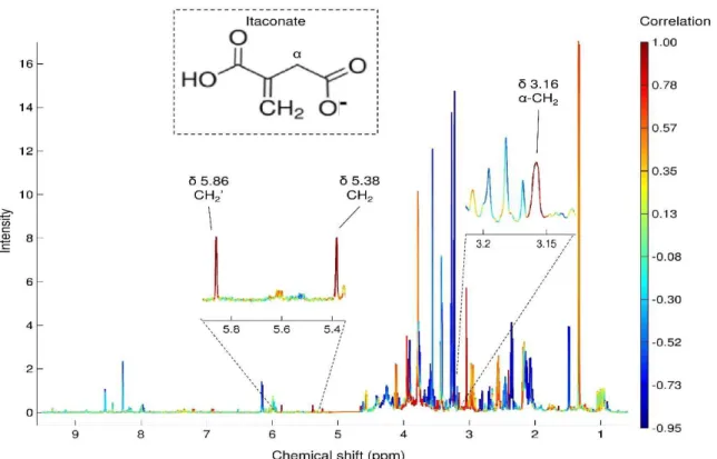
Chapter 6 addresses the impact of Cit30 AgNPs on the metabolome of murine RAW 264.7 macrophages, at concentrations causing minimal (~5 and 20%) decreases in cell viability. Exposed cells were suggested to upregulate glycolysis (at the low exposure concentration), to reprogram the TCA cycle (resulting in marked production of itaconate and succinate and in ATP depletion, consistent with a pro-inflammatory response), to activate gluconeogenesis, to promote GSH synthesis, and to increase the
creatine/phosphocreatine pool. Changes putatively related to osmoregulation and membrane modification were also observed. Notably, macrophages exposed to Ag+ showed common features to AgNPs-exposed cells (e.g. increased intracellular glucose suggesting gluconeogenesis), but also several distinct effects, for instance in metabolites involved in the TCA cycle, energy transfer processes or lipid metabolism. Furthermore, the cellular metabolome responded differently to H2O2 exposure (e.g. trend for downregulated
glycolysis, no evidence of gluconeogenesis activation or of GSH upregulation), indicating that many of the AgNPs-induced effects were not necessarily mediated by oxidative stress.
Finally, based on the integration of the results presented along the previous chapters, the main conclusions of this work are presented and discussed in Chapter 7.
A.L., Oliveira, H., Santos, C., Gil, A.M., Duarte, I.F. Metabolomics of silver nanoparticles toxicity in HaCaT cells: structure-activity relationships and role of ionic silver and oxidative stress. Nanotoxicology 2016, 10(8): 1105-17.
Full paper: Carrola, J., Bastos, V., Ferreira De Oliveira, J.M., Oliveira, H., Santos, C., Gil,
A.M., Duarte, I.F. Insights into the impact of silver nanoparticles on human keratinocytes metabolism through NMR metabolomics. Archives of Biochemistry and Biophysics 2016, 589: 53-61.
Abstract: Carrola J., Gil A.M., Daniel-Da-Silva A.L., Bastos V., Oliveira H., Oliveira J.,
Santos C., Duarte I.F. Metabolic response of human keratinocytes to silver nanoparticles: A metabolomics study. Toxicology Letters 2013, 221: S242-S3.
Manuscripts in preparation:
Carrola J., Pinto R.J.B., Nasirpour M., Freire C.S.R., Gil A.M., Santos C., Oliveira H., Duarte I.F. Metabolic profiling of liver (HepG2) cells exposed to silver nanoparticles suggests activation of metabolism-mediated protective mechanisms.
Carrola J., Oliveira H., Gil A.M., Santos C., Duarte I.F. Differential effects of silver nanoparticles, ionic silver and hydrogen peroxide on the metabolome of RAW 264.7 macrophages.
Other publications related to this work
Book Chapter: Pinto R.J.B., Nasirpour M., Carrola J., Oliveira H., Freire C.S.R., Duarte I.F.
Antimicrobial properties and therapeutic applications of silver nanoparticles and nanocomposites. In: Grumezescu, A.M. (ed.) Antimicrobial Nanoarchitectonics. 2017. Elsevier.
Full paper: Bastos V., Ferreira-De-Oliveira J.M.P., Carrola J., Daniel-Da-Silva A.L., Duarte
I.F., Santos C. and Oliveira H. Coating independent cytotoxicity of citrate- and PEG-coated silver nanoparticles on a human hepatoma cell line. Journal of Environmental Sciences 2016 (in press).
Full paper: Jarak I., Carrola J., Barros A.S., Gil A.M., Pereira M.L., Corvo M.L., Duarte I.F.
Metabolism modulation in different organs by silver nanoparticles: an NMR metabolomics study of a mouse model. Submitted.
Abstract: Bastos V., Carrola J., Duarte I.F., Santos C., Oliveira H. Comparative in vitro
cytotoxicity of citrate-coated silver nanoparticles on skin, liver and blood cell lines.
Toxicology Letters 2016, 258(Supplement): S262.
Abstract: Marçal R., Carrola J., Jarak I., Corvo M.L., Duarte I.F., Pereira, M.D.L. Microscopic
studies of liver and kidney in mice exposed to silver nanoparticles. Microscopy and
List of abbreviations and symbols i
Chapter 1. Introduction 1
1.1. Silver nanoparticles (AgNPs) 1
1.1.1. Synthesis and physicochemical properties of AgNPs 1
1.1.2. Biological properties of AgNPs 3
1.1.3. Toxicity of AgNPs towards mammalian cells 7 1.2. Brief overview of major cellular metabolic pathways 10 1.2.1. Glycolysis and gluconeogenesis 11 1.2.2. The TCA cycle and oxidative phosphorylation 14 1.2.3. The pentose phosphate pathway (PPP) 15 1.2.4. Fatty acid synthesis and degradation 16
1.3. Metabolomics in nanotoxicology 17
1.3.1. The metabolomics approach 17
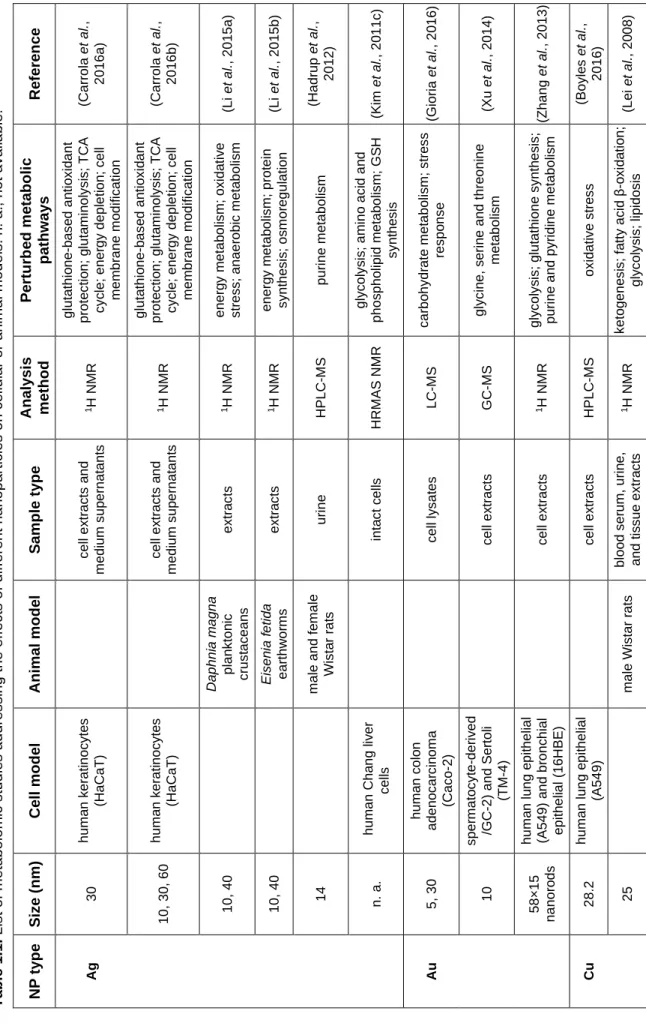
1.3.2. Considerations about NMR metabolomics of cultured mammalian cells 25 1.3.3. State-of-the-art of metabolomics applications in nanotoxicology 28
1.4. Scope and aims of this thesis 34
Chapter 2. Materials and methods 35
2.1. Silver nanoparticles: sources and characterisation 35
2.1.1. Sources of AgNPs 35
2.1.2. Transmission Electron Microscopy (TEM) 35 2.1.3. UV-Visible (UV-Vis) Spectroscopy 36 2.1.4. Dynamic Light Scattering (DLS) 36
2.1.5. Zeta potential 37
2.1.6. Inductively Coupled Plasma Optical Emission Spectrometry (ICP-OES) 38 2.2. Cell culture and biological assays 38
2.2.1. Chemicals 38
2.2.2. Cell lines 39
2.2.3. Routine cell culture maintenance 39
2.2.4. Cell viability assay 40
2.2.5. Reactive Oxygen Species (ROS) assay 41
2.3. NMR metabolomics 42
2.3.1. Cell culture and exposure for metabolomics 42
2.3.2. Sample collection 43
Discriminant Analysis (PLS-DA) 46 2.4.3. Spectral integration and univariate statistical analysis 47
2.4.4. Correlation analysis 47
Chapter 3. Metabolite profiling of cultured cells: keratinocytes (HaCaT), hepatoma
cells (HepG2) and macrophages (RAW 264.7) 49
3.1. Background and aims 49
3.2. Metabolic activity of cells assessed by the NMR analysis of culture media 50
3.3. Intracellular polar metabolites 57
3.4. Lipophilic metabolites 64
Supplementary information to Chapter 3 70
Chapter 4. Metabolomic analysis of human keratinocytes (HaCaT cells) exposed to
silver nanoparticles of different sizes and coatings 73
4.1. Background and aims 73
4.2. Physicochemical properties of the AgNPs tested 74 4.3. Viability of HaCaT cells exposed to AgNPs, Ag+ and H
2O2 76
4.4. Generation of reactive oxygen species (ROS) 79 4.5. Metabolic variations induced by AgNPs in HaCaT cells 79 4.6. Metabolic variations induced by the coating substances alone 84 4.7. Metabolic variations induced by Ag+ in HaCaT cells 84
4.8. Metabolic variations induced by H2O2 in HaCaT cells 87
4.9. Discussion of metabolic variations in HaCaT cells 88
Supplementary information to Chapter 4 94
Chapter 5. Metabolomic analysis of human hepatoma cells (HepG2) exposed to
citrate-coated and biogenic silver nanoparticles 111
5.1. Background and aims 111
5.2. Physicochemical properties of the AgNPs tested 112 5.3. Viability of HepG2 cells exposed to AgNPs, Ag+ and H
2O2 114
5.4. Generation of reactive oxygen species 117 5.5. Metabolic variations induced by AgNPs in HepG2 cells 118 5.6. Metabolic variations induced by Ag+ in HepG2 cells 124
5.7. Metabolic variations induced by H2O2 in HepG2 cells 127
5.8. Discussion of metabolic variations in HepG2 cells 129 Supplementary information to Chapter 5 136
Chapter 6. Metabolomic analysis of murine macrophages (RAW 264.7 cells) exposed
to citrate-coated silver nanoparticles 149
6.1. Background and aims 149
6.2. Physicochemical properties of the AgNPs tested 150 6.3. Viability of RAW 264.7 cells exposed to AgNPs, Ag+ and H
2O2 150
6.4. Generation of reactive oxygen species 152 6.5. Metabolic variations induced by AgNPs in RAW 264.7 macrophages 153 6.6. Metabolic variations induced by Ag+ in RAW 264.7 macrophages 157
6.7. Metabolic variations induced by H2O2 in RAW 264.7 macrophages 159
6.8. Discussion of metabolic variations in RAW 264.7 macrophages 161 Supplementary information to Chapter 6 168
Chapter 7. Integrated discussion and conclusions 175
i
List of abbreviations and symbols
1D one-dimensional2D two-dimensional Abs absorbance
ADP adenosine diphosphate AgNPs silver nanoparticles Ag+ ionic silver
AMP adenosine monophosphate ATP adenosine triphosphate BSA bovine serum albumin
BSA30 30 nm BSA-stabilised silver nanoparticles Cho choline
Cit10 10 nm citrate-stabilised silver nanoparticles Cit30 30 nm citrate-stabilised silver nanoparticles Cit60 60 nm citrate-stabilised silver nanoparticles CK choline kinase
CoA coenzyme A Cr creatine
CR classification rate
DMEM Dulbecco's modified Eagle's medium DMSO dimethyl sulfoxide
DNA deoxyribonucleic acid
EDTA ethylenediaminetetraacetic acid EG Eucaliptus globulus Labill.
FAD+ flavin adenine dinucleotide
FBP-1 fructose-1,6-bisphosphatase FBS foetal bovine serum
GC gas chromatography GDP guanosine diphosphate GPC glycerophosphocholine GSH reduced glutathione GSSG oxidised glutathione GTP guanosine triphosphate
GS30 30 nm biogenic silver nanoparticles HMDB human metabolome database
ii
HRMAS high resolution magic angle spinning HSQC heteronuclear single-quantum coherence IC5 5% inhibitory concentration
IC20 20% inhibitory concentration
IC50 half maximal inhibitory concentration
IDH isocitrate dehydrogenase
J-res J-resolved
LC liquid chromatography LDH lactate dehydrogenase LV latent variable
MCCV Monte Carlo cross-validation
m-Ino myo-inositol
MALDI matrix-assisted laser desorption/ionisation MIC minimum inhibitory concentration
MS mass spectrometry
MTT 3-(4,5-dimethylthiazol-2-yl)-2,5-diphenyltetrazolium bromide MVA multivariate analysis
NAA N-acetylaspartate
NAD+ nicotinamide adenine dinucleotide
NADP+ nicotinamide adenine dinucleotide phosphate
NMR nuclear magnetic resonance NPs nanoparticles
PBS phosphate buffer saline PC phosphocholine
PCA principle component analysis PEG polyethylene glycol
PEG10 10 nm PEG-stabilised silver nanoparticles PEG30 30 nm PEG-stabilised silver nanoparticles PTC phosphatidylcholine
PTE phosphatidylethanolamine
PLS-DA partial least squares-discriminant analysis PUFA polyunsaturated fatty acyl
PVP polyvinylpyrrolidone
r Pearson’s correlation coefficient RNA ribonucleic acid
iii
ROC receiver operating characteristic ROS reactive oxygen species
RSPA recursive segment-wise peak alignment SDH succinate dehydrogenase
STOCSY statistical total correlation spectroscopy SW spectral window
TCA tricarboxylic acid
TEM transmission electron microscopy TMS tetramethylsilane
TOCSY total correlation spectroscopy TSP 3-trimethylsilylpropionic acid UDP uridine diphosphate
UTP uridine triphosphate UV unit variance
UV-Vis UV-visible spectroscopy
1
Chapter 1. Introduction
1.1. Silver nanoparticles (AgNPs)
Silver has been used throughout history for a large variety of applications, including the manufacture of currency coins, ornaments, jewellery, tableware and utensils. Furthermore, its healing and preservative properties have been known since primordial civilisations, as attested by the use of silver films for water preservation by the Phoenicians, or of silver powders as anti-disease agents by ancient Egyptians (Murphy et al., 2015). Over the last century, the engineering of silver into nanoparticles has further extended the myriad of applications for this noble metal. Today, silver nanoparticles (AgNPs) represent one of the classes of nanomaterials with largest production and commercialisation (Tran et al., 2013). In particular, their well-documented antimicrobial properties has propelled their incorporation into numerous consumer goods, including plastics, textiles, cosmetics and food packaging materials, as well as in medical products, such as wound dressings, implants and catheters (Ge et al., 2014). Moreover, new therapeutic applications are increasingly being explored with a view to take advantage of other interesting biological properties of AgNPs, such as the antiviral or anticancer activities (Wei et al., 2015). On the other hand, given the high propensity for human exposure, there is a recognised need to better define the potential adverse effects of AgNPs to human health, and to understand their mechanisms of action in eukaryotic cells and higher organisms. In the following sections, the main physicochemical and biological properties of AgNPs as well as the current knowledge on their toxicity will be reviewed, aiming at establishing a sound and informed background that will aid in the interpretation of the results presented in this thesis.
1.1.1. Synthesis and physicochemical properties of AgNPs
AgNPs are nanoscale clusters of metallic silver (20-15000 atoms), with sizes typically below 100 nm in at least one dimension. They can be synthesised by numerous methods, usually divided into three main categories: chemical, physical and biological methods (Wei et al., 2015; Pinto et al., 2017). Chemical methods comprise the reduction of silver ions (Ag+) in aqueous or organic solutions, forming metallic silver (Ag0) atoms that
subsequently form clusters, yielding colloidal silver in the nanometric range. In particular, the chemical synthesis of AgNPs can be achieved through chemical or electrochemical reduction, hydrothermal, microemulsion and photochemical methods (Pinto et al., 2017).
2
Physical methods include, among others, laser ablation, whereby AgNPs are synthesised through the ablation of metallic bulk materials by laser pulses, evaporation-condensation, and arc discharge between titanium electrodes in a silver nitrate solution (Pinto et al., 2017). Finally, biological synthetic routes for AgNPs make use of different organisms like bacteria, fungi, algae, yeasts or plants. These are generally safer and more economic than chemical or physical methods, since the solvent medium is preferably water, and the reducing and stabilising agents are non-toxic and environmentally friendly (Rajan et al., 2015; Rauwel et
al., 2015).
Depending on the fabrication method, AgNPs can present different shapes, including spheres, rods, wires and plates (Figure 1.1a), as well as various sizes. For instance, strong reducing agents usually produce small monodisperse particles, while weak reductants tend to generate larger polydisperse particles (Galdiero et al., 2011).
Figure 1.1. a) Differently shaped AgNPs (nanoComposix, 2016a). b) Surface area:volume ratios of nanospheres, according to their diameter (nanoComposix, 2016a). c) Schematic representation of surface oxidation and dissolution of the oxide layer in AgNPs; reprinted with permission from (Li et al., 2012), Copyright © 2012 American Chemical Society.
Since nanosized materials have a significant number of atoms on the surface, they are characterised by high surface area to volume ratio, which increases with decreasing nanoparticle size (Fig 1.1b). Consequently, the chemical reactivity and many of the physical properties of the nanoparticles, such as solubility and tendency to aggregate, are dominated
3
by the nature of the nanoparticle surface. Silver nanoparticles are characterised by the presence of an oxide layer (Ag2O) on the surface of the metallic core (Ag0) (Figure 1.1c),
which dissolves under certain conditions to release silver ions (Ag+) (Li et al., 2010; Li et al.,
2012). Thus, it is not surprising that surface coating affects the dissolution behaviour of AgNPs (Li et al., 2012). The coating agent is also determinant for nanoparticle stabilisation and prevention of agglomeration, either through electrostatic (e.g. citrate coating) or steric interactions (e.g. polyvinylpyrrolidone (PVP) or polyethylene glycol (PEG) coating). Furthermore, it is important to stress that the surface of most nanoparticles is dynamic and strongly influenced by the local environment.
Another size-dependent property of AgNPs is their ability to absorb and scatter light with great efficiency. This is due to the collective oscillation of conduction electrons on the metal surface when they are excited by incident light at specific wavelengths, producing the so called surface plasmon resonance (SPR). These unique optical properties allow AgNPs stability to be accurately monitored using UV-Vis spectroscopy (Zook et al., 2011).
1.1.2. Biological properties of AgNPs Antibacterial activity
Silver has been used as a bactericidal agent since ancient times, especially until the appearance of antibiotics. Then, the evolution of antibiotic-resistant strains of bacteria (Rai
et al., 2012), led to a renewed interest in the antibacterial properties of silver, namely in the
nanoparticulate form. AgNPs have shown high efficacy against a vast range of bacteria, including Gram negative bacteria, such as Escherichia, Vibrio or Pseudomonas, and Gram positive bacteria, such as Staphylococcus, Enterococcus or Bacillus, with reported minimum inhibitory concentrations (MIC) typically varying in the range 1-100 μg/mL (Tran
et al., 2013). Moreover, the efficacy of AgNPs alone or in combination with antibiotics
against several multidrug-resistant (MDR) strains (Rai et al., 2012) and the ability of AgNPs to prevent and disrupt bacterial biofilms (Franci et al., 2015), which can be responsible for serious infections, present a gateway for the development of new antibacterial therapies.
The bactericidal activity of AgNPs is greatly influenced by their physicochemical properties, namely size, surface charge and shape. Smaller nanoparticles have been shown to possess stronger antibacterial activity than larger nanoparticles (Morones et al., 2005; Choi and Hu, 2008; Agnihotri et al., 2014), possibly due to their higher surface area and reactivity. In terms of surface charge, the strong electrostatic interactions between the
4
bacterial membrane and positively-charged AgNPs are probably responsible for the higher efficacy of these nanoparticles against Bacillus species, when comparing to negatively charged ones (El Badawy et al., 2011). The influence of shape has also been tested, revealing that silver nanoplates present higher bactericidal activity than spherical or rod-shaped AgNPs (Pal et al., 2007; Sadeghi et al., 2012); this was proposed to be related to the NPs crystallographic surface structure, since the top basal plane of a truncated triangular nanoplate is a {111} facet, which is a high-atom-density surface that favours silver reactivity, whereas spherical and rod-shaped AgNPs are known to predominantly have {100} facets with a small percentage of {111} facets.
Some works have attributed the antibacterial activity of AgNPs to intrinsic properties (Morones et al., 2005; Choi and Hu, 2008), while silver ions (Ag+), formed by oxidative
dissolution of AgNPs or through the release of chemisorbed ions at the particles surface (Le Ouay and Stellacci, 2015), have been shown to play a key role in AgNPs toxicity (Xiu
et al., 2012). Indeed, Xiu and co-workers found that the viability of E. coli was not affected
under anaerobic conditions, in which dissolution to Ag+ did not occur, even when the
concentration of AgNPs was much higher than the minimum lethal concentration measured under aerobic conditions.
Even though the mechanism of silver interaction with bacterial cells is not fully understood, several processes are thought to be involved (Eckhardt et al., 2013), as illustrated in Figure 1.2. The direct physical damage to the cell membrane, resulting from attachment and penetration of AgNPs and Ag+ into the bacterial cell wall, can lead to
increased cell permeability and subsequent cell death. In particular, it was proposed that membrane destabilisation could result in collapsed membrane potential, dissipation of proton motive force and depletion of intracellular ATP levels (Dibrov et al., 2002; Lok et al., 2006). Furthermore, the uptake and intracellular accumulation of AgNPs and Ag+ (facilitated
by membrane poration) enables their interaction with vital enzymes and phosphorous-containing nucleobases, affecting major cell functions, such as cell division and DNA replication (Li et al., 2013b), resulting in bacterial apoptosis (Bao et al., 2015). Oxidative stress, with consequent damage to proteins, lipids and DNA, is another possible mechanism of AgNPs toxicity; high levels of reactive oxygen species (ROS) have been found in bacteria exposed to AgNPs (Kim et al., 2007; Park et al., 2009; Xu et al., 2012), and have been suggested to arise either from over-production, or from disruption of ROS-scavenging enzymatic and non-enzymatic systems, as recently reviewed (Rizzello and Pompa, 2014).
5
Figure 1.2. Scheme illustrating the proposed mechanisms of action on AgNPs on bacteria: 1A, diffusion; 1B, endocytosis; 2A, mitochondrial damage; 2B, direct generation of ROS; 2C, generation of ROS by damaged mitochondria; 3A, organelle damage via interaction with sulphur and phosphor groups; 3B, DNA damage via nucleotide oxidation; 3C, DNA and organelle damage via oxidative stress; 4A, electrostatic interaction of AgNPs with cell membrane; 4B, membrane damage (lipid oxidation) by Ag+ and ROS (Pinto et al., 2017).
Antifungal activity
The application of many antifungal medicines is often accompanied by toxicity and development of resistance (Sardi et al., 2013). Thus, AgNPs have appeared as alternative fungicidal agents, especially against Candida species, which are opportunistic pathogens in humans. The high anticandidal activity of AgNPs has been reported in several studies, with MIC values in approximately the same range as that of conventional antifungal agents (0.05 – 2 μg/mL) (Kim et al., 2009a; Panacek et al., 2009; Selvaraj et al., 2014; Bonilla et
al., 2015). Moreover, AgNPs have been shown to inhibit the formation and bioactivity of Candida albicans biofilms (Li et al., 2014b; Lara et al., 2015; Longhi et al., 2015). It has
been proposed that the antifungal mode of action of AgNPs against C. albicans involves the disruption of the cell membrane, with pore formation and subsequent inhibition of the normal budding process (Kim et al., 2009a). Nonetheless, the ultrastructural analysis of C.
6
microscopy (HRTEM), did not show severe damage to cell membranes (Vazquez-Munoz et
al., 2014). It showed, however, high extracellular accumulation of AgNPs, with release of
Ag+, and intracellular biosynthesis of smaller AgNPs (through reduction by cell organic
compounds). In another study, the increased ROS production in AgNPs-exposed C.
albicans was related with mitochondria-dependent apoptosis and DNA fragmentation
(Hwang et al., 2012).
Antiviral activity
Several works have demonstrated the activity of AgNPs against different viruses (Galdiero et al., 2011), such as those causing human immunodeficiency (Elechiguerra et
al., 2005), herpes simplex (Baram-Pinto et al., 2009), hepatitis B (Lu et al., 2008), Dengue
(Sujitha et al., 2015) and influenza (Xiang et al., 2011; Mori et al., 2013; Xiang et al., 2013). It has been suggested that the inhibition of viral infections by AgNPs could be due to an impaired attachment of viruses to host cells, resulting from the interaction of AgNPs with outer proteins in the viral envelope (Elechiguerra et al., 2005; Sun et al., 2008; Lara et al., 2010). Nonetheless, in the presence of AgNPs, the Vaccinia virus was capable of adsorbing to cells but was prevented from entering (Trefry and Wooley, 2013). On the other hand, AgNP-treated tacaribe virus (TCRV) was more efficiently internalised into Vero cells than untreated TCVR (Speshock et al., 2010); in that work, viral RNA production (and thus virus replication) was found to be inhibited upon AgNPs treatment, leading the authors to suggest the potential use of AgNPs in vaccine products.
Anti-inflammatory activity
The anti-inflammatory properties of AgNPs have been demonstrated both in vitro and in vivo. Recently, biosynthesised AgNPs with antibacterial activity were shown to have a cytoprotective effect towards hydrogen peroxide (H2O2)-induced inflammation in murine
macrophages (Manikandan et al., 2015). PVP-coated AgNPs also helped controlling inflammation triggered by a Chlamydia trachomatis infection in macrophages, through the regulation of several upstream surface receptors and downstream inflammatory pathway genes (Yilma et al., 2013). In another study, composites of AgNPs and dendrimers were shown to have synergistic anti-inflammatory activity in lipopolysaccharide-stimulated murine macrophages (Liu et al., 2014). Furthermore, in the presence of biogenic AgNPs, the production of pro-inflammatory cytokines by human keratinocytes exposed to UVB
7
radiation was decreased (David et al., 2014). Regarding in vivo studies, AgNPs were shown to reduce inflammation and/or accelerate healing in murine models of allergic airway diseases (Jang et al., 2012; Shin and Ye, 2012), a porcine model of contact dermatitis (Nadworny et al., 2008), postoperative peritoneal adhesion models (Wong et al., 2009; Zhang et al., 2014a) and a burn wound mouse model (Liu et al., 2014).
Anticancer activity
A recent review thoroughly describes the potential therapeutic applications of AgNPs in cancer (Wei et al., 2015). Angiogenesis, the formation of new blood vessels, is a critical process for tumour growth and spreading. It has been shown that AgNPs and a natural anti-angiogenesis molecule, contributed almost equally to the inhibition of the formation of new blood microvessels induced by vascular endothelial growth factor (VEGF) (Gurunathan et
al., 2009). Moreover, several studies have demonstrated the ability of AgNPs to directly kill
tumour cells. As described in the following section, AgNPs can cause dose-dependent toxicity in mammalian cells, including oxidative stress and DNA damage, ultimately leading to cell death. In particular, apoptosis has been often highlighted as a main mechanism through which AgNPs exert their cytotoxic action against tumour cells (Gurunathan et al., 2009; Krishnasamy et al., 2015; Gurunathan et al., 2015; Satapathy et al., 2015). Other potential applications of AgNPs in cancer treatment are based on their ability to sensitise tumour cells to radiation therapy (Lu et al., 2012; Swanner et al., 2015), and to show phototherapeutic properties under near-infrared (NIR) radiation (Boca et al., 2011; Mfouo-Tynga et al., 2014).
1.1.3. Toxicity of AgNPs towards mammalian cells
While in some applications the cytotoxicity of AgNPs is a desirable feature, e.g. when envisaging the killing of tumour cells, the successful use of AgNPs, e.g. as antimicrobial agents, often requires low toxicity towards human cells. Therefore, there is a great need to understand the mechanisms of AgNPs toxicity and the influencing factors that modulate toxic effects. In recent years, numerous studies have been carried out to address the toxicological potential of AgNPs towards mammalian cultured cells, as will be briefly reviewed below.
Several works have demonstrated that AgNPs can be readily taken up by various human-derived cell types, such as normal bronchial cells (Gliga et al., 2014), liver cells
8
(Song et al., 2012), epidermal keratinocytes (Monteiro-Riviere et al., 2013), mesenchymal stem cells (Greulich et al., 2011) and various tumour cell lines (Liu et al., 2010; Yu et al., 2013b). Similarly to other nanomaterials (Zhao et al., 2011), the uptake of AgNPs has been shown to occur mainly through active pathways, namely clathrin- and caveolin-dependent endocytosis, macropinocytosis and phagocytosis, although involvement of energy-independent uptake pathways has also been suggested (Jiang et al., 2014). Inside cells, AgNPs were often found to localise within membrane-bound structures, namely endosomes and lysosomes (Jiang et al., 2013; Milic et al., 2015; Gliga et al., 2014). Less frequently, AgNPs have also been found in the cell nuclei (AshaRani et al., 2009; Cronholm et al., 2013).
Silver nanoparticles have been found to negatively affect the viability of many cell types in a concentration- and time-dependent manner. Based mainly on mitochondrial metabolic activity (e.g. formazan-based MTT assay) or membrane leakage (e.g. LDH assay) endpoints, several studies have reported significant decreases in cell viability upon AgNPs exposure. Reported IC50 values (concentrations causing a 50% decrease in cell
viability) typically ranged from a few μg/mL (e.g. HepG2 cells exposed to 5 nm AgNPs (Avalos et al., 2014)) to over 50 μg/mL (e.g. HaCaT cells exposed to 30 nm AgNPs (Mukherjee et al., 2012)). This variability likely reflects not only the diverse susceptibility of the different cell types to AgNPs, but also the variable physicochemical properties of the AgNPs tested. For instance, the size of AgNPs was often shown to have a major influence on biological outcomes. In the majority of cases, smaller particles were more cytotoxic than larger ones, with AgNPs sized below 40 nm generally causing much higher toxicity (Liu et
al., 2010; Park et al., 2011; Prasad et al., 2013; Avalos et al., 2014; Wang et al., 2014; Gliga et al., 2014; Butler et al., 2015). The surface chemistry and shape of AgNPs were also
highlighted as determinant to particle toxicity (Stoehr et al., 2011).
DNA damage and chromosomal aberrations are other commonly reported outcomes of AgNPs exposure. In particular, DNA strand breaks and/or micronuclei formation have been observed in several AgNPs-exposed human cells, such as fibroblasts and glioblastoma cells (AshaRani et al., 2009), epidermal keratinocytes (Austin et al., 2011; Szmyd et al., 2012), renal cells (Kang et al., 2012), and white blood cells (Butler et al., 2015). Induction of bulky DNA adducts (Foldbjerg et al., 2011) and increased levels of 8-oxo-2'-deoxyguanosine (Jiang et al., 2013), a marker of oxidative DNA damage, have also been reported, summing up to recent alerting evidence on the mutagenic potential of AgNPs (Huk et al., 2014). Consistently with DNA damage, cell cycle arrest at the G2/M phase, to repair damaged DNA, has often been reported (AshaRani et al., 2009; Kang et al., 2012;
9
Song et al., 2012; Foldbjerg et al., 2012; Jiang et al., 2013; Xue et al., 2016). When cellular genetic material is damaged beyond repair, cells may initiate apoptotic cell death. Indeed, AgNPs have been shown to induce apoptosis in several human-derived cells (Piao et al., 2011; Szmyd et al., 2012; Xue et al., 2016). Necrosis has also been reported to occur in a few studies with AgNPs, to an extent which depended on the cell type (Kim et al., 2012), as well as on the time of exposure, size and concentration of AgNPs (Kumar et al., 2015).
The cells inability to cope with high levels of ROS and to counteract their harmful effects leads to oxidative stress. Many studies have proposed this condition to be the main mechanism underlying the cytotoxicity and genotoxicity of AgNPs. Indeed, ROS have been found to be significantly increased in several types of human cells exposed to AgNPs (Suliman et al., 2015; Avalos et al., 2014; Ahlberg et al., 2014; De Matteis et al., 2015). Additionally, exposure to AgNPs has been related to other markers of oxidative stress, namely lipid peroxidation, protein carbonylation, altered levels of reduced glutathione (GSH) and upregulated transcription of stress-related genes (Kim and Ryu, 2013). Together with studies showing that ROS scavengers could strongly reduce or eliminate AgNPs toxic effects (Hsin et al., 2008; Foldbjerg et al., 2011; Kim et al., 2011a; Kang et al., 2012), there is thus strong evidence for a major role of oxidative stress in AgNP-mediated toxicity. The causal relationship between AgNPs exposure and increased ROS is thought to be explained through multiple events (Foldbjerg et al., 2015): i) the AgNPs themselves and their oxidative dissolution to silver ions can directly generate free radicals (He et al., 2012; Ahlberg et al., 2014), ii) AgNPs (and/or liberated ions) can enhance ROS production by disrupting the mitochondrial electron transport chain, whereby escaped electrons are accepted by molecular oxygen and give rise to radical species, iii) antioxidant molecules may be depleted and antioxidant enzymes impaired by AgNPs (and/or liberated ions). On the other hand, a few studies have underlined the possible additional role of ROS-independent mechanisms. For instance, Gliga and co-authors found no evidence of ROS production preceding DNA damage and postulated that this could be due to direct interaction of AgNPs with DNA repair pathways (Gliga et al., 2014). In another work, pre-treatment of lung cells with a ROS scavenger prevented reduction of cell viability upon AgNPs administration but did not stop cell cycle changes, which appeared to be ROS-independent (Chairuangkitti et al., 2013).
The contribution of the particles per se and of released silver ions to AgNPs-mediated toxicity has been a matter of controversy. While some studies attributed toxicity solely to Ag+ (Bouwmeester et al., 2011; Prasad et al., 2013), others suggested that Ag+
10
2012; Grosse et al., 2013; Sambale et al., 2015). Moreover, it has been reported that the influence of Ag+ on nanosilver toxicity decreased with increasing particle size (Pratsinis et
al., 2013) and varied with the silver ion fraction in the AgNPs suspensions (Beer et al.,
2012). Recent works have provided key data to understand this interplay, by clearly distinguishing between the effects caused by extracellular and intracellular Ag+ (Cronholm
et al., 2013; Gliga et al., 2014; De Matteis et al., 2015). Silver ions present in the external
cell medium showed very low internalisation by cells and induced cell death through membrane damage (Cronholm et al., 2013; De Matteis et al., 2015). On the other hand, AgNPs were shown to be readily taken up by cells and to release Ag+ intracellularly, in a so
called ‘Trojan horse’ effect. Indeed, by taking advantage of a recently developed chemosensor, De Matteis and co-authors could experimentally demonstrate the gradual release of Ag+ inside living cells (De Matteis et al., 2015). Thus, the intracellular dissolution
of AgNPs, favoured by the acidic environment in the lysosomes and influenced by the physicochemical properties of the particles, appears to be a main driver of the AgNPs toxic effects on cells.
While many reports have addressed the acute toxic effects of AgNPs towards various cultured cells, there is still insufficient knowledge on possible sub-toxic alterations of cell function, more representative of real exposure scenarios. Omics technologies, namely genomics, transcriptomics, proteomics and metabolomics, constitute a valuable approach in this respect, as they offer the possibility to detect unsuspected subtle changes in genes, transcripts, proteins or metabolites before cytotoxic events are detectable by conventional methods. In this thesis, metabolomics has been the approach elected to detect and aid interpreting biochemical events triggered by exposure to low concentration of AgNPs. Thus, the principles of this approach and the state-of-the-art concerning its applications in the field of nanotoxicology will be presented in subsequent sections of this chapter. Before that, a brief overview of cell metabolism is presented, focusing on some major metabolic pathways, as this information is relevant to interpret the results and metabolic hypotheses generated within this work.
1.2. Brief overview of major cellular metabolic pathways
Cellular metabolism can be viewed as a complex network of chemical reactions, catalysed by enzymes and strictly regulated, which allow organisms to grow and reproduce, maintain their structures, and respond to environmental changes (Nelson and Cox, 2004).
11
As represented in Figure 1.3, these reactions are organised into many inter-dependent metabolic pathways (Kanehisa et al., 2016). Some of those pathways are briefly reviewed below, due not only to their central importance to animal cells, but also to their particular relevance in the context of the results presented in this thesis.
Figure 1.3. Diagram illustrating the integration of major metabolic pathways in animal cells. Pyruvate, from glycolysis, is either converted into lactate and released, or used to fuel the tricarboxylic acid (TCA) cycle. The TCA cycle can also be fuelled by several amino acids, which originate acetyl-CoA, pyruvate or cycle intermediates, or by acetyl-CoA resulting from fatty acid oxidation. Citrate, generated in the TCA cycle, and NADPH, from the pentose phosphate pathway (PPP), are then used for fatty acid synthesis. Finally, NADH and FADH2, produced in the TCA cycle and during fatty acid oxidation, contribute for ATP production from the electron transport chain. Green boxes, oxygen-dependent pathways; blue boxes, oxygen-inoxygen-dependent pathways. Adapted from (O'Neill et al., 2016).
1.2.1. Glycolysis and gluconeogenesis
Glycolysis is a ten-step process by which cells split one glucose molecule into two pyruvate molecules, yielding also two ATP and two NADH molecules (Figure 1.4a, left, green). It starts with the uptake of extracellular glucose and proceeds within the cell’s cytosol. At an initial phase, the six-carbon glucose molecule is phosphorylated to glucose-6-phosphate, which is converted to fructose-6-phosphate; then, another phosphorylation occurs, forming fructose-1,6-bisphosphate, which is split in two three-carbon molecules,
12
glyceraldehyde-3-phosphate and dihydroxyacetone phosphate (later isomerised to glyceraldehyde-3-phosphate). In these steps, two molecules of ATP are consumed. Then, each molecule of glyceraldehyde-3-phosphate is oxidised and phosphorylated by inorganic phosphate (Pi) to form 1,3-bisphosphoglycerate. This reaction is accompanied by the
reduction of NAD+ to NADH. The two molecules of 1,3-bisphosphoglycerate are then
dephosphorylated in successive steps, with formation of four ATP molecules (substrate-level phosphorylation), yielding two molecules of pyruvate. Pyruvate can subsequently be oxidised to yield the acetyl group of acetyl-coenzyme A, which enters the tricarboxylic acid (TCA) cycle, as part of cellular respiration (section 1.2.2).
The glycolytic pathway is under tight enzymatic control, phosphofructokinase (1) acting as the key regulatory and rate-limiting point of glycolysis (Berg et al., 2002). PFK-1 is inhibited by ATP and activated by AMP, thus making glycolytic activity dependent on the cell energetic status. PFK-1 is also regulated by metabolites which signal the availability of building blocks for biosynthesis, being inhibited by citrate (generated in the TCA cycle) and long-chain fatty acids. On the other hand, fructose-2,6-bisphosphate is the most potent allosteric activator of PFK-1, being able to elevate enzymatic activity even when ATP levels are high.
Although glycolysis is not highly efficient in energetic terms (net production of two ATP molecules per each glucose molecule oxidised), it plays essential roles in generating NADH molecules, used as electron carriers in multiple biochemical reactions, and in providing intermediates for biosynthetic purposes. Accordingly, rapidly proliferating cells (such as tumour cells) are often characterised by intense glycolytic activity (Mazurek, 2007). To maintain the glycolytic flux, cells must regenerate NAD+ from NADH. This can be
accomplished in the mitochondria through the electron transport chain, or, especially when oxygen is in short supply, through the reduction of pyruvate into lactate, catalysed by the enzyme lactate dehydrogenase (LDH) – lactic acid fermentation (Figure 1.4a, bottom, purple).
Another important pathway of glucose metabolism is gluconeogenesis, defined as the endogenous production of glucose from non-carbohydrate precursors, mainly lactate, certain amino acids and glycerol. These precursors are first converted into pyruvate, either directly (e.g. lactate and alanine) or through TCA cycle intermediates (e.g. oxaloacetate from aspartate), or enter the pathway at later stages (e.g. glycerol converted to dihydroxyacetone phosphate). Although many reactions in gluconeogenesis are the reverse of glycolytic steps, specific enzymes are needed to bypass the three irreversible reactions
13
of glycolysis (Figure 1.4a, right, blue). These are pyruvate carboxylase and phosphoenolpyruvate carboxykinase, required to form phosphoenolpyruvate from pyruvate, bisphosphatase, needed to hydrolyse the phosphate ester of fructose-1,6-bisphosphate and form fructose-6-phosphate, and glucose-6-phosphatase, which catalyses the hydrolysis of glucose-6-phosphate into glucose. Gluconeogenesis and glycolysis are usually reciprocally regulated so that one pathway is minimally active while the other is highly active (Berg et al., 2002).
Figure 1.4. a) Opposing pathways of glycolysis (left, green) and gluconeogenesis (right, blue), and lactic acid fermentation (bottom, purple). Circled grey numbers represent the enzymes catalysing each reaction: 1, hexokinase; 2, phosphohexose isomerase; 3, phosphofructokinase-1; 4, aldolase; 5, triose phosphate isomerase; 6, glyceraldehyde-3-phosphate dehydrogenase; 7, phosphoglycerate kinase; 8, phosphoglycerate mutase; 9, enolase; 10, pyruvate kinase; 11, pyruvate carboxylase; 12, phosphoenolpyruvate carboxykinase; 13, fructose-1,6-bisphosphatase; 14, glucose-6-phosphatase; 15, lactate dehydrogenase. b) Reactions of the TCA cycle. Enzymes are represented in grey. Adapted from (Nelson and Cox, 2004).
14
1.2.2. The TCA cycle and oxidative phosphorylation
The TCA cycle is a series of eight enzyme-catalysed reactions which take place in the mitochondria and form a key part of cellular aerobic respiration (Figure 1.4b). Acetyl-CoA from glycolysis-derived pyruvate, fatty acid oxidation, or amino acid metabolism, enters the TCA cycle by donating its acetyl group to the four-carbon oxaloacetate, forming the six-carbon citrate, in a condensation reaction. Next, in reversible steps, citrate is dehydrated yielding cis-aconitate, which is hydrated to form isocitrate. The oxidative decarboxylation of isocitrate, requiring NAD(P)+ as the electron acceptor, then forms α-ketoglutarate, NAD(P)H
and CO2. Another oxidative decarboxylation follows, in which α-ketoglutarate is converted
to succinyl-CoA and CO2. In this step, NAD+ also acts as the electron acceptor. Then,
succinate is reversibly formed through the hydrolysis of the thioester bond in succinyl-CoA. This reaction has intermediate steps where the enzyme is phosphorylated, and then the phosphoryl group is transferred to ADP/GDP to form ATP/GTP (substrate-level phosphorylation). Succinate is then reversibly oxidised to fumarate, and FADH2 is formed
from FAD. Following the reversible hydration of fumarate to malate, the latter is finally oxidised to oxaloacetate, with production of NADH, in a reversible step.
NADH and FADH2 are two major products of the TCA cycle, which transfer electrons
to molecular oxygen (reducing it to water) in a set of membrane proteins known as the electron transport chain. This leads to the formation of a proton gradient across the inner mitochondrial membrane, which powers the synthesis of ATP. Overall, this process of oxidative phosphorylation generates 26 (or 28) of the 30 (or 32) ATP molecules formed when one glucose molecule is completely oxidised to carbon dioxide and water. Notably, the tight coupling between electron transfer (with recycling of electron donors back to the TCA cycle) and ADP phosphorylation to ATP ensures that the rate of the TCA cycle matches the need for ATP (Berg et al., 2002).
The TCA cycle also has a role in anabolism, as it provides intermediates for biosynthesis, such as succinyl-CoA for the formation of porphyrins, α-ketoglutarate or oxaloacetate for the synthesis of amino acids, and citrate for the formation of fatty acids. This anabolic role is usually sustained by replenishment of TCA cycle intermediates by anaplerosis, mainly using amino acids as substrates, in order to keep the cycle operating.
15
1.2.3. The pentose phosphate pathway (PPP)
The pentose phosphate pathway occurs in the cytosol, as a parallel branch of glycolysis, and comprises a linear oxidative portion and a cyclic non-oxidative phase (Figure 1.5). The first step is the oxidative decarboxylation of glucose-6-phosphate, ultimately leading to: i) the formation of ribose-5-phosphate, used in the synthesis of nucleotides, coenzymes and nucleic acids, and ii) the generation of NADPH. This dinucleotide provides reducing equivalents for biosynthetic reactions (e.g. synthesis of fatty acids and sterols), as well as for regeneration of reduced glutathione (GSH) from its oxidised form (GSSG), in order to maintain an adequate cellular redox environment. The non-oxidative branch of the PPP consists of the reversible metabolism of five-carbon phosphosugars into phosphorylated three- and six-carbon sugars, which can be utilised by glycolysis.
Figure 1.5 Schematic representation of the pentose phosphate pathway (PPP): in the oxidative phase, glucose-6-phosphate is dehydrogenated and decarboxylated by NADP-dependent enzymes, yielding NADPH and ribulose-5-phosphate which is then converted to ribose-5-phosphate, the precursor for nucleotides, coenzymes and nucleic acids; in the nonoxidative phase, ribulose 5-phosphate is converted back to glucose 6-5-phosphate by a series of reactions catalysed mainly by transketolase and transaldolase. NADPH formed in the oxidative phase is used to recycle GSH from GSSG and to support reductive biosynthesis. Adapted from (Nelson and Cox, 2004).
16
1.2.4. Fatty acid synthesis and degradation
Fatty acids are synthesised in the cytosol from acetyl-CoA and NADPH, in a series of reactions catalysed by fatty acid synthases. The precursors in fatty acid synthesis may derive from glycolysis, the TCA cycle and the pentose phosphate pathway. In particular, for the synthesis of straight chain fatty acids, citrate originated in the TCA cycle passes through the mitochondrial inner membrane, through the citrate-malate shuttle, into the cytosol where it is cleaved, regenerating acetyl-CoA along with oxaloacetate in an ATP-dependent reaction. Acetyl-CoA is then carboxylated to form malonyl-CoA, the committed step in fatty acid synthesis, catalysed by acetyl-CoA carboxylase and allosterically regulated (through activation by citrate and feedback inhibition by palmitoyl-CoA). Elongation of fatty acyl chains proceeds with condensation of an acyl group (e.g. from acetyl-CoA) activated by fatty acid synthase and two carbons derived from malonyl-CoA, with elimination of CO2.
The nascent fatty acid chain is then reduced to an alcohol, dehydrated to create a double bond, and reduced to form a saturated product. The last four steps are repeated until, for example, palmitate, which is the precursor of other long-chain fatty acids, is synthesised. In the case of branched-chain fatty acid synthesis, branched-chain amino acids are used as substrates for elongation. Fatty acids are then used to generate lipids essential for cellular growth and proliferation, namely triacylglycerols and phospholipids.
Fatty acid degradation takes place in the mitochondria and involves three stages: in the first, named β-oxidation, fatty acids are transported into the mitochondria, either by diffusion (short-chain fatty acids) or through conjugation with carnitine (medium- and long-chain fatty acids), where they are oxidised to two-carbon fragments, yielding large amounts of acetyl-CoA, NADH and FADH2; then, if the supply of oxaloacetate is sufficient, the
resulting acetyl-CoA is oxidised to CO2 in the TCA cycle. Alternatively, acetyl-CoA can give
rise to ketone bodies; finally, the reduced electron carriers FADH2 and NADH produced
during β-oxidation transfer electrons to O2 through the electron transport chain, for ATP
generation (oxidative phosphorylation). The rate of fatty acid degradation is coupled to the need for ATP, as β-oxidation can continue only if NAD+ and FAD are regenerated, and is
inhibited by malonyl-CoA, which prevents the translocation of fatty acids into mitochondria by inhibiting the formation of acyl carnitine.
17
1.3. Metabolomics in nanotoxicology
1.3.1. The metabolomics approach
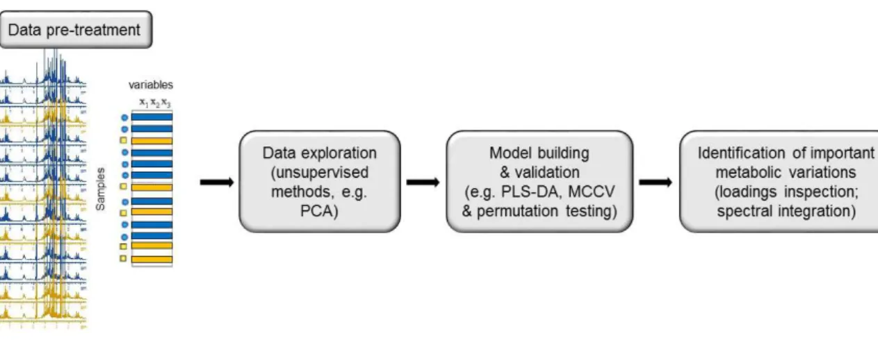
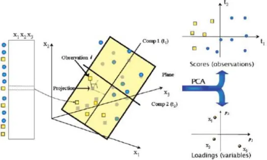
Metabolomics entails the comprehensive analysis of the inventory of endogenous small molecules (molecular mass < 1000 Da) present in a biological system as a result of intermediary cellular metabolism (Nicholson et al., 1999; Fiehn, 2002). The general aim is to detect/quantify fluctuations in this inventory, the so called metabolome, upon a given stimulus/perturbation, such as a disease, a toxicant, a pharmaceutical drug or a nanomaterial. Being modulated by the complex interplay between gene expression, enzymatic activities and fluctuations in metabolic fluxes, the metabolome closely reflects the cell’s functional status. Hence, the comprehensive description of metabolite changes, through metabolomics, has the potential to reveal unforeseen deviations from homeostasis and to identify new endpoint markers of effect. Moreover, since a metabolite is the same chemical entity irrespective of its origin, metabolic profiles of cultured cells can potentially be correlated to those of animal tissues/biofluids, thus enabling in vitro-in vivo correlations to be proposed.
The main analytical platforms used in metabolomic studies are nuclear magnetic resonance (NMR) spectroscopy and mass spectrometry (MS), the latter being usually coupled to liquid (LC) or gas chromatography (GC). These techniques enable the simultaneous detection of tens to hundreds of metabolites in complex mixtures like biofluids and tissue/cell extracts, providing a holistic approach which is clearly more powerful than the measurement of a few pre-established metabolites by classical biochemical methods (Lindon and Nicholson, 2008). Data treatment by multivariate statistics is then typically employed to deal with data complexity and search for consistent variation patterns or build classification models.
MS-based methods are generally more sensitive than NMR, enabling the detection of metabolites present at sub-nanomolar concentrations (Wishart et al., 2013). However, the wider view of the metabolome offered by MS methods does not always translate into a significant gain in biochemical information, as the resulting data may be extremely complex and difficult to interpret. On the other hand, despite inherent sensitivity limitations, high resolution NMR shows unparalleled analytical reproducibility and the ability to provide unequivocal structural and quantitative information on a wide range of metabolites, present in the micromolar-milimolar range. In this work, metabolic profiling of cultured cells has been performed through NMR metabolomics. Thus, the basic principles of NMR spectroscopy,
18
as well as of the multivariate analysis and univariate statistical methods employed, will be reviewed below.
NMR spectroscopy
The theory of NMR is thoroughly covered in the literature (Ross et al., 2007; Keeler, 2010; Zerbe and Jurt, 2014) and the aim of this subsection is not to provide an in depth account of NMR fundamentals and methods, but rather to briefly describe the basic principles underlying this technique.
NMR is a phenomenon which occurs when the nuclei of certain atoms are immersed in a static magnetic field (B0) and exposed to a second oscillating magnetic field (B1). Not
all nuclei experience this phenomenon, it depends on whether they possess a property called spin (I). Some nuclei carry a total spin (e.g. 1H, 2H, 13C, 15N, 19F, 31P) resulting in a
magnetic moment µ = γ I h / 2π (where γ is the gyromagnetic ratio of the atomic nucleus), while others, with even number of protons and neutrons (e.g. 12C, 14C, 16O), do not, thus
being undetectable by NMR spectroscopy.
When nuclear spins with I = ½ (such as protons) are exposed to an external homogenous magnetic field B0, usually defined along z, the magnetic moments align
parallel or anti-parallel with respect to B0, resulting in two spin states α and β with an energy
difference ΔE. The α and β states are filled according to the Boltzmann distribution, resulting in a slight excess of α states at equilibrium (Nα/Nβ = eΔE/kT). This excess of α states gives rise to a macroscopic magnetic moment, the so called net magnetisation M0, aligned with
the z axis.
To detect an NMR signal, M0 is flipped into the xy plane by applying a radiofrequency
(RF) magnetic field B1, orthogonal to z, for a defined time period (RF pulse). The
magnetisation M will align along the x-direction and precess with a resonance frequency
f0 = γ B0 / 2π. As soon as the RF pulse is turned off, the nuclear spins relax back to
equilibrium according to two processes: i) the recovery of the magnetization along the z axis (longitudinal relaxation characterised by the time constant T1) and ii) the loss of phase
coherence of spins precessing at different rates in the xy plane (transverse relaxation occurring with the time constant T2). Precession of magnetization in the xy plane induces
an oscillating signal in the detection coil, the so called free induction decay (FID), which is then converted into a frequency domain signal by Fourier transformation (FT) (Figure 1.6).