INSTITUTO UNIVERSITÁRIO EGAS MONIZ
MESTRADO INTEGRADO EM MEDICINA DENTÁRIA
PERIODONTAL STATUS IN PARKINSON’S DISEASE PATIENTS:
A RETROSPECTIVE STUDY
Trabalho submetido por
Patrícia Soares Lyra
para a obtenção do grau de Mestre em Medicina Dentária
INSTITUTO UNIVERSITÁRIO EGAS MONIZ
MESTRADO INTEGRADO EM MEDICINA DENTÁRIA
PERIODONTAL STATUS IN PARKINSON’S DISEASE PATIENTS:
A RETROSPECTIVE STUDY
Trabalho submetido por
Patrícia Soares Lyra
para a obtenção do grau de Mestre em Medicina Dentária
Trabalho orientado porProf. Doutor Luís Francisco Alexandrino Proença
e coorientado por
Prof. Doutora Catarina Afonso Godinho Prof. Doutor José João Baltazar Mendes
Dedicatory
To my beloved parents, Fátima and George
“Without the quest, there can be no epiphany”
Agradecimentos
Antes de mais, quero agradecer ao Prof. Doutor Luís Proença pela orientação neste projeto e pela disponibilidade demonstrada desde o primeiro dia. Levo como exemplo inegável todo o profissionalismo, rigor, experiência e conhecimento científico, pelos quais espero reger o meu percurso daqui em diante.
À Prof. Doutora Catarina Godinho, agradeço o incansável apoio e incentivo, por me ter sempre aconselhado e acompanhado com entusiasmo e por me despertar ainda mais o interesse pelo tema que desenvolvi.
Ao Prof. Doutor José João Mendes, quero agradecer a oportunidade de integrar pela primeira vez uma equipa e projeto de investigação tão desafiantes. Foi uma experiência única de crescimento pessoal e profissional que levarei para a vida. Agradeço o voto de confiança, todo o encorajamento, ajuda e carinho.
Ao Mestre João Botelho – um exemplo claro de determinação, motivação, interesse e trabalho árduo – quero dar o meu agradecimento especial. Sem a sua ajuda, nada do que idealizei desenvolver na tese de mestrado teria sido possível. Obrigada pelo interesse permanente, pelos conselhos amigos e pela orientação indispensável ao longo do último ano.
À Mestre Vanessa Machado, os meus sinceros agradecimentos por todo o apoio e conselhos prestados, que foram sem dúvida indispensáveis. O sentido de responsabilidade, foco e determinação transmitidos foram uma inspiração ao longo deste projeto.
À Doutora Josefa Domingues, investigadora especialista na Doença de Parkinson, quero agradecer todo o interesse e envolvimento demonstrados. Agradeço o seu papel na consolidação científica do projeto. Agradeço também à Associação Portuguesa de Doentes de Parkinson pela pronta disponibilidade e excelente acolhimento durante todo o trabalho de campo.
Ao Instituto Universitário Egas Moniz, a minha segunda casa nos últimos 5 anos, agradeço do fundo do coração. Aqui cresci, aprendi e consolidei a minha vocação na área da saúde. Agradeço ainda a todos os professores, funcionários e colegas que contribuíram para a minha formação e me serviram de exemplo.
passados juntas. Obrigada por me ajudares a evoluir e por me acompanhares desde o primeiro dia. Foram muitas horas de trabalho, entreajuda e amizade que nunca esquecerei. Aos meus queridos amigos – Gonçalo Santos, Miguel Alves, João Geraldes, Carolina Lopes, Carlota Simões, Carolina Tavares e, em especial, à minha querida amiga Margarida Ramos – agradeço individualmente o apoio imprescindível e todo o companheirismo. Guardo, com saudade, memórias inesquecíveis destes anos de faculdade. Agradeço ainda às minhas queridas amigas Catarina Marques, Margarida Pereira e Maria João Barbeito, sempre presentes, por quem tenho a maior admiração e o privilégio de partilhar momentos como este.
Finalmente, agradeço à minha mãe – Fátima – que é o meu maior exemplo de força e determinação; ao meu querido pai – George – com quem aprendi a valorizar o detalhe e a honrar o que me proponho a fazer; à minha irmã – Marta – que me ensina diariamente a sair da minha zona de conforto e a arriscar; e a toda a minha família, que sempre me acompanhou. Agradeço o amor incondicional e o incentivo constante para fazer mais e melhor. São os meus pilares.
Abstract
Background: People with Parkinson’s Disease (PD) may be at risk of having bad
periodontal status. A consistent periodontal examination is critical to investigate the impact on quality of life and if periodontitis in PD patients potentially causes systemic repercussions.
Aims: Our primary goal was to assess the association of periodontitis and PD, considering
patient’s self-perceived general quality of life, oral health-related quality of life (OHRQoL) and xerostomia. Secondly, we compared blood and standard biochemistry surrogates of PD patients with periodontitis with PD patients without periodontitis.
Material and Methods: Firstly, 28 individuals from the Portuguese Association of
Parkinson’s Disease Patients were consecutively enrolled, PD clinical manifestations were assessed, a full-mouth periodontal examination was performed and questionnaires on self-perceived quality of life in PD (PDQ-8), oral health impact profile (OHIP-14) and xerostomia (SXI-5) were applied. Secondly, National Health and Nutrition Examination Survey (NHANES) 2011-2012 dataset was analyzed, with PD participants being identified through specific PD reported medications, from a sample of periodontally assessed individuals, and blood levels and standard chemical laboratory profiles were compared according to the presence of periodontitis.
Results: The prevalence of periodontitis in the Portuguese sample was 75.0% with 46.4%
of severe cases. Upper extremities rigidity and hands postural and kinetic tremors were significantly correlated with worse periodontal status. PDQ-8 showed correlation with self-perceived OHRQoL and xerostomia levels. In the American sample, we found an association of periodontitis in PD patients with increased White Blood Cells (WBC), segmented neutrophils and basophils and lower Total Bilirubin levels. Furthermore, WBC, segmented neutrophils, Vitamin D2 and gender presented potential predictive value to infer periodontitis in PD individuals.
Conclusions: People with PD may have high prevalence of periodontitis. Deteriorated
levels of the upper extremities in advanced PD stages may influence the periodontal status and hygiene habits. Quality of life in PD appears to be associated with self-perceived OHRQoL and xerostomia. Periodontitis may cause systemic changes with predictive
value in PD patients. Future studies should further assess periodontitis impact on the quality of life of PD patients and its potential systemic inflammatory burden.
Keywords: Parkinson’s Disease; Periodontitis; Periodontal Disease; Quality of Life;
Resumo
Contexto: Indivíduos com doença de Parkinson (PD) podem estar em risco de apresentar
um mau estado periodontal. Uma avaliação periodontal consistente é fundamental para investigar o impacto na qualidade de vida dos pacientes, e se a periodontite em pacientes com PD desencadeia potenciais repercussões sistémicas.
Objetivos: O nosso principal objetivo foi a avaliação da associação entre a periodontite
e a PD, considerando a autopercepção da qualidade de vida global do paciente, da qualidade de vida relacionada com a saúde oral (OHRQoL) e de xerostomia. Secundariamente, comparámos marcadores sanguíneos e bioquímicos padrão de pacientes com PD com e sem periodontite.
Materiais e Métodos: Primeiramente, após a inclusão consecutiva de 28 indivíduos da
Associação Portuguesa de Doentes de Parkinson, avaliaram-se as manifestações clínicas da PD, o estado periodontal e questionários de autopercepção da qualidade de vida na PD (PDQ-8), do perfil de impacto da saúde oral (OHIP-14) e de xerostomia (SXI-5). Posteriormente, mediante análise de dados do National Health and Nutrition Examination Survey (NHANES) 2011-2012, de uma amostra de indivíduos avaliados periodontalmente, selecionaram-se doentes de Parkinson através de medicação específica relatada na PD, e compararam-se níveis sanguíneos e perfis bioquímicos padrão de acordo com a presença ou não de periodontite.
Resultados: A prevalência de periodontite na amostra portuguesa foi de 75,0%, com
46,4% de casos graves. Rigidez das extremidades superiores e tremores posturais e cinéticos das mãos foram significativamente correlacionados com um pior estado periodontal. O PDQ-8 mostrou correlação entre os níveis de autopercepção de OHRQoL e xerostomia. Na amostra Americana, encontrámos uma associação de periodontite com o aumento de glóbulos brancos, neutrófilos segmentados e basófilos, bem como níveis mais baixos de bilirrubina total, em pacientes com PD. Adicionalmente, fatores como a contagem de glóbulos brancos, neutrófilos segmentados, vitamina D2 e género apresentaram potencial valor preditivo para inferir a presença de periodontite em indivíduos com PD.
Conclusões: Indivíduos com PD apresentaram uma elevada prevalência de periodontite.
A maior incapacidade motora dos membros superiores em fases avançadas de PD podem influenciar o estado periodontal e os hábitos de higiene. A qualidade de vida na PD parece estar associada com a autopercepção de OHRQoL e xerostomia. A periodontite pode causar alterações sistémicas com valor preditivo em pacientes com PD. Estudos futuros devem explorar o impacto da periodontite na qualidade de vida dos pacientes com PD e na potencial carga inflamatória, a nível sistémico.
Palavras-chave: Doença de Parkinson; Periodontite; Doença Periodontal; Qualidade de
Index
I. INTRODUCTION ... 11
1. PARKINSON’SDISEASE ... 11
1.1. Clinical Manifestations ... 11
1.2. Diagnosis ... 12
1.3. PD Subtypes, Onset and Progression ... 14
1.4. Causal and Risk Factors ... 14
1.5. Etiopathogenic Mechanisms ... 17
1.6. Therapeutic Approaches ... 21
2. PERIODONTITIS ... 23
2.1. Clinical Manifestations ... 23
2.2. Onset and Progression ... 26
2.3. Causal and Risk Factors ... 27
2.4. Etiopathogenic Mechanisms ... 28
2.5. Periodontitis in the Portuguese Context ... 31
2.6. Periodontitis in the United States of America Context ... 32
3. PERIODONTITISANDPARKINSON’SDISEASEINTERPLAY ... 33
4. AIMS ... 34
4.1. Parkinson’s Disease, Periodontitis and Patient-Related Outcomes: A Cross-Sectional Study .. 34
4.2. Relationship between Blood and Standard Biochemistry Levels with Periodontitis in Parkinson’s Disease Patients: Data from the NHANES 2011–2012 ... 35
II. PARKINSON’S DISEASE, PERIODONTITIS AND PATIENT-RELATED OUTCOMES: A CROSS-SECTIONAL STUDY ... 37
III. RELATIONSHIP BETWEEN BLOOD AND STANDARD BIOCHEMISTRY LEVELS WITH PERIODONTITIS IN PARKINSON’S DISEASE PATIENTS: DATA FROM THE NHANES 2011–2012 ... 49
IV. GENERAL DISCUSSION ... 61
VI. FUTURE PERSPECTIVES ... 69 VII. REFERENCES ... 71
Figure Index
Figure 1 | Schematic representation of PD clinical development. ... 13
Figure 2 | Association between PD risk factors and pre-motor/prodromal signs with the decreased/increased risk of disease development and diagnosis. ... 17
Figure 3 | Representation of the nigrostriatal pathway and other dopaminergic pathways (yellow) in the brain. ... 18
Figure 4 | Loss of dopaminergic neurons in PD. ... 19
Figure 5 | Etiopathogenic intracellular mechanisms responsible for dopaminergic neuronal death in PD. ... 21
Figure 6 | Local effects of periodontitis. ... 25
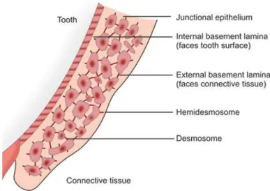
Figure 7 | Junctional epithelium permeability. ... 29
Figure 8 | Etiopathogenesis of periodontitis. ... 30
Figure 9 | Geographic distribution of the estimated prevalence of severe periodontitis in US adults. ... 32
Abbreviation Index 25OHD2 | 25-Hydroxy D2
BOP | Bleeding on Probing
CD14 | Cluster of Differentiation 14 CNS | Central Nervous System CT | Computed Tomography DJ-1 | Deglycase 1
DBS | Deep Brain Stimulation EDS | Excessive Daytime Sleepiness
eIF4G1 | Eukaryotic Translation Initiation Factor 4 G1 FCGR2A | Fc Fragment of IgG Receptor IIa
GBA | Glucocerebrosidase H&Y | Hoehn & Yahr IL1B | Interleukin 1 Beta IL6 | Interleukin 6
LRRK2 | Leucine-Rich Repeat Kinase 2 MCI | Mild Cognitive Impairment
MDS-UPDRS | Movement Disorder Society – Unified Parkinson’s Disease Rating
Scale
NHANES | National Health and Nutrition Examination Survey OHIP-14 | Oral Health Impact Profile
OHRQoL | Oral Health Related Quality of Life PARK2 | Parkin 2
PD | Parkinson’s Disease
PDQ-8 | Parkinson’s Disease Questionnaire PINK-1 | PTEN-induced Kinase 1
PET | Positron Emission Tomography PFC | Prefrontal Cortex
PIGD | Postural Imbalance Gait Disorder PMNs | Polymorphonuclear Neutrophils RBD | REM Sleep Behavior Disorder REM | Rapid Eye Movement
SNc | Substantia Nigra Pars Compacta SNCA | !-synuclein Encoding Gene
SXI-5 | Shortening the Xerostomia Inventory TNF | Tumoral Necrosis Factor
US | United States
WNT5A153 | Wnt Family Member 4A 153 WBC | White Blood Cells
Introduction
I. INTRODUCTION
1. PARKINSON’S DISEASE
Parkinson’s Disease (PD) is a chronic and progressive neurodegenerative process, clinically characterized by a variety of motor and nonmotor features which heavily affect patients’ quality of life (Obeso et al., 2010).
After Alzheimer’s disease, PD is one of the most frequent neurodegenerative conditions that mostly impacts the central nervous system (CNS) (de Lau & Breteler, 2006; Nussbaum & Ellis, 2003).
PD occurrence increases with age, being rare under 50, affecting 1% of individuals over 60 and reaching up to 4% in higher age groups (Tysnes & Storstein, 2017). Therefore, in unselected populations, at any time, PD prevalence varies from 0.1% up to 0.2% (Tysnes & Storstein, 2017). Also, about 0.24% of the portuguese population over 50 years of age has PD (Jesus-Ribeiro et al., 2017). An estimated total of 10 million individuals are suffering from PD today (Ball et al., 2019).
In a globally aged population, PD occurrence seems to be noticeably increasing, and is expected to duplicate in the next couple decades (Johnson et al., 2019; Dorsey et al., 2018). PD has an annual incidence of roughly 15 new cases in a 100 000 universe (it varies from less than 10 to more than 20 – this variation can be the result of PD under-diagnosing) (Tysnes & Storstein, 2017).
Also, PD tends to affect more men than women, since the risk of developing the disease is 1.5 to 2 times higher in the male gender (Alves et al., 2009; Ball et al., 2019; Ferreira et al., 2017; Schrag et al., 2000).
1.1. Clinical Manifestations
PD is considered a heterogeneous disorder due to the broad range of motor and non-motor features (Foltynie et al., 2002).
The classic motor manifestations are resting tremor, muscular rigidity and bradykinesia (lost capability to command movement, which becomes slow), all often targeted by
available levodopa-therapies (Nussbaum & Ellis, 2003; Obeso et al., 2010). An asymmetric onset and resting tremors are typical clinical signs of the sporadic form of this disease (Tysnes & Storstein, 2017).
Other motor and non-motor manifestations – such as loss of balance, gait dysfunction, swallowing and speech impairment, autonomic disturbances and cognitive decline (including hallucinations and delirium) – may be complications of the administration of dopaminergic drugs like levodopa, and end up being responsible for the disability in PD (Figure 1) (Obeso et al., 2010).
These motor and nonmotor features can eventually develop dementia in 30 to 80% of cases, which stands as the ultimate causal factor of disability (Obeso et al., 2010). Therefore, PD progression and treatment may interfere with daily-life activities and ultimately deteriorate patient’s overall quality of life (Kalia & Lang, 2015; Poewe et al., 2017).
1.2. Diagnosis
A positive diagnosis of PD is delivered when gradual onset of motor symptoms takes place, especially its three cardinal signs - resting tremor, rigidity and bradykinesia (Figure 1) (Obeso et al., 2010).
To date, PD clinical diagnosis is merely speculative, since an easy and reliable testing process is yet to be developed (Litvan et al., 2003; Nussbaum & Ellis, 2003). The use of single-photon-emission CT or PET is only useful in the diagnosis of isolated PD patients in specialized settings (de Lau & Breteler, 2006). Therefore, a definitive diagnosis only
Introduction
important factors to corroborate PD differential diagnosis (de Lau & Breteler, 2006). An accurate PD clinical diagnosis is less effective at an early stage, and implicates several long-term follow-up reavaluations in the course of the disease (de Lau & Breteler, 2006). However, several lines of evidence show that there might be a pre-motor phase of PD, usually beginning up to 20 to 30 years prior to diagnosis, with non-motor prodromal signs such as hyposmia (olfactory dysfunction), sleep abnormalities (REM-sleep behavior disorder), cardiac sympathetic denervation, constipation, depression and pain (Figure 1) (Heintz-Buschart et al., 2018; Obeso et al., 2010; Tysnes & Storstein, 2017). Evidence shows that constipated individuals have 2.7 times more risk of developing PD (Figure 1) (Tysnes & Storstein, 2017). Patients with the presence of this wide range of signs, still without any PD motor impairs, might be considered at high-risk of developing PD, and those non-motor prodomal signs might even stand as early biomarkers to PD diagnosis (Nair et al., 2018; Obeso et al., 2010).
Figure 1 | Schematic representation of PD clinical development.
Prior to PD diagnosis (at 0 year’s time mark), which usually occurs upon the establishment of the three cardinal motor symptoms (bradykinesia, rigidity and tremor), there might be a 20 plus year timespan of pre-motor/prodromal signs. Subsequently, further into PD establishment, other non-motor symptoms usually settle alongside the complications of dopaminergic therapy (such as fluctuations, dyskinesia and psychosis), and disability might emerge in advanced PD cases. RBD - REM Sleep Behavior Disorder; EDS - Excessive Daytime Sleepiness; MCI - Mild Cognitive Impairment (Adapted from Kalia & Lang (2015), by permission of corresponding author).
1.3. PD Subtypes, Onset and Progression
Despite being a heterogeneous condition, to this date, PD is known to have two major clinical subtypes based on the onset age, the predominance of clinical features and the rate of progression (Foltynie et al., 2002; Obeso et al., 2010):
● On the one hand, the early-onset form, mostly affecting young people (20 to 40 years of age), is usually a tremor-predominant form (Kalia & Lang, 2015); ● On the other hand, the late-onset form, often observed in the elderly (mostly over
60 to 70 years of age), is typically a non-tremor-predominant form also identified as Postural Imbalance Gait Disorder (PIGD), and akinesia (deterioration of voluntary movement), rigidity and gait and balance impairment are its typical signs (Thenganatt & Jankovic, 2014).
Usually the late-onset form is the most common one, since PD often starts to develop in patients around 65 to 70 years of age and an onset prior to the 4th decade of life happens in less than 5% of cases (Tysnes & Storstein, 2017).
Onset age is considered to be the best predictor for the rate of progression in PD: young patients subtype tend to have a slow decline in motor function, and elderly patients tend to present a quicker long-term progression (Thenganatt & Jankovic, 2014). Also, cognitive impairment tends to start earlier and is more common to appear in patients who are older at the start of clinical manifestations (Obeso et al., 2010).
Introduction
Usually, non-genetic (environmental and idiopathic) factors play a key role in PD, as they are mostly responsible for 90% of PD cases (which are the sporadic late-onset forms of the disease), while genetic factors end up being responsible for the minority of cases (typically in the familial early-onset subtypes) (Tysnes & Storstein, 2017). Nonetheless, the non-genetic factors involved in the etiology of sporadic PD seem to synergically relate to existing susceptibility genes (de Lau & Breteler, 2006; Kalia & Lang, 2015). Further, 5 to 10% of PD total population presents some kind of genetic influence and that the immediate family of a PD patients have a doubled or tripled risk of developing PD (Nussbaum & Ellis, 2003). Since the heterozygous state of PD is considerably more common than the homozygous state, PD is classically a hereditary autosomal dominant neurodegenerative disorder (Nussbaum & Ellis, 2003).
The risk of developing PD - which is particularly aggravated in men - has not only been linked to the generalized ageing of the world’s population, but also to several environmental risk factors (Ball et al., 2019; Tysnes & Storstein, 2017).
The occupational exposure to environmental toxins like pesticides and herbicides involved in farming and rural living, as well as to heavy metals in the welding industry (iron, copper, aluminium, zinc, etc.) have been hypothesised to increase the risk of developing PD, although further research is needed (Ball et al., 2019; Jankovic, 2005; Petrovitch et al., 2002; Priyadarshi et al., 2000). As was aforementioned, exposure to heavy metals increases oxidative stress due to free radical formation and heavy metal aggregates accumulation in the SNc (Substantia Nigra Pars Compacta) (de Lau & Breteler, 2006). Moreover, dietary habits like the high intake of satturated fatty acids, non-heme iron and dairy products (especially milk) have also been considered a risk factor in PD (Boulos et al., 2019).
Furthermore, inflammation seems to have a role in PD pathogenesis, since increased cytokine levels have been detected in the CNS of PD patients (brain and cerebrospinal fluid), as well as activated microglia in post-mortem evaluations (de Lau & Breteler, 2006). Actually, infectious and inflammatory agents in systemic circulation have been hypothesized to reach the brain, causing the activation of primed microglial cells (major innate immune-system cells in the CNS), which can initiate the neurodegenerative cascade found in neurodegenerative diseases such as PD, ultimately leading to the necrosis of dopaminergic neurons (Hashioka et al., 2019; Kaur et al., 2016). For instance,
microbial agents from the nasal cavity and the gastrointestinal tract (and their low-grade inflammatory states) are thought to instigate !-synuclein pathology both in the olfatary bulb and intestines, ultimately reaching the CNS and triggering neurodegeneration in PD (Heintz-Buschart et al., 2018; Nair et al., 2018). Enteric !-synuclein pathology usually causes the gastrointestinal dysfunctions that are typicall prodromal signs of PD (Nair et al., 2018). Therefore, the likelihood of PD development is presumably increased in scenarios where neuroinflammation or neurodegeneration are present, namely secondary inflammatory responses of chronic and infectious conditions (Chen et al., 2017; Hashioka et al., 2019).
Remarkably, higher urate levels, the consumption of tobacco (nicotine), coffee (caffeine), tea, beer, unsaturated fatty acids (arachidonic acid, omega 3 and ! −linolenic acid) and non-steroidal anti-inflammatory drugs may reduce the risk of developing PD due to their neuroprotective effects (Figure 2) (Asanuma et al., 2001; Boulos et al., 2019; Hernán et al., 2002; Noyce et al., 2016; Obeso et al., 2010; Quik, 2004; Youdim et al., 2000). Also, research has shown that not only estrogens, but also the intake of vitamin E, both associate with a lower risk of developing PD, since both show antioxidant properties that inhibit the pathogenic mechanism of oxidative stress in PD (de Rijk, 1997; Saunders-Pullman, 2003). Additionally, physically active individuals seem to present a lower chance of developing PD, since physical exercise is thought to be neuroprotective (Chen et al., 2005).
Ultimately, further studies evolving these risk factors will probably lead to more understanding on PD etiology (Obeso et al., 2010).
Introduction
Figure 2 | Association between PD risk factors and pre-motor/prodromal signs with the decreased/increased risk of disease development and diagnosis.
PD presents several idiopathic, environmental (orange) and genetic (red) risk factors, all of which synergically interact amongst each other, either to potentiate or diminish the risk of PD development. Specific PD pre-motor/prodromal signs (green) also mark higher/lesser risk of PD establishment. GBA - Glucocerebrosidase gene; LRRK2 - Leucine-Rich Repeat Kinase gene; PARK2 - Parkin 2 gene; PINK-1 - PTEN-induced kinase 1 gene; DJ-1 - Deglycase 1 gene (Adapted from Noyce et al. (2016), by permission of corresponding author).
1.5. Etiopathogenic Mechanisms
Neurodegeneration in PD is characterized by two major hallmarks: 1) the death of dopaminergic neurons in the SNc (Figure 4) and other brain sights; and 2) also the presence of Lewy bodies (abnormal ubiquinated protein deposits in the cytoplasm) and Lewy neurites (thread-like ubiquinated protein inclusions within the axonal processes) in the remaining neurons, in which !-synuclein is the main proteic component (Eriksen et al., 2003; Lebouvier et al., 2010; Nussbaum & Ellis, 2003). !-synuclein is a short presynaptic protein of unidentified function, tangled in the molecular cascade of events that culminate in some neurodegenerative disorders, including PD (Nussbaum & Ellis, 2003).
The main established etiopathogenic mechanism of PD is the selective neurodegeneration of the nigrostriatal pathway, which is a bilateral dopamine pathway (originating in the SNc and emerging in the dorsal striatum), composed by dopaminergic neurons (Figure 3) (Obeso et al., 2010). The latter secrete dopamine and synapse with GABAergic medium
spiny projection neurons, involved in the production of movement (Figure 3) (Poewe et al., 2017). Being a neurotransmitter, dopamine functions through a signalling transmission system between neurons in the CNS (Iversen & Iversen, 2007).
Therefore, the death of these dopaminergic nigrostriatal neurons in the SNc results in the marked decrease of striatal dopamine concentration, inhibiting GABAergic projections, causing the depletion of voluntary movements and ultimately being the key to motor manifestations in PD (Barone, 2010; Obeso et al., 2010; Tysnes & Storstein, 2017).
Figure 3 | Representation of the nigrostriatal pathway and other dopaminergic pathways (yellow) in the brain.
The nigrostriatal pathway originates in the substantia nigra pars compacta (SNc) and emerges into the dorsal striatum (composed of caudate nucleus and putamen) of the midbrain. The dopaminergic neurons of this pathway secrete dopamine and synapse with GABAergic medium spiny projection neurons, involved in the production of movement. PFC - Prefrontal cortex (Adapted from Telzer (2016) ©, used under the CC
Introduction
Figure 4 | Loss of dopaminergic neurons in PD.
This image schematically shows the neurodegeneration’s effects on the neural projections from the substantia nigra pars compacta (SNc) to the dorsal striatum (composed of caudate nucleus and putamen) upon PD installation (Adapted from Broski et al. (2014), by permission of Mayo Foundation for Medical Education and Research).
As aforementioned, the homeostatic balance in the SNc is susceptible to the influence of genetic, cellular and environmental factors. Although not yet fully understood, these factors (isolated or associated) may overtime trigger the intracellular mechanisms that ultimately result in neuronal death, which are as follows (Figure 5) (de Lau & Breteler, 2006; Nussbaum & Ellis, 2003; Obeso et al., 2010):
● Mitochondrial dysfunction, consequent oxidative stress and intracellular toxicity; ● Abnormal protein degradation through proteosomal and lysosomal dysfunction
and the compromised ubiquitination process (ubiquitin-mediated metabolism); ● Abnormal aggregation of !-synuclein, which forms lewy bodies and lewy neurites
and ultimately causes intracellular toxicity (most common).
Gene mutations also play a role in PD pathogenesis, although the means by which they cause SNc cell death and Lewy body aggregations remains unknown. Some of the mutated loci result in inherited autosomal dominant PD (with only one mutant allele) and others in autosomal recessive PD (if both alleles of the mutated gene are altered) (Nussbaum & Ellis, 2003).
For instance, gene mutations in SNCA (!-synuclein encoding gene), LRRK2 (Leucine rich repeat kinase 2 gene) and eIF4G1 genes are responsible for about 2-3% of the late-onset, autossomal dominant forms of PD. Likewise, mutations in GBA, PRKN, PINK-1 and DJ-1 genes associate with 50% of the early-onset, autossomal recessive forms (Figure 2) (Obeso et al., 2010). Actually, the LRRK2-G2019S mutation is thought to be the most frequent PD causal factor, both in familial and sporadic forms of PD (Ferreira et al., 2017).
Besides gene mutations, excitotoxicity (neural receptors overactivations by excitatory neurotransmitters) and inflammation may also be involved in this progressive neural degeneration (Obeso et al., 2010).
Additionally, with PD progression, !-synuclein aggregation into lewy bodies becomes more widespread, extending to other brain sites (Tysnes & Storstein, 2017). Also, other affected neurotransmission systems (involving glutamate, acetylcholine and #-aminobutyric acid (GABA)) ultimately translate into PD clinical manifestations (Barone, 2010).
Introduction
Figure 5 | Etiopathogenic intracellular mechanisms responsible for dopaminergic neuronal death in PD.
Mitochondrial dysfunction (causing oxidative stress and intracellular toxicity), lysosomal (not represented) and proteosomal dysfunction (affecting the ubiquitin-mediated metabolism and originating abnormal protein degradation), abnormal !-synuclein aggregation (forming lewy bodies which cause intracellular toxicity) and further intertwined gene mutations are here schematically represented, as they are supposed etiopathogenic intracellular mechanisms responsible for neuronal death in PD (Adapted by permission from Springer Nature Customer Service Centre GmbH: Nature Springer, Nature Medicine, Missing pieces in the Parkinson’s disease puzzle, Obeso et al., © (2010)).
The degeneration of nondopaminergic transmitter systems is now known to partially cause the nonmotor features in PD, and it may present similar cellular mechanisms in its origin: it not only relates with the !-synuclein gene copy number, but also with gene and protein overexpression, though the latter seem to be especially toxic to dopaminergic neurons (Obeso et al., 2010).
1.6. Therapeutic Approaches
There are pharmacological and non-pharmacological treatments for PD (Lang & Lozano, 1998).
Levodopa has been the primary pharmacological treatment, while other dopaminergic drugs like dopamine agonists and atypical neuroleptics (antipsychotics) have been later introduced in treatment plans (Obeso et al., 2010).
Although useful in stabilizing the progression of motor symptoms, the chronic use of dopaminergic drugs like levodopa has the tendency to become less efficient overtime, as well as to increase disability and lead up to complications such as motor and non-motor fluctuations, dyskinesias and behavioral changes (Kalia & Lang, 2015). Hence, levodopa treatment may ultimately be responsible for shifting motor to nonmotor dysfunction in PD (Obeso et al., 2010).
Also, deep brain stimulation (DBS) is therapeutically used when patients present unmanageable levodopa related motor complications, and functional neurosurgery is an option in severe disabling PD cases (Poewe et al., 2017).
The focus of these standard treatments is in attenuating symptoms to hopefully improve PD patients quality of life and longevity, although tending to be a universal solution to a non-uniform disease (Johnson et al., 2019; Kalia & Lang, 2015; Schapira, 2009). Lately, ongoing research has been directed in the development of individualized disease-modifying therapies that target the etiopathogenic mechanisms responsible for the core neurodegenerative process, and aim to prevent or slow down PD installation and progression, as well as the consequent non-motor complications (Kalia & Lang, 2015; Schapira, 2009). For instance, whilst its causes and consequences are not fully comprehended, the aggregation of !-synuclein and its increased neuronal levels seem to be a primary factor to PD. Therefore, a potential therapeutic approach may involve the depletion of α-synuclein expression (Obeso et al., 2010).
Introduction
2. PERIODONTITIS
Periodontitis is a chronic, infectious and inflammatory disease of the periodontium, where a dysbiotic microflora triggers an immune response in the supporting structures of the teeth (Hajishengallis, 2015). Clinically, periodontitis is characterized by chronically inflamed gingivae and alveolar bone destruction (Darveau, 2010).
Periodontitis is part of the periodontal disease family, alongside other conditions, such as gingivitis, necrotising periodontal disorders, periodontal manifestations of systemic conditions, periodontal abscesses and endo-perio lesions (Armitage, 1999; Caton et al., 2018).
From an epidemiologic standpoint, periodontitis is one of the most prevalent inflammatory diseases in the adult population (with its severe form ranked as the 6th most prevalent disease worldwide in 2010), affecting up to 50% of the world’s population alongside other periodontal diseases (Kassebaum et al., 2014; Nazir, 2017; Tonetti et al., 2017).
Moreover, periodontitis slightly affects more men than women, with both its prevalence and severity being age-related (Ebersole et al., 2016; Eke et al., 2016).
2.1. Clinical Manifestations
In a healthy periodontium, the oral epithelium connects itself to tooth surfaces through a specialized junctional epithelium (Figure 6) (Nibali, 2018). The slight space resulting from such union forms the gingival sulcus, which is normally populated with a polymicrobial niche and filled by gingival crevicular fluid (a serous exudate containing pro-inflammatory cytokines, enzymes leukocytes, oral bacteria, etc.) (Donos, 2018; Nibali, 2018). Underneath, the alveolar bone and surrounding connective tissue completely brace tooth roots (Figure 6) (Darveau, 2010).
The daily accumulation of dental plaque in the oral cavity is inevitable, in which aggregates of diverse microbial species from the saliva or jugal mucosa attach onto coronal and radicular tooth surfaces, and ultimately participate in the homeostatic battle held with the host’s immune system (Marsh, 2006; Socransky et al., 1977). The dental
plaque is an organized biofilm that mimics tissues, not only through its systematized architecture, but also through its codependent and metabolically integrated polymicrobial community function (Marsh, 2004; Marsh, 2006). It lodges a characteristic channel system and a quorum sensing mechanism: a signalling system between species that allows a rapid density-dependent and gene regulated adaptation of the biofilm when exposed to environmental stress (Marsh, 2004).
Whereas upon periodontal disease installation, some bacterial species in the subgingival dental plaque manage to invade the ulcerated epithelium, to breakdown the periodontal ligament fibers and to destroy the underlying connective tissue, creating the so-called periodontal pocket – a crevice of destruction (Figure 6) (Donos, 2018; Nibali, 2018). A typical sign of periodontitis is bleeding on probing (BOP), which is currently accepted as a proxy of tissue inflammation and consistent with bad oral hygiene habits (Tonetti et al., 2018).
Eventually, periodontitis exacerbation might progress into permanent alveolar bone loss (which stands as the major disease hallmark), causing consequent gum recession, root furcation involvement, tooth mobility or, eventually, tooth loss (Figure 6) (Darveau, 2010; Haffajee & Socransky, 1986; Newman et al., 2018).
Introduction
Figure 6 | Local effects of periodontitis.
On the left side a healthy periodontal insertion is schematized, where a specialized junctional epithelium properly connects the gingival sulcus epithelium to tooth surfaces and the alveolar bone is fully supporting tooth roots. Whereas, on the right side, the local effects of periodontitis are shown. Microbial species in the subgingival dental plaque destructively detach intact sulcus epithelium, breakdown the periodontal ligament fibers and destroy the underlying connective tissue and alveolar bone (Adapted by permission from Springer Nature Customer Service Centre GmbH: Nature Springer, Nature Reviews Microbiology, Periodontitis: a polymicrobial disruption of host homeostasis, Darveau, R. P., © (2010)).
The involvement of the hole dentition rarely occurs in periodontitis, since periodontal damage is usually site-specific and therefore constricted to certain teeth or tooth sites in the oral cavity (Haffajee & Socransky, 1986; Slots, 2017).
The burden of periodontitis may result in severe consequences to the patients, such as masticatory dysfunction and impaired patient’s oral health-related quality of life (OHRQoL) (Buset et al., 2016), which can be restored after successful periodontal treatment (Botelho et al., 2020) However, self-perception of periodontitis is often misunderstood by patients (Machado et al., 2019) and has repercussions in the prediction of periodontal therapy adherence (Machado et al., 2020), hence the education of patients towards periodontal health is key to obtaining good clinical control of this disease.
Apart from its local effects as a peripheral inflammatory process of the oral cavity, periodontitis can also instigate slight systemic inflammation, which end up setting off or aggravating other chronic systemic inflammatory diseases, for instance cardiovascular diseases such as atherosclerosis and infective endocarditis (Muñoz-Aguilera et al., 2020; Sanz et al., 2020), diabetes mellitus and obesity (Preshaw et al., 2012; Winning & Linden, 2017), renal diseases, adverse pregnancy outcomes and ultimately cancer (Hajishengallis, 2015; Michaud et al., 2017). Additionally, periodontitis has been consistently associated with solid organ transplants and stress (Botelho et al., 2020; Machado et al., 2020), and very recently with polycystic ovary syndrome (Machado et al., 2020).
Recent evidence raised the possibility of periodontitis inducing neuroinflammation through the activation of microglia (CNS immune cells) (Hashioka et al., 2019). Hence, activated microglia in the brain are thought to be a common feature in the neuropsychiatric pathologies spectrum, which include both psychiatric disorders, such as Schizophrenia and Major Depression, and neurodegenerative disorders, such as Alzheimer’s Disease (Dominy et al., 2019), Multiple Sclerosis, Amyotrophic Lateral Sclerosis and PD (Hashioka et al., 2019).
2.2. Onset and Progression
While presenting a high prevalence within all age groups, the risk of developing periodontitis exponentially increases with age (Flemmig, 1999). Apart from the immunological impairments associated with aging, the higher risk of periodontitis in the elderly has also strongly to do with long-term exposure to certain environmental risk factors (Borrell & Papapanou, 2005; Ebersole et al., 2016; Neely et al., 2001).
Introduction
The microbiology, immunology and clinical features of each compromised periodontal site in this transient condition is dependent on individual factors (Kamma et al., 2001). Notwithstanding, a persistent and unresolved status of periodontitis endures a persistent subgingival infection and associated inflammation, which can be clinically compatible with gingivitis (Slots, 2017).
2.3. Causal and Risk Factors
Periodontitis onset and progression are thought to be triggered by inadequate oral hygiene behaviors or motor hygiene impairments, which inevitably become more prevalent with age (Ebersole et al., 2016; Eke et al., 2016). Nevertheless, although being necessary factors, bad hygiene habits and pathogenic biofilm build-up seem insufficient to solely instigate periodontitis (Hajishengallis, 2015; Kornman, 2008; Meyle & Chapple, 2015; Newman et al., 2018).
Accordingly, a multitude of etiological factors and risk factors (modifiable or non-modifiable) make periodontitis a complex condition (Stabholz et al., 2010).
The association of dental plaque build-up, specific destructive dysbiosis and the host’s immune response may collectively be considered periodontitis’ etiological factors (Slots, 2017). That is to say, neither microbial dysbiosis or biofilm formation in the oral cavity will solely cause periodontitis in prone hosts (Hajishengallis, 2015).
Some examples of patient-specific risk factors, which play a role in the asseberbation of the host’s immune response, are as follow (Borrell & Papapanou, 2005; Hajishengallis, 2015; Laine et al., 2012; Meyle & Chapple, 2015; Slots, 2010; Stabholz et al., 2010):
● Genetic predisposition (main responsible for the host’s susceptibility); ● Age and gender;
● and Environmental factors, such as:
○ Systemic health status (uncontrolled diabetes mellitus, obesity or immunosuppression followed by an herpesvirus outbreak or HIV infection);
○ Medication; ○ Stress; ○ Diet;
○ Nocive behaviours (smoking, drinking alcohol and bad hygiene habits); ○ and Socioeconomic status and educational levels.
Additionally, retentive local anatomy and restorative treatments with overflowing margins exemplify site-specific risk factors (Meyle & Chapple, 2015).
Regarding the genetic influence in periodontal diseases, periodontitis majorly stands as a polygenic condition, meaning that multiple genes determine individual risk of disease settlement (for example, IL1B, IL6, TNF, CD14, FCGR2A and WNT5A153 genes) (Laine et al., 2012). On the other hand, there are also reports of a monogenetic form of periodontitis, usually associated with young aggressive cases (Hajishengallis, 2015).
2.4. Etiopathogenic Mechanisms
The periodontium is closely related to a polymicrobial community present in the oral cavity, which exceed 800 different bacterial species (Nazir, 2017).
Knowing that junctional epithelial cells are merely attached through some desmosomes and gap junctions, forming a rather porous barrier with major intercellular spaces, periodontium defense machinery mostly relies on well organized immune defence mediators to fight the perpetual stimuli produced by microorganisms of the dental plaque (Darveau, 2010).
Introduction
Figure 7 | Junctional epithelium permeability.
Junctional epithelial cells are connected through a few scattered desmosomes, forming large intercellular spaces between them (Adapted from Bathla, 2011, by permission of the publisher).
The permeable junctional epithelium allows for the constant flow of non-resident polymorphonuclear neutrophils (PMNs) and other immune cells (summoned by previously secreted proinflammatory mediators), which end up forming a barrier between the periodontium and the dental plaque biofilm (Darveau, 2010). Consequently, subgingival bacteria in healthy periodontal tissues trigger a chronic low-grade controlled defense response from the host’s innate immune system, preventing further pathogenic colonization and disease installation (Newman et al., 2018).
On other hand, periodontitis oftentimes is the development of an established reversible gingivitis condition, through dysbiotic mechanisms (Hajishengallis, 2014). Hence, the consistent accumulation of non-disrupted supragingival dental plaque begins to favor commensal quorum-sensing bacteria (like Fusobacterium nucleatum) (Meyle & Chapple, 2015). The latter release chemical signalling and ultimately develop an inflammatory process, while also altering available nutrients (like heme) (Meyle & Chapple, 2015). Such process overtime potentiates a switch in the normal oral microflora, lessening commensal bacteria and/or asseberbating periodontopathic bacterial colonization that reaches subgingival tissues, or even through diminishing the host’s defensive mechanisms (Hajishengallis, 2015; Newman et al., 2018).
Therefore, upon installation of a pathologic state, a transition from a previous majority of gram-positive bacteria to gram-negative bacteria occurs, who stand as major periodontopathic agents (Darveau, 2010).
Traditionally, red-complex bacteria (Porphyromonas gingivalis, Treponema denticola and Tannerella forsythia) were thought to specifically be key pieces to periodontal pathogenesis, negatively modulating the host’s innate immune response (Hajishengallis, 2015). However a change in paradigm now helds polymicrobial synergy and dysbiosis accountable for the debilitation of the host’s innate immune response, the consequent imbalance of destructive proinflammatory cytokines exaggeratedly allocated to the region and the consequent irreversible destruction of periodontal soft and mineralized tissues, which end up being a paradoxical side effect to the defense response itself (Darveau, 2010; Hajishengallis, 2014; Newman et al., 2018).
Figure 8 | Etiopathogenesis of periodontitis.
Introduction
2.5. Periodontitis in the Portuguese Context
The oral health status of the adult Portuguese population has been reportedly weak, regardless of the existing regional and socioeconomic discrepancies, and especially when compared to other European counterparts (Marques et al., 2000; Melo et al., 2017). Recent data showed that the estimated prevalence of periodontitis in the adult Portuguese population of the South Lisbon Metropolitan Area stands at 59.9%, although known risk factors such as age, socioeconomic status, educational levels, smoking habits and systemic health issues like Diabetes Mellitus seemed to be heavily present in the evaluated sample (Botelho & Machado et al., 2019). The majority of the confirmed periodontitis cases presented moderate to severe disease forms (Botelho & Machado et al., 2019) With that reported, the authors find that an updated and thorough investigation at a national level is necessary, following strict full-mouth periodontal examination protocols and the new American Association of Periodontology (AAP)/European Federation of Periodontology (EFP) case definition of periodontal diseases, to find the real prevalence of periodontitis in Portugal (Botelho & Machado et al., 2019).
In addition, Machado et al. (2018) reported that severe cases of generalized periodontitis seem to be more prevalent in the male gender. Also, approximately 68% of adults in Portugal show some level of tooth loss and 6% are fully edentulous, both of which are directly associated with deficient oral hygiene habits (OMD, 2017).
Recognizing the need for treatment and seeking professional help, as well as maintaining good oral hygiene habits, are aspects still far from meeting clinician’s expectations in the Portuguese scenario (with greater prevalence in men, lower income individuals and in the elderly) (Melo et al., 2017; Santos et al., 2019).
Therefore, a widespread promotion of oral educational programs, as well as the need for a universal dental health care accessibility, stand as a major current priority (Santos et al., 2019).
2.6. Periodontitis in the United States of America Context
According to the National Health and Nutrition Examination Survey (NHANES, 2009‐ 2012), the burden of periodontitis in the adult United States (US) population stands at about 46%, with the predominance of non-severe forms of periodontitis (Eke et al., 2015). There is reason to believe, both due to the aging of the population and to the chronicity of periodontitis, that the tendency moving forward is that of increasing numbers in periodontitis cases (Eke et al., 2020).
Expectably, poorer oral health status in US citizens also follows socioeconomic disparities, such as ethnicity (higher periodontitis prevalence among Mexican Americans), low income and low educational levels (Dye et al., 2012; Eke et al., 2012). Furthermore, periodontitis increased with age, and affected both more men and active smokers (Eke et al., 2012).
Also, nationwide geographic distribution affects the total prevalence of periodontitis (and in severe cases as well), with the southeastern and southwestern states being heavily affected by this disease, as is Hawaii and secluded regions in western Alaska (Eke et al., 2020).
Introduction
Overall, periodontitis is considered a relevant public health issue in the US, and the active decrease of periodontitis cases is a present-day top priority (Healthy People 2020).
3. PERIODONTITIS AND PARKINSON’S DISEASE INTERPLAY
The motor degeneration PD patients face tends to affect their ability to perform everyday life activities, such as swallowing and the performance of their oral hygiene habits (Hashioka et al., 2019; Kalia & Lang, 2015; Poewe et al., 2017). Also, the frequency of oral hygiene care may be compromised due to the cognitive impairment PD patients might experience (Hashioka et al., 2019).
Furthermore, although PD is normally associated with sialorrhea, the daily intake of numerous medications (mostly of PD) may diminish the salivary flow rate and the quality of the secreted saliva, possibly contributing to an impaired oral condition (Kaur et al., 2016).
Therefore, it has been speculated that PD clinical signs and symptoms have a negative effect in the periodontium, which causes the deterioration of patient’s oral status and can ultimately lead to the development and progression of periodontal disease (Kaur et al., 2016).
Although PD individuals are supposably at high risk of developing periodontitis, the oral status in people with PD remains poorly studied, even with a number of observational studies reporting weakened oral health status and reduced oral hygiene care in PD patients (Einarsdóttir et al., 2009; Hanaoka & Kashihara, 2009; Nakayama et al., 2004; Schwarz et al., 2006; van Stiphout et al., 2018). The periodontal assessment performed in these studies was inadequate because partial mouth strategies were employed that increases the risk of reporting bias (Botelho et al., 2019; Machado et al., 2018).
Accordingly, assessing the periodontal status in PD patients with a consistent periodontal examination method is of the utmost importance, as well as the impact of self-perceived oral-health related quality of life (OHRQoL) and xerostomia in the quality of life of PD individuals.
4. AIMS
With all being said, the aim of the present study is twofold:
● To investigate the association of periodontitis and PD, along with self-reported quality of life and xerostomia (Section II);
● To investigate whether periodontitis might cause systemic changes in PD patients via blood and biochemical levels (Section III).
4.1. Parkinson’s Disease, Periodontitis and Patient-Related Outcomes: A Cross-Sectional Study
Firslty, to address the primary aim of the study, we proposed the PECO question: “What is the prevalence of periodontitis in PD patients?” (A), in order to evaluate the periodontal status of people with PD through the following statements:
● P (Population): PD patients; ● E (Exposure): Periodontitis;
● C (Control): PD patients without periodontitis; ● O (Outcome): Prevalence of periodontitis.
Additionally, to assess the relationship between periodontal clinical measures and clinical characteristics of PD, we proposed another PECO question: “In PD patients is periodontitis associated with worse levels of PD clinical characteristics?” (B), with the following statements:
Introduction
Lastly, we proposed the PECO question: “In PD patients how does the quality of life in PD correlate with self-perceived levels of OHRQoL and self-perceived levels of xerostomia?” (C), serving the primary aim of the study with the following statements:
● P (Population): PD patients;
● E (Exposure): Quality of life in PD; ● C (Control): Not applicable;
● O (Outcome): Self-perceived levels of OHRQoL and self-perceived levels of xerostomia.
4.2. Relationship between Blood and Standard Biochemistry Levels with Periodontitis in Parkinson’s Disease Patients: Data from the NHANES 2011–2012
To assess the presence of altered biomarkers in PD individuals with periodontitis, we advanced the secondary aim of the study with the PECO question: “Are there altered blood and standard biochemical surrogates in PD patients diagnosed with periodontitis and PD patients without periodontitis?” (D), with the statements:
● P (Population): PD patients; ● E (Exposure): Periodontitis;
● C (Control): PD patients without periodontitis;
Parkinson’s Disease, Periodontitis and Patient-Related Outcomes: A Cross-Sectional Study
II. PARKINSON’S DISEASE, PERIODONTITIS AND PATIENT-RELATED OUTCOMES: A CROSS-SECTIONAL STUDY
Adapted from:
Lyra, P.; Machado, V.; Proença, L.; Domingos, J.; Godinho, C.; Mendes, J.J.; Botelho, J. Parkinson’s Disease, Periodontitis and Patient-Related Outcomes: A Cross-Sectional Study. Medicina 2020, 56, 383.
medicina
Article
Parkinson’s Disease, Periodontitis and
Patient-Related Outcomes: A Cross-Sectional Study
Patrícia Lyra1, Vanessa Machado1,2 , Luís Proença3 , Josefa Domingos4, Catarina Godinho1, José João Mendes1 and João Botelho1,2,*
1 Clinical Research Unit (CRU), Centro de Investigação Interdisciplinar Egas Moniz (CiiEM),
Instituto Universitário Egas Moniz, 2829-511 Caparica, Portugal; patricialyra10@gmail.com (P.L.); vmachado@egasmoniz.edu.pt (V.M.); cgodinho@egasmoniz.edu.pt (C.G.);
jmendes@egasmoniz.edu.pt (J.J.M)
2 Periodontology Department, Clinical Research Unit (CRU), CiiEM, Egas Moniz, CRL,
2829-511 Caparica, Portugal
3 Quantitative Methods for Health Research Unit (MQIS), CiiEM, Egas Moniz, CRL,
2829-511 Caparica, Portugal; lproenca@egasmoniz.edu.pt
4 Laboratory of Motor Behavior, Sport and Health Department, Faculty of Human Kinetics,
University of Lisbon, 1495-751 Lisbon, Portugal; domingosjosefa@gmail.com * Correspondence: jbotelho@egasmoniz.edu.pt; Tel.: +351-969848394
Received: 12 June 2020; Accepted: 29 July 2020; Published: 30 July 2020 !"#!$%&'(!"#$%&'!
Abstract: Background and objectives: People with Parkinson’s disease (PD) may be at risk of having bad periodontal status. A consistent periodontal examination is critical to investigate how it impacts on PD quality of life. We aimed to assess the periodontal status of people with PD, and its association with quality of life and self-perceived xerostomia. Materials and Methods: To this end, from February to March 2020, we consecutively enrolled 28 PD individuals, and motor and non-motor symptoms of PD were assessed using the Movement Disorder Society Unified Parkinson’s Disease Rating Scale (MDS-UPDRS). We performed full-mouth periodontal examination and gathered information on self-perceived quality of life in PD, oral health impact profile (OHIP-14) and xerostomia. Results: The prevalence of periodontitis was 75.0% and most cases were identified as severe (46.4%). Upper extremity rigidity, hand posture and kinetic tremors were significantly correlated with worse periodontal status. PDQ-8 showed to be correlated with self-perceived oral health-related quality of life and xerostomia levels. Conclusions: This group of people with PD had a high prevalence of periodontitis. Deteriorated levels of the upper extremities in advanced stages of PD were associated with worse periodontal status and hygiene habits. Quality of life in PD appears to be associated with self-perceived OHRQoL and xerostomia.
Keywords: Parkinson’s disease; movement disorders; Parkinsonian disorders; oral health;
Parkinson’s Disease, Periodontitis and Patient-Related Outcomes: A Cross-Sectional Study
Medicina 2020, 56, 383 2 of 11
speech impairment, autonomic disturbances and cognitive impairments [5]. PD progression may interfere with daily activities, among them oral hygiene habits, increasing the risk for oral diseases.
Periodontitis is a chronic, polymicrobial and inflammatory disease of the oral cavity, which is characterized by chronic inflamed gums and bone destruction surrounding the teeth due to a dysbiotic
microflora [6,7]. Periodontitis is one of the most prevalent diseases, being its severe form the 6th most
prevalent condition worldwide [8,9]. The onset and progression are triggered by inadequate oral hygiene
behaviours and motor hygiene impairments, becoming more prevalent with age [10,11]. Periodontitis
is associated with masticatory dysfunction and impacts negatively on the patient’s oral health-related
quality of life (OHRQoL) [12], which can be restored after successful periodontal treatment [13].
Periodontitis has been consistently associated with a number of chronic diseases, such as
diabetes mellitus [14], cardiovascular diseases [15,16], neurological diseases such as Alzheimer’s [17],
rheumatoid arthritis [18], solid organ transplants [13] and stress [19]. Nevertheless, the oral status in
people with PD is poorly studied. A number of observational studies reported weakened oral health
status and reduced oral hygiene care in PD patients [20–23], although the periodontal assessment
performed in these studies was inadequate because partial mouth strategies were employed that
increases reporting bias risk [13,24]. Furthermore, data from Taiwan’s National Health Insurance
Research Database revealed that periodontal inflammatory disease may increase the risk of developing
PD [25], though clinical definitions followed the ninth revision of the International Classification of
Diseases (ICD-9-CM) and lack scientific robustness. Therefore, assessing the periodontal status in PD patients with a consistent periodontal examination method is of the utmost importance. As well, the impact of self-perceived oral-health related quality of life (OHRQoL) and xerostomia in the quality of life of PD individuals have never been investigated within this purpose.
Our primary aim was to investigate the periodontal status of people with PD. Secondly, we assess the relationship between periodontal clinical measures and clinical characteristics of PD and how quality of life in PD correlates with self-perceived levels of OHRQoL and xerostomia.
2. Experimental Section
2.1. Study Design
We recruited individuals from the Portuguese Parkinson’s Disease Patient Association (Lisbon branch), between February and March 2020. Inclusion criteria were as follows: people with PD and other parkinsonisms. Exclusion criteria included: unwilling to participate; edentulous; cerebrovascular disease; periodontal treatment during the past six months; and treatment with
immunosuppressive chemotherapy. All participants were taking dopaminergic medications,
and L-DOPA equivalent doses were calculated for each patient [26].
The study was approved on February 2020 by the Egas Moniz Ethics Committee (Institutional Review Board, protocol 824), and all participants gave informed written consent to the study procedures. We followed the STrengthening the Reporting of OBservational studies in Epidemiology (STROBE)
guidelines (Supplementary Table S1) [27].
2.2. PD Assessment
Motor and non-motor symptoms were assessed using the Movement Disorder Society Unified
Parkinson’s Disease Rating Scale (MDS-UPDRS) [28], up to one month prior to the periodontal
evaluation. Patient’s motor impairment severity was assessed by the Modified Hoehn and Yahr
(H and Y) scale [29]. We categorized H and Y stages as mild (1–2.5) and moderate to severe (3.0–5.0).
To assess health-related quality of life (HRQoL), we used the Portuguese version of the eight-item PD
Questionnaire (PDQ-8) [30,31]. The PDQ-8 is calculated from eight items representing eight di↵erent
dimensions. All items are scored on a five-point Likert scale ranging from 0 (“never”) to 4 (“always”). The summed score is divided by total possible score and given in a percentage score out of 100, and higher scores indicate worse HRQoL.
Medicina 2020, 56, 383 3 of 11
2.3. Periodontal Examination and Diagnosis
Full-mouth periodontal examination was performed using a manual periodontal North Carolina probe by two trained and calibrated examiners (VM and JB) (Hu-Friedy; Chicago, IL, USA).
The following parameters were circumferentially measured at six sites per tooth (mesiobuccal, buccal, distobuccal, mesiolingual, lingual and distolingual) in all teeth (except third molars, implants and retained roots): plaque index (PI) [32], gingival recession (REC), periodontal pocket depth (PPD) and bleeding on probing (BoP). PPD referred to the distance from the free gingival margin to the bottom of the pocket. REC was the distance from the cementoenamel junction (CEJ) to the free gingival margin and this assessment was assigned a negative sign if the gingival margin was located coronally to the CEJ. CAL was the algebraic sum of REC and PD measurements for each site. The measurements
were rounded to the lowest whole millimetre (mm). Tooth mobility was further appraised [33].
Intra-class correlation coefficient (ICC) values were 0.98 and 0.99, for CAL and PPD, respectively. The intra-examiner ICC ranged from 0.97 to 0.99, for both PPD and CAL.
Periodontitis cases were defined if: interdental CAL 2 non-adjacent teeth, or Buccal or Oral
CAL 3 mm with PD > 3mm is detectable at 2 teeth [34]. Then, periodontitis staging was defined
according to the 2018 World Consensus. Staging was defined as:
• CAL at site of greatest loss of 1–2 mm—Stage 1 or mild;
• CAL at site of greatest loss of 3–4 mm—Stage 2 or moderate;
• and, CAL at site of greatest loss of 5 mm—Stage 3/4 or severe/advanced.
2.4. Sociodemographic and Oral Health Covariates
By means of a structured questionnaire, we collected information regarding: (1) gender, age, marital status, educational level, occupation; (2) smoking habits; (3) oral hygiene-related behaviors (toothbrush type, toothbrushing frequency, and interproximal cleaning); (5) attitudes and awareness towards oral health; and (6) diabetes mellitus (DM).
We categorically registered Education levels according to the 2011 International Standard Classification of Education (ISCED-2011): elementary (ISCED 1–2 levels), middle (ISCED 3–4 levels), higher (ISCED 5–8 levels). Smoking status was defined as non-smoker (category 0), former smoker (category 1); or active smoker (category 2). DM was based on insulin regimen and/or oral hypoglycaemic medications and was confirmed through haemoglobin A1c (HbA1c).
2.5. OHRQoL and Xerostomia Self-Perception Questionnaires
Before the periodontal examination, we used the Portuguese versions of the Oral Health Impact
Profile-14 (OHIP-14-PT) [35] and the Summated Xerostomia Inventory (SXI-5) to assess OHRQoL and
dry mouth symptoms, respectively.
We measured OHRQoL through the Portuguese version of the Oral Health Impact Profile-14
Parkinson’s Disease, Periodontitis and Patient-Related Outcomes: A Cross-Sectional Study
Medicina 2020, 56, 383 4 of 11
for sample subsets, according to the patient’s motor impairment severity, given by the Modified H and Y scale. The Mann–Whitney test was used to compare the periodontal clinical measures between these subgroups. Spearman’s rank-order correlation coefficient (rho) was used to analyze the correlation among questionnaires scores (MDS-UPDRS, PDQ-8, OHIP-14 and SXI-5) and between these and the periodontal clinical variables. Data were analysed using IBM SPSS Statistics, v. 25, (Armonk, New York, NY, USA). A level of significance of 5% was considered in all inferential analyses.
3. Results
3.1. Sample Description
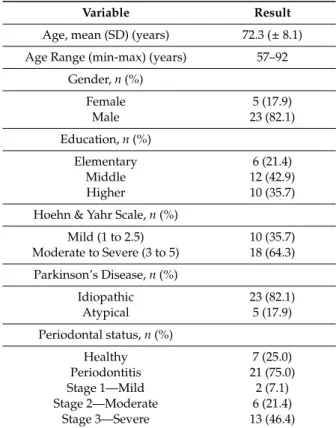
From a total of 33 individuals with PD, five participants were excluded because they were edentulous. A final sample of 28 participants were enrolled, with a mean age of 72.3 (±8.1) years, meeting the required inclusion criteria (Table1). The group was composed mostly by men (82.1%), with idiopathic PD (82.1%) and diagnosed with moderate to severe patient’s motor impairment (H and Y) (64.3%). The prevalence of periodontitis was high (75.0%), and the majority were severe cases (stage III) (46.4%). On average, the participants had 12 teeth missing, and one tooth with pathological mobility. The average percentage of plaque and gum inflammation in the whole mouth were 37.0% (±29.4) and 19.3% (±21.1), respectively. The majority of patients report the use of a manual toothbrush (75.0%) and a last dental visit within the last 6 months (64.3%).
Table 1. Participant characteristics.
Variable Result
Age, mean (SD) (years) 72.3 (± 8.1) Age Range (min-max) (years) 57–92
Gender, n (%) Female 5 (17.9) Male 23 (82.1) Education, n (%) Elementary 6 (21.4) Middle 12 (42.9) Higher 10 (35.7)
Hoehn & Yahr Scale, n (%)
Mild (1 to 2.5) 10 (35.7) Moderate to Severe (3 to 5) 18 (64.3) Parkinson’s Disease, n (%) Idiopathic 23 (82.1) Atypical 5 (17.9) Periodontal status, n (%) Healthy 7 (25.0) Periodontitis 21 (75.0) Stage 1—Mild 2 (7.1) Stage 2—Moderate 6 (21.4) Stage 3—Severe 13 (46.4)
Medicina 2020, 56, 383 5 of 11
Table 1.Cont.
Variable Result Teeth with mobility, mean (SD) 1 (2)
Missing teeth, mean (SD) 12 (7) Plaque Index, mean (SD) (%) 37.0 (29.4)
BoP, mean (SD) (%) 19.3 (21.1) Mean PPD, mean (SD) (mm) 2.1 (0.8) Mean CAL, mean (SD) (mm) 3.2 (1.8) Mean REC, mean (SD) (mm) 1.2 (1.2)
Toothbrush type, n (%)
Manual 21 (75.0)
Electric 7 (25.0)
Last dental visit, n (%)
< 6 months 18 (64.3)
6–12 months 4 (14.3)
> 12 months 6 (21.4)
Toothbrushing
Once a day 9 (32.1)
Twice or more a day 19 (67.9) Interproximal cleaning Never 9 (32.1) No 8 (28.6) Often/Yes 11 (39.3) Smoking habits, n (%) Never 16 (57.1) Former smoker 7 (25.0) Active smoker 5 (17.9) Diabetes Mellitus, n (%) 3 (10.7)
BoP—Bleeding on Probing; CAL—Clinical Attachment Loss; PD—Probing Depth; REC—Recession; SD—Standard Deviation.
3.2. Relationship between PD Staging and Periodontal Status
We found no di↵erences between PD stages (Table2). Mild PD presented lower prevalence of
severe periodontitis than more advanced PD stages (Table3). An increased number of missing teeth
was associated with a worsening of speech and eating tasks (Table4). Further, worse periodontal