L
aser
A
ssisted
E
ndodontic
T
reatment
» Blind Randomized Clinical Trial «
Orientador:
Prof. Doutor Manuel Fontes de Carvalho
Co-Orientador:
Candidature dissertation for the Doctorate Degree in Dental Medicine,
Prof. Doutor Afonso Manuel Pinhão Ferreira (Prof. Catedrático)
Prof. Doutor Américo dos Santos Afonso (Prof. Associado c/agregação)
Prof. Doutor António Cabral Campos Felino (Prof. Catedrático)
Prof. Doutor César Fernando Coelho Leal Silva (Prof. Associado c/agregação)
Prof. Doutor Germano Neves Pinto Rocha (Prof. Associado)
Prof. Doutora Irene Graça Azevedo Pina Vaz (Prof. Associado)
Prof. Doutora Inês Alexandra Costa Morais Caldas (Prof. Auxiliar)
Prof. Doutor João Carlos Antunes Sampaio Fernandes (Prof. Catedrático)
Prof. Doutor João Carlos Gonçalves Ferreira de Pinho (Prof. Associado c/agregação)
Prof. Doutor João Fernando Costa Carvalho (Prof. Catedrático)
Prof. Doutor Jorge Manuel Carvalho Dias Lopes (Prof. Catedrático)
Prof. Doutor José António Macedo Carvalho Capelas (Prof. Associado c/agregação)
Prof. Doutor José Carlos Reis Campos (Prof. Auxiliar c/ agregação)
Prof. Doutor José Mário Castro Rocha (Prof. Auxiliar)
Prof. Douto Manuel José Fontes de Carvalho (Prof. Associado)
Prof. Doutora Maria Cristina P. C. M. Figueiredo Pollmann (Prof. Associado)
Prof. Doutora Maria Helena Guimarães Figueiral da Silva (Prof. Catedrática)
Prof. Doutora Maria Helena Raposo Fernandes (Prof. Catedrático)
Prof. Doutora Maria Lurdes Ferreira Lobo Pereira (Prof. Auxiliar)
Prof. Doutor Mário Augusto Pires Vaz (Prof. Associado - personalidade convidada)
Prof. Doutor Mário Jorge Rebolho Fernandes Silva (Prof. Catedrático)
Prof. Doutor Mário Ramalho Vasconcelos (Prof. Associado c/agregação)
Prof. Doutor Miguel Fernando Silva Gonçalves Pinto (Prof. Catedrático)
Prof. Doutor Paulo Rui Galrão Ribeiro Melo (Prof. Associado c/ agregação)
Prof. Doutor Adão Fernando Pereira (Prof. Catedrático)
Prof. Doutor Amilcar Almeida Oliveira (Prof. Associado)
Prof. Doutor António Manuel Machado Capelas (Prof. Associado )
Dr. António Ulisses Matos dos Santos (Assistente Convidado)
Prof. Doutor Durval Manuel Belo Moreira (Prof. Associado c/Agregação)
Prof. Doutor Francisco António Rebelo Morais Caldas (Prof. Catedrático)
Dr. José Maria Vaz Osório (Assistente Convidado)
Prof. Doutor José Serra Silva Campos Neves (Prof. Catedrático)
Prof. Doutor Manuel Desport Marques (Prof. Associado Convidado )
Prof. Doutor Manuel Guedes de Figueiredo (Prof. Associado)
Retired Professors
Prof. Doutor António Manuel Guerra Capelas (Prof. Auxiliar)
Prof. Dr. Artur Manuel Osório de Araújo (Prof. Associado Convidado)
Prof. Doutor Fernando Jorge Morais Branco (Prof. Catedrático)
Prof. Doutor Fernando José Brandão Martins Peres (Prof. Catedrático )
Prof. Doutor José Albertino Cruz Lordelo (Prof. Associado c/ agregação)
Prof. Doutor José Carlos Pina Almeida Rebelo (Prof. Catedrático)
Prof. Doutor Manuel Pedro da Fonseca Paulo (Professor Catedrático)
Prof. Doutora Maria Adelaide Macedo Carvalho Capelas (Prof. Associada )
Prof. Doutora Maria Purificação Valenzuela Sampaio Tavares (Prof. Catedrática)
Prof. Doutor Rogério Serapião Martins Aguiar Branco (Prof. Catedrático)
TO MY PARENTS,
Maria do Rosário Antunes Rodrigues Martins
Miguel André Duarte Martins
To all my friends and colleagues…
…who have supported and encouraged such personal achievement.
Muito para além do seu conteúdo científico, esta tese tem um enorme conteúdo emocional pois foram muitas as pessoas que contribuíram com a sua sabedoria mas, sobretudo, com a sua forte amizade.
Neste contexto, quero expressar de uma forma simples, mas sincera e merecida, o meu agradecimento a quem - directa ou indirectamente - contribuiu para a consolidação e realização do meu trabalho, por vezes constante, outras pontualmente, abrindo-me portas e ajudando a trilhar caminhos para (mais) uma conquista profissional.
Ao Professor Doutor Manuel José Fontes de Carvalho, meu orientador e responsável directo, um especial reconhecimento pela sua confiança que se converteu numa boa amizade. De início, poucos seriam aqueles que, no panorama nacional, aceitariam entrar comigo num mundo tão vasto e ao mesmo tempo tão desconhecido como o dos lasers dentários. Mais ainda pelo facto de saber antecipadamente as dificuldades que se iriam deparar na minha vontade expressa em realizar uma investigação endodôntica eminentemente clínica. Assim, agradeço profundamente pela sua paciência, pelos seus conselhos e ensinamentos (soluções) que me foi proporcionando em todos os trâmites deste longo percurso. Sem a sua diplomacia e estímulo permanente teria sido muito difícil ou mesmo impossível travar todas as batalhas superando todos os obstáculos que, de uma forma mais ou menos esperada, se foram colocando à nossa frente. Esta investigação tem o seu cunho pessoal pela enorme vontade em querer fazer de mim, alguém…
Ao Professor Doutor Norbert Gutknecht, meu co-orientador e inspirador, um agradecimento muito especial. Poderei talvez afirmar que a sua presença no meu percurso académico foi provavelmente a mais marcante: de facto, nunca irei esquecer o brilhar dos seus olhos durante as intensas palestras a que tive o prazer de assistir durante dois anos em Aachen. Essas primeiras palestras foram, porventura, a ignição de toda esta paixão pela Light
Amplification by Stimulated Emission Radiation. Reviu todo o meu percurso académico no seu,
sendo pioneiro na introdução dos lasers dentários na RWTH Aachen Uniklinikum e posterior divulgação por todo o mundo. Assim, foi-me ensinando a lidar com todas as naturais adversidades do mundo académico. Foi assim que, com o seu apoio científico e confiança absoluta na minha capacidade de trabalho, se tornou tecnicamente e financialmente possível
XIV realizar toda esta investigação. Apostou no primeiro pupilo português e, com a sua inexcedível cooperação, deixou-me crescer a seu lado…
[To Prof. Dr. Norbert Gutknecht, my inspiring co-supervisor, one very special
acknowledgment. I can probably quote that his presence in my academic career was indeed the most determinant: in fact, I will never forget the shining of his eyes during those outstanding lectures I have been the pleasure to attend during two years, in Aachen. Those classes were by chance the ignition for all my Light Amplification by Stimulated Emission Radiation passion. He has saw in my academic path huge similarities with his own, being pioneer on the introduction of lasers in the RWTH Aachen Uniklinikum and through all over the world afterwards. As consequence he taught me how to deal with natural challenges of the academic world. Hence, it was just with all his - more than - scientific support and personal trust in work capacity that was technically and financially possible to start and complete all this investigation. Prof. Dr. Gutknecht took his chances on his first Portuguese pupil and, with all his unsurpassed furtherance, allowed me to grow on his side…]
Ao Professor Doutor José António Capelas pela pronta colaboração, pelo privilégio da sua enorme amizade e por todos os seus (cirúrgicos) conselhos que me foram guiando ao longo desta cruzada académica. Do lazer ao laser, foi-me apoiando pessoalmente e cientificamente tornando-me, sem dúvida, mais perspicaz e adulto numa vivência universitária recheada de ilusões. A sua influência sempre positiva e entusiasta transformou todos estes períodos num enorme prazer.
À Professora Doutora Irene Graça Azevedo Pina Vaz que me acolheu e acompanhou de perto toda a evolução do meu trabalho. Agradeço-lhe a amizade, a disponibilidade e o exemplo de dedicação à investigação endodôntica. Sendo - para mim - uma referência científica, fui aprendendo atentamente as melhores formas de investigar e também de ensinar. Sendo um modelo de dedicação para com o mundo académico, foi um privilégio enorme trabalhar, evoluir e conviver a seu lado.
Ao Professor Doutor Afonso Pinhão Ferreira que, na qualidade de Director da Faculdade de Medicina Dentária da Universidade do Porto, desde início possibilitou e desbloqueou a concretização desta investigação. Acreditou no seu potencial científico, na minha capacidade de a concretizar, e no meu espírito positivo ao querer fazer algo realmente ambicioso.
amizade acabou por incutir-me o gosto pela endodontia.
A todos os meus colegas do Departamento de Endodontia. Juntos partilhamos não só os sonhos, ambições e conquistas como também as algo esperadas…controvérsias “endodônticas”. Assim é um privilégio acordar para trabalhar e depois…almoçar! Para a Prof. Doutora Cláudia Rodrigues, Drª. Eva Salgueirinho, Drª. Joana Barros e Prof. Doutora Rita Noites, expresso o meu profundo agradecimento por toda a boa disposição, apoio e carinho.
À Drª Daniela Abreu que colaborou na parte estatística de uma forma profissional e sobretudo amiga. Para que tudo estivesse de acordo com o que se pretendia, e nos timings que estabelecia, o seu empenho e paciência foram significativamente….inesgotáveis!
Referindo-me aos participantes, o meu obrigado pois permitiram “pacientemente” ser o alvo de toda a investigação. Sem qualquer tipo de incentivo financeiro, compartilharam o seu tempo e apresentaram-se na Faculdade sempre a que foram chamados para os devidos controlos. Sempre colaborantes foram, sem dúvida, a parte fulcral e indispensável deste projecto e foi com enorme satisfação que fui comunicando - individualmente - os bons resultados deste estudo.
À Faculdade de Medicina Dentária da Universidade do Porto, onde me formei como Médico Dentista, por me ter possibilitado realizar este trabalho junto de inúmeras referências tanto profissionais como académicas. Neste capítulo, manifesto um agradecimento indispensável a todas as Assistentes Técnicas, Administrativas e Operacionais (expressamente à D. Maria Alice Rio e D. Maria Eugénia Costa) que, com um sorriso diário sempre presente, foram uma preciosa ajuda no que concerne a toda a parafernália clínica necessária para atender os participantes incluídos na investigação.
Ao Aachen Dental Laser Center (AALZ Institute, RWTH Aachen University, e ao seu Coordenador Académico Leon Vanweersch que, desde o início apoiou e depositou na minha pessoa os requisitos necessários para que este trabalho viesse a alcançar os êxitos desejados. Através deste Instituto e das suas actividades científicas tenho vindo a granjear amizades pelo mundo. Várias destas amizades têm vindo a acompanhar o meu trajecto profissional, sendo
XVI frequentemente os espectadores mais atentos das palestras que tenho proferido em congressos internacionais. Aqui, deixo um agradecimento pela sua atenção e companhia.
À Biolase, CA, USA., principalmente ao seu representante Europeu Sr. Pedro Morales e ao Chema Cid Alvarez. Foi através desta equipa que todo o equipamento e material acessório me foram cedidos - como fiel depositário - calibrados e reparados. Por tal, a sua ajuda foi das mais determinantes. A confiança que o Sr. Pedro Morales depositou inicialmente em mim, na altura como aluno da RWTH Aachen University, foi-se solidificando numa amizade omnipresente em todos os eventos científicos nos quais marquei presença.
Para a Ritinha que bem me acompanha no meu lado mais emocional. Lado a lado redigimos teses, discutimos pormenores, limámos arestas e…fomos construindo uma relação. Apesar de sermos distintos na forma de abordar adversidades, o sentimento que nos une permitiu, muitas vezes, ser o apoio mútuo e imprescindível a tanto comprometimento com o trabalho. Assim, creio que tudo aquilo que fomos idealizando e que ficou por fazer durante este período, será com certeza um óptimo motivo para continuarmos a passar óptimos momentos, a dois!
Por fim, e porque é o agradecimento mais difícil, obrigado, muito, muito obrigado à minha Família. É difícil agradecer porque não há palavras que possam traduzir, realmente, a minha gratidão. Na minha tese de Mestrado escrevi nos agradecimentos: All the efforts
applied in the present study were only possible to be accomplished due to the support and sacrifice of my family. Their experience, education and guidance were crucial for all these “light dreams” slowly became real…!
Pode não ser a forma mais original mas gostava que este trabalho pudesse representar e demonstrar que mereci tudo aquilo que me deram até agora. Num país onde a Educação Superior e Pós-Graduada é – infelizmente - um privilégio ao alcance de muito poucos, foram muitos os sacrifícios para que, aos 31 anos, pudesse ter no Currículo tantas páginas. Incluído nesses sacrifícios encontra-se naturalmente o meu irmão que, por ser mais novo, de forma indirecta pode ter sido privado de alguma atenção, certas regalias e/ou extravagâncias. Assim, ao meu irmão, obrigado por fazer parte da nossa vida. Não é preciso estar presente em todos os momentos nem falar a toda a hora para ser indispensável e parte de mim…
Uma referência merecida ao meu Pai pois no que toca a opções profissionais foi sempre o meu timoneiro. Com a concordância de Mãe, foi por ele aconselhado que me iniciei na actividade docente e da pós-graduação bem como na partida à descoberta dos lasers,
Por fim, obrigado aos meus pais pela dedicação incondicional; obrigado por nunca estarem cansados ou indisponíveis para atenderem a mais um pedido meu. Esta tese pode ser o resultado do meu trabalho, apoiado por todos a quem já agradeci e outros que aqui não referi, mas só foi possível porque os meus pais estiveram sempre ao meu lado. Por isso, esta tese é acima de tudo deles…
TABLE OF CONTENTS
TITLE ... 3 ABSTRACT ... 5 Key Words: ... 5 RESUMO ... 7 JUSTIFICATION ... 9 INTRODUCTION ... 13Historical context of lasers in Dentistry and the paradigm-shift ... 15
Cleaning and disinfecting the root canal system ... 18
Necrotic pulp condition: Bacterial aspects & E .faecalis role ... 22
Apical Periodontitis: Microbiological aspects ... 26
Apical Periodontitis: Radiological features & Anatomical considerations ... 30
Sodium hypochlorite (NaOCl) as irrigation solution ... 36
Hazardous effects of sodium hypochlorite ... 40
Calcium hydroxide (CaOH) paste as inter-appointment dressing ... 42
Root canal preparation techniques ... 43
The role of different lasers in Endodontics ... 44
Different Lasers for Endodontic Treatments ... 47
Limitations Associated to Laser Tips ... 52
Erbium Lasers ... 56
The Er,Cr:YSGG Laser ... 58
Light Transmission System(s) ... 60
Tapered Fibers and the Radial Firing Tip (RFT) ... 61
Endodontic Radial Firing Tip: concept and state of art ... 64
Er,Cr:YSGG LASER IN ENDODONTICS ... 72
Er,Cr:YSGG laser safety & temperature considerations ... 72
Er,Cr:YSGG laser Bactericidal Properties ... 77
Er,Cr:YSGG laser mechanisms for debridement and smear layer removal ... 82
MATERIALS AND METHODS ... 91
OUTCOMES & HYPOTHESIS ... 93
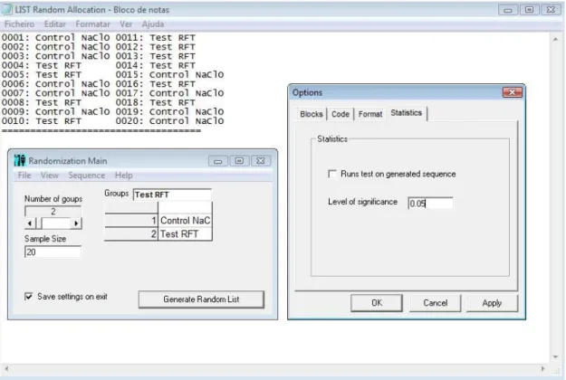
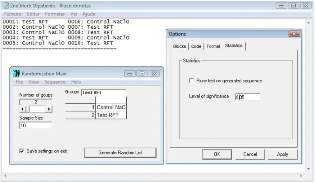
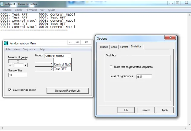
RANDOMIZATION PROCESS & ALLOCATION ... 98 INTERVENTIONS ... 101 OUTCOME CLASSIFICATION AND DATA ANALYSIS ... 106
RESULTS ... 109
CALIBRATION RESULTS ... 112 6 MONTHS FOLLOW-UP RESULTS ... 113 12 MONTHS FOLLOW-UP RESULTS ... 116 Additional records ... 118
STATISTICAL ANALYSIS ... 119
6 MONTHS (T6) FOLLOW-UP STATISTICS ... 126
12 MONTHS (T12) FOLLOW-UP STATISTICS ... 133 DISCUSSION ... 145 Analysis of Cost-Effectiveness ... 175 SOURCES OF FUNDING ... 177 ANNEXES ... 179 ANNEX I ... 181 ANNEX II ... 185 ANNEX III ... 187 ANNEX IV ... 189 ANNEX V ... 191 ANNEX VI ... 193 ANNEX VII... 197 ANNEX VIII ... 205 ANNEX IX ... 225 ANNEX X ... 231
TITLE
Efficacy of the Er,Cr:YSGG laser
in the
Laser Assisted Endodontic Treatment
ABSTRACT
Introduction: Clinical studies conducted to explore the safety and efficacy of new therapies is
considered an important focus in endodontic research. The primary objective of this randomized clinical study was to compare radiographic evidences of periapical healing after root canal therapy assisted by the Er,Cr:YSGG laser Radial Firing Tips (RFT) versus the concomitant use of 3% sodium hypochlorite and interim calcium hydroxide paste in necrotic teeth with chronic apical periodontitis. We hypothesized to find similar outcomes in both groups in order to assess the predictability of this new laser assisted endodontic protocol.
Methods: 36 and 43 (anterior and premolar) teeth were randomly assigned for the 6 and
12-month analyses. In group 1 teeth were manually prepared and irrigated with 3% sodium hypochlorite and calcium hydroxide inter-appointment dressing was applied; in group 2 teeth were manually prepared with saline solution and irradiated with Er,Cr:YSGG laser using the RFT2 (140µs, 37.5mJ, 20Hz) and the RFT3 (140µs, 62.5mJ, 20Hz) at the first and second appointment respectively, four times each, moving at 2mm.s-1 from apical to coronal. The primary outcome measure was change in apical bone density either at 6 and 12 months of follow up, using the periapical index (PAI) for blind radiographic assessment. Results: At the 6-month assessment 29 patients were subjected to statistical analysis (12 in group 1 and 17 in group 2) while at the 12-month assessment 30 teeth were analyzed (12 in group 1 and 18 in group 2). There was one treatment failure after 6 months and two failures after 12 months, all in group 1; treatment failures were not included for analyses. In the 6 and 12-month outcomes, both groups exhibited a statistically significant decrease in mean PAI score. However, no differences were found between groups. Conclusion: The present findings suggest that this laser assisted protocol might achieve equally predictable and effective outcomes, while overcoming potential hazards and limitations associated to conventional therapies.
Key Words:
Chronic apical periodontitis, Er,Cr:YSGG laser, radial firing tip, sodium hypochlorite, calcium hydroxide, periapical index.
RESUMO
Introdução: Estudos clínicos que pretendam explorar a segurança e eficácia de novos
procedimentos são considerados um foco primordial na investigação endodôntica. O objectivo principal deste estudo clínico randomizado consistiu em analisar evidências radiográficas de cicatrização apical após o tratamento canalar assistido pelo laser de Er,Cr:YSGG (Fibras de Dispersão Radial - RFTs) em comparação com a utilização simultânea de hipoclorito de sódio a 3% e aplicação de hidróxido de cálcio entre consultas em dentes necróticos com periodontite apical crónica. A hipótese consistiu em encontrar resultados similares em ambos os grupos de modo a atestar a predictibilidade deste novo protocolo endodôntico assistido por laser.
Métodos: Para o controlo dos 6 e 12 meses, 36 e 43 dentes (anteriores e pré-molares) foram
aleatoriamente distribuídos. No grupo 1, os dentes foram preparados manualmente, irrigados com hipoclorito de sódio a 3% e, entre consultas, foi aplicado um revestimento de hidróxido de cálcio; no grupo 2 os dentes foram manualmente preparados, irrigados com soro fisiológico e irradiados com o laser de Er,Cr:YSGG utilizando a fibra RFT2 (140µs, 37.5mJ, 20Hz) e a fibra RFT3 (140µs, 62.5mJ, 20Hz) na primeira e segunda sessão respectivamente. Cada fibra foi introduzida quatro vezes, à velocidade de 2mm.s-1, desde apical até coronal. Os objectivos principais desta investigação consistiram na avaliação cega das diferenças radiográficas referentes à densidade óssea apical, após 6 e 12 meses, utilizando o índex periapical (PAI).
Resultados: Após 6 meses, 29 dentes foram submetidos a análise estatística (12 no grupo 1 e
17 no grupo 2) enquanto após 12 meses, 30 dentes foram submetidos à mesma análise (12 no grupo 1 e 18 no grupo 2). Registou-se um insucesso aos 6 meses e dois insucessos aos 12 meses, ambos no grupo 1; Os insucessos, porém, não foram submetidos a análise estatística. Na avaliação após 6 e 12 meses, ambos os grupos exibiram uma redução estatisticamente significativa na média de resultados. Contudo, não existiram diferenças significativas entre os grupos. Conclusão: Até à presente data, os resultados sugerem que este protocolo assistido por laser poderá atingir resultados igualmente seguros e preditíveis, ultrapassando potenciais riscos e limitações associados às terapias convencionais.
Palavras-chave:
Periodontite apical crónica, Er,Cr:YSGG laser, radial firing tip, hipoclorito de sódio, índex periapical.
JUSTIFICATION
The man of science has learned to believe in justification,
not by faith, but by verification.
Thomas H. Huxley (1825-95) English biologist
The primordial objective of any endodontic treatment is to achieve microorganism eradication from the root canal system, obtain a proper canal conformation achieved by mechanical means and finish with an efficient tridimensional obturation.
If every one of these requirements is necessary to achieve the success of the endodontic therapy, the first one can be considered truly the most important. It is unanimous that the effective elimination of all microorganisms inside the root canal it´s the main factor that can determine the success or the failure of the endodontic therapy.
The disinfection or sterilization of all root canal system it´s traditionally done by innumerous antiseptic agents which can act against the microorganisms when introduced and maintained, for a certain while, inside the main canal. In first instance that can be done with rinsing solutions as sodium hypochlorite, and in second instance through the application of semisolid formulas as calcium hydroxide, just as an example. There are also other physical means to achieve canal disinfection – which have being used and studied – such as ultrasounds, electro surgery or even laser assisted therapies.
The aim of this research was to test a new root canal system disinfection method using the Erbium, Chromium doped Yttrium, Scandium, Gallium, Garnet (Er,Cr:YSGG) laser with Radial Firing Tips recently developed for endodontics. With this technology it should became possible to tri-dimensionally disinfect the root canal system, overcoming hazardous effects and limitations commonly associated to chemical solutions such as sodium hypochlorite.
During conventional endodontic treatment, the laser application as disinfection mechanism should be considered not as a substitute but as complement for traditional endodontic procedures. However, due to the inherent costs it should be done a proper evaluation of its risk-benefits along with its polyvalence. Hence, a primary indication for laser adjunctive application can be those cases in which - under some circumstances – chemicals can be potentially hazardous or ineffective (e.g. wide apical constrictions or persistent infections).
Although innumerous publications can demonstrate the bactericidal potential of different wavelengths – diode, CO2, Nd:YAG and Erbium lasers– in assisting root canal treatments just few are in fact in vivo studies. Though, some controversies remain while choosing the ideal parameters for each laser, resulting in relevant discrepancies in terms of safety and efficacy.
In our case, following several precedent studies performed in vitro which could attest the safety and efficacy of all procedures, we could define the up-to-date ideal parameters and perform a randomized clinical trial. As endodontic researchers, we found interesting and a scientific challenge to compare clinical outcomes obtained by Er,Cr:YSGG - Radial Firing Tips laser assisted endodontic treatment, with those achieved by a conventional endodontic treatment protocol (3% sodium hypochlorite irrigation and calcium hydroxide dressing), in necrotic teeth.
INTRODUCTION
Every sentence I utter must be understood
not as an affirmation, but as a question.
Niels H. Bohr (1885-1962) Danish physicist
Historical context of lasers in Dentistry and the paradigm-shift
Since Theodore H. Maiman produced the first laser based on a synthetic ruby crystal in 1960, the use of lasers has undergone a technological revolution. Dentistry, for example, has witnessed a paradigm-shift as technological breakthroughs have enabled a wide range of hard and soft tissue procedures with improved patient outcomes, combined with the absence of tissue direct contact, vibrations or pain as well as significant reductions in post-operative symptoms and complications.
Based on a theory originally postulated by Albert Einstein, T. Maiman created a device where a solid-state ruby crystal medium was stimulated (pumped) by a flash lamp as source of energy, leading to the emission of laser light. A year later, Snitzer developed the first neodymium laser (Nd:YAG laser). However, early dental research focused on laser systems was largely ignored.
In fact, experiments done by Stern and Sognnaes in 1964 found that ruby laser was not an effective wavelength for cutting enamel or dentin. In overall, hard tissue applications for these lasers were not promising; hence research focused on soft tissue surgery where argon, carbon dioxide, and Nd:YAG lasers proved to be successful for cutting and coagulation.
The first reported oral surgical application using a CO2 laser occurred in 1977 by Lenz et
al. Nevertheless, it took a decade longer for the first Food and Drug Administration clearance
for the CO2 laser in oral surgery (January 1987). This could be considered the true beginning for the acceptance and viability of using lasers in oral cavity in clinical environment.
From then, innumerous rounds of research into the laser field lead to broader oral applications that have accomplished a definitive dental laser revolution. This technology refinement is actually expanding the scope of procedures that a dentist can offer their patients, shaping an ineluctable standard of preventive, effective and minimally invasive dentistry.
To understand the physical background of lasers is not strictly necessary to be aware of their technical set-up; however it is mandatory to have the knowledge background regarding properties of light generated according to the laser principle. For instance, “laser” is an acronym that completely describes the whole physical process of light generation: Light
Amplification by Stimulated Emission of Radiation. It is equally important to understand
several terms and concepts which can be briefly described.
Lasers are usually named for their “active medium” that is charged with energy inside the laser unit (resonator) to create laser light. Though, laser is a physical phenomenon
traduced by a unique energy transformation where different kinds of energy are transformed into a new kind of optical energy with peculiar properties. This energy form is completely artificial and cannot be found in nature.
It can also be stated that a laser device transforms energy of “low quality” into a kind of energy that has “high quality”. The problem that inhabits in this transformation, which generates a high degree of energy order, is that this causes a decline in entropy, limiting the efficiency of laser light generation itself.
Another key concept is that different wavelengths react with different tissues in different ways, as the biological interaction between laser light and tissue is strongly dependent of the tissues optical properties – absorption coefficients. The absorption of the laser energy can be illustrated by the absorption spectrum for each wavelength in the targeted tissue or tissue components. Apart from wavelength and absorption, reflection, scattering and transmission (degree in which the laser´s energy is able to penetrate into the tissue) are phenomenon’s that also play a crucial role in laser-tissue interaction (Fig. 1).
That is the main reason why the extensive study of enamel, dentin and soft tissues composition is demanded in order to find the most suitable wavelength to work with.
Enamel is considered a highly mineral tissue, composed of 85% mineral, 12% water and 3% organic material, by volume (2). Dentin however, has a significantly higher content of organic material (33%) and water (20%) whereas only 47% is mineral, by volume. The mineral content is composed by carbonated hydroxyapatite crystals organized in enamel rods of approximately 5µm diameter (3). Due to their composition, enamel and dentin demonstrate high absorption coefficients for wavelengths in the mid infrared range, between 2.6-3µm (coincident with Erbium lasers peak emissions). In this spectrum region, scattering is almost negligible and the energy deposition is mainly determined by absorption coefficients and tissue reflectance (4). In this context, at a wavelength of 2.94µm there is a strong absorption in water (800cm-1) while for a wavelength of 2.78µm the peak absorption is coincident with a narrow hydroxyapatite absorption band (400cm-1) (5). The highest absorption peak from dental mineral bands is coincident with the phosphate groups observed at 9.6µm (6500cm-1) for the CO2 laser (6).
Currently, distinct laser sources – wavelengths - are available each having its own specific properties. The correct understanding of these properties, as light behavior and tissue interactions, is a precondition for using dental lasers appropriately in safe conditions. Thus, the aim for dentists should be to know what kind of laser(s) could be suited for a specific indication in order to achieve the maximum benefit through the implementation of laser technology in their dental practice.
Figure 1: Absorption coefficients (µA) of several wavelengths (µm) in dental materials. (Illustration gently provided by AALZ Institute, Aachen University – Germany)
Although it can be practical to operate accordingly with the instructions provided by the manufacturer without notions of the real clinical benefits and hazardous potential, to select a wavelength from the various laser systems which are at the dental practitioner´s disposal requires an advanced training concomitant with a good understanding of each wavelength characteristics and its biological interactions.
Demands for New Strategies in Endodontic Procedures
Cleaning and disinfecting the root canal system
Root canal infection occurs frequently concomitant with dental caries. In addition, bacterial penetration and colonization of intact pulp can be due to either dental treatment or trauma.
Although the biological diversity of oral microbiota can enormous in terms of number of species, just approximately 50 strains were reported to be predominant in root canal infections (7, 8). However in this context, it must be understood that the methodologies of microbiological root canal sampling are complex and that the diagnostic accuracy is still poorly known. Still, if microorganisms hiding in biofilms or in untreated parts of the canal system may be hard to sample and detect, remnants of the previous medication might also depress laboratory analysis (9).
As bacterial contamination is considered the primal etiologic factor for the development of pulpal and periapical lesions, to obtain the root canal system free of irritants has been showing to be endodontic therapy primordial goal (10-12).
The idea that an absence of cultivable microbes at the time of obturation will favor healing is consistent with the notion that microorganisms are the primary cause of persistent apical periodontitis. Fabricius et al. (2006) reported results from an extensive experiment conducted on 175 monkey root canals supporting this position (13). Several studies have reported a tendency towards more favorable outcomes in teeth yielding negative cultures before the root canal filling (14-16); Accordingly, other investigators have suggested that the presence of microbes at the time of root filling will adversely affect the outcomes (17-19).
The bactericidal effects of conventional irrigation strategies during and after root canal preparation with solutions such as hydrogen peroxide (H2O2) or sodium hypochlorite (NaOCl) has been studied by numerous investigators such as Bystrom et al. in 1985, Smith et al. in 1986, and Orstravik et al. in 1990 (20-22).
NaOCl is used for endodontic applications at concentrations ranging from 0.5% to 5.25% (v/v), and has been showing to be the most reliable endodontic irrigant from several decades until the present. Although it is reported that it can exert its maximum capabilities as an antibacterial agent and solvent of organic substances at a concentration of 5,25% (23), the most effective concentration and temperature for reducing the root canal system bacterial load may vary according to the author and methodology. In fact, the ideal concentration and
temperature of NaOCl in root canal therapy remains as controversy and topic of debate within endodontists (20-22, 24).
Hence, in order to penetrate more readily into non-instrumented areas of the root canal system and increase the agent´s efficiency, recent improvements in the NaOCl chemical structure have been tested. These chemical modifications allow practitioners to lower its surface tension, increasing contact with dentinal walls and possibly enhancing not only its antimicrobial effectiveness as well as its ability to dissolve pulp and organic tissue (25).
Another relevant finding is that mechanical-chemical enlargement with irrigation solutions such as NaOCl is limited to effective bacterial reduction up to a depth of 100µm whereas heavy E. faecalis infection was found 800µm deep into the canal lumen. Additionally, in some of the samples, bacterial propagation into the dentinal tubules has reached up to 1100µm in depth (22, 26-28).
However, sodium hypochlorite use is not risk free since it is unselective and damages human cells, dentine, and/or periodontal tissues, with clinical consequences. Under adverse clinical situations, it may affect branches of the facial nerves or the inferior alveolar nerve, leading to severe sequelae (29, 30).
Although 5.25% NaOCl is thought to be the most effective concentration, it has recently been found to be associated with a significant increase in apically extruded debris after rotary preparation as compared to lower concentrations and chlorhexidine (31).
Nevertheless, the potential replacement of NaOCl by other antimicrobial agents such as chlorhexidine is matter of further discussion (32). Whereas no significant differences were found between the alternative irrigation with 0.12% chlorhexidine solution when compared with 2.5% NaOCl in a clinical trial (33), Vahdaty et al. suggested that chlorhexidine and NaOCl were equally effective antibacterial agents against E. faecalis. In addition, both irrigants were capable of effectively reducing bacterial counts in the superficial 100µm of dentinal tubules but still failed to disinfect up to 50% of dentine samples (27).
Biomechanical instrumentation of the root canal system aim to remove infected root canal dentin and various techniques have been successfully reported and clinically adopted over the years. Moreover, while shaping the canal, either by manual or mechanical preparation, we could enlarge the root canal space in order to facilitate the chemical action and penetration of irrigation solutions. Concomitantly due to the complexity of the root canal system it has been shown to be virtually impossible to completely eliminate debris and sterilize the intricate parts of the root morphology.
During the canal enlargement proceedings, a smear layer is mechanically produced, covering the instrumented walls of root canal (34-38).
The smear layer is known to be a superficial layer on the surface of the root canal wall approximately 1-2µm thick with a deeper layer trapped in the dentinal tubules up to a depth of 40µm (37). Along with organic and inorganic substances, it also includes microorganisms and necrotic debris (39). Together with the possibility that the smear layer itself may be infected, it can also protect the bacteria harbored in the dentinal tubules by preventing the application and effective infiltration of successful intra-canal disinfection solutions (28, 37, 39).
Fogel et al. has consistently reported that the formation of a smear layer reduces the root dentin permeability from 25% to 49% (40). Hence, it is generally accepted that the complete removal of the smear layer would be consistent with the elimination of irritants from the root canal system (41). Root canal disinfection will be inefficient in the presence of smear layer as most current intracanal medications not only have limited antibacterial spectrums but also have shown limited ability to diffuse into the dentinal tubules (42).
Regardless of the instrumentation system or technique applied, it has been reported that, with the root canal preparation and enlargement, a substantial removal of pathogens can be achieved (43). However, it has also been clearly demonstrated that more than 35% of the canal surface area remained unchanged/untouched in a study that evaluated four distinct Nickel-Titanium preparation techniques (44).
Though, it has been suggested that newer treatment strategies designed to eliminate microorganisms from the root canal system should be considered. These must include agents that can penetrate the dentinal tubules and destroy the microorganisms beyond the host defense mechanisms, where they cannot be reached by systematically administered antibacterial agents (42). Therefore, other alternative possibilities such ozone treatment (45-47), ultrasonic and laser assisted treatments are being suggested as suitable, alternative methods to achieve endodontic disinfection, possibly overcoming the limitations of commonly used chemical solutions as well as any hazardous effects (48-50).
Depending on the wavelength, lasers have been mostly used in endodontics for either vaporizing the smear layer or eradicating pathogens, not only in the main root canal but especially deep in the root canal tubular network where rinsing solutions could never efficiently reach.
These can be considered the true goals for the adjunctive application of lasers in root canal therapy: the ability of infrared light to interact with water and efficiently remove the smear layer and debris from the root canal walls, together with the ability of light to propagate into the dentinal tubules further than any chemical solution, thus providing deep disinfection.
The goal of laser assisted endodontic treatments (LAET) is thus to provide increased outcomes, namely in cases of persistent infections or per-operative obstacles (e.g. isthmus, recurrent canals, internal resorptions, root canal perforations, or wide apical constrictions) which are often associated to either lower or compromised clinical expectations.
The increase of reported research suggesting the efficacy of such procedures has been proving the potential of LAET as a powerful and versatile method to achieve predictable endodontic outcomes.
Necrotic pulp condition: Bacterial aspects & E .faecalis role
Injuries to the pulp may lead to complete tissue breakdowns. The non-vital (or necrotic) pulp is defenseless against microbial invasion and allows indigenous microorganisms to reach the pulp chamber, either via direct exposure, uncovered dentinal tubules or cracks in the enamel or dentine. Lateral canals exposed as a result of progressive marginal periodontitis may also serve as pathways for bacteria to reach the pulp (51).
The specific environment in the root canal, characterized by the degrading pulp tissue and lack of oxygen, results in a microbiota dominated by proteolytic and anaerobic bacteria. Via the apical foramen, microbes and their by-products may reach the periapical tissue promoting an inflammatory response. This response induces reabsorption of the surrounding bone, which is often visible by x-ray assessment as localized periapical radiolucency. Such inflammatory reaction may progress and stimulate epithelial cells in the periodontal membrane to proliferate and form a periapical cyst (52, 53).
The first observation of these microorganisms was carried out by Anthony van Leeuwenhoek. In 1683, with a homemade microscope he was enabled to make the first drawings of dental plaque bacteria. However, it took about 200 years before root canal microorganisms received further scientific investigation, by the “father” of oral microbiology, Willoughby D. Miller (1953-1907).
A classical study in germ-free and conventional rats in 1965 demonstrated the essential role of microorganisms in the pathogenesis of periapical lesions (10).
Even before Kakehashi´s experiments, Miller described in 1890 the clinical effects of gangrenous tooth-pulps as centers of infections varying from hardly perceptible periapical inflammation to severe local and general symptoms, sometimes even with fatal outcomes. He cultured and characterized bacteria from necrotic pulps and studied their pathogenic potential in animal experiments (54, 55).
In some cases of infected necrotic pulp, an open pathway for the entry of bacteria is found in the form of pulp exposure due to caries or fractures. On the other hand, infections and apical periodontitis also occur in cases of closed necrosis, even in apparently intact teeth. Although there are potential forms of entry (e.g. dental caries, trauma, periodontal disease and anachoresis), no data is available concerning the relative frequency of these entry routes in clinical situations.
Nevertheless, the intact dentine-pulp complexes are considered a highly efficient defense system, often capable of preventing and suppress the entry of microorganisms.
Therefore, with the event of bacteria entering a vital pulp, their survival depends on their number and virulence as opposed to the defense mechanisms of the pulp (56).
The necrotic pulp, in turn, provides excellent growth conditions for microorganisms and is the major location for bacteria causing periapical lesions. Mixed microbial masses consisting of cocci, rods, filamentous bacteria, spirochetes and yeasts have been demonstrated in necrotic pulps using light microscopy of histological tissue sections, as well as by transmission electron microscopy of root fragments (57-59).
Generally, microorganisms adhere to some areas of the root canal walls, either as dense aggregates or as thin, single, or multilayered condensations. When the pulp becomes non-vital, the organisms can also extend for some distance into the dentinal tubules. In fact, the presence of bacteria in the dentinal tubules of infected teeth has been seen as reaching approximately half the distance between the root canal walls and the cement-dentinal junction (60-62). If bacteria are confined usually to the inner third, Sen et al. reported bacteria and fungi penetration between 10 to 150µm in most of their samples (63).
The invasion or penetration of the dentinal tubules by bacteria is essentially due to microbial multiplication and not necessarily by their movements. External pressure can also encourage migration, in which the microbes that were in the external part of the dentinal tubes penetrate into the interior - passive migration. Bae and colleagues have inclusively shown that most pulp infections are related to immobile bacteria progression (64).
Thus, in cases of periapical lesions, microorganisms are always found in these intra-radicular locations, often walled off by neutrophil granulocytes or an epithelial plug at the apical foramen (57).
The resident oral microflora compromise more than 300 species of cultivable bacteria and an unknown number of species that is impossible to grow with usual methods. The special environment of the root canal, however, selects for certain frequent species. A mixture of several typical ones has been cultured from necrotic pulp samples prior the start of treatment (65, 66).
The microorganisms in the root canal samples from deciduous and permanent teeth are predominantly the same bacteria as those found in dental plaque, periodontal pockets and carious lesions. Most isolates in initial cultures are obligate anaerobic bacteria. These have constituted 91% of the isolates from closed necrosis (67), 90% of isolates from necrotic pulps of deciduous teeth (68), and 68% from the apical part of necrotic pulps in carious teeth (69). A large proportion of these anaerobes are asaccharolytic, peptide and amino acid-degrading bacteria (70).
Thus, many genera and species currently identified in root canal samples comprise obligate anaerobic and facultative anaerobic oral bacteria. Among the streptococci, species of the anginosus group (S. anginosus, S. intermedius, S. constellatus) and mitis group (S. mitis, S.
oralis, S. gordonii, S. sanguis, S. parasanguis) are common, and in carious teeth S. mutans is
also prevalent. It has been suggested that S .sanguis and S. salivarius often occur in root canal cultures due to contamination with saliva or invasion through leaking temporary fillings (71).
Actinomyces israelli and other Actinomyces species may be present and actinomycotic
periapical lesions may sometimes develop for periodontal reasons (72).
The Enterococcus faecalis is known to be a facultative Gram-positive anaerobic coccus and endodontic-related pathogen, as it is frequently recovered from the root canals of teeth with post-treatment infections (73, 74). It is frequently isolated from saliva, mucosal surfaces, and supra-gingival dental plaque; consequently it has been associated with several conditions, such as oral mucositis, aggressive periodontitis and other infections including endodontic–related ones (75, 76).
It is thought that the biological properties of E. faecalis allow the bacterium to survive in adverse conditions inside the canal, where concentrations of nutrients are low and alkaline conditions are prevalent (it can resist high pH values, up to 11.5)(77). E. faecalis also has the particular characteristic that it can survive in canals as a single species without the support of other microorganisms (78). In addition, they are able to form intra and extra-radicular biofilms making it even harder to control or eradicate them (79, 80).
Bacteria may also invade the dentinal tubules, depending on the morphological factors of the bacterial species. Some bacteria including E. faecalis might migrate deeper into the lateral root canal or dentine tubules than others, which may protect them from effective disinfection (81).
Although experimental demonstrations have shown that this specific bacteria can invade as far as 800µm into the dentinal tubules of root canal walls (28), it has also been reported that this microorganism has the ability - under specific conditions - to infect the full length of the tubules within two days (22). These characteristics often contribute to therapy resistant cases that end up as long-term failures after endodontic treatment.
Sodium hypochlorite and chlorhexidine, i.e., have proven to be effective against E.
faecalis in vitro, but they require direct contact (82, 83). Furthermore, whereas pathogenic
microorganisms are able to penetrate more than 1mm (Streptococcus mutans were found in dentinal tubules of open infected root canals up to a depth of 1100µm), rinsing solutions, such
as NaOCl, only reach a depth of 100µm and under special conditions (e.g. temperature) (26, 62).
E. faecalis has also been found to resist to inter-appointment medications, including the most common intracanal treatment, calcium hydroxide (22, 84-86).
An additional and crucial factor that may prevent the complete elimination of E. faecalis relates to the intricate anatomy of the root canal system, consisting of lateral canals, fins, apical ramifications (deltas), accessory canals and isthmuses (87, 88).
Altogether, such findings may clearly justify the necessity to develop effective means of removing the smear layer and all known pathogenic bacteria from root canal dentin following biomechanical treatment. Consequently, the development of an alternative to traditional disinfection protocols (such as a laser-assisted approach) may seem useful and appropriate to be under investigation in order to effectively eradicate microorganisms harbored deep within the dentinal tubules and complex root canal ramifications.
Apical Periodontitis: Microbiological aspects
Dental pulp reacts to the different external irritant factors (microbes, thermal, mechanical, and chemical injuries) through characteristic inflammatory responses including vasodilatation. It can frequently lead to an increase of internal pressure which affects the pulp circulatory system; as it is a terminal circulatory type, it is often associated as the direct cause for the pulp necrosis (89).
Necrotic root canal ambient can afford bacteria space and a moist of warm, nutritious, and anaerobic environment that is relatively well protected from host defenses. Even so, only a restricted assortment of oral bacteria can be found in an infected canal, suggesting that selective pressures can favor the establishment of some species and inhibit others to proliferate in the root canal system (90).
Following the formation of a periapical inflammatory lesion secondary to pulpal necrosis, chronic apical periodontitis (granuloma) is considered the next step in the progression of these inflammatory events showing replacement of adjacent tissue with an inflammatory cell infiltrate that usually occurs at the expense of the surrounding bone. In addition to the inflammatory cells, it typically contains fibrous tissue and cholesterol crystals (91).
Over time, due to inflammatory stimulation and proliferation of the epithelial rests of Malassez (residual epithelial cells in the periodontal ligament), an inflammatory cyst can develop at the root apex and through the bone (92). If the lumen of the cyst is continuous with the infection source at the pulpal entry, it may not be self-sustained (“pocket” cyst); this will heal following infection source elimination. On the other hand, if the cyst is completely encased by epithelium and removed from the source of infection, it may be self-sustained (“true” cyst) and become refractory to treatment except by surgical excision (93).
Cysts most appear as round or pear-shaped, unilocular, radiolucent lesions in the periapical region. They are usually classified when become bigger than 1cm in diameter, being bordered by a thin rim of cortical bone. Cysts may displace adjacent teeth or cause mild root resorption (94).
The differentiation between radicular cysts and granulomas is difficult or impossible by traditional radiographic techniques, even if several radiographic features have been proposed to make this distinction; these may include the lesion size and the presence of a radiopaque rim lining the cystic lesion (95, 96). While the probability of a lesion being a cyst may increase with its size, a reliable diagnosis still remains based on histology (95, 97).
Chronic Apical Periodontitis (CAP) is arguably one the most common forms of biofilm-induced human diseases (98). This condition is usually asymptomatic and can be often found by radiographic means in patients between 30 and 50 years old (99, 100).
This condition usually develops after dental pulp necrosis and infection as a result of caries, trauma, or iatrogenic clinical procedures. The environmental conditions in the necrotic root canal are conducive to the establishment of a microbiota typically dominated by anaerobic bacteria. However, bacterial profiles may vary from individual to individual i.e., each one harbors a unique biofilm in terms of species richness and abundance. This also indicates that apical periodontitis has a heterogeneous etiology, where no single species can be considered to be the main endodontic pathogen, and multiple bacterial combinations can play a demanding role in the disease development (101).
The literature contains significant divergence regarding histological results in terms of apical lesions, with the prevalence of granulomas ranging from 9% (102) to 87% (103) and cysts prevalence ranging from 7% to 59% (92, 104).
The discrepancy between prevalence in the literature is probably attributed to the different criteria used in the various histological studies. Histological diagnosis based on samples with few sections can also lead to the incorrect definition of epithelialized lesions as cysts. As example, Ricucci et al. established the diagnosis of cysts with the presence of a cavity completely or partially delimited by epithelium. This epithelium was found in 21 of 50 lesions, whereas only 16 were indeed classified as cysts (105).
In most studies, neither radiographic size nor the presence of radiopaque lamina alone can be considered sufficient evidence on which to base a diagnosis of periapical pathology. A histological study is mandatory to definitively identify the type of periapical lesion. Authors such as Hirsch et al. cited that most periapical lesions are either cysts or granulomas and only few are keratocysts. Therefore this controversy can be found quite irrelevant from a surgical point of view and - as that the prognosis does not depend on the type of lesion -histological diagnosis in considered unnecessary. While periapical cysts usually demonstrate the largest radiographic areas, chronic apical periodontitis is the most common type of apical lesions, followed by scar tissue (106).
Early studies of microbiota associated with apical periodontitis were conducted using a broad-range of culture methods. Culture studies have first demonstrated that primary endodontic infections are characterized by a mixed consortium dominated by anaerobic bacteria and composed of a mean number of 2.6 to 5.4 taxa per root canal (8, 107, 108).
Culture studies were followed by a number of studies employing molecular detection methods such species-specific PCR and the original checkerboard DNA-DNA hybridization. These methods allowed the inclusion of some culture-difficult species in the set of candidate endodontic pathogens. Afterwards, the adoption of 16S rRNA gene clone library analysis allowed even more comprehensive investigations of bacterial biofilms in endodontic infections. Through this technique, not only cultivable species but also as-yet-uncultivated and uncharacterized bacteria could be identified, revealing that 40 to 55% of the bacterial taxa found in primary endodontic infections have not been cultivated and validly named/identified (109, 110).
After about 1 decade of application of molecular biology methods to endodontic microbiology research, specific knowledge regarding bacterial diversity involved with apical periodontitis has been substantially refined and redefined. In addition, to strength the associations of several cultivable species with chronic apical periodontitis, new findings using large scale analysis allowed to constantly improve the inclusion of some newly named species and as-yet-uncultivated phylotypes in the set of candidate pathogens associated with this disease.
The broad-range molecular analyses of bacteria present within the root canals of teeth with chronic apical periodontitis have revealed higher figures than those demonstrated by culture methods: 7 taxa in denaturing gradient gel electrophoresis analyses (101), 11 taxa in terminal restriction fragment length polymorphism analyses (109), 10 to 12 taxa in clone library analyses (111), and 20 taxa in combined culturing and clone library analyses (110). Therefore, data gathered from culture studies tend to underestimate the number of bacteria taxa in infected canals, as a result of difficulties or even impossibilities in cultivating a significant proportion of the endodontic microbiota.
Another important finding was related to the mean number of taxa per canal that was clearly in direct proportion to the lesion size: small lesions (<5mm) harbored 11,7 taxa, lesions from 5 to 10mm harbored 16 taxa, and lesions >10mm harbored about 20 species (112). These differences in species richness help explain the long-held concept that the endodontic treatment of teeth with large lesions have lower success rates than treatment of teeth with smaller or no lesions (113).
Another example of 16S rRNA gene-based nested or heminested PCR assays effectiveness is that O. uli only recently was recognized as a frequent member of the endodontic microbial consortium of teeth with apical periodontitis (114). In corroboration, and analyzing the presence and relative levels of 83 oral bacterial species, Siqueira et al. have adopted an innovating reverse-capture checkerboard hybridization assay; associations
between the most frequently detected taxa were also recorded. The most prevalent taxa detected were O. uli, E. corrodens, P. endodontalis, and P. anaerobius. O. uli was, in fact, present in about three-fourths of the samples, confirming that this species is a very regular member of the microbiota associated with chronic apical periodontitis (112).
E. corrodens has been detected in endodontic infections mainly by molecular methods
and the 63% frequency - at which this species was found in Fouad et al. study - is probably the highest one, while assessing endodontic infections. P. endodontalis was primarily found in endodontic infections by culture means, but its association with apical periodontitis has been strengthened by findings obtained from molecular studies, where higher prevalence values have been reported (115).
It has also been demonstrated by scanning electron microscopy that periapical bacterial plaque is a coating of various microbial forms embedded in a structureless material on the outer root surface, near the main apical foramen. In resorption lacunae bacteria and yeast cells sometimes could be detected (116, 117).
In ultrastructural studies, micro-organisms are generally not found in the soft tissue lesion in cases of chronic apical periodontitis or in periapical cysts (when the cavity completely encases in an epithelial lining so that no communication to the root canal exists). An exception to this rule is the occasional finding of typical actinomyces-containing colonies in granulomas, radicular cysts (118) and in cases of periapical abscess with or without sinus (fistula), which sometimes various bacterial forms and yeasts may be present inside the lesion and/or inside phagocytes (119).
The stages in development and healing of chronic apical periodontitis, granulomas and cysts are, depending on several circumstances, reflected by changes in the radiographic appearance of periapical areas. Generally, the prognosis for complete healing of endodontically treated teeth with diagnosis of apical periodontitis is approximately 10%-15% lower than teeth without apical periodontitis (34, 120). Thus, if with ideal conditions for root canal therapy the success rate can reach over 90%, for teeth with periapical radiolucency, the success rate can decrease to about 80% (121). So, the real challenge for endodontists regarding disinfection of the root canal system would be gangrenous teeth in addition with chronic apical periodontitis which the pathological reaction caused by certain (and yet to be determined) bacteria harbored in the root canal thrive to produce the clinical picture of periapical infection (122).
Apical Periodontitis: Radiological features & Anatomical considerations
Radiological features: In several dental situations, the use of radiographs is considered
mandatory for precise diagnostic judgment. Specifically in endodontics, radiographs arguably represent the most important single diagnostic aid due to its reliability as method to obtain relevant information concerning the pulp canal space and periapical tissues.
The periodontal ligament provides the space for the initial cell infiltration. It serves as starting point for resorption processes as well as the end of healing ones. Then, a widened periodontal ligament space can be associated with either initial or residual chronic inflammation (123).
If teeth with increased mobility due to bruxism or marginal periodontitis may also present records of widened periodontal ligament, in the case of apical periodontitis it is restricted to the infected area near the apex (124).
The lamina dura appearance in radiographs varies, as it is a continuation of the jawbone cortex, which encases the root in a cortical bone socket. There is a considerable intra- and inter-individual range in its thickness and density. Even related to the fact that the bone is frequently thin in the region of maxillary canines, the lamina dura is almost impossible to discern around these teeth.
Lamina dura may be described as irregular indistinct or serrated, but none of these
changes is pathognomonic in the early or healing stages of apical periodontitis (125).
Thus, its appearance is mainly determined by the shape and position of the root in relation to the X-ray beam rather than the density and integrity of the lamina dura itself. As it can also vary with the amount of occlusal load or stress, some lesion in lamina dura may produce radiographic detection earlier than in cancellous bone because more minerals are removed at that site (126).
Unfortunately, normal variations in thickness and continuity of lamina dura make the diagnosis by these single criteria inappropriate. In more advanced stages however, other radiographic changes become prominent and those can offer better distinction between stages and degrees of inflammation in terms of pathognomonic signs of apical periodontitis.
As inflammatory reactions of periapical tissues often proceed without any clinical symptoms, apical periodontitis cases are frequently diagnosed just by radiographic means. Numerous investigators have studied the sensitivity of periapical radiography; a common approach was to create artificial bone lesions in cadavers to access the minimum amount of bone loss necessary to result in a visible radiolucency. One reference study set up by Bender &
Seltzer reported that bone lesions were not visible until the cortex or the interface between cortical and cancellous bone was involved. In addition bone destructions were always found larger then suspected from studying the correspondent radiographs (127).
In fact, changes in mineralization and structure of the bone adjacent to the site of inflammation constitute the basis of radiographic diagnostic procedures for the detection and monitoring chronic apical periodontitis. Nevertheless, radiographic appearance depends as well on its evolution stage. While studying human autopsies, Brynolf compared the radiology and histology of periapical areas in upper incisors and reported a high frequency of radiographically undetectable inflammatory lesions (128).
Figdor et al. generally address to apical periodontitis as a very prevalent problem (98). Typically, these lesions are located at the root apex, but communications may exist at various levels along the root surface, and though can develop from lateral and furcal locations to sinus or further adjacent structures.
By its turn, Moreira et al. in 1998 performed a portuguese epidemiological study - in Porto area – and their results indicated a prevalence of apical periodontitis in 27% of the 322 residents which were assessed (129).
Anatomical considerations: Although being very prevalent, the location of periapical lesions in
the oral cavity was found quite similar in different populations. A study by Nobuhara and Del Rio (130) found the majority in the anterior maxilla (47,3%), followed by the posterior maxilla (28,7%), posterior mandible (15,3%), and anterior mandible (8,7%). These findings are in agreement with Spatafore et al., who found 46,5% of lesions in the anterior maxilla, 20,7% in the posterior maxilla, 18,3% in the posterior mandible, and 14,3% in the anterior mandible (131). These prevalence results are also similar to the distribution found and reported by Carrillo et al. (132).
Radiographic imaging of CAP is presented on a background of superimposed, normal osseous and other structures. By its turn, radiographic diagnosis of apical periodontitis is based on deviations from the normal anatomy. So, periapical changes can only be interpreted properly considering some important anatomical aspects that are closely related to the location of the lesions and different types of bone.
Because cortical bone is more mineralized than cancellous bone, the demineralization or resorption process will manifest radiolucent changes, i.e. minerals are lost sooner and more readily in more calcified tissues than in less calcified ones. Though, lesions in cortical bone are easier to detect and assume prominence for their clarity and size in radiographs.


![Figure 5: The absorption curve of water in the middle infrared region [adapted from (282)]](https://thumb-eu.123doks.com/thumbv2/123dok_br/15919100.1093633/77.892.281.620.854.1039/figure-absorption-curve-water-middle-infrared-region-adapted.webp)