UNIVERSIDADE DA BEIRA INTERIOR
Ciências da Saúde
Desenvolvimento de novos micro- e nano-sistemas
responsivos para entrega de drogas anti-tumorais
André Ferreira Moreira
Dissertação para obtenção do Grau de Mestre em
Ciências Biomédicas
(2º ciclo de estudos)
Orientador: Prof. Doutor Ilídio Joaquim Sobreira Correia
Coorientador: Mestre Vítor Manuel Abreu Gaspar
iii
“You, me or nobody is going to hit as hard as life. But it ain't about how hard you're hit, it is about how hard you can get hit and keep moving forward, how much can you take and keep moving forward. That's how winning is done!” Rocky Balboa (2006)
v
Acknowledgements
Firstly, I would like to thank to my supervisor the Professor Ilídio Correia for the opportunity to develop my master thesis in his group, and for all the efforts and time spent to help me to improve myself.
I thank to Vítor Gaspar, my co-supervisor for always making me go further and also for his friendship, guidance and endless support. Our constant work discussions contributed to develop my focus and technical skills as a researcher and also, improved my ability to interpret and criticize results. His continuous orientation was very important in the development of my work and in my growth as a person.
Also, I would like to thank to Elisabete Costa and to Duarte Diogo, for their fellowship, trust and endless support. They accompanied me throughout this year helping me to surpass all the barriers and frustrations. One word also for the other elements of this research group, thank our joyful conviviality it was very easy to work and become integrated.
I thank all my family, especially to my father, mother and brother for their support, patience, love and affection.
Lastly, and foremost, a very special thanks to my girlfriend, Ana Catarina, for all her love, comprehension, patience and support. Also for her continuous encouragement to me in order to continue to move forward during this difficult year.
vii
Abstract
Cancer is a major health care problem with growing incidence, not only at a national level but also worldwide. Due to this urgency in reducing cancer prevalence, the scientific community has put forward a great attention in the search for novel anti-cancer treatments, particularly, in the development of nanocarriers capable to control and promote drug delivery to target cells. These drug delivery systems are capable to overcome the limitations presented by the conventional chemotherapeutic treatments. Among the various types of nanocarriers developed so far, mesoporous silica nanoparticles (MSNs) possess unique structural properties that make them highly suitable to encapsulate and deliver drugs to cancer cells. However, for these specialized nanocarriers to be applied in cancer therapies it is still of critical importance to control the time frame of drug release at the tumor microenvironment or inside cancer cells, in order to maximize the therapeutic effect and reduce unspecific cytotoxicity. One alternative to control drug release is to endow the nanocarriers with a pH responsive drug release that takes advantage of the naturally acidic tumor microenvironment and also of the acidic pH of lysosomes.
In this thesis the development of dual drug loaded pH-responsive mesoporous silica nanoparticles (MSNs) with a calcium carbonate-based coating is presented as an effective alternative to deliver drugs to prostate cancer cells. This approach allowed the simultaneous co-encapsulation of a non-steroidal anti-inflammatory drug (Ibuprofen) and Doxorubicin (an anti-tumoral drug), with high efficiency. Furthermore, the idealized calcium carbonate coating successfully promoted a pH sensitive drug release from the MSNs matrix. The delivery systems proved to be capable of maintaining the drugs inside their mesoporous structure under physiological pH, and to prompt its release in acidic environments. The resulting dual loaded MSNs coated with calcium carbonate have spherical morphology and a mean size of 167 nm, presenting therefore, good characteristics to be applied as nanocarriers. Such, is supported by the cytotoxicity studies where the idealized MSNs produced a 93% higher anti-proliferative effect than the non-coated silica nanoparticles, being even more effective than the dual free drug administration, as well. Overall, the carbonate coating of MSNs showed to be a simple and cost-effective approach for cancer therapy, in particular for a pH-triggered drug delivery. Moreover, the versatile nature of these nanocarriers allows surface modifications that can improve the selectivity to target cells, allow imaging or even a combination of them both.
viii
Keywords
Calcium carbonate, cancer therapy, co-delivery of drugs, mesoporous silica nanoparticles, pH responsive release.
x
Resumo Alargado
O cancro é atualmente um dos maiores problemas que afeta a saúde pública, tanto ao nível nacional como mundial. Apesar de grande parte das terapias convencionais possuírem a potencialidade de eliminar a maioria das células cancerígenas, estas apresentam vários problemas associados. Um dos que mais se destaca é a falta de especificidade, o que se traduz frequentemente em danos de células e tecidos saudáveis, que constituem efeitos secundários nefastos. Aliado a este facto, normalmente, também se verifica uma baixa biodisponibilidade e, por isso, são muitas vezes utilizadas concentrações mais elevadas dos agentes terapêuticos na tentativa produzir algum efeito benéfico para o paciente. Estes aumentos nas concentrações administradas acarretam consigo um acréscimo dos efeitos nocivos. Como tal, esta doença tem atraído a atenção da comunidade científica para o desenvolvimento de novas terapias. Uma boa abordagem para ultrapassar estas desvantagens é a entrega combinada de diferentes agentes terapêuticos. Esta múltipla administração utiliza os compostos em quantidades inferiores à da sua aplicação isolada, tentando assim tirar partido de um possível efeito terapêutico sinérgico resultante da combinação do ataque a diferentes caraterísticas chave das células cancerígenas. Contudo, mesmo esta abordagem não tem conseguido ultrapassar os efeitos secundários produzidos nas células saudáveis.
Nos últimos anos, uma estratégia que tem sobressaído é a utilização da Nanotecnologia para a administração destes agentes terapêuticos. A utilização de nanotransportadores concede a oportunidade de ultrapassar algumas das limitações apresentadas anteriormente. De fato, no geral os nanoveículos são capazes de aumentar a solubilidade dos agentes terapêuticos, protegê-los e transportá-los na circulação sanguínea. Simultaneamente, também podem controlar a libertação destes compostos bioativos, aumentando a seletividade e penetração/absorção dos mesmos no tecido alvo. Dentro dos diferentes tipos de nanopartículas que têm vindo a ser estudados as nanopartículas mesoporosas de sílica (MSNs) apresentam características estruturais que as tornam muito adequadas para esta aplicação. Estas partículas possuem uma estrutura porosa singular, com um grande número de poros que nunca se interconectam, aliada à capacidade de armazenarem uma grande quantidade de agentes terapêuticos. Além disso, as MSNs apresentam uma estrutura rígida muito resistente à temperatura, pH e stress mecânico o que lhes garante uma elevada estabilidade. Contudo, apesar das boas propriedades que as MSNs apresentam, é ainda necessário conferir-lhes a capacidade de libertarem a sua carga na presença de um determinado estímulo para que os agentes terapêuticos sejam apenas libertados quando cheguem a um ambiente que possua esses estímulos. O estímulo pode ter como origem alterações no pH, luz, enzimas, temperatura entre outros. A sensibilidade ao pH é um dos estímulos que melhor se adequa para ser utilizado na terapia do cancro, pois as diferenças de pH observadas no microambiente tumoral e também nas vias endocíticas no interior das células cancerígenas podem ser aproveitadas para
xi
desencadear a libertação dos agentes terapêuticos. Nas MSNs esta sensibilidade a estímulos é geralmente conseguida através da ligação de polímeros na sua superfície. Porém, esta estratégia apresenta algumas desvantagens como a necessidade de utilização de processos de purificação complexos, custos elevados e um potencial de aplicação clínica limitado.
Assim sendo, o trabalho de investigação desenvolvido nesta tese descreve não só o desenvolvimento de nanopartículas de sílica mesoporosas carregadas com dois agentes terapêuticos, Doxorrubicina e Ibuprofeno mas, também a nova aplicação do carbonato de cálcio para tornar as MSNs sensíveis ao pH. O carbonato de cálcio forma-se preferencialmente nos poros das MSNs impedindo assim a libertação da sua carga, e quando em meio ácido este sofre uma rápida degradação desimpedindo os poros e permitindo a libertação da Doxorrubicina e do Ibuprofeno. Este sistema foi desenvolvido e testado para a entrega de agentes terapêuticos a células do cancro da próstata. As nanopartículas produzidas apresentaram um tamanho na ordem dos 160 nm e uma morfologia esférica uniforme. Além disto, os estudos efetuados demostraram que as partículas são capazes de armazenar grandes quantidades de Doxorrubicina e Ibuprofeno na sua matriz porosa. Por outro lado, apenas perdas residuais destes agentes terapêuticos foram detetadas nos passos subsequentes ao seu armazenamento nas MSNs. Os resultados obtidos demostraram também que o revestimento de carbonato de cálcio é sensível ao pH, visto que a um pH acídico (5,6) os agentes terapêuticos apresentaram uma rápida libertação e a um pH fisiológico (7,4) a libertação foi retardada. Os estudos realizados in vitro com células do cancro da próstata (PC-3) mostraram que estas partículas eram capazes de penetrar nas células e entregar os agentes terapêuticos no seu local de ação. Em particular, foi comprovado que uma quantidade substancial de Doxorrubicina se localizava no núcleo das células tumorais após administração. Estes resultados são essenciais para verificar a eficácia desta estratégia uma vez que este agente anti-tumoral atua no núcleo ao nível do ADN. Adicionalmente, as partículas de sílica revestidas com carbonato de cálcio contendo os fármacos apresentaram uma maior atividade citotóxica do que os agentes terapêuticos na forma livre e mesmo do que as nanopartículas não revestidas.
Em geral, o revestimento de carbonato de cálcio mostrou-se capaz de imprimir um comportamento sensível ao pH por parte das nanopartículas de sílica, e futuramente permitir a sua utilização na terapia do cancro. Além disto, a versatilidade que este sistema apresenta, permite modificações futuras que podem melhorar a sua seletividade para as células de interesse ou mesmo adicionar funções permitindo por exemplo o diagnóstico e a terapia em simultâneo.
xii
Palavras-chave
Carbonato de cálcio, co-entrega, nanopartículas de sílica mesoporosas, responsividade ao pH, terapia do cancro.
xiv
List of Publications
Articles in peer reviewed international journals:
André F. Moreira , Vítor M. Gaspar, Elisabete C. Costa, Duarte de Melo-Diogo, Paulo Machado, Catarina M. Paquete and Ilídio J. Correia, “Preparation of end-capped pH-sensitive mesoporous silica nanocarriers for on-demand drug delivery”, European Journal of Pharmaceutics and Biopharmaceutics (3.826), submitted.
Duarte de Melo-Diogo, Vítor M. Gaspar, Elisabete C. Costa, André F. Moreira , David Markl, Eugénia Gallardo and Ilídio J. Correia, “Combinatorial delivery of Sildenafil-Crizotinib-Palbociclib by TPGS-PLA micelles for improved cytotoxic effect in lung cancer”, European Journal of Pharmaceutics and Biopharmaceutics (3.826), submitted.
Posters communications:
André F. Moreira, Vítor M. Gaspar, Elisabete C. Costa, Duarte de Melo-Diogo, Paulo Machado, Catarina M. Paquete and Ilídio J. Correia, Synthesis and characterization of MCM-41 type sílica nanoparticles by a Stöber modified method, Encontro Bienal das Divisões Técnicas da Sociedade Portuguesa de Materiais (SMP), 4th of May, Covilhã, Portugal.
Elisabete C. Costa, Vítor M. Gaspar, Duarte de Melo-Diogo, André F. Moreira, João F.G. Marques, Paula Coutinho and Ilídio J. Correia, Evaluation of nanoparticles uptake in breast cancer co-cultures, Encontro Bienal das Divisões Técnicas da Sociedade Portuguesa de Materiais (SMP), 4th of May, Covilhã, Portugal.
Awards:
INESPO II project (2014) - 3000 Euros Grant winner for prototypes development, and enrollment in CEBT Ibérico - Competências Empreendedoras De Base Tecnológica.
xvi
Index
Chapter 1 1
Introduction 1
1. Cancer 2
1.1. Cancer development and main hallmarks 2
1.2. Prostate cancer prevalence and development 5
1.3. Anti-tumoral drugs used in prostate cancer 7
2. Nanotechnology and drug delivery systems 12
2.1. Nanosized delivery systems for delivery of bioactive molecules 12
2.2. Classes of nanocarriers 13
2.2.1. Mesoporous silica nanoparticles 16
2.2.1.1. Chemical production of MSNs – The Stöber modified method 18
2.2.1.2. Surface functionalization of MSNs 18
2.2.1.3. Mesoporous silica nanoparticles uptake and biocompatibility 18
2.3. Administration routes and barriers 20
2.4. Nanocarriers targeting to tumor tissues 21
2.4.1. Passive Targeting 21 2.4.2. Active Targeting 22 2.5. Stimuli-responsive nanocarriers 24 2.5.1. pH-responsiveness nanocarriers 25 Aims 28 Chapter 2 29
Materials and Methods 29
2. Materials and Methods 30
2.1. Materials 30
2.2. Methods 30
2.2.1. Mesoporous silica nanoparticles synthesis 30
2.2.2. Drug loading 31
2.2.3. Mesoporous silica nanoparticles coating 31
2.2.4. Mesoporous silica nanoparticles morphological characterization 31 2.2.5. Mesoporous silica nanoparticles size and zeta potential characterization 32
2.2.6. Mesoporous silica nanoparticles porosity analysis 32
2.2.7. X-ray powder diffraction of mesoporous silica nanoparticles 33 2.2.8. Energy dispersive X-ray spectroscopy of mesoporous silica nanoparticles 33
2.2.9. Fourier transform infrared spectroscopy analysis 33
2.2.10. Drug release analysis 34
2.2.11. Cytotoxicity assays 34
xvii
2.2.13. IC50 determination 35
2.2.14. Anti-tumoral activity of drug loaded mesoporous silica nanoparticles 35
2.2.15. Statistical analysis 35
Chapter 3 36
Results and Discussion 36
3. Results and Discussion 37
3.1. Synthesis of mesoporous silica nanoparticles 37
3.2. Morphological characterization mesoporous silica nanoparticles 39 3.3. Size and zeta potential characterization of mesoporous silica nanoparticles 40
3.4. Porosity analysis of mesoporous silica nanoparticles 41
3.5. X-ray powder diffraction of mesoporous silica nanoparticles 42
3.6. Energy dispersive X-ray spectroscopy of mesoporous silica nanoparticles 42
3.7. Fourier transform infrared spectroscopy analysis 44
3.8. Drug loading and release analysis 45
3.9. Cytotoxicity assays 47
3.10. Mesoporous silica nanoparticles cellular uptake 49
3.11. IC50 determination 53
3.12. In vitro anti-tumoral activity of drug loaded mesoporous silica nanoparticles 54
Chapter 4 57
4. Conclusions and Future Perspectives 58
xix
Figure Index
Figure 1 – Evolution of cancer concept. ... 2
Figure 2 – Major components of the tumor microenvironment. ... 3
Figure 3 – Cancer hallmarks and therapeutic targets of each key characteristic in cancer cells. ... 5
Figure 4 –Most commonly diagnosed types of cancers worldwide in men, in 2008. ... 6
Figure 5 – Representation of Prostate anatomic zones. ... 6
Figure 6 – Doxorubicin molecular structure and the representation of its mechanisms of action. ... 10
Figure 7 – Ibuprofen molecular structure and COX-2 role in tumor development. ... 11
Figure 8 – General nanocarrier-based strategies employed for drug delivery, and their structure representation. ... 14
Figure 9 – Mesoporous silica nanoparticles general structure, and their cargo loading and possibilities of surface functionalization. ... 17
Figure 10 – Pathways used by mesoporous silica nanoparticles for cellular internalization. ... 19
Figure 11 – Physical characteristic of nanoparticles that determine their biocompatibility and capacity to surpass certain barriers. ... 21
Figure 12 – Nanocarriers targeting, passive vs active targeting strategies. ... 23
Figure 13 – Summary of some overexpressed biomolecules on prostate cancer. ... 24
Figure 14 – Major strategies employed in the development of pH responsive nanocarriers. ... 26
Figure 15 – Schematic representation of MSNs and Dox-Ibu-MSN-CaCO3 synthesis. ... 39
Figure 16 – Morphology analysis. ... 40
Figure 17 – Size and zeta potential characterization of MSNs particles. ... 41
Figure 18 - X-ray diffraction spectra of MSN and MSN-CaCO3. ... 43
Figure 19 - Energy-dispersive X-ray spectroscopy (EDX) analysis of MSNs. ... 44
Figure 20 - FTIR spectra of CTAB, MSNs+CTAB, MSNs and Dox-Ibu-MSNs. ... 45
Figure 21 - Drug encapsulation efficiency analysis. ... 46
Figure 22 - pH-sensitive release kinetics of (A) Doxorubicin and (B) Ibuprofen from Dox-Ibu-MSNs-CaCO3. ... 47
Figure 23 - Evaluation of the cytotoxic profile of MSNs. ... 48
Figure 24 - Confocal microscopy images of MSNs uptake in PC-3 cancer cells. ... 50
Figure 25 - Time course uptake analysis of Dox-Ibu-MSNs-CaCO3. ... 52
Figure 26 - Comparison between Doxorubicin mean fluorescence intensity (MFI) in the nucleus and in the cytoplasm, at 6 h. ... 53
Figure 27 - Doxorubicin and Ibuprofen IC50 determination. ... 54
Figure 28 - Evaluation of MSNs anti-tumoral activity in PC-3 prostate cancer cells. ... 56 …
xxi
Table Index
Table 1. Porosity analysis of non-purified MSNs (MSNs+CTAB), MSNs after purification step
xxiii
List of abbreviations
ATP Adenosine Triphosphate
Bcl-2 B-cell lymphoma 2
BET Brunauer–Emmett–Teller
BJH Barrett-Joyner-Halenda
CaCl2 Calcium Chloride
CaCO3 Calcium Carbonate
CO2 Carbon Dioxide
COX-1 Cyclooxygenase 1
COX-2 Cyclooxygenase 2
CTAB Hexadecyltrimethylammonium Bromide
DGS Direção Geral de Saúde
DLS Dynamic Light Scattering
DMEM-F12 Dulbecco's Modified Eagle Medium: Nutrient Mixture F-12
DNA Deoxyribonucleic Acid
Dox Doxorubicin
Dox-Ibu-MSNs Doxorubicin and Ibuprofen loaded Mesoporous Silica Nanoparticles Dox-Ibu-MSNs-CaCO3 Doxorubicin and Ibuprofen loaded Mesoporous Silica Nanoparticles coated with Calcium Carbonate
Dox-MSNs Doxorubicin loaded Mesoporous Silica Nanoparticles
EDX Energy Dispersive X-Ray
EMA European Medicines Agency
EPR Enhanced Permeability and Retention
FBS Fetal Bovine Serum
FDA Food and Drugs Administration
FGF Fibroblast Growth Factor
FibH Primary Normal Human Dermal Fibroblasts
FITC Fluorescein Isothiocyanate
FTIR Fourier Transform Infrared Spectroscopy
HCl Hydrochloric Acid
HSA Human Serum Albumin
Ibu Ibuprofen
Ibu-MSNs Ibuprofen loaded Mesoporous Silica Nanoparticles
MCM-41 Mobil Crystalline Materials
MFI Mean Fluorescence Intensity
MSNs Mesoporous Silica Nanoparticles
MSNs+CTAB Non-Purified Mesoporous Silica Nanoparticles
MSNs-CaCO3 Calcium Carbonate coated Mesoporous Silica Nanoparticles
MTS 3-(4,5-Dimethylthiazol-2-Yl)-5-(3-Carboxymethoxyphenyl)-2-(4-Sulfophenyl)-2H-Tetrazolium
Na2CO3 Sodium Carbonate
NAD(P)H Nicotinamide Adenine Dinucleotide Phosphate-Oxidase
NaHCO3 Sodium Bicarbonate
NaOH Sodium hydroxide
NSAID Non-Steroidal Anti-Inflammatory Drug
xxiv
PBS Phosphate Buffered Saline
PC-3 Human Prostate Cancer Cells
PCA3 Prostate Cancer Antigen 3
PDFG Platelet Derived Growth Factor
PDI Polydispersity index
PEG Polyethylene Glycol
PGE E series prostaglandins
PMS Phenazine Methosulfate
PSA Prostate Specific Antigen
PSCA Prostate Stem Cell Antigen
PSMA Prostate Specific Membrane Antigen
PTEN Phosphatase and Tensin Homolog
RES Reticuloendothelial System
RPMI-1640 Roswell Park Memorial Institute 1640
SEM Scanning Electron Microscopy
SLN Solid Lipid Nanoparticles
TEM Transmission Electron Microscopy
TEOS Tetraethyl Orthosilicate
TMOS Tetramethyl Orthosilicate
USA United States of America
UV-vis Ultraviolet–visible
VEGF Vascular Endothelial Growth Factor
WGA Wheat Germ Agglutinin
1
Chapter 1
Introduction
2
1. Cancer
1.1. Cancer development and main hallmarks
Cancer is a major health care problem with growing incidence around the globe (Lozano et al., 2012). It is estimated that cancer is responsible for 25% of total deaths in the United States of America (USA). Furthermore, a total of 1,665,540 new cases are expected to be diagnosed in 2014, which is equivalent to more than 4,500 newly diagnosed cancers each day (Siegel et al., 2014). Moreover, in Europe, in 2012, there were an estimated 3,450,000 new cancer cases and around 1,750,000 deaths (Ferlay et al., 2013). These numbers helps to understand the efforts put in the development of new cancer treatments that are more effective than those currently available.
Cancer is a disease that is originated from normal cells that by accumulating multiple transformations can become malignant. When this transformed phenotype is acquired these abnormal cells can affect the function of any organ of the body. Cancer cells generally present features like loss of differentiation and uncontrolled proliferation (Floor et al., 2012). Also, these cells are often capable of invasion of surrounding tissues or even the extravasation to other sites in the body, by a process termed metastasis (Floor et al., 2012). However, this minimalistic concept of cancer has been evolving (Figure 1), instead of a single mass of cancer cells in proliferation, cancer is now considered as much more complex tissue surrounded by the tumor microenvironment (Hanahan and Weinberg, 2000).
Figure 1 – Evolution of cancer concept. From reductionist a view (A) to tumor microenvironment (B) (Adapted from Joyce and Pollard, 2009).
3
This complex rich tumor microenvironment is established by resident tumor associated fibroblasts, macrophages, endothelial cells, pericytes, leukocytes, and extra-cellular matrix (Pietras and Ostman, 2010). The individual functions of the various microenvironment elements are summarized in Figure 2 (Hanahan and Coussens, 2012). In general, the cross-talk between tumor cells and their microenvironment elements triggers pro-survival, proliferation and invasion pathways in cancer cells (Liotta and Kohn, 2001, Quail and Joyce, 2013).
Figure 2 – Major components of the tumor microenvironment. Major cell subtypes and their key functions for tumor development (Adapted from Hanahan and Coussens, 2012).
This combined interaction between the microenvironment elements and cancer cells helps them to maintain certain key characteristics that were described by Hanahan et al. as “hallmarks of cancer” (Figure 3) (Hanahan and Weinberg, 2011). One of the first proposed hallmarks and one of the most important, is cancer cells capacity to sustain proliferative signaling, a unique characteristic achieved by the capacity to deregulate growth-promoting pathways (Hanahan and Weinberg, 2000, Daroqui et al., 2012, Quail and Joyce, 2013, Cheng et al., 2008a). However, in order to achieve this sustained proliferation, cancer cells also have to be capable of resisting anti-proliferation signals like those mediated by retinoblastoma protein and its two relatives, p107 and p130 (Hanahan and Weinberg, 2000, Costa et al., 2013, Di Fiore et al., 2013). Another strategy that allows continuous cancer proliferation is the cells ability to avoid programmed cell death, i.e., apoptosis (Hanahan and Weinberg, 2000, Evan and Vousden, 2001). This exceptional capacity arises from the ability to bypass pro-apoptotic signals
4
commonly present in healthy cells. This gain of function is generally obtained by the loss of p53 function derived from gene mutation (Wade et al., 2013, Muller and Vousden, 2013), or by overexpression of anti-apoptotic proteins like those of the B-cell lymphoma 2 (Bcl-2) family (Kelly and Strasser, 2011). Other important characteristic is cancer cells limitless replicative potential (Hanahan and Weinberg, 2000). In fact, cancer cells can acquire the capacity to surpass senescence, by up-regulating telomerase expression (Shay and Wright, 2011). Telomerase is a deoxyribonucleic acid (DNA) polymerase that adds repeat segments to telomeric DNA ends, its expression is almost absent in non-immortalized cells (i.e., the majority of cells that compose our organs), but with significant levels of expression in cancer cells (Mocellin et al., 2013). Telomerase overexpression prevents DNA damage and cell death associated to end-to-end fusion of chromosomes (Saharinen et al., 2011). Adding to this, like other tissues, cancer cells require the continuous supply of nutrients, oxygen and means to dispose of all the metabolic waste and carbon dioxide produced during their life (Chung et al., 2010). This nutrient supply/waste exchange mechanism is primarily supported by the tumor surrounding vasculature (Chung et al., 2010). In order to achieve a sustained angiogenesis, cancer cells activate the angiogenic cascade through changes in the balance of angiogenesis inducers and inhibitors (Hanahan and Weinberg, 2000). Tumors appear to have an increased vascular endothelial growth factor (VEGF) expression and other secreted pro-angiogenic factors like fibroblast growth factor (FGF), platelet-derived growth factors (PDGF) and angiopoietins (Weis and Cheresh, 2011). This overexpression results in heterogeneously distributed blood vessels, enlarged, tortuous, with excessive ramifications, large fenestrations (400-600 nm), leakiness, erratic blood flow and abnormal levels of endothelial cell apoptosis (Serres et al., 2014, Fukumura and Jain, 2008). Moreover, at certain point, cancer cells acquire the capacity to invade, survive and proliferate in other tissues and generate metastasis, a characteristic responsible for around 90% of cancer associated mortality (Hanahan and Weinberg, 2011, Chaffer and Weinberg, 2011). A well-known alteration in invasive cancer cells is the down-regulated expression of the protein E-cadherin, which plays an important role in cell-to-cell adhesion (Canel et al., 2013). Other characteristics that influence cancer cell invasion are the modifications in cellular morphology, the expression of matrix-degrading enzymes (matrix metalloproteinases) and an increased cell motility (Yilmaz and Christofori, 2009, Sahai, 2005). Finally, recently, two additional characteristics were proposed as cancer hallmarks, the cells ability to reprogram their metabolism and the ability to avoid immune system mediated destruction (Hanahan and Weinberg, 2011).
5
Figure 3 – Cancer hallmarks and therapeutic targets of each key characteristic in cancer cells (adapted from Hanahan and Weinberg, 2011).
1.2. Prostate cancer prevalence and development
Prostate cancer is the most incident cancer in men (Figure 4), with 233,000 expected new cases in USA in 2014 (Siegel et al., 2014). Furthermore, prostate cancer will be responsible for around 30,000 deaths, being the second deadliest cancer for men in USA (Siegel et al., 2014). In Portugal, according to official data from Direção Geral de Saúde (DGS), in 2007, prostate cancer presented an incidence of 114 cases per 100,000 and resulted in 1,654 deaths, a mortality rate of around 37%.
Prostate cancer is predominantly diagnosed at an old age, being rare before 50 years of age. More than 75% of men over the age 75 presently have been diagnosed with this type of malignancy (Arcangeli et al., 2012, Siegel et al., 2014). Thus the leading risk factor for prostate cancer is advanced age, followed by race (Grönberg, 2003). The African-American men presents the highest rate (137 cases per 100,000), followed by North American and Scandinavian individual. On the opposite side, the prevalence of prostate cancer in Chinese people is relatively low in comparison to other countries (1.9 cases per 100,000) (Center et al., 2012). Despite these relevant differences, a common risk factor to prostate cancer is family history, i.e. genetic predisposition to prostate cancer. Since, the probability to be diagnosed with prostate cancer increases two-fold for men who have or had a first degree relative that suffered from it (Loeb and Schaeffer, 2009).
6
Figure 4 –Most commonly diagnosed types of cancers worldwide in men, in 2008 (adapted from Center et
al., 2012).
The prostate is a glandular and muscular organ that works as a reproduction accessory gland. It is located in the lower pelvis around the beginning of the urethra (Lee et al., 2011a). The prostate has 5 anatomic zones (Figure 5) the peripheral, central, transition, fibromuscular, and periurethral gland region and its primary function is to secrete a fluid, which aids in motility and nourishment of the sperm (McNeal, 1981).
7
Prostate cancer development is generally characterized by a phenotype transition (So et al., 2003). In an initial phase of development, prostate cancer is mainly composed by a mass of androgen dependent cells, where growth and survival signaling are closely regulated by the androgen receptors (Taplin, 2007, Jenster, 1999). During cancer progression a phenotype transition to androgen independent cells is observed, and is strongly correlated with genetic modifications like Bcl-2 overexpression, oncogenes activation and inactivation of tumor suppressor genes (Feldman and Feldman, 2001). This phenotype modification, normally originates a much more aggressive type of prostate cancer with great metastatic capacity (Feldman and Feldman, 2001). Furthermore, in prostate cancer several molecules responsible for cell cycle control, cell growth and proliferation have their expression diminished (De Marzo et al., 2007). Some examples are the p27 (an inhibitor of cell cycle progression), the NKx3.1 gene (a prostate cell growth suppressor gene) and the PTEN (a tumor suppressor) (De Marzo et al., 2007, Shen and Abate-Shen, 2003). On the other side, various molecules produced either by the original tumor or in response to the malignant cells presence can be used as biomarkers for this disease (Romero Otero et al., 2014). Some examples for prostate cancer are PCA3 (prostate cancer antigen 3) and PSA (prostate-specific antigen), being the latter extensively used for prostate cancer screening (Romero Otero et al., 2014).
The most common prostate cancer and representing more than 95% of prostate cancers arises from the prostate gland epithelial cells (Goldstein et al., 2010). Nonetheless, there are other types of prostate cancers like the transitional cell cancer, the squamous cell prostate cancer, the carcinoid of the prostate and the small cell prostate cancer (Goldstein et al., 2010). Normally, prostate cancer is asymptomatic, particularly in the early development stages making difficult its early detection (Shen and Abate-Shen, 2003). Moreover, prostate cancers retain many of the healthy prostate properties, including their ability to form the secretory proteins and, ejaculate major components (Shen and Abate-Shen, 2003).
1.3. Anti-tumoral drugs used in prostate cancer
Currently there are several strategies that can be applied for prostate cancer treatment, these include: radiation, proton beam therapy, chemotherapy, hormonal therapy, cryosurgery, and high intensity focused ultrasound (Porche, 2011).
Chemotherapy arises as a first-line therapy for prostate cancers in advanced stages, it has shown some improvements in pain reduction and increase the life quality of prostate cancer patients (Picard et al., 2012). Some of the most commonly used chemotherapeutic drugs for prostate cancer treatment include Docetaxel, Doxorubicin, Mitoxantrone, Paclitaxel, Vinblastine, and others (Saad and Miller, 2014). In the USA, Europe, and Canada the standard chemotherapeutic treatment is Docetaxel, which is administered every 3 weeks in combination with corticosteroids (Prednisone) (Saad and Hotte, 2010). Docetaxel is a member of taxane family, which promotes tubulin assembly in microtubules and inhibits their depolymerization
8
(Lavelle et al., 1995). The administration of Docetaxel has shown to improve patient survival average in 2 months, at the cost of significant toxicity like some cardiac dysfunctions, fatigue, and sensory neuropathy (Tannock et al., 2004). Other alternative drug commonly used is Mitoxantrone, a DNA intercalator that causes crosslinks and strand breaks (Fox, 2004). It is less toxic, but not so effective in cancer treatment, presenting only palliative benefits (Tannock et al., 2004). The additionally used drugs presented above namely, Doxorubicin, Paclitaxel and Vinblastine can also be effectively in the treatment of prostate cancer (Saad and Miller, 2014). However, the side effects (e.g. cardiotoxicity and hepatoxicity) resulting from their administration restrains their widespread use in clinical practice (Monsuez et al., 2010). In fact, it is generally recognized that free drug administration (e.g., administration of bioactive compounds without any pharmaceutical excipient) shows some disadvantages that reduce its effectiveness and discourage long-term application (Evans and McLeod, 2003). Some of the conventional problems of free drug formulations are: i.) low specificity (side effects), ii.) poor solubility and iii.) tissue partitioning that consequently lowers drug bioavailability (Allen and Cullis, 2004). These sub-optimal physicochemical characteristics often lead to the necessity to administer higher doses in order to produce a therapeutic effect, which in turn increases the probability of severe side effects. A potential solution that might contribute for the use of lower drug concentration is their combined administration, in order to achieve a synergistic therapeutic effect, when the drug combination produces an effect greater than the sum of their individual components (Nabholtz and Riva, 2001). Since the combination of two or more anti-tumoral pharmaceutics unlocks the possibility to simultaneously target different intracellular pathways, or even different cancer hallmarks such as those that support cell survival (Ferlini et al., 1997).
Examples of this co-delivery concept for application in prostate cancer treatment is the mainstream treatment of Docetaxel/Prednisone. Moreover, ongoing phase III clinical trials involving their combination with other therapeutic agents are also trying to discover novel drug synergies (Saad and Miller, 2014). Quinn and coworkers, 2013, tested the combination of Docetaxel/Prednisone with Atrasetan (endothelin A receptor antagonist). Their results showed that no additional benefits for patient survival were obtained with this combination, moreover, similar toxicity to Docetaxel/Prednisone alone was verified (Quinn et al., 2013). Other combinatorial formulation tested was Dasatinib conjugated with Docetaxel/Prednisone (Saad and Miller, 2014). This ongoing phase III clinical trial is estimated to have 1,500 patients, and is supported for its promising data in phase I/II trial (Araujo et al., 2012). This combination showed higher tumor response than the studies with Docetaxel alone, followed by reduction of some tumor biomarkers (Araujo et al., 2012). However, in these studies no significant improvements in Docetaxel/Prednisone derived toxicity was observed (Allen and Cullis, 2004). Other chemotherapeutic drugs like Doxorubicin and Ibuprofen can also be used for the treatment of prostate cancer. Doxorubicin is a first line cancer therapy that is routinely used
9
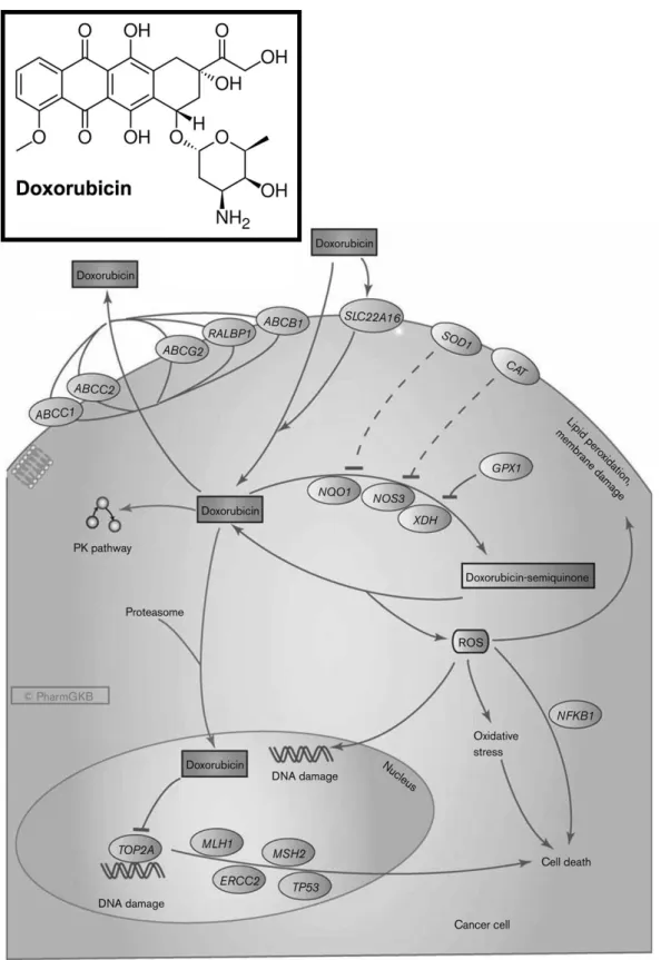
in the treatment of breast, lung, and gastric cancers (Thorn et al., 2011). Doxorubicin (Figure 6) belongs to a class of compounds called anthracyclines and has a planar structure that intercalates between neighboring DNA pairs anchored to one side through a covalent bond to one or more sugar units, and establishes formaldehyde and hydrogen bonds with a guanine on the opposing strand (Yang et al., 2014). The Doxorubicin intercalation in DNA promotes an increase in torsional stress, which can affect the nucleosomes structure and dynamics (Yang et al., 2014). Two major doxorubicin associated mechanisms of action (Figure 6) are generally described: i.) the disruption of topoisomerase-II repair and ii.) the generation of free radicals (Thorn et al., 2011, Yang et al., 2014).
Topoisomerases are enzymes responsible for regulate the DNA topology to facilitate DNA replication, transcription, and other nuclear processes (Nitiss, 2009). Particularly, topoisomerase II activity involves DNA entangling, and the cleavage of one strand of DNA duplex and the subsequent passage to a second duplex, through a transient cleavage (Swift et al., 2006). The anti-tumoral drug Doxorubicin impairs this cleavable complex, inhibiting the reconnection of the cleaved strands (Yang et al., 2014), which in turn triggers programmed cell death, i.e., apoptosis.
Other mechanism by which Doxorubicin can led to cell death is through the generation of free radicals (Keizer et al., 1990). The quinone structure can be oxidized by a number of nicotinamide adenine dinucleotide phosphate (NAD(P)H) oxidoreductases, the resulting semiquinones react quickly with oxygen and generate superoxide and hydrogen peroxide (Yang et al., 2014, Keizer et al., 1990). Doxorubicin easily binds to iron, and the formed complex catalyzes the hydrogen peroxide conversion into hydroxyl radicals (Thorn et al., 2011). The formed radicals can damage cell membranes, DNA, and proteins that can promote cell death.
10
Figure 6 – Doxorubicin molecular structure and the representation of its mechanisms of action (Adapted from Thorn et al., 2011).
Ibuprofen (Figure 7) is a non-steroidal anti-inflammatory drug (NSAID) that inhibits the cyclooxygenases 1 and 2 (COX-1 and COX-2). It has been applied in the treatment of several pathologies. Moreover, NSAIDs have been associated with cancer prevention, and NSAIDs such
11
as Aspirin and Ibuprofen promoted a significant anti—cancer activity (Marques et al., 2014, Baek et al., 2002). COX-2 expression activates the body inflammatory response, in the presence of stimulus such as traumas, foreign bodies, toxins, and bacteria, and their expression quickly results in the production of E-series prostaglandins (PGE) particularly PGE-2 (Harris et al., 2012). This inflammatory response, as all processes in human body is tightly controlled, but the continuous overexpression of COX-2 could initiate and promote carcinogenesis by several pathways (Figure 7) (Dannenberg et al., 2001). One option is the increase production of PGE-2 and other factors that promote cell proliferation. Also, the overexpression of COX-2 could increase the production of malondialdehyde and other oxygen reactive species (Nie et al., 2001). Moreover, it can stimulate the production of VEGF and PDGF promoting angiogenesis and metalloproteinases production, thus enhancing the invasive potential of cancer cells (Harris et al., 2012). Moreover, COX-2 can stimulate malignant cell proliferation through Bcl-2 stimulation, and also at the same time, contribute to inhibit the proliferation of B and T lymphocytes, reducing their antineoplastic activity (Nie et al., 2001, Harris et al., 2012).
Figure 7 – Ibuprofen molecular structure and COX-2 role in tumor development, cell-directed and microenvironment general effect (Adapted from Gupta and Dubois, 2001).
12
2. Nanotechnology and drug delivery systems
However, even using these combined therapies the side effects associated to drug administration are still prevalent. Moreover, the expected augmented therapeutic effect is not always obtained, due to problems that include rapid metabolism, poor solubility and inconsistent bioavailability (Greco and Vicent, 2009). These facts demonstrate the necessity to develop alternatives to conventional drug administration, in order to enhance their in vivo efficacy.
2.1. Nanosized delivery systems for delivery of bioactive
molecules
The application of Nanotechnology in healthcare is becoming a very common strategy. Moreover, it arises as one of the most compelling solutions to the problems faced by biotechnological and pharmaceutical industries in the development efficient and non-toxic cancer therapeutics (Akhter et al., 2013). The development of the so-termed nanomedicines offers the opportunity to overcome the several limitations associated to conventional drug delivery (Cho et al., 2008). These nanocarriers, i.e. delivery vehicles with nanoscale size can be easily tailored to possess unique compositions and functionalities that will improve the transported cargo therapeutic effect (Wang and Wang, 2014). Therefore, the nanocarriers can offer many advantages over free drug administration. They have the capacity to increase the solubility and at the same time protect bioactive molecules from premature degradation and interaction with blood components such as serum albumin (Davis et al., 2008). Furthermore, nanocarriers can improve the tissue penetration and accumulation, intracellular penetration and drug absorption in a selected tissue, improving their bioavailability (Alonso, 2004). Finally, the nanocarriers have the capacity to transport a large drug payload and control its release (Ganta et al., 2008). These advantages decrease the toxic side effects and promote an enhanced therapeutic outcome. Nevertheless, in order to be applied as delivery systems the nanocarriers need to possess an array of key properties that must be taken into account during nanodevices production process (Davis et al., 2008). One of the most important characteristics is particle size, that should be in the range of 10-200nm (Ernsting et al., 2013). The lower bound is the estimated size threshold where the particles are readily eliminated by kidneys, being excreted in urine (Ernsting et al., 2013). On the other side, the upper limit it is not so well defined but it is influenced by the tumor permeability and splenic filtration (Davis et al., 2008). Other important feature is morphology, in fact it has been described that nanocarriers geometry and surface orientation influence their cellular uptake (Herd et al., 2013). The surface properties are also a very important characteristic (Davis et al., 2008). Due to the high surface-volume ratios presented by nanocarriers, their surface properties play an important role in the interactions with the complex biological environment. Characteristics like
13
hydrophobicity and surface charge will influence the nanocarrier biological processing and fate (Ernsting et al., 2013). Changes in these parameters will modify their interaction with cells, proteins and even influence particle-particle agglomeration. Gessner et al., studied nanoparticles with decreasing surface hydrophobicity and their influence on plasma protein adsorption (Gessner et al., 2000). In this study the authors verified that a reduction in surface hydrophobicity led to decrease in protein adsorption (Gessner et al., 2000). The surface charge effect in nanocarrier interaction with cells is dependent of the cell type, probably because the differences verified in the molecules present in cell surface that will influence the cell-nanoparticle interaction (He et al., 2010a). But, in general, the particles with surface charge within ±10 mV showed optimal properties, exhibiting lower reticuloendothelial system interaction and extended circulation time (Ernsting et al., 2013).
2.2. Classes of nanocarriers
Due to the unique characteristics presented by nanosized systems, in the last years, several different types of nanocarriers have been developed to be applied in different therapies. The major classes of nanocarriers comprise (Figure 8): i.) liposomes, ii.) solid lipid nanoparticles, iii.) dendrimers, iv.) micelles, v.) polymeric nanoparticles and vi.) inorganic nanoparticles (including iron, gold, carbon and silica).
14
Figure 8 – General nanocarrier-based strategies employed for drug delivery, and their structure representation (Adapted from Mo et al., 2014). (A) Lipid-based nanocarriers; (B) Polymeric nanocarriers; and (C) Inorganic nanocarriers.
Liposomes were the first nanocarriers used to deliver drugs to cancer cells that were approved by European Medicines Agency (EMA) and Food and Drugs Administration (FDA) for cancer treatment (Wang and Thanou, 2010). Examples of these systems are Doxil® (Doxorubicin
encapsulated in a PEGylated liposome), DepoCyt (Cytarabine loaded liposome), Myocet (Doxorubicin loaded liposome) and Daunoxome (Daunorubicin loaded liposome) systems that have been used to treat cancer and other diseases (Zhang et al., 2011). They present a huge diversity of structure and compositions, but in general they are closed spherical vesicles constituted by a membranous lipid bilayer that surrounds an aqueous core compartment (Figure 8 A) (Al-Jamal and Kostarelos, 2011). It is worth to notice that the vesicles can be organized in single or multiple concentric bilayers (Allen and Cullis, 2013). Furthermore, the lipid bilayer
15
can be made from natural or synthetic phospholipids and cholesterol. These various combinations will in turn affect the liposome physicochemical properties, including their permeability, charge density and steric hindrance (Zhang et al., 2011). Other important characteristic is the liposomes capacity to load hydrophilic or hydrophobic bioactive molecules (Yang et al., 2011). Although, it is important to notice that liposomes present some limitations in their in vivo application. Some of the observed problems are correlated with short blood circulation time, in vivo instability, low solubility, opsonization and content leakage (Akbarzadeh et al., 2013).
Solid lipid nanoparticles (SLNs) are made from solid lipids stabilized by surfactants (Mehnert and Mader, 2001). SLNs are solid at room temperature and body temperature, can be comprised highly purified triglycerides (tricaprin, trilaurin, tripalmitin and others), complex glyceride mixtures (glyceryl palmitostearatea and glyceryl monostearate) or even waxes (cetyl palmitate) (Wissing et al., 2004). SLNs generally form structures that have a solid hydrophobic core having a layer of phospholipid coating (Figure 8 A) (Mehnert and Mader, 2001). Being the cargo dissolved or dispersed in the solid matrix, they possess the ability to carry lipophilic or hydrophilic bioactive compounds (Kaur et al., 2008). The solid nanoparticle properties are mainly influenced by their lipid composition, production method and surfactant type (Mehnert and Mader, 2001). But they present some advantages like their composition (physiological compounds), biocompatibility, and potential for large scale production (Mehnert and Mader, 2001). Furthermore, their content release can be modulated depending on the drug loading process (Almeida and Souto, 2007). On the other hand, these nanocarriers present some disadvantages namely their low drug loading capacity and presence of alternative colloidal structures, beyond nanoparticles micelles, liposomes and drug nanocrystals can also be formed (Mehnert and Mader, 2001). Moreover, the lipids can suffer transformations after the production process, also the sample dilution or water removal can change the particle stability and these modifications can originate premature drug release (Wissing et al., 2004).
Dendrimers are globular nanosized macromolecules with a characteristic branched structure that can be divided in three domains (Wijagkanalan et al., 2011). A core consisting in an atom or molecule, the interior shell formed by branches deriving from the core, and the terminal functional groups (Figure 8 B) (Frechet, 1994). These three domains can be tailored to serve various purposes, such as drug and gene delivery (Somani et al., 2014, Kesharwani et al., 2014). The high level of control over the dendrimer architecture, branching length and density, makes it easy to tailor their size, shape, and surface functionality (Svenson and Tomalia, 2005). However, they present immunogenicity, and also cationic dendrimers are highly cytotoxic hindering their application in the clinic (Lee et al., 2005).
Micelles are formed by blocks of copolymers consisting in hydrophilic and hydrophobic monomer units (Yih and Al-Fandi, 2006). Their hydrophobic core functions as a reservoir for poorly water-soluble drugs and the hydrophilic shell protects and controls the release of entrapped bioactive
16
molecules (Figure 8 B) (Zhang et al., 2011). Polymeric micelles have been reported as physiologically stable, biodegradable, with a surface suitable to be functionalized with cell targeting ligands, and with a long half-life in the body (Cho et al., 2008). Despite these valuable properties, micelles still show poor penetration into solid tumors, and also a burst drug release is verified in some micellar formulations (Miller et al., 2013).
Polymeric nanoparticles can be formed by synthetic or natural polymers. Moreover, the drugs can be immobilized on their surface or encapsulated in the polymeric structure, which gives the possibility to transport a wide range of therapeutics including drugs, proteins and nucleic acids (Faraji and Wipf, 2009). Most polymeric nanoparticles are biodegradable and biocompatible, present a surface suitable to be functionalized with various moieties and tunable drug release (Parveen et al., 2012).
Inorganic nanoparticles (Figure 8 C) comprise carbon nanotubes, gold nanoparticles, magnetic nanoparticles, mesoporous silica nanoparticles (MSNs), and quantum dots (Ladj et al., 2013). These different types of inorganic nanocarriers possess unique features to be used as delivery carriers, like a robust and stable structure, high loading capacity and a surface easily modified with different components to give them multifunctional capabilities (Jia et al., 2013). Furthermore, inorganic nanoparticles can exhibit imaging capacities through their magnetic properties and photothermal capabilities (Liong et al., 2008). However, the inorganic nanocarriers present some drawbacks, since they have a low biocompatibility and some aggregation issues (Ladj et al., 2013).
2.2.1. Mesoporous silica nanoparticles
Among the different carriers types presented above, ceramic particles have also been presented as a very interesting carriers. They have been highlighted due to their mechanical strength, chemical stability, porosity, relative biocompatibility and their resistance to microorganisms (Rosenholm et al., 2010). Moreover, the ceramic matrix does not suffer swelling or porosity changes, and also it is capable to protect the guest molecules from the action of enzymes and degradation resulting from pH or temperature (Rosenholm et al., 2010).
Inside the different ceramic particles, mesoporous silica nanoparticles have attracted a significant research attention for their potential application in Nanomedicine (Figure 9). A particular type of MSNs, mobil crystalline materials (MCM-41), contain a characteristic honeycomb-like porous structure with a large number of empty channels (mesopores) running from one end of the structure to the other without interconnectivity. They also possess unique properties like tunable particle size, stable and rigid framework (compared to polymer based nanocarriers, MSNs are more resistant to pH, heat and mechanical stress), a high surface area (>700m2/g), large pore volume (>0.6cm3/g), uniform and tunable pore size (2-10nm) and good
chemical and thermal stability (Li et al., 2012, Tang et al., 2012). Moreover, their large surface area, pore volume and the possibility to use the optimal solvent with no negative consequences
17
for the particle allows high loadings of therapeutic biomolecules with great efficacy (Slowing et al., 2008).
Figure 9 – Mesoporous silica nanoparticles general structure, and their cargo loading and possibilities of surface functionalization (Adapted from Rosenholm et al., 2010). Silica nanoparticles are capable to encapsulate several different biomolecules and are easily to functionalize with polymers and other components that will confer specific properties.
18
2.2.1.1.
Chemical production of MSNs – The Stöber modified method
In 1968, Stöber and collaborators applied an effective method for the controlled growth of uniform silica particles, which involves the hydrolysis of tetra alkyl silicates in a mixture of alcohol and water using ammonia as a catalyst (Stöber et al., 1968). Actually, most of the reported synthesis processes for mesoporous silica nanoparticles are based in the Stöber method. Generally they involve the use of an organosilane precursor (e.g. tetramethyl orthosilicate (TMOS) and tetraethyl orthosilicate (TEOS)), a cationic surfactant hexadecyltrimethylammonium bromide (CTAB), that will work as a structure guiding agent, water as solvent, and sodium hydroxide as morphological catalyst (Slowing et al., 2008). Afterwards, the template surfactant (CTAB) is removed by solvent extraction (hydrochloric acid (HCl) in alcohol solution) or calcination to originate nanopores. The particle formation in this process occur by base-catalyzed sol–gel condensation around the hexagonally packed micelle structures.
2.2.1.2.
Surface functionalization of MSNs
Beyond the above presented characteristics, MSNs also present a modifiable surface, which is easy to functionalize with various types of biomolecules, including fluorescent dyes, antibodies, peptides, proteins, surface charge tuning molecules and others (Figure 9) (Wu et al., 2014). Moreover, it can be considered that MSNs have two surfaces that can be functionalized, an internal surface (cylindrical surface pores) and external surface (exterior particle surface) (Slowing et al., 2008). This interestingly feature allows a selective particle functionalization, where the surface to functionalize can be chosen accordingly to a particular application and also allows the use of multiple moieties in external and internal surfaces (Slowing et al., 2008). Regarding surface functionalization two different methods are generally used, condensation and chemical grafting (Slowing et al., 2008). In the condensation method, organic alkoxysilanes are added to the synthesis reaction and bonded to the particle during its assembly (Radu et al., 2005). In the grafting method, the functionalization occurs post synthesis, and the chosen moiety binds to the particle surface silanol groups (He et al., 2010c). In order to use the grafting method, it is important to not use calcination as the purification process, since it promotes the condensation of MSNs silanol groups reducing the number of groups available for functionalization (Slowing et al., 2008).
2.2.1.3.
Mesoporous silica nanoparticles uptake and biocompatibility
The nanocarrier cellular uptake is a very important process in the delivery of anti-tumoral drugs via the action of nanocarriers. Unmodified MSNs present affinity for some of the head-groups of cell membrane phospholipids, particularly for the positive charged ones like 1,2-dioleoyl-3-trimethylammonium-propane. This affinity to the cell surfaces greatly facilitates the uptake process (Mornet et al., 2005). Moreover, further studies demonstrated that MSNs uptake is dependent on size, shape and surface functionalization, but it mainly occurs through the clathrin-coated endocytosis pathway and trough pinocytosis (Figure 10) (Huang et al., 2010).
19
Other uptake routes for MSNs can be also verified, like caveolin-dependent and receptor mediated (Li et al., 2012). The surface shape can also affect MSNs uptake, Trewyn et al. found that spherical and rod shape MSNs needed 180 min and 360 min, respectively, to be completely internalized by cells (Trewyn et al., 2008).
Figure 10 – Pathways used by mesoporous silica nanoparticles for cellular internalization (Adapted from Vivero-Escoto et al., 2010). The uptake pathway will be depend from the physicochemical properties possessed by the MSNs.
Other important parameter for assessing the applicability of MSNs is their biocompatibility. MSNs surface charge and size largely influence their toxicity. Concerning particle size it was demonstrated by Napierska and coworkers that particles with size lower than 50 nm induced cell death and even necrosis in human endothelial cells, whereas particles above 100 nm presented minor toxicity (Napierska et al., 2009). It is worth to notice that larger particles and even particles with rod morphology have higher cytotoxicity, since these particles cause a great disorder in F-actin formation and therefore disturbance in the organization of the cytoskeleton and cell membrane (Huang et al., 2010). This fact can lead to cell membrane disruption and cell death (Huang et al., 2010). Nanoparticle surface charge can also affect MSNs biocompatibility, Shahbazi et al. showed that negatively charged MSNs (-31 mV) produced less adenosine triphosphate (ATP) depletion and genotoxicity than those positively charged (32 mV) (Shahbazi et al., 2013).
In general, MSNs are reported to be safe in concentrations lower than 100 µg/mL, which is superior to the particle concentrations needed in most therapeutic treatments (Rosenholm et al., 2010). Furthermore, in this concentration range the morphology of healthy cells and
20
membrane integrity is conserved (Slowing et al., 2008). Also the growth rates remain unchanged indicating that no damage to the cells replication machinery occurs (Slowing et al., 2008).
2.3. Administration routes and barriers
Nanocarriers can be administered by several different administrations routes such as nasal, ocular, oral, intradermic and intramuscular or intravenous (Rabanel et al., 2012). Moreover, depending on the chosen route of administration the nanocarriers will have to surpass several barriers in order to reach the desired site (Ferrari, 2010). Therefore, as above mentioned, their size and surface properties assume a critical role in their ability to overcome these major obstacles upon delivery in human body (Figure 11).
One route of administration of MSNs is the intravenous injection. Which is the quickest and simplest method for delivering therapeutics to systemic circulation, and it is a relatively invasive approach that reduces the losses associated to other approaches like nasal, ocular and oral (Cheng et al., 2008b). However, this route has a variety of barriers associated with, that difficult an effective nanoparticles delivery.
The reticuloendothelial system (RES) is a global system comprised by phagocytic cells in the liver, spleen, and bone marrow, whose primary function is to eliminate foreign objects, such as microbes and also nanocarriers (Ernsting et al., 2013). The RES does not have the capacity to recognize these foreign bodies, first they have to be coated by a protein layer in a process called opsonization (Steichen et al., 2012). These proteins called opsonins adhere to the foreign particles by ionic, the electrostatic and hydrophobic forces and can be immunoglobulins, components from complement system (C3,C4 and C), fibronectin, and others (Steichen et al., 2012). The macrophages will recognize the opsonin coated particles and will attack them leading to their clearance from circulation (Elsabahy and Wooley, 2012).
Other important barrier is the first pass renal filtering, where the kidneys filter the blood through the glomerular wall, and normally particles with size smaller than 8 nm are rapidly eliminated from circulation (Ernsting et al., 2013). The particle excretion is also observed in the liver and spleen, where particles with size higher than 200 nm are cleared into bile, and then into feces (Elsabahy and Wooley, 2012). On the other hand it is also crucial that particles extravasation to the tumor site occur. This process is largely influenced by the heterogeneous blood flow and high tumor interstitial pressure (Ernsting et al., 2013). The heterogeneous blood flow arises as result of the characteristic aberrant an unorganized tumor vasculature, that will difficult the uniform particle dispersion in the tumor (Serres et al., 2014). The high tumor interstitial pressure is promoted by the high vascular permeability and lack of lymphatic drainage, and as the pressure increases in the tumor center it inhibits the drug accumulation and dispersion in the diseased tissues (Ernsting et al., 2013).
21
One last barrier that the particles have to surpass is intracellular trafficking to the site of action, where the particles must be internalized, transpose the cell membrane through the complex cell cytoplasm (Chithrani and Chan, 2007, Ruenraroengsak et al., 2010). Subsequently, the particles have to be capable of escaping from lysosomes and protect their cargo from the action of intracellular enzymes in order to assure its therapeutic efficacy.
Figure 11 – Physical characteristic of nanoparticles that determine their biocompatibility and capacity to surpass certain barriers (Adapted from Nel et al., 2009). Red representing likely toxicity, blue likely safety and blue–green–yellow intermediate levels of safety.
2.4. Nanocarriers targeting to tumor tissues
2.4.1. Passive Targeting
As described above the rapid vascularization in tumors results in leaky, and defective vasculatures and impaired lymphatic drainage (Nie et al., 2007). Therefore the combination between the large gap sizes in vessels (100 nm to 2 µm) with poor lymphatic drainage allows high retention times for particles that gain interstitial access to tumors, an effect known as Enhanced Permeability and Retention (EPR) effect (Figure 12 A) (Byrne et al., 2008). Nanoparticles smaller than the defective fenestrations (400-600 nm) can escape from the vasculature and accumulate in the tumor. Actually, the EPR effect is present in almost all the tumors with exception for the hypovascular ones, such as prostate or pancreatic tumors (Danhier et al., 2010).
In order to really benefit from the EPR effect and increase the possibilities to accumulate in the tumor, the nanocarriers need to remain in circulation as much time as possible (Ernsting et al., 2013). The most commonly chosen method is the nanocarrier PEGylation (Owens and Peppas, 2006). The nanocarrier PEGylation refers to the particle decoration by covalently grafting, entrapping or adsorbing polyethylene glycol (PEG) molecules (Owens and Peppas, 2006). PEG is FDA approved polymer described as a nontoxic, non-immunogenic, non-antigenic,
22
and a highly soluble in water (Veronese and Pasut, 2005). The PEG chains create a barrier layer that blocks opsonins adhesion, making the particles remain “camouflaged” or “invisible” to phagocytic cells (Greenwald et al., 2003). Furthermore, it will promote a prolonged residence in body and a decreased degradation by metabolic enzymes (Veronese and Pasut, 2005). He et al. studied the effect of MSNs PEGylation on nonspecific binding of serum proteins and cellular responses, applying PEGs with different sizes (He et al., 2010c). In their results they verified that all the tested molecular weights influenced the nonspecific binding to human serum protein (HSA), and also red blood cells hemolysis.
Another alternative to passively target MSN to tumors is the localized delivery (Parveen et al., 2012). In accessible tumors like breast, colon, prostate and neck can be realized a direct intra-tumoral delivery of nanocarriers or therapeutic agents (Parveen et al., 2012), avoiding systemic circulation and the majority of biological barriers.
2.4.2. Active Targeting
Active targeting is usually achieved by nanocarrier conjugation with a targeting component, which will promote a preferential accumulation in the tumor itself, in the tumor-bearing organ or in individual cancer cells (Figure 12 B) (Nie et al., 2007). This approach takes advantage of ligand-receptor, antigen–antibody and other forms of molecular recognition to privilege one specific site in the target cells (Steichen et al., 2012). The targeting component is chosen to bind to a unique molecule overexpressed by the tumor and at the same time it is not expressed or presents a limited expression in normal cells (Danhier et al., 2010). This active targeting strategy has the potentiality to enhance the therapeutic effectiveness, and at the same time decreases the delivery of chemotherapeutic molecules to healthy cells (Steichen et al., 2012). Consequently, minimizing the potential side effects.
23
Figure 12 – Nanocarriers targeting, passive vs active targeting strategies (Adapted from Danhier et al., 2010a). In (A) passive targeting, nanocarriers advantages over the free drug administration. In (B) possibility to target different cells associated to tumor development.
Focusing on prostate cancer, there are different molecules that can be used for nanoparticle targeting as shown in Figure 13. One of the major molecules associated to prostate cancer is the prostate-specific membrane antigen (PSMA) (Romero Otero et al., 2014). PSMA is a transmembrane protein produced nearly exclusively by prostate epithelial cells, and it is overexpressed in prostate cancer and other nonmalignant prostate conditions (Romero Otero et al., 2014). Other possible target is the prostate stem cell antigen (PSCA), this molecule is highly upregulated in prostate cancer and can be targeted by antibodies (Guo et al., 2013).