Pesq. agropec. bras., Brasília, v.50, n.2, p.97-105, fev. 2015 DOI: 10.1590/S0100-204X2015000200001
Marcos Fernando Basso(1), Álvaro Júlio Pereira(2), Hermano Monteiro de Barros Pereira(1),
Humberto Josué de Oliveira Ramos(3), Jorge Luiz Loyola Dantas(4), Elizabeth Pacheco Batista Fontes(3),
Eduardo Chumbinho de Andrade(4) and Francisco Murilo Zerbini(1)
(1)Universidade Federal de Viçosa (UFV), Departamento de Fitopatologia, CEP 36570‑900 Viçosa, MG, Brazil. E‑mail: [email protected], [email protected], [email protected] (2)Universidade Estadual do Ceará, Departamento de Ciências Biológicas, CEP 62500‑000 Itapipoca, CE, Brazil. E‑mail: [email protected] (3)UFV, Departamento de Bioquímica e Biologia Molecular. E‑mail: [email protected], [email protected] (4)Embrapa Mandioca e Fruticultura, Rua Embrapa, s/no, CEP 44380‑000 Cruz das Almas, BA, Brazil. E‑mail: [email protected], [email protected]
Abstract – The objective of this work was to produce a polyclonal antiserum against the coat protein (CP) of Papaya lethal yellowing virus (PLYV) and to determine its specificity and sensibility in the diagnosis of the virus, as well as to evaluate the genetic resistance to PLYV in papaya (Carica papaya) accessions and to investigate the capacity of the two-spotted spider mite Tetranychus urticae to acquire and transmit PLYV to the plants. Sixty‑five papaya accessions were evaluated. For each accession, ten plants were mechanically inoculated using PLYV‑infected plant extracts, and three plants were mock inoculated with phosphate buffer alone and used as negative controls. Ninety days after inoculation, newly‑emerging systemic leaves were collected from the inoculated plants, and viral infection was diagnosed by indirect Elisa, using polyclonal antiserum sensible to the in vitro-expressed PLYV CP. Viral transmission by T. urticae was evaluated in greenhouse. The experiments were repeated twice. Polyclonal antiserum recognized the recombinant PLYV CP specifically and discriminated PLYV infection from infections caused by other plant viruses. Out of the 65 papaya accessions evaluated, 15 were considered resistant, 18 moderately resistant, and 32 susceptible. The two‑spotted spider mite T. urticae was capable of acquiring PLYV, but not of transmitting it to papaya.
Index terms: Carica papaya, Elisa, genetic resistance, plant breeding, PLYV, two‑spotted spider mite.
Identificação de acessos de mamoeiro resistentes ao
Papaya lethal yellowing
virus
e capacidade de
Tetranychus urticae
em transmitir o vírus
Resumo – O objetivo deste trabalho foi produzir um antissoro policlonal contra a proteína capsidial (PC) do Papaya lethal yellowing virus (PLYV) e determinar sua especificidade e sensibilidade na diagnose do vírus, bem como avaliar a resistência genética de acessos de mamoeiro (Carica papaya) ao PLYV e investigar a capacidade do ácaro rajado Tetranychus urticae em adquirir e transmitir o vírus às plantas. Foram avaliados 65 acessos de mamoeiro. Para cada acesso, dez plantas foram submetidas à inoculação mecânica com extratos de plantas infectadas com PLYV, e três plantas receberam inoculação apenas com tampão de fosfato e foram usadas como controle negativo. Noventa dias após a inoculação, novas folhas sistêmicas emergentes foram coletadas das plantas inoculadas, e a infecção viral foi diagnosticada por Elisa indireto, com uso de antissoro policlonal sensível à PC do PLYV expressa in vitro. A transmissão viral por T. urticae foi avaliada em casa de vegetação. Os experimentos foram repetidos duas vezes. O antissoro policlonal reconheceu a PC do PLYV especificamente e discriminou a infecção pelo PLYV de infecções causadas por outros vírus. Dos 65 acessos de mamoeiros avaliados, 15 foram considerados resistentes, 18 moderadamente resistentes e 32 suscetíveis. O ácaro rajado T. urticae foi capaz de adquirir o PLYV, mas não de transmiti‑lo para o mamoeiro.
Termos para indexação: Carica papaya, Elisa, resistência genética, melhoramento de plantas, PLYV, ácaro rajado.
Introduction
Papaya (Carica papaya L.) produced in the Brazilian Northeast might be affected by a lethal yellowing caused by Papaya lethal yellowing virus (PLYV), a tentative
member of the genus Sobemovirus (Nascimento et al.,
greenish spots that become yellow with ripening.
PLYV infection is one of the most significant viral infections in this Brazilian region (Lima et al., 2013) and may cause significant losses worldwide.
PLYV has been tentatively assigned to the genus Sobemovirus (Truve & Fargette, 2012), which is comprised by viruses with isometric particles of
approximately 30 nm in diameter and a genome composed of one single‑stranded, positive‑sense RNA molecule. The PLYV genome is 4,145 nt long and is organized in four open reading frames (ORFs): ORF1, putative movement protein and silencing suppressor; ORF2a, serine protease and VPg; ORF2b, RNA‑dependent RNA polymerase; and ORF3, coat protein. The coat protein (CP) consists of 278 amino
acids and has a deduced molecular weight of
approximately 37 kDa (Pereira et al., 2012).
PLYV is easily mechanically transmitted to plants of the Caricaceae family and it can also be transmitted
by contaminated hands, which shows the high stability of the virus (Amaral et al., 2006; Saraiva et al., 2006). PLYV is found in contaminated soils, irrigation water, dry leaves, leaf and root debris, and on seed surface of infected fruit, but there are no reports of transmission through the seed embryo (Camarço et al., 1998; Saraiva et al., 2006; Nascimento et al., 2010). To date, no vector has been identified for this virus (Lima et al., 2013). The
bugs Myzus persicae Sulz, Diabrotica bivitulla Kirke, and D. speciosa Kirke were not capable of transmitting
it (Kitajima et al., 1992; Lima et al., 2001).
The two-spotted spider mite Tetranychus urticae (Arachnida: Tetranychidae) is an economic pest
worldwide, which may also be capable of transmitting plant viruses (Thomas, 1969). Given the high
incidence of this mite in papaya and the fact that three different types of vectors have already been reported
for sobemoviruses (aphids, beetles, and a mirid) (Truve & Fargette, 2012), it would be reasonable to
test T. urticae capability to acquire and transmit PLYV to papaya.
To detect PLYV, the reverse transcription‑polymerase
chain reaction (RT-PCR) and enzyme-linked
immunosorbent assay (Elisa) methods are used. Although Elisa is less sensitive than RT‑PCR, it is more
suitable for routine use with a large number of samples.
However, it requires a high‑quality antibody to achieve specificity and sensitivity. The quality of an antibody
is directly related to the purity and structural integrity
of the antigen. Recombinant proteins expressed in prokaryotic systems (generally Escherichia coli) are
frequently used in research because they are stable, abundant, and easily purified. Due to the low genetic
variability in the CP gene among isolates of PLYV
(Daltro et al., 2012), an antiserum against the CP of a specific virus isolate may detect different isolates. Furthermore, the antiserum would constitute an
important tool for papaya germplasm screening for genetic resistance to PLYV.
The only effective control measure for PLYV is
the eradication of symptomatic plants. Therefore, for viral diseases, planting resistant cultivars is the most effective control method (Gómez et al., 2009). Currently, almost all the areas dedicated to commercial
papaya production in Brazil are planted with only three
cultivars, which are members of two groups: the Solo Group, represented by the Sunrise Solo and Improved Sunrise Solo 72/12 cultivars; and the Formosa Group, represented by the Tainung number 1 cultivar. This evidences an extremely narrow genetic base, which
renders this crop much vulnerable to diseases and
other stresses (Santos, 2009). Efficient and consistent detection methods and the identification of sources of
resistance could contribute to reduce disease incidence and economic losses due to PLYV.
The objective of this work was to produce a polyclonal antiserum against PLYV CP and to determine its
specificity and sensibility in the diagnosis of the virus,
as well as to evaluate the genetic resistance to PLYV in papaya accessions and to investigate the capacity of the two-spotted spider mite T. urticae to acquire and transmit PLYVto the plants.
Materials and Methods
Standard procedures were performed to produce polyclonal antiserum against the in vitro-expressed
PLYV CP (Sambrook & Russel, 2001). Viral RNA was extracted from the leaves of infected papaya plants,
displaying typical symptoms of lethal yellowing
disease, using the RNeasy Plant Mini Kit (Qiagen Biotecnologia Brasil Ltda., São Paulo, SP, Brazil). The full‑length CP ORF of PLYV isolate 21 (GenBank accession number JQ394925) was amplified by
RT-PCR with the oligonucleotides PLYVcpBamHI
Pesq. agropec. bras., Brasília, v.50, n.2, p.97‑105, fev. 2015 DOI: 10.1590/S0100‑204X2015000200001
ATT CTT ATA GGT TTA GAG CAG ATG‑3’, reverse), cloned into the BamHI and EcoRI site of the pRSET_A
expression vector (Life Technologies do Brasil Ltda., São Paulo, SP, Brazil), and sequenced to confirm the integrity and correct orientation of the insert. For in vitro expression, the construct pRSET‑CP‑PLYV was
transformed into the E. coli strain BL21:DE3 using a
heat‑shock procedure, and a single colony was grown at 37°C in 200 mL LB/ampicillin until it reached an OD600 of approximately 0.5, when the synthesis
of the recombinant protein was induced with
2 mmol L‑1 isopropyl‑β‑D‑thiogalactopyranoside (IPTG). Six hours after induction, bacterial cells were collected by centrifugation at 5.000 g for 10 min. A total protein extract was obtained by re-suspension in
lysis buffer (50 mmol L‑1 Tris‑HCl, 100 mmol L‑1 NaCl, 2 mmol L‑1 EDTA, pH 8.0), lysozyme treatment, and sonication (Fajardo et al., 2007). The total protein extract was re‑suspended in 1 mL of 100 mmol L‑1 NaHCO3, pH 9.0, plus 0.5% SDS (w/v), and the recombinant CP was purified by affinity chromatography using a Ni‑NTA column (Qiagen Biotecnologia Brasil Ltda., São Paulo, SP, Brazil), according to the manufacturer’s instructions, under nondenaturing conditions. After dialysis in 10 mmol L‑1 phosphate buffer, pH 7.4, plus 0.425% NaCl (w/v), protein integrity was analyzed by SDS‑Page and quantification was performed with the NanoDrop ND‑1000 spectrophotometer (Thermo Fisher Scientific Brasil Instrumentos e Processo Ltda., São Paulo, SP, Brazil), according to the manufacturer’s
instructions. Protein identity was determined using ion
trap mass spectrometry, according to Shevchenko et al. (2006).
For immunization, 250, 400, 550, 700, and 850 µg
of the in vitro-expressed protein were injected
intramuscularly into the hind legs of two white, 40‑day‑old, New Zealand rabbits, at weekly intervals. The first injection was performed with complete Freund’s adjuvant (1:1 v/v), and the four remaining injections, with incomplete Freund’s adjuvant (1:1 v/v). One week after the last injection, seven weekly bleedings were carried out (25–30 mL per bleeding). Blood samples were incubated for 1 hour at 37ºC, followed by 2 hours at 4ºC, in order to coagulate, and then were centrifuged at 3.500 g for 10 min. The supernatant (antiserum) was
aliquoted and stored at ‑20ºC.
The antiserum specificity against the recombinant PLYV CP was confirmed by Western blot (Fajardo
et al., 2007). The specificity, sensitivity, and optimal
concentration of the anti-CP serum were also evaluated
using indirect Elisa, according to Almeida & Lima (2001). The antiserum was diluted to 1:500, 1:1,000, 1:5,000, and 1:10,000, and tested against protein
extracts from newly-emerging symptomatic papaya leaves. Samples were considered infected when the
absorbance at 405 nm was at least twice the average
value of the negative controls (extract from healthy
papaya). To confirm the antiserum specificity, an additional indirect Elisa was carried out for total
protein extracts from papaya plants infected with the potyvirus Papaya ringspot virus (PRSV).
Sixty‑five papaya accessions were evaluated, obtained from the active germplasm bank of Embrapa Mandioca e Fruticultura, located at Cruz das Almas, in the state of Bahia, Brazil (Table 1). The experiment was conducted in greenhouse, on the campus of Universidade Federal de Viçosa, in the state of Minas Gerais, Brazil. Ten plants of each accession were inoculated in a completely randomized design, and the experiment was repeated twice. Plants with 5–8 leaves were mechanically inoculated twice (15 day interval) with PLYV isolate 21 (JQ394925). The inoculation was performed via PLYV‑infected plant extract, ground in 0.05 mol L‑1 sodium phosphate buffer, pH 7.5, with 0.1% sodium sulfite in 1:2 proportions (weight of
leaves per volume of buffer). The extract was rubbed on the surface of leaves previously sprinkled with
aluminum oxide (600 mesh). Three plants of each
accession were mock inoculated with phosphate buffer
alone (no PLYV‑infected plant extract), and was used
as negative controls.
Ninety days after the first inoculation,
newly-emerging systemic leaves (non-inoculated)
were collected, and viral infection was diagnosed by indirect Elisa, initially using a polyclonal PLYV CP
antiserum kindly provided by Professor José Albérsio
de Araújo Lima from Universidade Federal do Ceará,
and then using the antiserum produced in the present study. Samples with an absorbance value at least twice as higher than that of the negative controls (healthy plants) were considered positive for the presence of PLYV. A given accession was considered “resistant” if
less than 25% of the inoculated plants were infected; “moderately resistant” if 25–50% of the inoculated
plants were infected; and “susceptible” when more
Table 1. Characteristics of the papaya (Carica papaya) accessions from the active germplasm bank of Embrapa Mandioca e Fruticultura, evaluated as sources of resistance to Papaya lethal yellowing virus (PLYV).
Accession Species Common name Original source Origin Classification(1)
CMF 11 Carica papaya DCG4403 Cenargen, Brasília Costa Rica Moderately resistant
CMF 12 Carica papaya DCG5956 Cenargen, Brasília Malaysia Resistant
CMF 14 Carica papaya DCG5908 Cenargen, Brasília Malaysia Resistant
CMF 15 Carica papaya DCG5863 Cenargen, Brasília Malaysia Resistant
CMF 18 Carica papaya DCG4246 Cenargen, Brasília Taiwan Resistant
CMF 20 Carica papaya DCG4244 x 4391 Cenargen, Brasília Brazil Susceptible CMF 21 Carica papaya Solsun Cenargen, Brasília Brazil Moderately resistant CMF 22 Carica papaya DCG5903 – Sunrise Cenargen, Brasília Malaysia Moderately resistant
CMF 24 Carica papaya Conchita EBDA, Conceição do Almeida, Bahia Costa Rica Moderately resistant CMF 26 Carica papaya DCG4224 EBDA, Conceição do Almeida, Bahia Taiwan Resistant CMF 27 Carica papaya DCG432 EBDA, Conceição do Almeida, Bahia -(2) Moderately resistant CMF 28 Carica papaya DCG439 EBDA, Conceição do Almeida, Bahia Costa Rica Susceptible CMF 30 Carica papaya DCG4344 EBDA, Conceição do Almeida, Bahia - Resistant CMF 31 Carica papaya DCG441 EBDA, Conceição do Almeida, Bahia Costa Rica Susceptible CMF 33 Carica papaya DCG539 EBDA, Conceição do Almeida, Bahia - Resistant CMF 36 Carica papaya Guinea – GoldxSel.Mexicana EBDA, Conceição do Almeida, Bahia Brazil Resistant CMF 38 Carica papaya JS3 EBDA, Conceição do Almeida, Bahia Brazil Moderately resistant CMF 44 Carica papaya JS21 EBDA, Conceição do Almeida, Bahia Brazil Resistant CMF 46 Carica papaya S3 EBDA, Conceição do Almeida, Bahia Brazil Resistant CMF 47 Carica papaya S15 EBDA, Conceição do Almeida, Bahia Brazil Resistant CMF 52 Carica papaya Solo EBDA, Conceição do Almeida, Bahia Brazil Moderately resistant CMF 54 Carica papaya PRI065 x Tailândia EBDA, Conceição do Almeida, Bahia Hawaii Resistant CMF 56 Carica papaya 7212 x Maradol EBDA, Conceição do Almeida, Bahia Brazil Moderately resistant CMF 59 Carica papaya Malaysian Yellow 422 EBDA, Conceição do Almeida, Bahia Hawaii Susceptible CMF 60 Carica papaya Sunrise Cross 2 EBDA, Conceição do Almeida, Bahia Hawaii Moderately resistant CMF 65 Carica papaya K77xJSI2 EBDA, Conceição do Almeida, Bahia Brazil Moderately resistant CMF 72 Carica papaya FERREIRA 87 EBDA, Conceição do Almeida, Bahia - Susceptible
CMF 76 Carica papaya MangaMourão - - Moderately resistant
CMF 82 Carica papaya Hortus Gold University of Natal South Africa Susceptible
CMF 88 Carica papaya Kapoho Purple Hawaii (Hilo) Hawaii Susceptible
CMF 92 Carica papaya Kapoho Green Hawaii (Hilo) Hawaii Susceptible
CMF 94 Carica papaya - Cenargen, Brasília Brazil Susceptible
CMF 102 Carica papaya - Cruz das Almas, Bahia Brazil Susceptible
CMF 108 Carica papaya SEED546 - South Africa Moderately resistant
CMF 114 Carica papaya SEED1216 - South Africa Susceptible
CMF 115 Carica papaya SEED1250 - South Africa Susceptible
CMF 116 Carica papaya SEED1291 - South Africa Susceptible
CMF 120 Carica papaya Faz. Caminhoá Cruz das Almas, Bahia Brazil Moderately resistant
CMF 121 Carica papaya - - - Moderately resistant
CMF 123 Carica papaya Vermelho Thai Cenargen, Brasília Thailand Susceptible CMF 129 Carica papaya - Embrapa Amazônia Ocidental, Amazonas Brazil Susceptible
CMF 130 Carica papaya - - - Susceptible
CMF 132 Carica papaya Seleção ≠2 Cenargen, Brasília Hawaii Susceptible
CMF 142 Carica papaya 16x17 - - Susceptible
CMF 145 Carica papaya Sergipe Verde x 6 - - Susceptible
CMF 150 Carica papaya Golden Teixeira de Freitas, Bahia Brazil Resistant CMF 154 Carica papaya Maradol Gua Cenargen, Brasília Guatemala Susceptible CMF 155 Carica papaya FRF1421 – Common papaya Barra do Garças, Mato Grosso do Sul Brazil Susceptible CMF 164 Jaracatia spinosa FRF1434 Jaracatia Antônio João, Mato Grosso do Sul Brazil Susceptible CMF 165 Jaracatia spinosa FRF1435 Jaracatia Bela Vista, Mato Grosso do Sul Brazil Susceptible CMF 166 Carica papaya FRF1436 – Common papaya Bela Vista, Mato Grosso do Sul Brazil Susceptible CMF 172 Carica papaya FRF1442 – Common papaya Parnaíba, Mato Grosso do Sul Brazil Resistant CMF 175 Carica papaya FRF1445 – Common papaya Rio Verde, Goiás Brazil Susceptible CMF 176 Carica papaya FRF1446 – Common papaya Rio Verde, Goiás Brazil Moderately resistant CMF 183 Carica papaya FRF1427 – Common papaya Cenargen, Brasília Brazil Susceptible CMF 186 Carica papaya FRF1434 – Common papaya Cenargen, Brasília Brazil Susceptible CMF 187 Carica papaya FRF1435 – Common papaya Cenargen, Brasília Brazil Susceptible CMF 200 Carica papaya FRF1454 – Common papaya Cenargen, Brasília Brazil Susceptible CMF 204 Carica papaya FRF1473 – Common papaya Cenargen, Brasília Brazil Moderately resistant CMF 206 Carica papaya FRF1475 – Common papaya Cenargen, Brasília Brazil Susceptible CMF 220 Carica papaya FRF1508 – Common papaya Cenargen, Brasília Brazil Moderately resistant CMF 223 Carica papaya FRF1520 – Common papaya Cenargen, Brasília Brazil Resistant CMF 230 Carica papaya Solo papaya – Ouromel Porto Seguro, Bahia Brazil Susceptible CMF 234 Carica papaya Solo papaya – BS – Faz. SF Teixeira Freitas, Bahia Brazil Moderately resistant CMF 235 Carica papaya Solo papaya – JTA Teixeira Freitas, Bahia Brazil Susceptible
(1)The accessions were evaluated by indirect Elisa at 90 days after inoculation and considered as: resistant, <25% of the inoculated plants were infected;
Pesq. agropec. bras., Brasília, v.50, n.2, p.97‑105, fev. 2015 DOI: 10.1590/S0100‑204X2015000200001 Figure 1. Expression of Papaya lethal yellowing virus (PLYV) coat protein (CP) in Escherichia coli (A): molecular weight markers in kDa (M), E. coli-produced and purified movement protein of Tomato yellow spot virus (1), total protein extract from IPTG‑induced E. coli culture transformed with pRSET‑CP‑PLYV (2), purified PLYV CP (3 and 4); and Western blot analysis using the anti‑PLYV CP polyclonal antiserum produced against the in vitro-expressed protein (B): PLYV CP expressed in E.coli (1), GFP expressed in E. Coli (2), total protein extract from healthy papaya (3), and total protein extract from PLYV-infected papaya (4).
To evaluate the capacity of the two-spotted spider mite T. urticae to acquire and transmit PLYV to
papaya, transmission experiments were conducted
in a greenhouse using two susceptible accessions
(CMF 130 and CMF 145), in a completely randomized
design. The experiment was repeated twice. The mites were reared on healthy papaya plants and transferred to PLYV-infected papaya during an acquisition access
period of 30 days; then, they were transferred to
susceptible healthy papaya and stayed on the plants until the end of the experiment. Plants were examined
for PLYV infection by indirect Elisa at 90 and 150 days
after the transfer of the spider mites. The spider mites maintained in infected plants were analyzed by
RT‑PCR, using the PLYVcpBamHI and PLYVcpEcoRI primers, and also by indirect Elisa, using the polyclonal
antiserum produced in the present study.
Viral RNA was isolated from samples comprised
of either ten adult spider mites or 30 nymphs, using TRIzol Reagent (Life Technologies do Brasil Ltda., São Paulo, SP, Brazil). The cDNA was synthetized
with SuperScript III Reverse Transcriptase kit (Life
Technologies do Brasil Ltda., São Paulo, SP, Brazil), and PCR was performed with the GoTaq DNA Polymerase kit (Promega, Madison, WI, USA), according to the manufacturer’s instructions. The amplified DNA was cloned into the pGEM‑T Easy vector and sequenced. The Elisa tests were performed as described by Almeida & Lima (2001), using 70 spider mites – adults or nymphs – processed in 200 µL of sodium carbonate 50 mmol L‑1, pH 9.6. Spider mites kept in healthy
papaya were used as negative controls in both RT-PCR
and Elisa assays.
Results and Discussion
The expression of the PLYV CP in E. coli BL21:DE3
resulted in an average protein yield of 17.32 μg mL‑1.
Recombinant fusion protein had a molecular mass
of approximately 38 kDa, which corresponds to the PLYV CP, plus 3 kDa from amino acid residues tagged
to its N-terminus. The molecular weight and identity
of the purified protein was confirmed by SDS‑Page (Figure 1 A) and mass spectrometry. The titer of
the polyclonal antiserum was initially evaluated by
Western blot analysis. The dilution of 1:1,000 resulted in high sensitivity and specificity for detecting in
vitro-expressed protein and viral protein from extracts
of PLYV‑infected papaya (Figure 1 B). Indirect Elisa
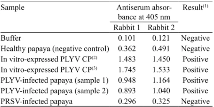
showed that the anti‑CP serum at a dilution of 1:1,000 was specific and sensible for detecting PLYV infection,
since no cross reaction was observed with protein
extracts from plants infected with PRSV (Table 1).
The expression of recombinant PLYV CP in E. coli
Table 2. Results of indirect Elisa to test polyclonal antiserum
produced against the in vitro-expressed coat protein (CP) of Papaya lethal yellowing virus (PLYV).
Sample Antiserum
absor-bance at 405 nm Result
(1)
Rabbit 1 Rabbit 2
Buffer 0.101 0.121 Negative
Healthy papaya (negative control) 0.362 0.491 Negative In vitro-expressed PLYV CP(2) 1.483 1.450 Positive In vitro-expressed PLYV CP(3) 1.745 1.533 Positive
PLYV‑infected papaya (sample 1) 0.948 1.164 Positive
PLYV‑infected papaya (sample 2) 0.893 1.040 Positive PRSV-infected papaya 0.296 0.325 Negative
(1)Readings performed 30 min after addition of substrate, with antiserum
diluted to 1:1,000 (v:v) in substrate buffer. (2)0.216 ng. (3)0.433 ng. PRSV,
Papaya ringspot virus (genus Potyvirus, family Potyviridae).
immunization of rabbits for antiserum production. The obtained results were similar to the average yields of recombinant coat proteins found for other viruses
(Fajardo et al., 2007; Basso et al., 2010). Good quality antiserum is essential for the efficiency of serological detection, particularly for a low titer virus such as PLYV (Nascimento et al., 2010). The PLYV CP antiserum
obtained in the present study showed sensitivity and absence of cross-reactions with other viruses or with plant extracts from healthy papaya. Similar results
were found with antiserum produced from purified viral particles (Nascimento et al., 2010).
An advantage of an antiserum produced against recombinant protein expressed in E. coli is that the E. coli culture can be stored indefinitely at ‑80oC and can be used to express protein for the production of
a new batch of antiserum, whenever necessary. This
allows not only for the production of large quantities
of antiserum, but also guarantees a high degree of
uniformity due to the easily replicable process of
protein expression and purification. This is not the case with purified virus preparations, which require a large
amount of infected leaf material and often display variable yields and degrees of purity.
Most of the accessions (32) were susceptible to PLYV (Table 2). Fifteen accessions had low infection or absence of PLYV and were, therefore, classified as resistant, whereas 18 were moderately resistant.
This variation reveals high diversity within papaya
germplasm, regarding resistance to PLYV. Resistant accessions, approximately 23% of the total, may constitute resistance sources to PLYV. Among them,
CMF 27, CMF 36, and CMF 44 are the strongest candidates, since the inoculated plants remained symptomless, with no virus detected during the entire
evaluation period.
Of the 65 accessions evaluated, approximately 80%
were considered moderately susceptible or susceptible to the virus. Two aspects related to this response to
the lethal yellowing disease are noteworthy: firstly,
the infection was not established in some plants of
susceptible accessions, and some plants of resistant accessions developed symptoms, with the infection confirmed by Elisa; secondly, the proportion of accessions classified as resistant can be considered high
when compared to other pathosystems. These results are probably due to genetic variability among plants of each accession and suggest that PLYV resistance
has an oligo‑ or a polygenic inheritance. Therefore,
putative resistance genes might be segregating among
these plants, making some of them behave as resistant.
Segregation of putative resistance genes is also the most likely explanation for the occurrence of infected
plants in accessions classified as resistant. Ramos et al. (2011) suggested that the Golden cultivar consists of a fixed non‑genetic type and, consequently, is subject to
high rates of segregation.
Most of the searches for sources of virus resistance in accessions from germplasm banks identified a small number of resistant accessions, if any. Specifically for papaya, no resistance sources to the potyvirus PRSV, the causal agent of papaya ringspot, were reported (Magdalita et al., 1997; Dillon et al., 2005; O’Brien & Drew, 2009). Similarly, Nascimento et al. (2012) found
that all commercial varieties of squash (Cucurbita spp.)
were susceptible to PRSV‑W (Papaya ringspot virus, watermelon strain) and just one genotype and its endogamic progeny were resistant.
Papaya cultivars grown in Brazil are products from
breeding programs conducted in other countries, which do not consider resistance to PLYV, since it occurs exclusively in Brazil (Silva et al., 2007; Nascimento et al., 2010). However, considering the restricted
occurrence of lethal yellowing disease in some states
of the Brazilian Northeast region, it is plausible that
sources of resistance may be present in local Carica
germplasm, as indicated by the results obtained in the
present study.
Pesq. agropec. bras., Brasília, v.50, n.2, p.97‑105, fev. 2015 DOI: 10.1590/S0100‑204X2015000200001 Figure 2. RT‑PCR based amplification using specific
primers for the coat protein (CP) gene of Papaya lethal yellowing virus (PLYV) from Tetranychus urticae spider mites collected from healthy and PLYV-infected papaya (Carica papaya). M, size marker (1Kb plus DNA ladder); 1, amplification from spider mites kept in healthy papaya; 2, amplification from PLYV RNA (positive control); 3, amplification from spider mites kept in PLYV‑infected papaya (sample comprised of ten spider mites); and 4, amplification from spider mites kept in PLYV‑infected papaya (sample comprised of 30 spider mites).
Table 3. Results of indirect Elisa from potentially viruliferous
Tetranychus urticae spider mites kept in papaya (Carica papaya) plants infected with Papaya lethal yellowing virus (PLYV).
Sample Absorbance
at 405 nm Result
Phosphate buffer 0.310 Negative
Healthy papaya 0.432 Negative
T. urticae maintained in healthy papaya 0.451 Negative PLYV-infected papaya 1.192 Positive
T. urticae maintained in PLYV-infected papaya
1.605 Positive or of encapsulated virus in T. urticae spider mites, showing that they are capable of acquiring the virus
(Table 3 and Figure 2). However, susceptible papaya
plants remained asymptomatic and tested negative for
the presence of PLYV by Elisa, after exposed to the spider mite. Therefore, the two‑spotted spider mite is able to acquire PLYV during feeding punctures, but is
not capable to retain or transmit the virus to papaya.
Equivalent results of acquisition but no transmission were also reported by Orlob (1968) and Granillo & Smith (1974) with five different plant viruses.
Although different acquisition or inoculation periods
were not tested in the present study, the long time spent for these purposes (30 days) should have been more
than enough to warrant acquisition and inoculation by the arthropods.
It is likely that the retention and transmission of the
virus by the vector are associated with the specificity of
protein-protein interactions between the viral particles
and proteins from the vector (Peng et al., 1998; Seo et al., 2010). It has been shown that the aphid
M. persicae and the beetles D. bivitulla and D. speciosa are not capable to transmit PLYV to papaya (Kitajima
et al., 1992; Lima et al., 2001). Therefore, the vector of PLYV remains to be identified, with the possibility that
the virus may not have one.
Conclusions
1. The antiserum produced against the in
vitro-expressed coat protein (CP) of Papaya lethal yellowing virus (PLYV) has high sensitivity and
specificity for PLYV CP, and is able to discriminate
PLYV infection from infections by other plant viruses.
2. The two‑spotted spider mite Tetranychus urticae
is able to acquire, but not to transmit PLYV to papaya
(Carica papaya).
3. There is a high diversity within papaya germplasm,
regarding resistance to PLYV.
Acknowledgments
To Fundação de Amparo à Pesquisa do Estado de Minas Gerais (Fapemig), to Coordenação de Aperfeiçoamento de Pessoal de Nível Superior (Capes), and to Conselho Nacional de Desenvolvimento Científico e Tecnológico (CNPq), for financial support;
and to Instituto Nacional de Ciência e Tecnologia em
Interações Planta‑Praga (INCT), for support on the
coordenation of the research.
References
ALMEIDA, A.M.R.; LIMA, J.A. de A. Princípios e técnicas de
diagnose aplicados em fitovirologia. Londrina: Embrapa Soja;
AMARAL, P.P.; RESENDE, R.O.; SOUZA JUNIOR, M.T. Papaya lethal yellowing virus (PLYV) infects Vasconcellea cauliflora.
Fitopatologia Brasileira, v.31, p.517, 2006. DOI: 10.1590/
S0100‑41582006000500014.
BASSO, M.F.; FAJARDO, T.V.M.; EIRAS, M.; AYUB, R.A.; NICKEL, O. Produção de antissoro policlonal utilizando a proteína
capsidial recombinante do Rupestris stem pitting-associated virus. Ciência Rural, v.40, p.2385‑2388, 2010. DOI: 10.1590/
S0103‑84782010001100022.
CAMARÇO, R.F.E.A.; LIMA, J.A.A.; PIO‑RIBEIRO, G. Transmissão e presença em solo do Papaya lethal yellowing virus.
Fitopatologia Brasileira, v.23, p.453‑458, 1998.
DALTRO, C.B.; PEREIRA, Á.J.; CASCARDO, R.S.; ALFENAS‑ZERBINI, P.; BEZERRA‑JUNIOR, J.E.A.; LIMA, J.A.A.; ZERBINI, F.M.; ANDRADE, E.C. Genetic variability of
papaya lethal yellowing virus isolates from Ceará and Rio Grande
do Norte states, Brazil. Tropical Plant Pathology, v.37, p.37‑43,
2012. DOI: 10.1590/S1982‑56762012000100004.
DILLON, S.; RAMAGE, C.; DREW, R.; ASHMORE, S. Genetic
mapping of a PRSV-P resistance gene in “highland papaya” based
on inheritance of RAF markers. Euphytica, v.145, p.11‑23, 2005.
DOI: 10.1007/s10681‑005‑8361‑3.
FAJARDO, T.V.M.; BARROS, D.R.; NICKEL, O.; KUHN, G.B.; ZERBINI, F.M. Expression of Grapevine leafroll-associated virus
3 coat protein gene in Escherichia coli and production of polyclonal antibodies. Fitopatologia Brasileira, v.32, p.496‑500, 2007. DOI:
10.1590/S0100‑41582007000600007.
GÓMEZ, P.; RODRÍGUEZ‑HERNÁNDEZ, A.M.; MOURY, B.; ARANDA, M.A. Genetic resistance for the sustainable control of plant virus diseases: breeding, mechanisms and durability.
EuropeanJournal of Plant Pathology, v.125, p.1‑22, 2009. DOI:
10.1007/s10658‑009‑9468‑5.
GRANILLO, C.R.; SMITH, S.H. Tobacco and tomato ringspot viruses
and their relationships with Tetranychus urticae. Phytopathology,
v.64, p.494‑499, 1974. DOI: 10.1094/Phyto‑64‑494.
KITAJIMA, E.W.; OLIVEIRA, F.C.; PINHEIRO, C.S.R.; SOARES, L.M.; PINHEIRO, K.; MADEIRA, M.C.; CHAGAS, M. Amarelo letal do mamoeiro solo no Estado do Rio Grande do
Norte. Fitopatologia Brasileira, v.17, p.282‑285, 1992.
LIMA, J.A.A.; NASCIMENTO, A.K.Q.; LIMA, R.C.A.; PURCIFULL, D.E. Papaya lethal yellowing virus. The Plant
Health Instructor, 2013. DOI: 10.1094/PHI‑I‑2013‑0123‑01.
LIMA, R.C.A.; LIMA, J.A.A.; SOUZA JUNIOR, M.T.; PIO‑RIBEIRO, G.; ANDRADE, G.P. Etiologia e estratégias de
controle de viroses do mamoeiro no Brasil. Fitopatologia Brasileira,
v.26, p.689‑702, 2001. DOI: 10.1590/S0100‑41582001000400001. MAGDALITA, P.M.; PERSLEY, D.M.; GODWIN, I.D.; DREW, R.A.; ADKINS, S.W. Screening Carica papaya x C. cauliflora hybrids for resistance to Papaya ringspot virus type P. Plant
Pathology, v.46, p.837‑841, 1997. DOI: 10.1046/j.1365‑3059.1997.
d01‑90.x.
NASCIMENTO, A.K.Q.; LIMA, J.A.A.; NASCIMENTO, A.L.L.; BESERRA JUNIOR, E.A.; PURCIFULL, D.E. Biological, physical, and molecular properties of a Papaya lethal yellowing
virus isolate. Plant Disease, v.94, p.1206‑1212, 2010. DOI:
10.1094/PDIS‑11‑09‑0733.
NASCIMENTO, I.R. do; SANTOS, L.B. dos; SARMENTO, R. de A.; FIGUEIRA, A. dos R.; OLIVEIRA, G.I.S. de; AGUIAR, R.W. de S. Reação fenotípica de genótipos de abóboras ao vírus da mancha anelar do mamoeiro, estirpe melancia (Pappaya ringspot virus, strain watermelon, PRSV‑W). Bioscience Journal, v.28,
p.191‑197, 2012. DOI: 10.1071/BT09111.
O’BRIEN, C.M.; DREW, R.A. Potential for using Vasconcellea
parviflora as a bridging species in intergeneric hybridisation
between V. pubescens and Carica papaya. AustralianJournal of
Botany, v.57, p.592‑601, 2009. DOI: 10.1071/BT09111.
ORLOB, G.B. Relationships between Tetranychus urticae Koch and some plant viruses. Virology, v.35, p.121‑133, 1968. DOI:
10.1016/0042‑6822(68)90312‑7.
PENG, Y.H.; KADOURY, D.; GAL‑ON, A.; HUET, H.; WANG, Y.; RACCAH, B. Mutations in the HC‑Pro gene of Zucchini yellow mosaic potyvirus: effects on aphid transmission and binding to purified
virions. Journal of General Virology, v.79, p.897‑904, 1998.
PEREIRA, A.J.; ALFENAS‑ZERBINI, P.; CASCARDO, R.S.; ANDRADE, E.C.; ZERBINI, F.M. Analysis of the full‑length
genome sequence of Papaya lethal yellowing virus (PLYV),
determined by deep sequencing, confirms its classification in the
genus Sobemovirus. Archives of Virology, v.157, p.2009‑2011,
2012. DOI: 10.1007/s00705‑012‑1384‑x.
RAMOS, H.C.C.; PEREIRA, M.G.; SILVA, F.F. da; VIANA, A.P.; FERREGUETTI, G.A. Seasonal and genetic influences on sex
expression in a backcrossed segregating papaya population. Crop
Breeding and Applied Biotechnology, v.11, p.97‑105, 2011. DOI:
10.1590/S1984‑70332011000200001.
SAMBROOK, J.; RUSSEL, D.W. Molecular cloning: a laboratory
manual. 3rd ed. New York: Cold Spring Harbor Laboratory, 2001.
3v. Irregular pagination.
SANTOS, V.J. Avaliação de resistência de genótipos de
mamoeiro a Asperisporium caricae. 2009. 57p. Dissertação
(Mestrado) – Universidade Federal do Recôncavo da Bahia, Cruz
das Almas.
SARAIVA, A.C.M.; PAIVA, W.O. de; RABELO FILHO, F.A.C.; LIMA, J.A.A. Transmissão por mãos contaminadas e ausência de transmissão embrionária do vírus do amarelo letal do mamoeiro.
Fitopatologia Brasileira, v.31, p.79‑83, 2006. DOI: 10.1590/
S0100‑41582006000100014.
SEO, J.K.; KANG, S.H.; SEO, B.Y.; JUNG, J.K.; KIM, K.H. Mutational analysis of interaction between coat protein and helper
component-proteinase of Soybean mosaic virus involved in aphid transmission. Molecular Plant Pathology, v.11, p.265‑276, 2010.
DOI: 10.1111/j.1364‑3703.2009.00603.x.
SHEVCHENKO, A.; TOMAS, H.; HAVLIS, J.; OLSEN, J.V.; MANN, M. In gel digestion for mass spectrometric characterization
of proteins and proteomes. Nature Protocols, v.1, p.2856‑2860,
2006. DOI: 10.1038/nprot.2006.468.
Pesq. agropec. bras., Brasília, v.50, n.2, p.97‑105, fev. 2015 DOI: 10.1590/S0100‑204X2015000200001
Received on June 11, 2014 and accepted on January 23, 2015 (Carica papaya L.) biology and biotechnology. Tree and Forestry
Science and Biotechnology, v.1, p.47‑73, 2007.
THOMAS, C.E. Transmission of Tobacco ringspot virus by
Tetranychus sp. Phytopathology, v.59, p.633‑636, 1969.
TRUVE, E.; FARGETTE, D. Genus Sobemovirus. In: KING,
A.M.Q.; ADAMS, M.J.; CARSTENS, E.B.; LEFKOWITZ, E.J. (Ed.). Virus taxonomy: ninth report of the International Committee