Mucosal delivery of liposome-chitosan nanoparticles
complexes
Edison LS Carvalho
1#, Ana Grenha
2#, Carmen Remuñán-López
1,
Maria José Alonso
1, Begoña Seijo
1*
1Dep. Pharmaceutical Technology, Faculty of Pharmacy, Campus Sur, University of
Santiago de Compostela, Santiago de Compostela, Spain, e-mails: [email protected], [email protected], [email protected]; phone: +34 981563100, fax: +34 981547148; 2CBME-Center for Molecular and Structural Biomedicine, IBB – Institute for Biotechnology and Bioengineering, University of Algarve, Campus de Gambelas, Faro, Portugal, e-mail: [email protected], phone: +351 289800077, fax: +351 289800066;
#Equally contributing authors
*Corresponding author
Tel.: +34 981563100 - 14881 Fax.: +34 981547148
E-mail: [email protected]
Abstract
Designing adequate drug carriers has long been a major challenge for those working in drug delivery. In fact, since drug delivery strategies have evolved for mucosal delivery, as the outstanding alternative to parenteral administration, many new drug delivery systems have been developed which evidence promising properties to address specific issues. Colloidal carriers, such as nanoparticles and liposomes, have been referred to as the most valuable approaches. However, they still present some limitations, which can become more inconvenient as a function of the specific characteristics of administration routes. In order to overcome these limitations, we developed a new drug delivery system that results from the combination of chitosan nanoparticles and liposomes, in an approach of combining their advantages, while avoiding their individual limitations. These lipid/chitosan nanoparticles complexes are, thus, expected to protect the encapsulated drug from harsh environmental conditions while, concomitantly, providing its controlled release. To prepare these assemblies, two different strategies have been applied, one focusing on the simple hydration of a previously formed dry lipid film with a suspension of chitosan nanoparticles; and the other relying on the lyophilisation of both basic structures, nanoparticles and liposomes, with a subsequent step of hydration with water. The developed systems are all able to provide a controlled release of the encapsulated model peptide, insulin, evidencing release profiles which are dependent on their lipidic composition. Moreover, satisfactory in vivo results were obtained, confirming the potential of these newly developed drug delivery systems as drug carriers through distinct mucosal routes.
Introduction
The efficient delivery of therapeutic peptides and proteins through routes other than the parenteral, has been one of the major scientific challenges in drug delivery research. Mucosal administration of these molecules has a number of advantages and many design strategies have been explored to administer these biomacromolecules by routes such as the oral, pulmonary and ocular (Jorgensen et al., 2006). The most valuable approach to address this purpose consists on the application of colloidal carriers like nanoparticles and liposomes (de la Fuente et al., 2008).
Polymeric nanoparticles have reduced dimensions which provide them with extremely increased surface-to-volume ratio and surface functionality (Silva et al., 2007). Furthermore, they have been reported to increase drug absorption by reducing the resistance of the epithelium to drug transport in a localised area or by carrying the drug itself across the epithelium (Csaba et al., 2006). In this context, mucoadhesive polymers, such as chitosan, have been proven adequate materials to design suitable nanoparticulate carriers, facilitating their interaction with mucosal surfaces (Takeuchi et al., 2001b). Chitosan is a polysaccharide with reported ability to improve the permeation of macromolecules across epithelial barriers and chitosan nanoparticles have demonstrated excellent capacity for protein entrapment and to increase their absorption through the nasal, intestinal and ocular mucosa (Alonso and Sánchez, 2003; de Campos et al., 2001; Fernández-Urrusuno et al, 1999a; Paolicelli et al., 2009). However, one of the major limitations of these nanoparticles is their limited stability in the biological fluids, such as the gastrointestinal media (Issa et al., 2005). Liposomes are versatile structures which enable the protection of the encapsulated material and tend to be relatively innocuous, because they are comprised of naturally-occurring lipids that are metabolised at endogenous level (Jiang et al., 2007; Torchilin, 2005). Their organised structure (an aqueous core enclosed within one or more phospholipid bilayers) permits the association of drugs to both the aqueous and lipid compartments and drug release can usually be controlled, depending on the bilayers
number and composition (Courrier et al., 2002; Kirby and Gregoriadis, 1999). Moreover, their aqueous core ensures the preservation of proteins structure and conformation, while the external lipids might help improving absorption across biological barriers (El-Maghrabya et al., 2008; Fenske et al., 2008; Gregoriadis, 1988). Nevertheless, one of the most reported problems of liposomes is their lack of stability in terms of leakage of the encapsulated drug (Gabizon, 1995). In fact, if liposomes’ inner core was solid instead of liquid, leakage would decrease dramatically, since drug release would imply an extra step of release from the solid core, followed by the traditional diffusion across the lipid bilayer (Campbell et al., 2004; Huang et al., 2005). In this manner, we have decided to combine the advantages of each of the described colloidal systems under the form of a single and new drug delivery system, which assembles the chitosan nanoparticles in lipid vesicles (liposome-chitosan nanoparticles complexes) (Carvalho et al., 2001). This system should permit an efficient encapsulation of therapeutic macromolecules, ensuring at the same time their stability and, ideally, providing a controlled release. As expected, as the phospholipidic bilayer comprises an extra barrier that should be overpass before release, phospholipids provided a controlled release of the encapsulated model protein, insulin (Grenha et al., 2008a), and also permitted the stability in biological fluids. Moreover, the complexes demonstrated very low toxicity in ocular epithelial cells (Diebold et al., 2007).
Depending on the administration objective (oral, ocular or lung delivery), different methodologies have been established to prepare the lipid/chitosan nanoparticles complexes, which are described in detail in this paper.
Methods
Preparation of liposome-chitosan nanoparticle (L/CS-NP) complexes
Preparation of chitosan nanoparticles (CS-NP)
CS-NP are prepared according to the procedure developed by our group, based on the ionotropic gelation of CS with tripolyphosphate (TPP), in which the positively charged amino groups of CS interact with the negatively charged TPP (Calvo et al., 1997). To do so, CS (hydrochloride salt, Protasan Cl 110 or Cl 213, FMC Biopolymer, Norway) is dissolved in purified water in order to obtain solutions of 1 or 2 mg/mL and the final CS/TPP mass ratio is adjusted to 6:1, by using TPP (Sigma Chemicals, USA) aqueous solutions of 0.42 and 0.84 mg/mL, respectively, for each concentration of chitosan. The spontaneous formation of nanoparticles occurs upon incorporation of 1.2 mL of the TPP solution in 3 mL of the CS solution, under mild magnetic stirring (Plate A-13 Serie D, SBS, USA) at room temperature. Insulin-loaded CS-NP are obtained following dissolution of the protein (bovine insulin, Sigma Chemicals, USA) in NaOH 0.01 M (0.9 mg insulin/0.6 mL NaOH or 3 mg insulin/0.6 mL NaOH) and it’s subsequent incorporation in the TPP solution (0.6 mL TPP solution + 0.6 mL insulin solution). When insulin-loaded nanoparticles are produced, TPP solution is prepared in double concentration as compared with that of the blank particles, to achieve the same final concentration as used in those, upon mixing with the insulin solution. The insulin concentration in the TPP solution is calculated in order to obtain CS-NP with a theoretical content of 30% or 50% (w/w) insulin respective to CS.
CS-NP are afterwards concentrated by centrifugation at 16000g on a 10 µl glycerol (Sigma Chemicals, USA) layer for 30 min at 15 ºC (Beckman Avanti 30, Beckman, USA). The supernatants are discarded and nanoparticles (sediment) are resuspended in 100 µl of purified water. When the final goal is lung administration, the application of the described small scale (3 mL CS + 1.2 mL TPP) to produce CS-NP is restricted to nanoparticles characterisation, while the production of nanoparticles for the assembly of lipidic complexes is achieved by means of a large scale production. In this case,
preparation of CS-NP involves adding 12 mL of the TPP solution to 30 mL of the CS solution (10-fold scaling-up) and maintaining the stirring conditions. Centrifugation is performed at 10000×g on a 100 µl glycerol layer for 40 minutes at 15 ºC and the particles are re-suspended in 300 µl of purified water. In all other applications, low scale production is used.
Recommendation: the stirring conditions (speed and type of vial, most importantly) used in the moment of nanoparticles’ assembly and the conditions of centrifugation (speed, duration and amount of glycerol layer) are the most important in obtaining suitable nanocarriers both in the moment of formation and in that of resuspension. Therefore, a correct optimisation of the process depending on the involved materials, vials, etc., is advised.
Another important detail is the type of chitosan to be used in the production of the particles. As chitosan exists under many different chemical structures (base, type of salt, molecular weight) and the chitosan type of structure determines the nanoparticles preparation variables, it is thus very important to make a complete optimisation of the process of obtaining adequate nanoparticles, concerning CS and TPP concentrations, CS/TPP mass ratio, time and speed of centrifugation, volume of resuspension, etc.
Preparation of liposomes
Lipid vesicles (empty liposomes) are produced by the technique of hydration of a dry lipid film (Beaulac et al., 1999; Marier et al., 2002), using water as hydrating solution. In some cases, extrusion is applied upon obtaining multilamellar liposomes in order to have size homogenization and liposomes with approximately the same size of the chitosan nanoparticles. The extrusion process (Lipex Biomembrane Inc., Vancouver, Canada) is performed under a nitrogen pressure of 100 – 500 lb/in2 at 60 ºC, and
liposomes are extruded five times through polycarbonate membranes of 0.8 µm pore size and, consecutively, other five times through pores of 0.4 µm, until the desired vesicle size of around 0.4 µm is obtained. Various lipid mixtures are selected,
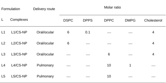
according to the corresponding L/CS-NP complexes to be prepared, using different combinations of dipalmitoylphosphatidylcholine (DPPC), diesteroylphosphatidylcholine (DSPC), dipalmitoylphosphatidylserine (DPPS), dimiristoylphosphatidylglycerol (DMPG) (Lipoid GMBH,Germany) and cholesterol (Sigma Chemicals, USA). In this manner, as shown in Table 1, three different liposomal formulations are produced for oral and ocular administration, using the following lipid combinations and molar ratios: L1 – DSPC:DPPS:Chol (6/0.1/4 molar ratio); L2 – DPPC:Chol (6/4 molar ratio); L3 – DSPC:Chol (6/4 molar ratio); while two formulations are prepared as controls for pulmonary administration; L4 – DPPC:DMPG (10:1 molar ratio) and L5 – DPPC.
Table 1
The selection of these combinations of lipids, in the case of the oral delivery, was driven by the necessity of having gastrointestinal resistant vesicles, since DSPC and DPPC possess saturated, relatively long fatty acid chains and high phase transition temperatures, making them stable in acidic and intestinal media (Aramaki et al., 1993). Moreover, the inclusion of negatively charged lipids aims to favour the interaction with chitosan nanoparticles and cholesterol is included in the formulations to increase the stability and the rigidity of the system (Kirby and Gregoriadis, 1999; Takeuchi et al., 2001a). Respect to the lipids applied in the formulations intended for lung delivery, DPPC and DMPG were chosen as they are endogenous to the lung and principal constituents of the pulmonary surfactant. Furthermore, the molar ratios correspond approximately to the phospholipids proportions in the referred surfactant (McAllister et al., 1996; Wright and Clements, 1987) and, in this manner, the overall in vivo lung environment is simulated. It is important to mention that, in some cases, liposomes were used as controls of the L/CS-NP complexes, while in other occasions liposomes were produced as a part of the technology applied to produce the lipid/chitosan nanoparticles complxes, as occurs in the complexes formulated by lyophilisation.
In order to produce the liposomes, the referred mixtures of lipids are dissolved in 20 mL of chloroform in a round bottom flask, to a final lipid concentration of 0.3 mM and 0.12 mM for cholesterol, when applicable (formulations for oral and ocular delivery – L1, L2 and L3). Afterwards, 50 g of glass beads are added in order to increase the surface available for the formation of the dry lipid film. Subsequently, the organic solvent is evaporated under a nitrogen stream in a rotary evaporator (Buchi® R-114, Buchi,
Switzerland) for a period of 3 hours, at temperatures between 55 ºC and 60 ºC and a homogeneous film is formed. Nitrogen is used to ensure removing all the traces of organic solvent. The resulting thin film is then hydrated for 30 min, using 10 mL of water, previously heated to a temperature above that of phase transition and the obtained vesicles are filtered under vacuum to separate them from the glass beads. Formulations prepared for oral and ocular delivery are subsequently extruded according to the procedure described above. All the formulations of liposomes are stored at 4 ºC until use.
The applied temperatures of evaporation are established to be above the phase transition temperature of the lipids or lipid mixtures, so that more flexible vesicles can be obtained (Rodríguez and Xamaní, 2003). These transition temperatures can be easily found in the literature and correspond, for example, to 41 ºC for DPPC and 58 ºC for DSPC (Delattre et al., 1993).
Recommendation: phase transition temperature of lipid mixtures is different from that of the individual lipids. Therefore, when using lipid mixtures, there is the need to find the transition temperature of the specific mixture and, in the absence of that information, the highest transition temperature of the lipids applied in the mixture should be considered, all the work being performed above that temperature.
Preparation of L/CS-NP complexes by hydration
The preparation of L/CS-NP complexes by the method of hydration of a lipid film consists of adding a suspension of previously prepared CS-NP to a dry lipid film. As
can be observed in Table 1, each of the presented lipidic combinations is used to produce the liposomal vesicles and the corresponding L/CS-NP complexes. However, instead of hydrating the dry lipid film with water, as for the liposomes, the assembly of L/CS-NP complexes is achieved by using a suspension of chitosan nanoparticles (unloaded or insulin-loaded) as hydrating phase (volume of 10 mL). Complexes for oral or ocular administration are produced with lipid/nanoparticles mass ratio of 2/1, while those intended for lung administration count with a mass ratio of 3/1.
Figure 1 displays a schematic demonstration of the methodology of the assembly of the L/CS-NP complexes. In the case of the complexes for lung delivery, DPPC or a mixture of DPPC and DMPG (10:1 molar ratio) are dissolved in 20 mL of chloroform and the same procedure and characteristics described before for the preparation of liposomes are applied until obtaining a dry lipid film. This film is then hydrated for 30 min with a suspension of the CS-NP (unloaded or insulin-loaded), forming the L/CS-NP complexes (3/1, w/w). Immediately afterwards, the complexes are filtered under vacuum to allow their separation from the glass beads.
Figure 1
In order to provide an efficient pulmonary administration and simultaneously, increase the long-term stability of the formulation, the complexes are submitted to a further step of spray-drying to encapsulate these structures in dry powders with adequate properties for systemic lung delivery. In brief, a suspension of complexes is mixed with mannitol (mannitol/complexes = 80/20 (w/w), final solids content 2.1%), which acts as inert carrier, and this mixture is spray-dried (Buchi Mini Spray Drier B-290, Buchi, Switzerland) at an inlet temperature of 160 ºC. For further details please consult the original paper (Grenha et al., 2008a).
Complexes for oral and ocular delivery are prepared in a similar manner, except for the absence of the process of spray-drying and the application of different combinations of lipids, as observed in Table 1.
As referred for the production of liposomes, the temperatures used for evaporation are, in all cases, adjusted in order to ensure that the complexes are assembled at temperatures above the phase transition temperature of all phospholipids of the formulation, enabling the production of more flexible structures (Rodríguez and Xamaní, 2003). The selection of phospholipids to be included in the lipid film surrounding the inner core composed of nanoparticles, which can have very different properties, especially concerning surface charges, dictates the type of covering, which can result either complete or only partial. Therefore, as demonstrated in Figure 2, we propose the production of different structures, depending on the phospholipids composing the film.
Recommendation: different mass ratios between lipids and nanoparticles should be tested and optimised for each case and for each lipid composition, since the interaction of the various combinations of lipids with the nanoparticles might be different and determine the production of complexes with different properties.
Figure 2
Preparation of L/CS-NP by lyophilisation
The preparation of L/CS-NP complexes by the method of lyophilisation involves the separate preparation of the basic systems, chitosan nanoparticles and lipid vesicles (liposomes), with a subsequent mixing of both structures in a suspension.
Upon mixing aliquots of chitosan nanoparticles and phospholipid vesicles suspensions, a procedure of freeze-drying is applied in the presence of 5% trehalose, which acts as a cryoprotectant. The trehalose solution is prepared in double concentration in order to obtain the final concentration of 5 % upon mixing with the suspension of the basic systems.
Two different lipid/nanoparticles mass ratios are used to prepare the complexes by this method, 1/1 and 2/1. In this review only results of the ratio 2/1 are shown. The obtained mixtures are freeze-dried (Labconco freeze-dryer, United States) under the following
conditions: a primary drying step of 48 h at -30 ºC and a secondary drying step until the temperature gradually ascends to +20 ºC. After obtaining the lyophilised powder, the L/CS-NP complexes are prepared by hydration of the powder with purified water, under vigorous vortexing. As shown in Figure 3, the complexes obtained by this method, are the result of a “sandwich” structure which is formed by the drying of the liposomes intercalated with the nanoparticles, assembling in the form of complexes upon hydration, which obliges the lipids to rearrange into bilayers surrounding the inner core composed of nanoparticles. Albumin labelled with fluorescein isothyocyanate (FITC-BSA) was, in some cases, associated to chitosan nanoparticles, acting as a marker molecule.
Figure 3
Recommendation: One important detail in the freeze-drying step is the type of cryoprotectant to be used in the production of the systems. In previous studies, our group tested different cryoprotectants, such as dextran, sucrose, glucose and trehalose, all applied to optimise the freeze-drying of chitosan nanoparticles (Fernández-Urrusuno et al., 1999b). Specifically concerning the preparation of L/CS-NP complexes, glucose and trehalose were tested, and trehalose demonstrated to be the most adequate to preserve the integrity of freeze-dried L/CS-NP complexes. The selection of the cryoprotectant has demonstrated to play a major role in the final properties of the developed systems and, thus, this optimisation is highly recommended.
Characterisation of L/CS-NP complexes
Morphological examination
The morphological examination of the complexes is conducted by optical (Olympus BH-2, Japan) and transmission electron microscopy (TEM) (CM12 Philips, The Netherlands). For TEM observation, samples are mounted on copper grids previously
covered with Formvar films. To obtain adequate samples for viewing, three different steps should be optimised: sample addition to the grid, staining process and final washing. All these steps should be performed with drops of approximately 10 µL. Initially, the 10 μL sample should be placed in contact with the grid surface for a pre-determined period, which is usually of 10 seconds. Afterwards, the drop is dried using a little piece of filter paper. The second step consists on the sample staining with 10 µL of a solution of 2% (w/v) tungstophosphoric acid (Sigma Chemicals, Germany) that contacts with the sample deposited on the grid for other 10 seconds. Finally, after drying the staining material, a subsequent step of washing with 10 µL of water is advised to remove the excess of tungstophosphoric acid (10 seconds once again). Upon the final drying of water, the grid should be placed on a proper grid storage box and stored in a dessicator, for a minimum of 12 hours, until use. An optimisation of observations can be performed by changing the contact time of the samples with the grid and of the staining material with the samples, and also the time of washing (for instance, washing for 20 seconds instead of 10). Moreover, more than one washing step can be performed if desired.
Size measurements
The size of formulations (liposomal vesicles and complexes) developed for oral or ocular delivery is determined by photon correlation spectroscopy using a Zetasizer 3000 HS (Malvern, UK). In this case, the samples should simply be diluted to the appropriate concentration (setup by the equipment) with water and placed in an adequate cell for the measurement. Those formulations developed for lung delivery, as they have not been extruded and, thus, present a larger size, are analysed for their size using a Coulter counter (Coulter® Multisizer II, Coulter Electronics, UK), equipped
with a tube with an orifice aperture of 50 μm. Each particle produces a voltage alteration when passing through the orifice, according to its volume, which is transformed in a size value. To perform the measurements, 20 μL of the complexes or
liposomes suspension are dispersed in 100 mL of the electrolyte Isoton II (filtered, phosphate-buffered saline solution PBS). The instrument is previously calibrated using Isoton II and monodisperse latex microspheres of 13 μm, both supplied by Coulter. Nanoparticles’ size was, in all cases, determined by photon correlation spectroscopy, using the Zetasizer 3000 HS and following the procedure described above for sample preparation.
Zeta potential measurements
Zeta potential measurements are performed by laser Doppler anemometry, using the Zetasizer® 3000 HS (Malvern, UK). Samples are diluted with KCl 0.1 mM to an
adequate concentration, which is set by the equipment, and placed in the electrophoretic cell, where a potential of ± 150 mV is established. KCl must be filtered (0.22 µm) before dilution, as does the water, which is used to clean the cell between each measurement. Samples are prepared to approximately 5 mL and are introduced in the cell using a syringe.
Surface characterisation
The complexes’ surface was analysed in order to determine whether the lipids were completely or only partially coating chitosan nanoparticles, using two different and complimentary techniques, X-ray photoelectron spectroscopy (XPS, VG Escalab 250 iXL ESCA, VG Scientific, UK) and static time-of-flight secondary ion mass spectrometry (TOF-SIMS, TOF-SIMS IV, Ion-Tof GmbH, Germany). CS-NP, DPPC and DMPG control vesicles, and L/CS-NP complexes (DPPC/CS-NP and DPPC-DMPG/CS-NP) are placed on polished monocristaline silicon wafers, which are used as sample holders. XPS measurements are carried out using non monochromatic Al-Kα radiation (hν = 1486.62 eV) and photoelectrons are collected from a take off angle of 90º relative to the sample surface. Measurements are performed in a Constant Analyser Energy mode (CAE) with a 100 eV pass energy for survey spectra and 20eV
pass energy for high resolution spectra. Charge referencing is done by setting the lower binding energy C1s photopeak at 285.0 eV C1s hydrocarbon peak. The high resolution spectra fitting is based on “Chi-squared” algoritm used to determine the goodness of a peak fit. The chemical functional groups identity is obtained from the high-resolution peak analysis of carbon-1s (C1s), oxygen-1s (O1s) and nitroge-1s (N1s)
envelopes. The experimental conditions (X-ray source, power and analysis area) are kept constant for each analysis.
For TOF-SIMS analyses, a pulsed Gallium primary ion beam (69Ga+) generated with a
liquid metal ion gun working at 15 kV, is used to bombard the samples with 45º incidence respect to the sample surface. The obtained secondary ions are extracted with a 10 KV voltage and their time of flight from the sample to the detector is measured in a reflectron mass spectrometer. Electron flood gun charge compensation is necessary during measurements. A raster size of 500 µm × 500 µm is used and at least three different spots are analyzed under the “static” condition with ion doses of about ≈1012 ions/cm2. The calibration of the mass spectra in the positive mode is based
on hydrocarbon peaks such as CH2+, CH3+, C2H2+, and C3H5+. The experimental
conditions (ion type, beam voltage and primary ion dose) are maintained constant for each experiment and for compared spectra.
In vitro release studies
To estimate the release rate of insulin from CS-NP and L/CS-NP complexes, suspensions of these systems are incubated under dynamic conditions at 37 ºC in PBS pH 7.4 (which simulates the lung lining fluid) or simulated gastric fluid (U.S. Pharmacopoeia). Briefly, 0.1 mL of each formulation are incubated with 0.9 mL of gastric medium or 0.5 mL with 4.5 mL of PBS pH 7.4. At appropriate time intervals, individual samples are filtered with a low protein binding filter (0.22 µmMillex® GV,
Millipore Ibérica, Spain) and the amount of insulin released is evaluated by the MicroBCA protein assay (Pierce, USA), measuring the absorbances by
spectrophotometry (Shimadzu UV-Visible Spectrophotometer UV-1603, Japan) at 562 nm. A calibration curve is made at each time interval using unloaded-nanoparticles and L/CS-NP complexes.
Physical stability in simulated lacrimal fluid
The physical stability of each formulation of L/CS-NP complexes developed for ocular delivery (L1, L2 and L3) was assessed in simulated lacrimal fluid composed of 0.18% KCl, 0.63% NaCl, 0.006% CaCl2.2H2O and 0.01% MgCl2.6H2O in distilled water, pH =
7.4 ± 0.1 (van Haeringer, 1981). An aliquot of each diluted suspension of complexes was incubated in the fluid at 37 ºC, for 2 h, after which the complexes size was measured by photon correlation spectroscopy using the Zetasizer® 3000 HS.
Evaluation of complexes cytotoxicity using the IOBA-NHC cell line
The cytotoxicity of L/CS-NP complexes (L1, L2 and L3) was determined upon incubation with IOBA-NHC cells, an immortalized epithelial cell line from normal human conjunctiva. To do so, the 2,3-bis[2-methoxy-4-nitro-5-sulfophenyl]-2H-tetrazolium-5-carboxyalinide (XTT) test (XTT assay kit, Sigma Chemicals, USA) was applied, measuring the production of yellow formazan crystals upon cleavage of XTT by the mitochondrial dehydrogenases of viable cells. Briefly, cells were seeded on 96-well plates at a density of 4 × 10⁴ cells/well and incubated until confluency (approximately
20h) with the appropriate culture medium (DMEM-F12 supplemented with 10% fetal bovine serum, 5000 U/mL penicillin, 5 mg/mL streptomycin, 2 ng/mL human EGF, 1 μg/mL bovine insulin, 0.1 μg/mL cholera toxin and 0.5 μg/mL hydrocortisone). Afterwards, the cells were washed with supplement-free culture medium for 1 hour and, subsequently, 30 μL of CS-NP or L/CS-NP (0.25, 0.5 and 1 mg/mL) suspensions were added over the cells. After 15, 30 or 60 min incubation, the cells were washed three times with phosphate buffered saline (PBS) pH 5.0, containing 0.27% glucose. The cells were then incubated with 20 L of XTT solution composed of 1 mg/mL XTT in 100
L of phenol red-free RPMI culture medium. A solution of 0.005% benzalkonium chloride in DMEM/F-12 culture medium was used as a positive control. The cytotoxicity of the different L/CS-NP complexes tested was expressed as viability, calculated using the following formula:
Cell viability (%) = 100 – [(ODtest / ODc) x 100] (1)
where ODtest is the optical density of those wells exposed to CS-NP or L/CS-NP complexes suspensions; and ODc is the optical density of those wells treated with supplement-free DMEM/F-12 medium.
Cell uptake studies
To evaluate the ability of L/CS-NP complexes to cross the plasma membrane and enter the cells, IOBA-NHC cells and primary cultures (PCs) of human conjunctival epithelium were exposed to the three L/CS-NP formulations developed for ocular delivery. Confluent monolayers of IOBA-NHC cells and 3-week-old PCs were washed out for 1 h with supplement-free DMEM/F-12 culture medium and then incubated for 15, 30 or 60 min with 0.25, 0.5 or 1 mg/mL of the L/CS-NP formulations. L/CS-NP complexes uptake by the cells was analyzed immediately after incubation and after a 24 h recovery period in supplemented culture medium.
After incubation, cells were washed three times with PBS pH 5.0, containing 0.27% glucose and, subsequently, with cold PBS, pH 7.4. They were then fixed in cold methanol (-20º C) for 10 min and cold acetone for 3 sec, followed by incubation with PBS pH 7.4, containing 0.27% glucose and 0.2% Tween-20® for 30 min. Cells were
then counterstained with tetramethyl-rhodamine isothiocyanate (TRITC)-conjugated phalloidin (1:200) to identify the actin component of the cytoskeleton. Preparations were mounted and examined with a confocal laser scanning microscope (Carl Zeiss LSM310, Jena, Germany) equipped with a krypton-argon laser. FITC and TRITC were
excited with a 488 and 543 nm emission laser beam, respectively. Controls included culture medium-treated IOBA-NHC cells and PCs. To confirm the intracellular LCS-NP complexes location, stacks of serial 10 µm optical sections were captured along the Z-axis, first using the 488 nm laser for green FITC emissions and then the 543 nm laser for red TRITC emissions. Z-series images were then projected as profiles superimposed with green and red images.
In vivo hypoglycaemic effect: oral administration
The in vivo evaluation of the therapeutic response to insulin upon oral administration of the L/CS-NP complexes was carried out using normal male Wistar rats (200-210 g), fasted overnight before the administration, but with free access to water. The rats were previously acclimatised in the place where the experiments are performed and the baseline blood glucose was determined. Only rats with normal blood glucose are used in the experiment. Four experimental groups were established, each composed of seven animals, which correspond to four different formulations to be administered: 1) control water, 2) insulin control solution in PBS pH 7.4, 3) insulin-loaded CS-NP, and 4) insulin-loaded L/CS-NP complexes. All the formulations are administered in a single dose directly to the stomachs of conscious rats, by means of a glass syringe fitted to a gastric cannula. In all cases, the required dose of insulin (10 UI/kg) is administered to the rats in a total volume of 1 mL. Blood samples are collected from the tail vein at different times after dosing (1, 2, 4, 6, 10 and 24 hours) and glycaemic levels are determined in the samples by the glucose-oxidase method (Glucose GOD-PAD kit, Spinreact, Spain). The serum glucose level at time zero is taken as a 100% glucose level.
In vivo tolerance assay: ocular administration
Female albino New Zealand rabbits weighing 2.0-2.5 kg were used to study the acute ocular tolerance to CS-NP, L1/CS-NP and L3/CS-NP complexes. Only the
concentration evidencing the best in vitro results (0.5 mg/mL) was studied. Rabbits are randomly divided into three groups of five animals, each receiving 30 µL of one of the three formulations in the right eye every 30 min for 6 h. The contralateral eye is used as control and receives no treatment. The animals are then euthanized by air embolism after being deeply anesthetized with an intramuscular overdose of anesthetic and paralyzing mixture of xylazine (20 mg/kg) and ketamine (200 mg/kg). Eyeball and lid tissues are removed and fixed in Davison’s fixative solution composed of 1 part glacial acetic acid, 2 parts 37% formaldehyde, 3 parts 95% ethanol and 3 parts distilled water. Following fixation, they are embedded in paraffin for pathology. Eyeball and lid sections (6 µm) are evaluated in a masked fashion according to the following criteria: alteration in any of the ocular surface epithelia (cornea, limbus, conjunctiva), edema in lid tissues, presence of inflammatory cells (eosinophils, neutrophils, mast cells, and lymphocytes) and any other abnormality.
Results
CS-NP (unloaded and insulin-loaded) developed to assemble the L/CS-NP complexes presented sizes around 400 nm and strong positive zeta potentials (from + 34 to + 44 mV); and insulin was associated with high efficiencies (68 - 96%).
Pulmonary route
Control vesicles and L/CS-NP complexes composed of DPPC or DPPC:DMPG present sizes around 2 µm. As expected, given the inherent charges of the phospholipids, the physicochemical properties of the complexes are influenced by the phospholipid composition. In this respect, systems comprised of DPPC present a neutral or close to neutral charge (-7 to 0 mV), while those with DMPG present a strong negative charge (-54 to -36 mV).
As compared to chitosan nanoparticles, which display a burst release of insulin in 15 minutes, the L/CS-NP complexes provide a controlled release of the peptide, which is
more effective for the complexes containing the mixture DPPC-DMPG. In a general manner, the controlled release of the complexes, as compared to the nanoparticles, is explained by the presence of the phospholipid external layer, which represents an extra barrier for the drug diffusing from the nanoparticles before releasing completely from the drug delivery system. The ability of the complexes comprised of the mixture of phospholipids, which present an overall negative charge, to provide a further control over drug release, is attributed to the fact that the interaction with the positively charged nanoparticles is favoured, providing a complete lipid coating of these. In fact, the occurrence of a complete coating could be confirmed by the surface analysis of the complexes by the techniques of XPS and TOF-SIMS, which have also demonstrated the partial coating of complexes formulated with only DPPC. While complexes containing DPPC and DMPG evidence on their surface only chemical signals attributed to the phospholipids, those comprised of only DPPC, display some signals assigned to the chitosan nanoparticles, demonstrating that, in this latter case, only a partial lipidic coating is achieved (Grenha et al., 2008b). In this manner, the major role of electrostatic interactions as driving forces to control the organisation of the lipid/nanoparticles assemblies is clearly evident.
It is important to mention that mannitol microspheres developed to act as carriers of the insulin-loaded L/CS-NP complexes to the lung present adequate aerodynamic properties for an efficient delivery of the complexes to the alveolar zone, where systemic absorption of the model peptide is expected to occur. The referred microspheres, which contain 20% of complexes and 80% of mannitol, display aerodynamic diameters between 2.1 and 2.7 µm and apparent tap densities as low as 0.4 – 0.5 g/cm3, depending on the formulation of complexes to be encapsulated.
Moreover, the L/CS-NP complexes are easily recovered from the mannitol microspheres by incubation in physiological medium simulating the lung lining fluid (PBS pH 7.4), without significant alterations in both the morphology and physicochemical characteristics. In addition, the microencapsulation process does not
have any effect on the insulin release profile (Grenha et al., 2008a). A previous work from our group concerning the microencapsulation of non-complexed insulin-loaded chitosan nanoparticles as a strategy in lung protein administration has proven successful following intratracheal administration to rats (Al-Qadi et al., 2009) and the in vivo evaluation of the presently developed complexes is currently being setup.
Oral route: L/CS-NP complexes prepared by hydration
The L/CS-NP complexes present sizes within 1.5 and 3 µm. L1/CS-NP and L2/CS-NP formulations have a negative zeta potential of –17.7 and –3.1 mV, respectively, while L3/CS-NP complexes are positively charged (+15.9 mV).
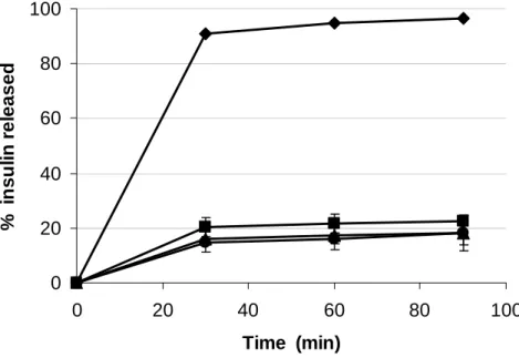
Figure 4 displays the insulin release profile from NP and L/CS-NP formulations in artificial gastric juice. As expected, CS-NP release their payload very rapidly, due to chitosan behaviour in acidic pH (dissolution), but all L/CS-NP complexes formulations exhibit the ability to control the release of the peptide in a similar manner, which is justified by the presence of the lipidic coating, as was confirmed in the complexes designed for lung administration.
Figure 4
These formulations of complexes were administered in vivo to rats and, as depicted in Figure 5, different lipidic compositions reflect different hypoglycaemic responses. In concordance with the observed release behaviour, the formulation containing a negatively charged phospholipid (DPPS) (L1/CS-NP), exhibits a more pronounced and rapid reduction in plasma glucose levels (P < 0.05), which decreased to about 50% of the baseline, remaining unaltered for at least 24 hours. This profile suggests that, as also indicated by the zeta potential data and as confirmed by the XPS and TOF-SIMS analysis performed in the complexes for lung delivery, a complete lipidic coating of the NP was achieved, which comprises an extra barrier that has to be overcome before complete release. The behaviour of these complexes is noticeably different than that of
the other two formulations (L2/CS-NP and L3/CS-NP), although those also registered punctual significant differences as compared to the profiles displayed by the insulin solution and CS-NP. As expected, the administration of an insulin solution does not lead to significant hypoglycaemic effect, given the rapid degradation in the gastric fluid prior to absorption.
Figure 5
Oral route: L/CS-NP complexes prepared by lyophilisation
CS-NP applied to prepare L/CS-NP complexes by lyophilisation have the same characteristics of those used in the hydration method. Liposomes display sizes around 300-360 nm, with neutral or negative charge, the latter reflecting the presence of DPPS. The size and zeta potential of the produced L/CS-NP complexes reflect the interaction of lipids with CS-NP. The most important data to refer on these characteristics is that L1/CS-NP complexes have a zeta potential of +31 mV, while the other two are close to neutral.
Again (as reported for the first method), in contrast with the CS-NP behaviour, all L/CS-NP complexes exhibit the ability to control insulin release along time (Figure 6); the profile varying according to the composition of vesicles used to form the complexes. The formulation with the negatively charged surface (L1/CS-NP) displays the more rapid release; while the other two formulations provide a more controlled release of the peptide.
Figure 6
Zeta potential data previously suggested that the incorporation of the negatively charged phospholipid in the complexes (L1/CS-NP) does not favour the lipidic coating of the nanoparticles (as happened in the hydration method), because the complexes zeta potential in this case is very similar to that presented by the nanoparticles. The release profile confirms this, because this formulation is in fact the more ineffective in
controlling drug release. This behaviour seems to indicate that the complexes formation by the method of lyophilisation is very different from that of hydration, probably due to a much lower time for interaction between the different parts (lipids and nanoparticles).
The described in vitro behaviour was in perfect agreement with the in vivo results. Upon oral administration of the complexes formulations to rats, although a different pattern of change in serum glucose levels was observed in each case (Figure 7), L2/CS-NP and L3/CS-NP complexes were those providing the more effective and long-lasting hypoglycaemic effect. Glucose levels decreased to about 70% of basal, remaining unaltered until 24 h.
Figure 7
These studies have shown that the complexes formed by both methodologies (hydration and lyophilisation) present different structures, which are affected by the system composition and by the applied methodology.
Ocular route
To evaluate the application of the L/CS-NP complexes as drug vehicles by the ocular route, the complexes were prepared by lyophilisation. The systems were loaded with FITC-BSA with the goal of assessing the penetration of the complexes in the ocular epithelium, their cytotoxicity and in vivo tolerance. FITC-BSA loaded CS-NP present sizes around 400 nm, while liposomes vary within 290 and 420 nm, as a function of their lipid composition; resulting in L/CS-NP complexes between 400 and 750 nm. The complexes zeta potential varied within +5.8 and +14.7 mV. All the complexes formulations have demonstrated to remain stable in simulated lacrimal fluid for at least 2 h and are present inside the cells as early as 15 min after incubation and more clearly at 30 min. The systems exhibited negligible toxicity in vitro and a good tolerance in vivo. These results point out L/CS-NP complexes as potentially useful drug delivery
carriers through the ocular mucosa. For further details please consult the original paper (Diebold et al., 2007).
Conclusions and Prospects
We have developed attractive and simple methodologies to efficiently associate/incorporate chitosan nanoparticles within lipid vesicles. By means of a lyophilisation procedure or by performing the hydration of a dry lipid film, we obtain new assemblies (liposome-chitosan nanoparticle complexes) that can display different properties, as a function of the lipid composition applied in their formation. As expected, these complexes permit the efficient encapsulation of therapeutic molecules like insulin; offer great stability in biological fluids and provide a controlled release of their payload. Furthermore, their in vivo behaviour can be considered very promising. The oral administration of these newly developed complexes loaded with insulin, demonstrates their ability to provide an important reduction in plasma glucose levels. Furthermore, these complexes exhibit negligible in vitro toxicity in conjunctival epithelial cells and a good tolerance in vivo, which corroborates their potential as drug carriers to the epithelial surfaces (i.e. ocular and lung (alveolar) surface).
Our strategy can be applied to other naturally-occurring polymeric nanoparticles that, in this manner, should benefit from the existence of an extra lipidic barrier, which will probably take place as a bilayer, being an excellent means of controlling the release pattern. This structure is favoured by the interaction between nanoparticles and phospholipids, since it has been reported that the solid cores have a strong ordering effect on the phospholipid molecules.
References
Al-Qadi, S., Grenha, A., Seijo, B., Goycoolea, F., Alonso, M.J., and Remuñan-Lopez, C. Chitosan nanoparticle-based inhalable dry powders for protein lung delivery: in vivo
evaluation of microencapsulated insulin-loaded nanoparticles in rats. 2009. Venice, Italy. Proceedings 9th International Conference of the European Chitin Science (in press).
Alonso, M.J., Sánchez, A., 2003. The potential of chitosan in ocular drug delivery. J. Pharm. Pharmacol. 55, 1451-1463.
Aramaki, Y., Tomizawa, H., Hara, T., Yachi, K., Kikuchi, H., Tsuchiya, S., 1993. Stability of liposomes in vitro and their uptake by rat Peyer’s patches following oral administration. Pharm. Res. 10, 1228-1231.
Beaulac, C., Sachetelli, S., Lagacé, J., 1999. Aerosolization of low phase transition temperature liposomal tobramycin as dry powder in an animal model of chronic pulmonary infection caused by Pseudomonas aeruginosa. J. Drug Target. 7, 33-41. Calvo, P., Remuñan-Lopez, C., Vila-Jato, J.L., Alonso, M.J., 1997. Novel hydrophilic Chitosan-Polyethylene Oxide nanoparticles as protein carriers. J. Appl. Polym. Sci. 63, 125-132.
Campbell, A., Taylor, P., Cayre, O.J., Paunov, V.N., 2004. Preparation of aqueous gel beads coated by lipid bilayers. Chem. Commun. 21, 2378-2379.
Carvalho, E.L.S., Seijo, B., Alonso, M.J. Formation and characterization of chitosan nanoparticles-phospholipid complexes. 2001. Angers, France. Proceedings 13th
International Symposium of Microencapsulation.
Courrier, H.M., Butz, N., Vandamme, T.F., 2002. Pulmonary drug delivery systems: recent developments and prospects. Crit. Rev. Ther. Drug Carr. Syst. 19, 425-498. Csaba, N., Garcia-Fuentes, M., Alonso, M.J., 2006. The performance of nanocarriers for transmucosal drug delivery. Exp. Opin. Drug Deliv. 3, 463-478.
De Campos, A., Sanchez, A., Alonso, M.J., 2001. Chitosan nanoparticles: a new vehicle for the improvement of the delivery of drugs to the ocular surface. Application to cyclosporin A. Int. J. Pharm. 224, 159-168.
de la Fuente, M., Csaba, N., Garcia-Fuentes, M., Alonso, M.J., 2008. Nanoparticles as protein and gene carriers to mucosal surfaces. Nanomedicine 3, 845-857.
Delattre, J., Couvreur, P., Puisieux, F., Philippot, J.R., Schuber, F., 1993. Les liposomes: aspects technologiques, biologiques et pharmacologiques. INSERM, Paris. Diebold, Y., Jarrín, M., Sáez, V., Carvalho, E.L.S., Orea, M., Calonge, M., Seijo, B., Alonso, M.J., 2007. Ocular drug delivery by liposome-chitosan nanoparticle complexes (LCS-NP). Biomaterials 28, 1553-1564.
El-Maghrabya, G.M., Barryc, B.W., Williams, A.C., 2008. Liposomes and skin: From drug delivery to model membranes. Eur. J. Pharm. Sci. 34, 203-222.
Fenske, D.B., Chonn, A., Cullis, P.R., 2008. Liposomal nanomedicines: an emerging field. Toxicol. Pathol. 36, 21-29.
Fernandez-Urrusuno, R., Calvo, P., Remuñan-Lopez, C., Vila-Jato, J.L., Alonso, M.J., 1999a. Enhancement of nasal absorption of insulin using chitosan nanoparticles. Pharm. Res. 16, 1576-1581.
Fernandez-Urrusuno, R., Romani, D., Calvo, P., Vila-Jato, J.L., Alonso, M.J., 1999b. Development of a freeze-dried formulation of insulin-loaded chitosan nanoparticles intended for nasal administration. S. T. P. Pharm. Sci. 9, 429-436.
Gabizon, A.A., 1995. Liposome circulation time and tumor targeting: implications for cancer chemotherapy. Adv. Drug Deliv. Rev. 16, 285-294.
Gregoriadis, G., 1988. Liposome as drug carrier: recent trends and progress. John Wiley & Sons, Chichester.
Grenha, A., Seijo, B., Carvalho, E.L.S., Remuñan-Lopez, C., 2008a. Microspheres containing lipid/chitosan nanoparticles complexes for pulmonary delivery of therapeutic proteins. Eur. J. Pharm. Biopharm. 69, 83-93.
Grenha, A., Seijo, B., Serra, C., Remuñán-López, C., 2008b. Surface characterisation of lipid/chitosan nanoparticles assemblies, using X-ray photoelectron spectroscopy and time-of-flight secondary ion mass spectrometry. J. Nanosci. Nanotechnol. 8, 358-365. Huang, Y.Z., Gao, J.Q., Liang, W.Q., Nakagawa, S., 2005. Preparation and characterization of liposomes encapsulating chitosan nanoparticles. Biol. Pharm. Bull. 28, 387-390.
Issa, M., Koping-Hoggard, M., Artursson, P., 2005. Chitosan and the mucosal delivery of biotechnology drugs. Drug Discov. Today: Technol. 2, 1-6.
Jiang, W., Kim, B.Y., Rutka, J.T., Chan, W.C., 2007. Advances and challenges of nanotechnology-based drug delivery systems. Exp. Opin. Drug Deliv. 4, 621-633. Jorgensen, L., Moeller, E.H., van de Weert, M., Nielsen, H.M., Frokjaer, S., 2006. Preparing and evaluating delivery systems for proteins. Eur. J. Pharm. Sci. 29, 174-182.
Kirby, C.J., Gregoriadis, G., Liposomes. In: Mathiowitz, E. (Ed.), Encyclopedia of Controlled Drug Delivery, John Wiley & Sons, New York, 1999, pp. 461-492.
Marier, J.-F., Lavigne, J., Ducharme, M.P., 2002. Pharmacokinetics and efficacies of liposomal and conventional formulations of tobramycin after intratracheal administration in rats with pulmonary Burkholderia cepacia infection. Antimicrob. Agents Chemother. 46, 3776-3781.
McAllister, S.M., Alpar, H.O., Teitelbaum, Z., Bennett, D.B., 1996. Do interactions with phospholipids contribute to the prolonged retention of polypeptides within the lung? Adv. Drug Deliv. Rev. 19, 89-110.
Paolicelli, P., de la Fuente, M., Sanchez, A., Seijo, B., Alonso, M.J., 2009. Chitosan nanoparticles for drug delivery to the eye. Exp. Opin. Drug Deliv. 6, 239-253.
Rodríguez, R. Xamaní, M., Liposomes prepared by high-pressure homogenizers. In: Düzgünes, N. (Ed.), Liposomes, Vol. 367. Elsevier Academic Press, San Diego, 2003, pp. 28-45.
Silva, G.A., Coutinho, O.P., Ducheyne, P., Shapiro, I.M., Reis, R.L., 2007. Starch-based microparticles as carriers for the release of active platelet-derived growth factor. Tissue Eng. 13, 1259-1268.
Takeuchi, H., Kojima, H., Yamamoto, H., Kawashima, Y., 2001a. Evaluation of circulation profiles of liposomes coated with hydrophilic polymers having different molecular weights in rats. J. Control. Release 75, 83-91.
Takeuchi, H., Yamamoto, H., Kawashima, Y., 2001b. Mucoadhesive nanoparticulate systems for peptide drug delivery. Adv. Drug Deliv. Rev. 47, 39-54.
Torchilin, V.P., 2005. Recent advances with liposomes as pharmaceutical carriers. Nat. Rev. Drug Discov. 4, 145-160.
van Haeringen, NJ., 1981. Clinical biochemistry of tears. Surv. Ophthalmol. 26, 84-96. Wright, J.R., Clements, J.A., 1987. Metabolism and turnover of lung surfactant. Am. Rev. Resp. Dis. 135, 426-444.
Table 1. Lipid molar ratio used in the preparation of liposomes (L) and respective liposome/chitosan nanoparticles (L/CS-NP) complexes.
Formulation Delivery route Molar ratio
L Complexes DSPC DPPS DPPC DMPG Cholesterol L1 L1/CS-NP Oral/ocular 6 0.1 4 L2 L2/CS-NP Oral/ocular 6 4 L3 L3/CS-NP Oral/ocular 6 4 L4 L4/CS-NP Pulmonary 10 1 L5 L5/CS-NP Pulmonary 10
DPPC – dipalmitoylphosphatidylcholine; DSPC – diesteroylphosphatidylcholine; DPPS - dipalmitoylphos-phatidylserine; DMPG - dimiristoylphosphatidylglycerol
Figure legends
Figure 1. Schematic preparation of liposome/chitosan nanoparticles (L/CS-NP) complexes by hydratation.
Figure 2. Hypothetical structures corresponding to complexes obtained by the method of hydration of a lipid film: a) complete coating with one lipid layer; b) complete coating with more than one lipid layer; c) partial coating of the nanoparticles; d) coating of several particles within the same complex; e) absence of coating, two separated systems coexisting (nanoparticles and liposomes).
Figure 3. Schematic preparation of liposome/chitosan nanoparticles (L/CS-NP) complexes by lyophilisation.
Figure 4. Insulin release profile from () chitosan nanoparticles, () L1/CS-NP, () L2/CS-NP and () L3/CS-NP complexes prepared by the hydration methodology, in artificial gastric juice.
Figure 5. Hypoglycemic effect following oral administration of () water, () insulin solution, () CS-NP, () L1/CS-NP complexes, () L2/CS-NP complexes and () L3/CS-NP complexes to rats (insulin dosis = 10 UI/kg, data represent mean SEM, n = 7).
Figure 6. Insulin release profiles from () CS-NP, () L1/CS-NP, () L2/CS-NP and () L3/CS-NP complexes prepared by the lyophilisation method, in artificial gastric juice.
Figure 7. Hypoglycemic effect following oral administration of () water, () insulin solution, () CS-NP, () L1/CS-NP, () L2/CS-NP and () L3/CS-NP complexes to rats (insulin dosis = 10 UI/kg, data represent the mean SEM, n = 7).
Figure 1
30 min 55 - 60 ºCSpray-drying
L/CS-NP complexes Pulmonary + mannitol (mannitol/complexes = 8:2, w/w) L3 L3/CS-NP L2 L2/CS-NP L1 L1/CS-NP L5 L5/CS-NP L4 L4/CS-NP L/CS-NP complexes Oral/ocular Lipids dissolved in chloroform EvaporationDry lipid film
Chitosan NP
suspension 2:1 (Oral/ocular)3:1 (Pulmonary) Lipid:NP ratio (w/w):
Hy
d
ra
tio
n
30 min 55 - 60 ºCSpray-drying
L/CS-NP complexes Pulmonary + mannitol (mannitol/complexes = 8:2, w/w) L3 L3/CS-NP L2 L2/CS-NP L1 L1/CS-NP L5 L5/CS-NP L4 L4/CS-NP L/CS-NP complexes Oral/ocular 30 min 55 - 60 ºC 30 min 55 - 60 ºC 30 min 55 - 60 ºC 30 min 55 - 60 ºCSpray-drying
L/CS-NP complexes Pulmonary + mannitol (mannitol/complexes = 8:2, w/w) L3 L3/CS-NP L2 L2/CS-NP L1 L1/CS-NP L5 L5/CS-NP L4 L4/CS-NP L/CS-NP complexes Oral/ocularSpray-drying
L/CS-NP complexes PulmonarySpray-drying
Spray-drying
L/CS-NP complexes Pulmonary L/CS-NP complexes Pulmonary + mannitol (mannitol/complexes = 8:2, w/w) L3 L3/CS-NP L2 L2/CS-NP L1 L1/CS-NP L5 L5/CS-NP L4 L4/CS-NP L/CS-NP complexes Oral/ocular + mannitol (mannitol/complexes = 8:2, w/w) + mannitol (mannitol/complexes = 8:2, w/w) L3 L3/CS-NP L2 L2/CS-NP L1 L1/CS-NP L5 L5/CS-NP L4 L4/CS-NP L/CS-NP complexes Oral/ocular L3 L3/CS-NP L2 L2/CS-NP L1 L1/CS-NP L5 L5/CS-NP L4 L4/CS-NP L3 L3/CS-NP L2 L2/CS-NP L1 L1/CS-NP L5 L5/CS-NP L4 L4/CS-NP L/CS-NP complexes Oral/ocular Lipids dissolved in chloroform EvaporationDry lipid film
Chitosan NP
suspension 2:1 (Oral/ocular)3:1 (Pulmonary) Lipid:NP ratio (w/w):
Hy
d
ra
tio
n
Lipids dissolved in chloroform Evaporation Lipids dissolved in chloroform Evaporation EvaporationDry lipid film
Chitosan NP
suspension 2:1 (Oral/ocular)3:1 (Pulmonary) Lipid:NP ratio (w/w):
Hy
d
ra
tio
n
Dry lipid film
Chitosan NP suspension
Dry lipid film Dry lipid film
Chitosan NP suspension Chitosan NP
suspension 2:1 (Oral/ocular)3:1 (Pulmonary) Lipid:NP ratio (w/w):