F
ACULDADE DEC
IÊNCIASD
EPARTAMENTO DEB
IOLOGIAA
NIMALINFLUENCE OF
E
RF3
A
/GSPT1 GENE EXPRESSION
ON SUSCEPTIBILITY TO CARCINOGENESIS
J
OANA
F
ERREIRA
T
OMÉ
M
ALTA
V
ACAS
Tese orientada por:
Professor Doutor Rui Miguel Brito
Professora Doutora Maria Manuela Coelho
This study was supported by the Fundação Para a Ciência e a Tecnologia: PhD fellowship SFRH/BD/21468/2005, projects POCTI/MGI/40071/2001 and PTDC/SAU-GMG/67031/2006.
This dissertation should be cited as:
Malta-Vacas J. (2009) Influence of eRF3a/GSPT1 gene expression on susceptibility to carcinogenesis. PhD Thesis, University of Lisbon, Portugal.
“O erro é a noite dos espíritos e a armadilha da inocência” Luc de Clapiers, Marquês de Vauvenargues
N
OTAS
P
RÉVIAS
Nos termos do n.º 1 do Artigo 40, Capítulo V, do Regulamento de Estudos Pós-Graduados da Universidade de Lisboa, publicado no Diário da República – II Série N.º 153, de 5 de Julho de 2003, esclarece-se que na elaboração da presente dissertação foram usados integralmente artigos científicos já publicados (4) ou submetidos para publicação (1) em revistas indexadas de circulação internacional, os quais integram os Capítulos II e III da presente tese. Tendo os referidos trabalhos sido realizados em colaboração, a candidata esclarece que participou integralmente no planeamento e na elaboração de todos os trabalhos, assim como na análise e discussão dos resultados.
Esclarece-se ainda que a formatação dos vários artigos que integram a presente dissertação obedece às regras das revistas em que foram publicados ou submetidos para publicação. Por este motivo, não foi possível adoptar um critério uniforme ao longo dos vários capítulos.
C
ONTENTS
Agradecimentos / Acknowledgements xi Resumo xv Abstract xix 1. CHAPTER I – INTRODUCTION 1 1.1. Identification of eRF3 11.2. Characterization of human eRF3’s genes and proteins 2
1.3. Functions of eRF3a 5
1.3.1. The canonical role of eRF3a in translation termination 5
1.3.2. Other roles translation-related 7
1.3.3. Cytoskeleton assembly 9
1.3.4. Apoptosis regulation 10
1.4. Cancer 11
1.4.1. Translation and cancer 15
1.4.2. Trinucelotide repeats and cancer 17
1.5. Aims and Thesis Structure 19
1.6. References 21
2. CHAPTER II – ERF3A/GSPT1 POLYMORPHISMS AND GENE EXPRESSION 33
ALTERATIONS IN CANCER 2.1. Paper 1 35
Malta-Vacas J, Ramos S, Aires C, Costa P, Conde AR, Martins AP, Monteiro C, Brito M. (2005) Differential expression of the eukaryotic release factor 3 (eRF3/GSPT1) according to gastric cancer histological type. J Clin Pathol 58: 621–625. 2.2. Paper 2 41
Brito M, Malta-Vacas J, Aires C, Costa P, Carmona B, Gaspar G, Monteiro C. (2005) Polyglycine Expansions in eRF3/GSPT1 are Associated With Gastric Cancer Susceptibility. Carcinogenesis 26: 2046-2049.
3. CHAPTER III – ROLE OF ERF3A DEREGULATION IN TUMORIGENESIS 45
3.1. Paper 3 47
Malta-Vacas J, Chauvin C, Gonçalves L, Nazaré A, Carvalho C, Monteiro C, Bagrel D, Jean-Jean O, Brito M (2009) eRF3a/GSPT112GGC allele increases the susceptibility for breast cancer development. Oncol Rep 21: 1551-1558. 3.2. Paper 4 55
Malta-Vacas J, Ferreira P, Monteiro C, Brito M. (submitted) eRF3a/GSPT1 (GGC)n alleles differential expression in cancer. 3.3. Paper 5 75
Malta-Vacas J, Nolasco S, Monteiro C, Soares H, Brito M (2009) Translation termination and protein folding pathway genes are not correlated in gastric cancer. Clin Chem Lab Med 47:427-31. 4. CHAPTER IV – DISCUSSION 81
4.1. Translation and cancer 82
4.2. eRF3a/GSPT1 DNA polymorphisms 83
4.2.1. eRF3a/GSPT1 polymorphisms in cancer 83
4.2.2. eRF3a/GSPT1 microssatelite polymorphism in inflammation 86
4.3. eRF3a/GSPT1 gene expression 87
4.4. eRF3a/GSPT1 as a proto-oncogene 89
4.4.1. eRF3a roles in translation 89
4.4.2. eRF3a role in cytoskeleton dynamics 91
4.4.3. eRF3a role in apoptosis and proliferation rates 93
4.5. References 95
A
GRADECIMENTOS
Chegada ao fim deste percurso de trabalho de vários anos não poderia deixar de agradecer a todos quantos me ajudaram a percorrer este caminho. Para que ninguém fique esquecido, começo por agradecer a TODOS os que de alguma forma contribuíram para a realização deste trabalho. Em particular gostaria de agradecer:
Ao Professor Doutor Miguel Brito pela confiança que depositou em mim ao aceitar-me como sua aluna de doutoramento, pela ajuda, apoio e orientação, desde o início, na construção deste projecto de doutoramento e pelo trabalho que desenvolvemos nos últimos 7 anos. Pela enorme convicção, energia positiva e força de vontade, sem as quais o Grupo de Investigação da ESTESL nunca seria o que é…
Obrigada pela amizade e os bons momentos partilhados. Todo este trabalho seria, sem dúvida, muito mais pobre não fossem os conhecimentos, o incentivo e o entusiasmo que me foi transmitindo.
À Professora Doutora Manuela Coelho da Faculdade de Ciências da Universidade de Lisboa por ter aceite o desafio de me aceitar como sua aluna de doutoramento, pela sua disponibilidade e pelo acompanhamento dado nas questões burocráticas/ administrativas do processo.
Ao Professor Doutor Carolino Monteiro, que ficará sempre lembrado como “Pai” deste projecto, por todo o apoio prestado desde o início até ao final deste projecto, pelo espírito crítico e entusiasmo demonstrado e transmitido ao longo destes anos.
Ao Doutor Olivier Jean-Jean, por ter aceite receber-me no seu Laboratório na Unité de Biochimie Cellulaire, Université Pierre et Marie Curie, de que resultou uma excelente colaboração entre ambos os grupos. Agradeço ao Olivier e a todos os membros da sua equipa, nomeadamente à Celine Chauvin, pelo acompanhamento de todo o trabalho que realizei em Paris e à Samia Salhi pelas dicas e sugestões quer relativas ao trabalho laboratorial, quer em relação à minha estadia naquela maravilhosa cidade!
À Fundação para a Ciência e a Tecnologia agradeço a concessão de uma bolsa de doutoramento que permitiu o desenvolvimento desta dissertação.
À Fundação Calouste Gulbenkian agradeço a concessão de bolsas de viagem que permitiram a participação em reuniões científicas internacionais e a realização do estágio na Unité de Biochimie Cellulaire, Université Pierre et Marie Curie, coordenado pelo Doutor Olivier Jean-Jean.
Aos colegas de laboratório Bruno Carmona, Cátia Aires e Patrícia Costa que me acompanharam desde o nascimento do projecto. Juntos “demos à luz” o actual Laboratório de Genética Humana da ESTESL. Obrigada por toda a ajuda no trabalho laboratorial, pela ambiente criado, pelas discussões e críticas e construtivas e pela amizade criada.
À Prof. Doutora Helena Soares por me ter recebido por um curto período no seu laboratório no IGC, e por toda a ajuda dada na produção dos anticorpos e na optimização e interpretação das imunos e dos westerns.
A todos os colegas e amigos que entretanto foram passando pelo Laboratório de Investigação da ESTESL e que contribuíram um pouco para o sucesso deste trabalho, não só pela ajuda com todos os pormenores laboratoriais mas principalmente pelos bons momentos de convívio. Agradeço especialmente a: Alice Melão, Ana Moleirinho, Carina Ladeira, Carla Mota, Catarina Sousa Guerreiro, Dolores Prudêncio, Elisabete Costa, Filipa Quintaneiro, Gilberto Matias, Joao Gonçalves, João Lourenço, Lourent Brault, Rui Placido, Sandra Raicar e Susana Gonçalves.
À Paula Ferreira pela enorme ajuda e apoio prestados no laboratório, pela simpatia e disponibilidade, boa disposição, e pelo excelente trabalho de colaboração ao longo destes anos.
À Prof. Doutora Ana Rita Conde da Faculdade de Farmácia da Universidade de Lisboa por toda a ajuda prestada na execução do trabalho prático e pela disponibilidade e amabilidade constantes.
Ao João Gonçalves pela grande ajuda e apoio prestado no laboratório quer na ESTESL quer mais tarde no IGC, pela disponibilidade constante, e pelo exemplo de entusiasmo e entrega à Ciência.
À Sofia Nolasco pela disponibilidade para a realização de inúmeras imunos e westerns no IGC, pela simpatia e boa disposição constantes, pelas aliquotas disto e daquilo que generosamente foram cedidas e pelas inúmeras dicas e conselhos práticos.
A todos os colegas docentes e não-docentes da ESTESL, por toda a ajuda prestada para o bom desenvolvimento deste trabalho, nas suas componentes técnicas, burocráticas e administrativas. Uma palavra especial à Luísa Veiga, pela sua boa disposição e disponibilidade constantes; ao Gilberto Matias, pela coragem e grande ajuda na luta contra as parafinas e os anticorpos; aos Prof. Fernando Belém, Prof. Ana Almeida e Prof. Renato Abreu, pela ajuda a recolha de sangue aos dadores voluntariados; à Prof. Susana Viegas por ter sido a força motriz para o início dos testes de micronúcleos e pelas simpáticas palavras de apoio; à Fernanda Dantas, Mónica Júlio, Ana Oliveira e Ruth Joaquim, pela constante disponibilidade no apoio aos laboratórios; aos colegas da Biologia e da Química que contribuíram com dicas e sugestões úteis quando nem tudo corria bem: Anita Gomes, Dulce Azevedo, Helena Soares, Lisete Fernandes, Mário Pádua, Mário Gomes, Sofia Nolasco.
A todos os médicos que gentilmente colheram e cederam as amostras e dados clínicos dos seus doentes, que constituíram o material de base para a execução do presente trabalho, nomeadamente: Dra. Sancia Ramos e Dra. Ana Paula Martins do Hospital de Santa Cruz, Dra. Marília Cravo e Dr. António Pinto do Instituto Português de Oncologia Francisco Gentil, ao Dr. Carlos Carvalho, Dra. Lucília Gonçalves e Dr. Raul Lobato-Faria e do Hospital Fernando da Fonseca. Uma palavra especial para a Dra. Lucília Gonçalves pelo esforço de envolvimento de toda a sua equipa, indispensável para os bons resultados obtidos, e ao Dr. António Pinto pela execução das análises no citometro de fluxo.
À Lisete Fernandes pela preciosa ajuda na amplificação dos plasmídeos, incluindo as células competentes, e por muitas outras dicas e sugestões úteis ao longo dos anos.
Ao Prof. Shin-Ichi Hoshino da Universidade de Tokyo, pelo generoso envio do anticorpo e péptido contra o eRF3.
À Doutora Joana Diamond por nos colocar à disposição a ajuda dos membros da sua equipa no Centro de Investigação de Patobiologia Molecular do Instituto Português de Oncologia (CIPM-IPO), assim como a utilização do equipamento; à Sofia Fragoso, pelas dicas e ajuda com o isolamento de linfócitos, à Lara e à Joana Dionísio pelas horas de trabalho no sequenciador.
Ao Celso Cunha do Instituto de Higiene e Medicina Tropical pela utilização do aparelho de PCR em Tempo Real da unidade que então coordenava.
Aos elementos do grupo de investigação do Professor Doutor Carolino Monteiro na Faculdade de Farmácia da Universidade de Lisboa, nomeadamente à Susana Santos, Margarida Alves, Cátia Evangelista por todo o apoio, sempre que necessário.
À Joana Morais, pela amizade que nasceu do convívio diário no meu primeiro laboratório e atravessou todos estes anos. Pelas constantes palavras de apoio e incentivo, pela grande ajuda nas questões burocráticas e processoais envolvidas no processo, e por tudo o resto.
À Cristina Luís pelos sábios conselhos de quem já passou pelas mesmas experiencias e está sempre disposto a ajudar os amigos.
A todos os meus amigos que de alguma forma me ajudaram a passar por estes anos. Sem os bons momentos de descompressão o resultado não seria o mesmo!
Ao Paulo, pelo incentivo que me deu, fazendo-me olhar para o futuro. E por ter esperado (im)pacientemente pelo fim deste projecto.
À minha família, por estar sempre presente nos momentos importantes, em especial à minha Mãe por me dar tudo o que só uma Mãe pode dar, incondicionalmente.
R
ESUMO
O envolvimento dos componentes da maquinaria de tradução no desenvolvimento de várias neoplasisas é hoje amplamente reconhecido, principalmente no que diz respeito a factores de iniciação e factores de alongamento da tradução. Neste contexto, os factores de tradução têm sido também encarados como potenciais alvos para a inibição da proliferação celular e consequentemente como alvos de terapia contra o cancro. O factor de terminação da tradução eucariótico 3 (eRF3) é uma pequena GTPase que se associa com o factor de terminação da tradução eucariótico 1 (eRF1) e com o GTP para formar o complexo de terminação da tradução. A proteína eRF3 humana existe em duas isoformas, designadas por eRF3a e eRF3b, codificadas respectivamente pelos genes eRF3a/GSPT1 e eRF3b/GSPT2. Para além das funções que lhe são atribuídas no processo de tradução (terminação da tradução, reciclagem de ribossomas e iniciador da degradação do mRNA), este factor está também envolvido no controlo do ciclo celular, na organização do citoesqueleto e na regulação da apoptose. As proteínas eRF3a e eRF3b apresentam 87% de homologia, sendo a maior diferença entre ambas no seu domínio N-terminal. A extremidade amina do eRF3a apresenta uma expansão de poliglicinas codificada por uma expansão estável do tripleto GGC no exão 1 do gene eRF3a/GSPT1. Este polimorfismo está ausente no gene eRF3b/GSPT2. Vários genes contendo expansões de microssatélites exónicos têm sido associados ao desenvolvimento de cancro, sendo em alguns casos os polimorfismos STR (Short Tandem Repeat) relacionados com a transactivação da expressão dos genes respectivos.
O principal objectivo deste trabalho foi avaliar o gene eRF3a/GSPT1 como potencial gene de susceptibilidade para o desenvolvimento de cancro.
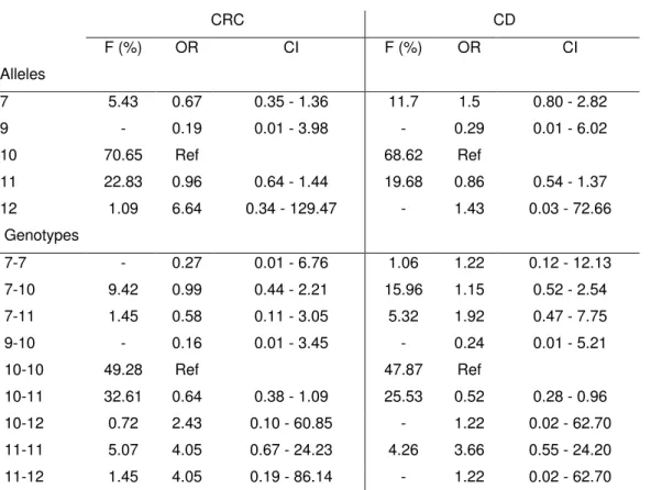
Pela análise do polimorfismo STR no gene eRF3a/GSPT1, foram detectados cinco alelos diferentes na população portuguesa contendo 7, 9, 10, 11 e 12 repetições GGC, sendo o alelo 10GGC o mais comum (F=68.5% na população controlo saudável). O alelo mais longo (12GGC) foi detectado exclusivamente em 5.1% dos pacientes com cancro (N=411), com uma frequência alélica de 3%, correspondendo a um risco 12 vezes aumentado de desenvolvimento de cancro (OR= 11.8; SE=1.43; C.I.=0.71-195.46). Demonstrou-se que não se trata de uma mutação somática, mas sim da linha germinal, e que o alelo 12GGC está ausente na população controlo e em indivíduos com Doença de Crohn, uma doença inflamatória com elevada predisposição para o desenvolvimento de cancro colorectal.
Recorrendo ao RT-PCR em Tempo Real, verificou-se que o gene eRF3a/GSPT1 se encontra sobre-expresso em vários tipos de cancro, nomeadamente em 70% dos tumores gástricos do tipo intestinal e em 44% dos tumores da mama. Mais ainda, demonstrou-se que existe uma variação significativa dos níveis de mRNA em função do tamanho dos alelos expressos, encontrando-se o alelo 12GGC sobre-expresso quer em linhas primárias de linfócitos (p<0.001) quer em células Jurkat transformadas (p<0.0001), quando comparado com o alelo 10GGC, tomado como referência. Os elevados níveis de expressão do gene eRF3a/GSPT1 detectados em tecidos
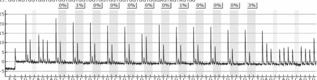
tumorais não estão relacionados com um aumento da taxa de tradução das células cancerígenas, visto que este aumento não se verifica no principal factor de terminação da tradução (eRF1). Também não foram detectadas alterações nos níveis de eRF3b/GSPT2 correlacionadas com as variações de expressão do eRF3a/GSPT1, pelo que as alterações de expressão não serão ajustadas por um mecanismo de compensação entre as duas isoformas da proteína. A dosagem génica do eRF3a/GSPT1 foi também foi determinada através de PCR em Tempo Real mas não foram detectadas amplificações/delecções do gene em tecidos tumorais associadas às alterações nos padrões de expressão. Os níveis de metilação dos locais CpG localizados na expansão GGC foram quantificados por pirosequenciação, mas a variação de 7 a 12 repetições GGC não se encontra associada a alterações nos níveis de metilação, independentemente do tipo de tecido analisado. Assim, o mecanismo responsável pela sobre-expressão do gene continua por esclarecer.
Estando envolvido em processos críticos e vitais para a célula, é de prever que alterações no domínio N-terminal do eRF3a possam ser altamente relevantes para a integridade celular. Para avaliar o efeito da variação do número de glicinas presentes no terminal amina do eRF3a foram analisados vários tipos de linhas celulares, expressando os diferentes alelos (GGC)n, e comparada a eficiência das proteínas codificadas, nas suas várias funções.
Dado que a função canónica do eRF3a é como factor de terminação da tradução, utilizou-se a linha celular HEK293, que contém um gene repórter com um codão stop prematuro, para comparar a eficiência das proteínas na terminação da tradução. Sendo o gene repórter o da β-galactosidase, a determinação da quantidade desta proteína activa nas células reflete o nível de readthrough do codão SOPT prematuro no respectivo gene. Através da quantificação dos níveis de readthrough em células expressando proteínas eRF3a com 7-12 resíduos de glicina no terminal amina não foram detectadas diferenças significativas de eficiência entre as proteínas de diferentes tamanhos.
Para além de outras funções relacionadas com o processo de tradução, sabe-se que o eRF3a está envolvido na passagem da fase G1 para a fase S do ciclo celular e também no controlo da apoptose. Apesar de, em ambos os casos, as vias em que o eRF3a participa não estarem ainda identificadas, pretendeu-se avaliar se proteínas com diferentes tamanhos poderiam ter eficiências diferentes nas funções que desempenham nas respectivas vias. Para tal, recorreu-se à citometria de fluxo de forma a determinar as taxas de proliferação e de apoptose em linhas primárias de linfócitos e em células Jurkat transformadas, expressando os cinco alelos diferentes. No entanto, não foram detectadas diferenças significativas entre as linhas celulares relativamente a qualquer dos parâmetros analisados quer em relação a índices de proliferação, quer no que diz respeito a taxas de apoptose.
Está descrito que, em diferentes espécies, alterações de expressão ou mutações no gene eRF3 levam a malformações do fuso acromático, erros na segregação dos cromossomas durante a meiose, e bloqueio da citocinese. Sendo estes eventos potencialmente geradores de um fenótipo maligno, pretendeu-se averiguar se o tamanho da proteína teria alguma influência no seu papel
de regulação do citoesqueleto. Através da realização de um teste de micronúcleos com bloqueio da citocinese (CBMN assay), foi determinada a frequência de micronúcleos (MN) em células binucleadas, em linhas celulares com diferentes genótipos. Os nossos resultados demonstram que as linhas celulares que expressam alelos mais longos, especificamente as que expressam o alelo 12GGC, apresentam maiores frequências de MN em células binucleadas, possivelmente como resultado de defeitos na formação do fuso acromático. Em consequência, é de prever que ocorra uma acumulação de erros a nível dos cromossomas, característica de células cancerígenas, levando à promoção da tumorigenese.
Apesar do envolvimento do gene eRF3a/GSPT1 no desenvolvimento de cancro não estar ainda totalmente esclarecido, os nossos resultados indicam que a presença do alelo 12GGC por si só poderá ser considerado como marcador de susceptibilidade para o desenvolvimento de cancro. Uma melhor compreensão dos mecanismos de regulação da expressão do gene eRF3a/GSPT1 e a sua associação com a proliferação celular poderá também contribuir para progressos quer ao nível do prognóstico da doença quer a nível de aplicações terapêuticas. Em conclusão, os nossos resultados indicam que o gene eRF3a/GSPT1 deve ser considerado um potencial proto-oncogene. Espera-se que, no seu todo, esta dissertação tenha contribuído para o desenvolvimento de futuras investigações sobre a regulação da expressão do gene eRF3a/GSPT1 e a sua contribuição na génese tumoral e possa vir a melhorar o nosso conhecimento sobre o diagnóstico, prognóstico e tratamento de cancro.
A
BSTRACT
The eukaryotic release factor 3 (eRF3) associates with eRF1 in a complex that mediates translation termination. In addition to its roles in translation, eRF3 is also involved in cell cycle regulation, apoptosis and cytoskeleton assemble. Human eRF3 has two distinct isoforms, eRF3a and eRF3b, encoded by eRF3a/GSPT1 and eRF3b/GSPT2 genes.
eRF3a/GSPT1 contains a stable (GGC)n expansion coding for proteins with different N-terminal extremities. We identified five alleles encoding 7, 9, 10, 11 and 12 GGC repeats in the Portuguese population, being the 10GGC allele the most frequent (F= 68.5% in the control population). The longer allele (12GGC) was exclusively detected in 5.1% of the cancer patients (N=411) with an allele frequency of 3%, corresponding to a 12-fold increased cancer risk. Our results show that the mRNA levels of eRF3a/GSPT1 are overexpressed in a significant proportion of different types of cancer. Moreover, the transcript levels of eRF3a/GSPT1 show variation between alleles, being the 12GGC allele significantly overexpressed (p<0.001). The levels of eRF3a/GSPT1 transcription are not associated with eRF3a/GSPT1 amplification neither with the methylation pattern of the GGC expansion region.
Using an in vivo assay for readthrough efficiency, we do not detect any difference in the activity of the eRF3a proteins encoded by the five different eRF3a/GSPT1 alleles. Also, no differences in the levels of apoptosis and proliferation rates were found between cells lines. Finally, using a cytokinesis-block micronucleos assay, we show that cells with the longer alleles have higher frequencies of MN, which is probably a result of defects in mitotic spindle formation.
Although the connection between eRF3a/GSPT1 and tumorigenesis is not completely elucidated, our data suggests that the presence of the 12GGC allele provides a novel risk marker for cancer. Taken together, our results show that eRF3a should be considered as a potential proto-oncogene.
1. CHAPTER I
INTRODUCTION
1.1. Identification of eRF3
Long before the identification of the eukaryotic Release Factor 3 (eRF3), it was known that the translation termination process in eukaryotes was GTP-dependent. However, no GTP binding motif was identified in eRF1, the main factor acting in translation termination, which suggested the existence of an additional unidentified protein involved in the process. This suspicion was further reinforced by the knowledge of the prokaryotic termination factor RF3, which has no release activity by itself, being a GTP stimulator of the termination step of translation (Zhouravleva et al., 1995).
In 1988, the Japanese group from Kikuchi isolated a new temperature sensitive mutant strain of Saccharomyces cerevisae, gst1 (G-to-S transition), which affected the G1-to-S phase transition of the cell cycle. A DNA clone complementing the gst1-1 mutation was isolated from a yeast gene library and the gene product was a protein of ~76 KDa which contained consensus sequence for GTPases and had extensive homology to polypeptide chain elongation factor EF1α (Kikuchi et al., 1988). In the same year, Kushnirov’s group in Russia cloned the sup35 gene (also named SUP2) from S. cerevisae when looking for omnipotent suppressor mutants. Mutations in both GST1 and SUP35 increased the level of translation ambiguity, suggesting that the gene product could be a regulator of translation accuracy. Both turned out to be the same gene. One year later, the Japanese group cloned the human homologue gene (GST1-Hs) from the cDNA library of human KB cells (Hoshino et al., 1989). This gene was latter renamed GSPT1 and mapped on human chromosome 16p13.1 (Ozawa et al., 1992). In this study, the authors also showed the existence of a homologous gene on the X chromosome.
In 1995, it was demonstrated that the Sup-35 like protein from Xenopus laevis directly interacts with the Sup45 (eRF1) and enhances its activity in a GTP-dependent manner (Zhouravleva et al., 1995). Thus, the gene product was finally named eRF3, following the nomenclature from the prokaryote factors.
Later, Hoshino et al. (1998) isolated two mouse GSPT genes, the counterpart of human GSPT1 and a novel member of the family, GSPT2. Both protein products interact with eRF1 to function as eRF3 in mammalian translation termination. Subsequently, the human GSPT2 gene has been mapped in Xp11.21-23 (Hansen et al., 1999), and the protein encoded characterized and described as eRF3b (Jakobsen et al., 2001).
Despite the diversity of names that were given to these genes and encoded proteins during the past two decades, it is now consensual to call eRF3a and eRF3b to the mammalian proteins, and eRF3a/GSPT1 and eRF3b/GSPT2 to the respective genes. This is the terminology that will be used in the present dissertation.
1.2. Characterization of human eRF3’s genes and proteins
Human eRF3 has two distinct isoforms, eRF3a and eRF3b, encoded by eRF3a/GSPT1 and eRF3b/GSPT2 genes, located in 16p13.1 (Ozawa et al., 1992) and Xp11.21-23 (Hansen et al., 1999), respectively. It was recently proposed that eRF3b/GSPT2 was originated through retrotransposition of processed eRF3a/GSPT1. The putative eRF3b/GSPT2 retroposon included a copy of the endogenous eRF3a/GSPT1 5’UTR sequence, therefore being a functional retrogene (Zhouravleva et al., 2006).
eRF3a/GSPT1 contains 15 exons and spans along 43,27Kb in chromosome 16p (Figure 1). Although the promoter region(s) remains to be characterized, there are two alternative initiation codons identified. Even so, it was demonstrated that translation is initiated at the first AUG encountered at the 5' end of eRF3a/GSPT1 (Jean-Jean et al., 1996). The corresponding mRNA (Accession Number: NM_002094; 2585 bp) is ubiquitously expressed in all tissues, and its level varies during the cell cycle, being its expression inducible under growth stimulation (Hoshino et al., 1998).
Figure 1: Transcript structure of eRF3a/GSPT1
(Src: http://www.ensembl.org/Homo_sapiens/geneview?gene=ENSG00000103342)
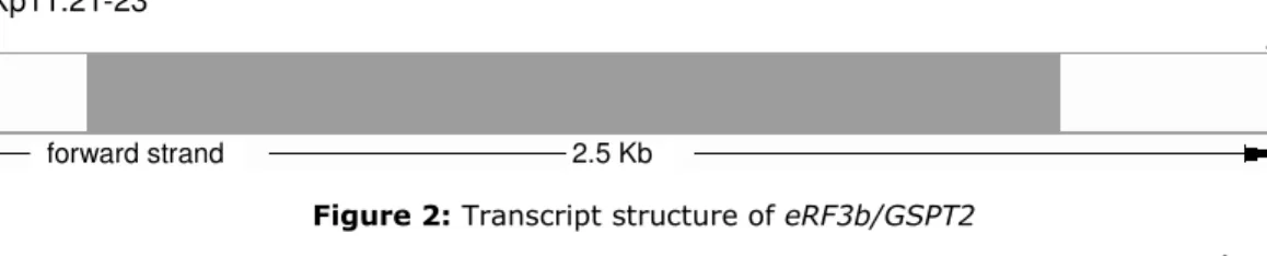
eRF3b/GSPT2 is an intronless gene that lies upon 2,5Kb in chromosome Xp (Figure 2). eRF3b/GSPT2 mRNA (NM_018094; 2503 bp) is poorly expressed in most mouse tissues tested except the brain, and does not fluctuate during the cell cycle (Hoshino et al., 1998). It’s expression in human tissues is still not clearly characterized.
16p13.1
reverse strand 43.27 Kb
Figure 2: Transcript structure of eRF3b/GSPT2
(Src: http://www.ensembl.org/Homo_sapiens/geneview?gene=ENSG00000189369)
The coding sequence of both genes share 88% homology, being the most important difference in the nucleotide sequences the presence of a GGC expansion close to the initiation codon in eRF3a/GSPT1 that eRF3b/GSPT2 lacks.
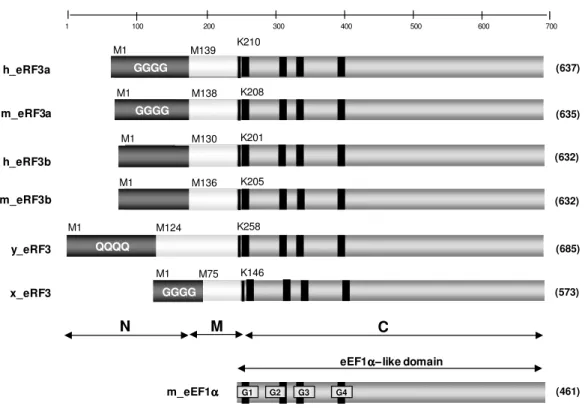
The eRF3 proteins consist of three main regions, a non-homologous amino (N) terminal domain (~200 residues), a middle (M) domain, and a conserved EF1α-like carboxyl (C) terminal domain of 428 residues (see Figure 3 for details). The three domains were defined based on amino acid features and functions. The amino acid sequence of the human eRF3a protein (Hoshino et al., 1989) presents extensive homology with other eukaryotes like the Xenopus laevis (Zhouravleva et al., 1995), Sacaromices cerevisae (Kushnirov et al., 1988), Pinchia pinus (Kushnirov et al., 1990), Mus musculus (Hoshino et al., 1998) and Podospora anserina (Gagny & Silar, 1998). All these proteins exhibit GTP-binding motifs in the C-terminal domain, which is similar to EF1α (Kushnirov et al., 1988) and to the prokaryotic RF3 (Grentzmann et al., 1994). However, eukaryotic eRF3 and the prokaryotic counterpart have a distinctive feature: the gene encoding the prokaryotic RF3 is nonessential (Grentzmann et al., 1994) while at least the C-domain of the eukaryotic eRF3 has been shown to be essential for cell viability in different species, including Homo sapiens (Kushnirov et al., 1988; Gagny & Silar, 1998; Frolova et al., 1996).
The C-terminal region of eRF3 proteins is highly conserved through evolution, and carries the four canonical GTP-binding motifs of the GTPase superfamily. The N-terminal region varies in both length and sequence among species.
Xp11.21-23
1 100 200 300 400 500 600 700 (685) (637) (635) (461) QQQQ h_eRF3a m_eRF3a m_eEF1αααα
eEF1αααα−−− like domain−
N
C
(632) h_eRF3b G1 G2 G3 G4 y_eRF3 K258 M124 M1 K210 M139 M1 GGGG K208 M138 M1 GGGG K201 M130 M1 (632) m_eRF3b K205 M136 M1 (573) x_eRF3 K146 M75 M1 GGGGM
Figure 3: Schematic representation of the eRF3 family. Proteins belonging to the eRF3 family could be divided into three regions: an amino terminal non-conserved region (N), a median domain (M) of
unknown function and a homologous C-terminal domain (C). h: H. sapiens; m: M. musculus; y: S. cerevisae; x: Xenopus laexis.
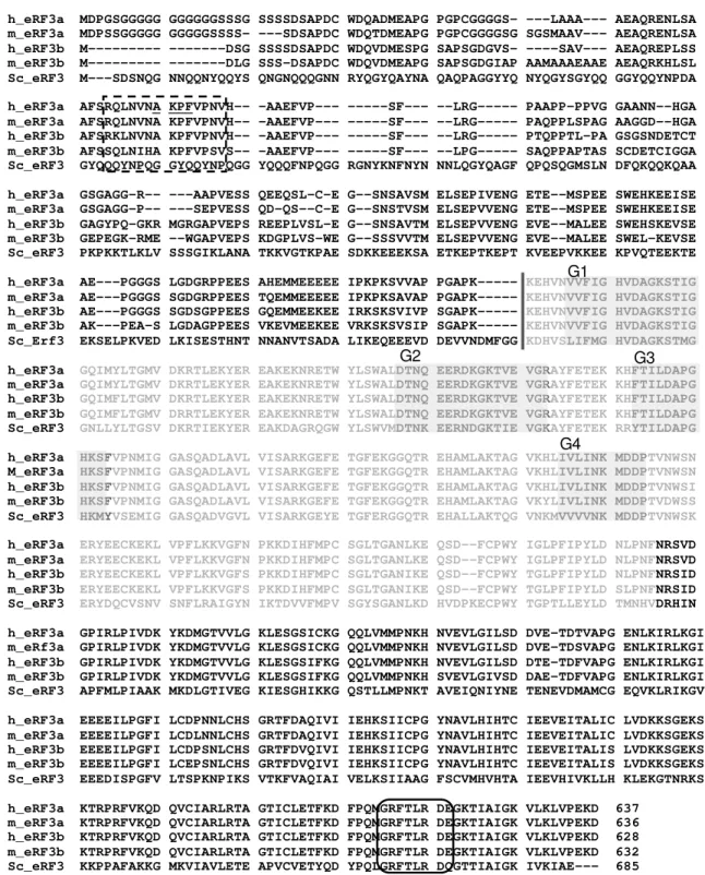
The human eRF3a and eRF3b protein sequences are about 87% identical (Figure 4) being the differences in amino acid sequence concentrated near the amino terminus. The N-domain of eRF3a is rich in acidic amino acids and contains a high percentage of Pro, Ser and Gly residues (10%, 15% and 20%, respectively). Also, eRF3a includes a stable polyglycine expansion encoded by a (GGC)n tract in eRF3a/GSPT1 gene, with five different known alleles (Riggins et al., 1992; see chapter 1.5 for details). eRF3a is abundant in all tissues, while eRF3b expression pattern still needs further elucidation, although it was reported to be absent in the majority of human cell lines tested (Chauvin et al., 2005).
From the first functional studies reported, the C-terminal domain of eRF3 has been described as essential for eRF1 binding and sufficient for the protein’s role as a translation termination factor, while the N-terminal domain remained poorly understood (Ter-Avanesyan et al., 1993; Zhouravleva et al., 1995; Kisselev & Frolova, 1995; Mugnier & Tuite, 1999) being sometimes viewed as functionally nonessential (Merkulova et al., 1999).
1.3. Functions of eRF3a
1.3.1. The canonical role of eRF3a in translation termination
Eukaryotic translation termination is governed by two release factors, eRF1 and eRF3. These factors associate in a complex which binds to the ribosomal A site. eRF1 is the key factor of translation termination, recognising the three stop codons in the mRNA (UAA, UAG and UGA), catalyzing the ester bond hydrolysis of the peptidyl-tRNA and promoting the release of the nascent peptide chain. eRF3 is a small GTPase protein that enhances eRF1 activity (Zhouravleva et al., 1995).
Hoshino et al. (1998) demonstrated that both mammalian eRF3a and eRF3b can interact with eRF1. Surprisingly, it was reported later that mouse eRF3b, but not eRF3a, could substitute for yeast eRF3 in vivo (Le Goff et al., 2002), which possible means that in mammalian cells eRF3a and eRF3b share the multiple functions fulfilled by yeast’s eRF3 (Inge-Vechtomov et al., 2003). GTP hydrolysis is required for fast and efficient termination of translation (Mitkevich et al., 2006). It was demonstrated that eRF3 is a GTP-binding protein capable of a negligible, if any, intrinsic GTPase activity that is greatly stimulated by the joint action of eRF1 and the ribosome (Salas-Marco & Bedwell, 2004). Neither eRF1 nor the ribosome displays this effect de per se (Frolova et al., 1996). Thus, eRF3 functions as a GTPase in the quaternary complex with the ribosome, eRF1, and GTP. Binding of eRF3 to eRF1, as revealed in vivo and in vitro (Stansfield et al., 1995; Zhouravleva et al., 1995), is mediated by the C-terminal domains of both proteins (Ebihara & Nakamura, 1999; Merkulova et al., 1999; Kononenko et al., 2008). This finding was further reinforced by Chauvin et al. (2005) observations that X. laevis eRF3, which is highly homologous to eRF3b in its N-terminal extension, can substitute for human eRF3a. In contrast, neither the entire S. cerevisiae eRF3, which is highly divergent from human and Xenopus eRF3’s in the N-terminal domain, nor the N-N-terminally truncated form of S. cerevisiae eRF3 carrying the conserved C terminal domain only, efficiently substitutes eRF3a in termination. The crystal structure of the eEF1α−like region of eRF3 from S. pombe led to the identification of the eRF1 binding region (Figure 4) and revealed that the N-terminal extension, rich in acidic amino acids, can block the proposed eRF1 binding site, potentially regulating eRF1 binding to eRF3 in a competitive manner (Kong et al., 2004).
h_eRF3a MDPGSGGGGG GGGGGGSSSG SSSSDSAPDC WDQADMEAPG PGPCGGGGS- ---LAAA--- AEAQRENLSA m_eRF3a MDPSSGGGGG GGGGGSSSS- ---SDSAPDC WDQTDMEAPG PGPCGGGGSG SGSMAAV--- AEAQRENLSA h_eRF3b M--- ---DSG SSSSDSAPDC WDQVDMESPG SAPSGDGVS- ----SAV--- AEAQREPLSS m_eRF3b M--- ---DLG SSS-DSAPDC WDQVDMEAPG SAPSGDGIAP AAMAAAEAAE AEAQRKHLSL Sc_eRF3 M---SDSNQG NNQQNYQQYS QNGNQQQGNN RYQGYQAYNA QAQPAGGYYQ NYQGYSGYQQ GGYQQYNPDA h_eRF3a AFSRQLNVNA KPFVPNVH-- -AAEFVP--- ---SF--- --LRG--- PAAPP–PPVG GAANN--HGA m_eRF3a AFSRQLNVNA KPFVPNVH-- -AAEFVP--- ---SF--- --LRG--- PAQPPLSPAG AAGGD--HGA h_eRF3b AFSRKLNVNA KPFVPNVH-- -AAEFVP--- ---SF--- --LRG--- PTQPPTL-PA GSGSNDETCT m_eRF3b AFSSQLNIHA KPFVPSVS-- -AAEFVP--- ---SF--- --LPG--- SAQPPAPTAS SCDETCIGGA Sc_eRF3 GYQQQYNPQG GYQQYNPQGG YQQQFNPQGG RGNYKNFNYN NNLQGYQAGF QPQSQGMSLN DFQKQQKQAA h_eRF3a GSGAGG-R-- ---AAPVESS QEEQSL-C-E G--SNSAVSM ELSEPIVENG ETE--MSPEE SWEHKEEISE m_eRF3a GSGAGG-P-- ---SEPVESS QD-QS--C-E G--SNSTVSM ELSEPVVENG ETE--MSPEE SWEHKEEISE h_eRF3b GAGYPQ-GKR MGRGAPVEPS REEPLVSL-E G--SNSAVTM ELSEPVVENG EVE--MALEE SWEHSKEVSE m_eRF3b GEPEGK-RME --WGAPVEPS KDGPLVS-WE G--SSSVVTM ELSEPVVENG EVE--MALEE SWEL-KEVSE Sc_eRF3 PKPKKTLKLV SSSGIKLANA TKKVGTKPAE SDKKEEEKSA ETKEPTKEPT KVEEPVKKEE KPVQTEEKTE h_eRF3a AE---PGGGS LGDGRPPEES AHEMMEEEEE IPKPKSVVAP PGAPK--- KEHVNVVFIG HVDAGKSTIG
m_eRF3a AE---PGGGS SGDGRPPEES TQEMMEEEEE IPKPKSAVAP PGAPK--- KEHVNVVFIG HVDAGKSTIG
h_eRF3b AE---PGGGS SGDSGPPEES GQEMMEEKEE IRKSKSVIVP SGAPK--- KEHVNVVFIG HVDAGKSTIG
m_eRF3b AK---PEA-S LGDAGPPEES VKEVMEEKEE VRKSKSVSIP SGAPK--- KEHVNVVFIG HVDAGKSTIG
Sc_Erf3 EKSELPKVED LKISESTHNT NNANVTSADA LIKEQEEEVD DEVVNDMFGG KDHVSLIFMG HVDAGKSTMG
h_eRF3a GQIMYLTGMV DKRTLEKYER EAKEKNRETW YLSWALDTNQ EERDKGKTVE VGRAYFETEK KHFTILDAPG
m_eRF3a GQIMYLTGMV DKRTLEKYER EAKEKNRETW YLSWALDTNQ EERDKGKTVE VGRAYFETEK KHFTILDAPG
h_eRF3b GQIMFLTGMV DKRTLEKYER EAKEKNRETW YLSWALDTNQ EERDKGKTVE VGRAYFETEK KHFTILDAPG
m_eRF3b GQIMFLTGMV DRRTLEKYER EAKEKNRETW YLSWALDTNQ EERDKGKTVE VGRAYFETEK KHFTILDAPG
Sc_eRF3 GNLLYLTGSV DKRTIEKYER EAKDAGRQGW YLSWVMDTNK EERNDGKTIE VGKAYFETEK RRYTILDAPG
h_eRF3a HKSFVPNMIG GASQADLAVL VISARKGEFE TGFEKGGQTR EHAMLAKTAG VKHLIVLINK MDDPTVNWSN
M_eRF3a HKSFVPNMIG GASQADLAVL VISARKGEFE TGFEKGGQTR EHAMLAKTAG VKHLIVLINK MDDPTVNWSN
h_eRF3b HKSFVPNMIG GASQADLAVL VISARKGEFE TGFEKGGQTR EHAMLAKTAG VKHLIVLINK MDDPTVNWSI
m_eRF3b HKSFVPNMIG GASQADLAVL VISARKGEFE TGFEKGGQTR EHAMLAKTAG VKYLIVLINK MDDPTVDWSS
Sc_eRF3 HKMYVSEMIG GASQADVGVL VISARKGEYE TGFERGGQTR EHALLAKTQG VNKMVVVVNK MDDPTVNWSK
h_eRF3a ERYEECKEKL VPFLKKVGFN PKKDIHFMPC SGLTGANLKE QSD--FCPWY IGLPFIPYLD NLPNFNRSVD m_eRF3a ERYEECKEKL VPFLKKVGFN PKKDIHFMPC SGLTGANLKE QSD--FCPWY IGLPFIPYLD NLPNFNRSVD h_eRF3b ERYEECKEKL VPFLKKVGFS PKKDIHFMPC SGLTGANIKE QSD--FCPWY TGLPFIPYLD NLPNFNRSID m_eRF3b ERYEECKEKL VPFLKKVGFS PKKDIHFMPC SGLTGANIKE QSD--FCPWY TGLPFIPYLD SLPNFNRSID Sc_eRF3 ERYDQCVSNV SNFLRAIGYN IKTDVVFMPV SGYSGANLKD HVDPKECPWY TGPTLLEYLD TMNHVDRHIN h_eRF3a GPIRLPIVDK YKDMGTVVLG KLESGSICKG QQLVMMPNKH NVEVLGILSD DVE-TDTVAPG ENLKIRLKGI m_eRf3a GPIRLPIVDK YKDMGTVVLG KLESGSICKG QQLVMMPNKH NVEVLGILSD DVE-TDSVAPG ENLKIRLKGI h_eRF3b GPIRLPIVDK YKDMGTVVLG KLESGSIFKG QQLVMMPNKH NVEVLGILSD DTE-TDFVAPG ENLKIRLKGI m_eRF3b GPIRLPIVDK YKDMGTVVLG KLESGSIFKG QQLVMMPNKH SVEVLGIVSD DAE-TDFVAPG ENLKIRLKGI Sc_eRF3 APFMLPIAAK MKDLGTIVEG KIESGHIKKG QSTLLMPNKT AVEIQNIYNE TENEVDMAMCG EQVKLRIKGV h_eRF3a EEEEILPGFI LCDPNNLCHS GRTFDAQIVI IEHKSIICPG YNAVLHIHTC IEEVEITALIC LVDKKSGEKS m_eRF3a EEEEILPGFI LCDLNNLCHS GRTFDAQIVI IEHKSIICPG YNAVLHIHTC IEEVEITALIC LVDKKSGEKS h_eRF3b EEEEILPGFI LCDPSNLCHS GRTFDVQIVI IEHKSIICPG YNAVLHIHTC IEEVEITALIS LVDKKSGEKS m_eRF3b EEEEILPGFI LCEPSNLCHS GRTFDVQIVI IEHKSIICPG YNAVLHIHTC IEEVEITALIS LVDKKSGEKS Sc_eRF3 EEEDISPGFV LTSPKNPIKS VTKFVAQIAI VELKSIIAAG FSCVMHVHTA IEEVHIVKLLH KLEKGTNRKS h_eRF3a KTRPRFVKQD QVCIARLRTA GTICLETFKD FPQMGRFTLR DEGKTIAIGK VLKLVPEKD 637
m_eRF3a KTRPRFVKQD QVCIARLRTA GTICLETFKD FPQMGRFTLR DEGKTIAIGK VLKLVPEKD 636 h_eRF3b KTRPRFVKQD QVCIARLRTA GTICLETFKD FPQMGRFTLR DEGKTIAIGK VLKLVPEKD 628 m_eRF3b KTRPRFVKQD QVCIARLRTA GTICLETFKD FPQMGRFTLR DEGKTIAIGK VLKLVPEKD 632 Sc_eRF3 KKPPAFAKKG MKVIAVLETE APVCVETYQD YPQLGRFTLR DQGTTIAIGK IVKIAE--- 685
G1
G2
G4
G3
Figure 4: Alignment of the amino acid sequences of human and mouse eRF3a and eRF3b and S. cerevisae eRF3 proteins. The eRF3 protein family is characterized by a non-homologous amino-terminal domain, and a conserved like C-amino-terminal domain of 428 residues. The start of the EF1α-like domain is shown in a vertical line. This domain is subdivided in the G domain (amino acid residues in grey) and the C-terminal domain. The conservative G1-G4 consensus sequences involved in GTP binding are indicated by shadow boxes. The consensus sequence for PABP interaction (RQLNVNAKPFVP) is indicated in a dotted line box; the IAP-binding motif (AKPF) is underlined, inside the box. The consensus sequence for eRF1 interaction (GRFTLRD) is indicated in a round box.
It was recently reported that the DEAD-box RNA helicase and mRNA export factor DBP5 controls the eRF3-eRF1 interaction and thus eRF3-mediated downstream events (Gross et al., 2007). DBP5 is reported to be required for efficient stop-codon recognition, directly binding to eRF1 to remodel the mRNA/protein complex to allow proper eRF1 positioning on the stop codon. Subsequent dissociation of DBP5 from eRF1 is followed by the entry of eRF3 into the complex. The precise mechanism by which eRF1 and eRF3 promote translation termination when a stop codon reaches the A site of the ribosome is still not completely elucidated. It is generally accepted that the mechanism is comparable with the elongation system, which is much better characterized: eRF1 structurally mimics the stem of an aminoacil-tRNA, whereas eRF3 mimics the function of an EF1α, which carries the aminoacil-tRNA to the A site in a GTP-dependent manner (Nakamura & Ito, 1998; Hoshino et al., 1998; Inge-Vechtomov et al., 2003). This model is consistent with the knowledge that only the EF1α−like C-domain of eRF3 is required for eRF1 interaction.
1.3.2. Other roles in translation
There is a functional conservation of eRF3 family from yeast to human proteins in what concerns its role as a release factor in translation termination. However, the differences in the amino domain of the eRF3 revealed interactions with several different proteins, pointing out the involvement of eRF3 in several cellular pathways.
When searching for new eRF3a binding proteins, Hoshino et al. (1999a) identified the polyadenylate-binding protein (PABP), whose amino extremity associates with the poli(A) tail of mRNAs. Both eRF3a and eRF3b directly interact with PABP via its amino terminal domains, through the carboxyl domain of PABP. This interaction results in the inability of PABP to multimerize and consequently inhibits it to perform the typical regularly spaced complexes on the poli(A) tail of mRNAs that regulate mRNA degradation. This may facilitate shortening of the poly(A) tail of mRNAs by an RNase (Hoshino et al., 1999a; Hoshino et al., 1999b; Hosoda et al., 2003). The PABP also mediates mRNA circularization through binding the eIF4F translation initiation complex, which is associated with the mRNA 5’ cap structure (Kozlov et al., 2001). Therefore, the termination of translation might regulate the next initiation step via the eRF3-PABP-eIF4G signalling cascade. These studies revealed that eRF3a may play an important role in mRNA stability, as an initiator of the mRNA degradation machinery, and/or in the recycling of ribosomes in successive cycles of translation (Hoshino et al., 1999a; Hoshino et al., 1999b; Uchida et al., 2002).
Another role of eRF3 in mRNA degradation, through the nonsense-mediated mRNA decay (NMD) pathway, was revealed by Czaplinski et al. (1998). The NMD pathway is a process that rids the cell out of transcripts that contain premature termination codons (PTCs). The biological and medical significance of NMD is highlighted by an increasing number of known genetic diseases that are caused by C-terminally truncated proteins that may be non-functional or could exert a
toxic effect in a dominant-negative manner (Holbrook et al., 2004; Kuzmiak & Maquat, 2006; Diop et al., 2007). Three core factors involved in NMD have been identified (UPF1, UPF2 and UPF3), but several other proteins are known to be involved in this pathway. It was demonstrated that these three UPF proteins and both release factors, eRF1 and eRF3, function as part of a “surveillance complex” that recognizes PTCs and triggers NMD in S. cerevisae. Direct interactions between UPF proteins and eRF1 and eRF3 were demonstrated in vitro (Czaplinski et al., 1998) and in vivo (Kobayashi et al., 2004).
Although the GTPase activity of eRF3 is not necessary for its binding with both UPF1 and PABP, the GTP/eRF3-dependent termination exerts direct influence on the subsequent mRNA degradation (Kobayashi et al., 2004).
In mammalian cells, translation termination codons context and exon–exon junctions are cis-acting elements that allow recognition of stop codons. Recently, Kashima et al. (2006) showed that the recognition of PTCs occurs in a complex named SURF (SMG1–UPF1–eRF1–eRF3 complex) associated with the EJC (exon junction complex) on mRNPs. The presence of translation termination factors eRF1 and eRF3 suggested that transient formation of SURF complex most likely occurs after recognition of the stop codon, and that the eRF1–eRF3 complex probably recruits UPF1 and SMG-1 to a PTC. The SURF associates with the post-splicing mRNA through UPF2–EJC to form the DECID (decay-inducing complex). Subsequent UPF1 phosphorylation finally leads to mRNA degradation (Behm-Ansmant & Izaurralde, 2006; Kashima et al., 2006).
This model was later considered simplistic because different alternative pathways were reported (Ivanov et al., 2008). The most recent publications suggest that the recognition of a PTC is mediated by competition between the 3’ UTR–associated factors, being the PABP a human NMD antagonizing factor inhibiting the interaction between eRF3 and UPF1 in vitro (Amrani et al., 2006; Eberle et al., 2008; Singh et al., 2008). The NMD appears to be triggered by a ribosome’s failure to terminate adjacent to a properly configured 3’-UTR (untranslated region), an event that may promote binding of the UPF/NMD factors to stimulate mRNA decapping. The physical distance between the stop codon and the PABP is considered a crucial determinant for PTC recognition. NMD can only take place when an extended 3’ UTR places PABP distally to the termination codon, leaving a downstream exon junction complex in between. The EJC acts enhancing NMD, likely through increasing the affinity of UPF proteins.
Although the precise model is still a matter of controversy, eRF3 plays a central role in the process of triggering mRNA decay upon the recognition of a termination codon, being part of a transient complex involved in the process.
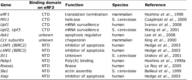
The outstanding feature of eRF3 is its ability to interact with many other factors, relevant or not to the translational machineries (summarized in Table 1). Therefore, it is tempting to speculate that eRF3 plays a molecular hub to regulate the translation termination complex functionality
through binding to a repertoire of cellular factors or to couple translation termination to other divergent cellular processes.
Table 1: Properties of known eRF3-binding proteins. Binding domain
on eRF3
eRF1 CTD translation termination mammalian Hoshino et al ., 1998
Mtt1 CTD helicase S. cerevisiae Czaplinski et al. , 2000
Upf1 CTD mRNA survaillence human Ivanov et al. , 2008
Upf2, Upf3 CTD mRNA survaillence S. cerevisiae Wang et al ., 2001
Ask1 unkown apoptosis regulator human Lee et al. , 2008
Cct subunits unkown chaperonin mammalian King et al ., 2000
cIAP1 (BIRC2) NTD inhibitor of apoptosis human Hedge et al. , 2003 cIAP2 (BIRC3) NTD inhibitor of apoptosis human Hedge et al ., 2003
Itt1 NTD Unknown S. cerevisiae Urakov et al ., 2001
Pabp1 NTD Poly(A) binding human Hoshino et al ., 1999a
RnaseL NTD Rnase human Le Roy et al ., 2005
Sla1 NTD actin assenbly S. cerevisiae Bailleul et al ., 1999
Xiap NTD inhibitor of apoptosis human Hedge et al ., 2003
Gene Function Species Reference
NTD: N-terminal domain; CTD: C-terminal domain
1.3.3. Cytoskeleton assembly
The cytoskeleton is a cytoplasmatic dynamic and complex network of filaments responsible for cell movement, shape, intracellular transport and cellular division, composed essentially of actin filaments, tubulin microtubules and intermediate filaments in the cytoplasm (Honore et al. 2005). After translation termination, the biogenesis process of some proteins, like actins and tubulins, involve interaction of nascent chains with the heterohexameric protein prefoldin (PFDN), whose function is to deliver non-native target proteins to the eukaryotic cytosolic chaperonins for facilitated folding (Simons et al., 2004) protecting them from aggregation while being transferred to chaperonin for the final step(s) in their folding (Vainberg et al., 1998). The target proteins are transferred by PFDN to the cytosolic chaperonin CCT (chaperonin containing TCP-1), which is a hetero-oligomeric complex with double-ring-like structure showing eightfold rotational symmetry of eight different subunits in mammalian somatic cells. The CCT complex is an important pathway that regulates microtubules stability and, consequently, cytoskeleton organization. CCT has been shown to assist in the folding of actin and tubulin in the presence of ATP in vitro (Tian et al., 1995; Farr et al., 1997) and to bind newly synthesized actin and tubulin in vivo (Thulasiraman et al., 1999). Each subunit of the complex recognizes specific target proteins and they collectively modulate the ATPase activity (Kubota et al., 1999).
An interaction between CCT subunits and eRF3 was first predicted using a theoretical model (Eisenberg et al., 2000), and experimentally confirmed by immunoprecipitation (King et al.,
2000; Martin Carden, personal comm). The CCT expression level correlate with growth rates in mammalian cultured cells, and is markedly up-regulated in the early S phase of the cell cycle (Yokota et al., 1999). Interestingly, the eRF3 protein is also essential for G1 to S phase transition of the cell cycle (Hoshino et al., 1998; Chauvin et al., 2007).
Using different organisms as study models, eRF3 has been shown to affect both tubulin and actin cytoskeleton. Deregulation of eRF3 expression and/or mutations in the N-terminal domain of the protein resulted in abnormal meiotic chromosome segregation and defects in the cytoskeleton assembly in spermatids of Drosophila melanogaster (Basu et al., 1998). In S. cerevisiae, eRF3 is also involved in regulatory interactions with intracellular structural networks, through interactions with the yeast cytoskeletal assembly protein SLA1, via its N-terminal domain (Bailleul et al., 1999). The eRF3 was also shown to affect the tubulin cytoskeleton, suggesting a role in the control of chromosome segregation at the anaphase (Borhsenius et al., 2000). More recently, its involvement in the assembly of the actin cytoskeleton in yeast was also demonstrated (Valouev et al., 2002), affecting the impairment of the mitotic spindle structure and chromosome segregation. Chai et al. (2006) showed that the eRF3 is distributed around the macronucleus and in the basal bodies in the cortex of Euplotes octocarinatus cells, suggesting that this protein may be involved in cytoskeleton organization.
From these studies, it is not clear whether the aberrant phenotypes reported are either an indirect consequence of disruptions of translation caused by deficient activity of eRF3 in translation termination, or, instead, reflect a more direct interaction of this protein with the microtubule cytoskeleton.
1.3.4. Apoptosis regulation
A proteolytically processed isoform of the eRF3a protein was reported to function as an inhibitor of apoptosis binding proteins (IAP-BPs) (Hedge et al., 2003; Verhagen et al., 2007). IAP-BPs are a group of proteins characterized by the presence of a conserved 4-residue IAP-binding motif (IBM) at their N termini, which allows them to bind to the Baculovirus IAP Repeat (BIR) domain of IAPs. The processed eRF3a protein contains the conserved N-terminal IAP-binding motif (AKPF), which is exposed after proteolytic cleavage of a 69-residue leader sequence (Figure 4). eRF3 directly interacts with IAPs (such as cIAP1, cIAP2 and XIAP), and can promote caspase activation, IAP ubiquitination and apoptosis (Hegde et al., 2003). In the model proposed, eRF3a could potentiate apoptosis by liberating caspases from IAP inhibition, and/or target IAPs and the processed eRF3a for proteasome-mediated degradation. Interestingly, the authors also demonstrated that the processed protein localized in both the cytoplasm and nuclear compartments of MCF-7 cells, suggesting that its association with the endoplasmic reticulum (ER) (which was expected and confirmed for the normal complete protein) is disrupted by deletion of its N terminus.
Two possible explanations for these findings were suggested: the processing of eRF3a might be triggered by ER stress or other cellular stress conditions or, alternatively, could be a regulatory mechanism to modulate the protein levels during the cell cycle by targeting it for proteosomal degradation via the IAP pathway. The last hypothesis is supported by Chauvin’s et al. (2008) results showing that eRF3a is degraded by the proteasome when not associated with eRF1. Nevertheless, disturbance of the correct interaction/balance between eRF3a and IAPs can release these proteins and consequently repress or even block the apoptosis pathways in which they are involved. Conversely, it can promote the apoptosis process targeting the IAPs for degradation. Recently, eRF3a was also reported to interact with the apoptosis signal-regulating kinase 1 (ASK1), a mitogen-activated protein kinase (MAPK) kinase kinase of the c-Jun N-terminal kinase (JNK) and p38 MAPK pathways (Lee et al., 2008). ASK1 plays a critical role in mediating apoptosis signals initiated by various stresses, such as reactive oxygen species (ROS), endoplasmic reticulum (ER) stress, hydrogen peroxide (H2O2) and tumor necrosis factor-α
(TNF-α). Although the authors did not clarify what signal activates eRF3a to regulate ASK1, they showed that the processing of eRF3a is not critical for ASK1 binding. In addition, they also show that H2O2 and TNF-α have no effect on the transcription of eRF3a/GSPT1.
1.4. Cancer
During the past century, the clinical behavior of human cancer has been predicted using its histological features. In the 1980s, at the dawn of the era of molecular medicine, researchers started to believe that cancer was caused by deregulation of a few oncogenes or tumour-suppressor genes. In the past decades, substantial progress has been made in discovering cancer-associated genes that are altered through point mutations, deletions, amplifications, rearrangements or other events. Therefore, it has become clear that human tumours are more complex and heterogeneous than expected, and are caused by defects in numerous pathways and factors that operate at many levels (Liotta & Petricoin, 2000; Hanash, 2004; Bodmer & Bonilla, 2008; Dong et al., 2008).
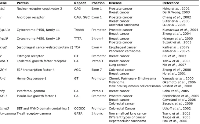
Cancer is a genetically and biologically highly heterogeneous disease that likely requires individually tailored therapies based on the patient’s individual genetic and biologic alterations. The multifactorial process of carcinogenesis involves mutations in oncogenes, or tumor suppressor genes, as well as the influence of environmental etiological factors. During the last few decades, extensive effort has been invested in identifying sources of genetic susceptibility to cancer. Common DNA polymorphisms in low penetrance genes have emerged as genetic factors that seem to modulate an individual's susceptibility to malignancy (Fearnhead et al., 2004; Kryukov et al., 2007; Iyengar & Elston, 2007; Milne & Benítez, 2008). Both the International Human Genome Sequencing Project and the International HapMap Project have generated a very large amount of data on the location, quantity, type and frequency of genetic variants in the
human genome. A large and increasing number of observational studies investigating the association between variants in candidate genes and cancer risk have emerged. Given their abundance and stability, single nucleotide polymorphisms (SNPs) hold great promise as markers for mapping disease susceptibility loci for common, complex disorders by association studies (Bodmer & Bonilla, 2008; Polychronakos, 2008). For this purpose the development of non-invasive, inexpensive, accurate, high-throughput methods for scoring large numbers of SNPs from hundreds of patients and controls is critical.
Despite the intensive effort devoted to the issue, the ability to detect the genetic components of cancer etiology has been very limited. Exceptions include genes that cause rare, mainly monogenic family cancer syndromes such as Brca1 and Brca2 in familial breast and ovarian cancer (Narod & Foulkes, 2004), Mlh1 and Mlh2 in hereditary nonpolyposis colorectal cancer (Plotz et al., 2006), Cdkn2a in familial melanoma (Bishop et al., 2007) and p53 in Li-Fraumeni syndrome (Evans & Lozano, 1997; Varley, 2003). However, such exceptions tend to account for only a small portion of disease heritability. For example, the breast cancer susceptibility genes Brca1 and Brca2 explain only a minority (<30%) of familial breast cancers and a negligible proportion of sporadic breast cancers (Narod & Foulkes, 2004). In conclusion, the genetic etiology of most cancers remains largely unclear.
Cancer is now a major public health problem in Europe, and the ageing of the European population will most certainly cause these numbers to continue to increase. According to the World Health Organization (WHO) cancer is the second cause of death in Portugal and so it will be until 2030, according to projections based on 2002 WHO burden of disease estimates (http://apps.who.int/infobase/report.aspx?iso=PRT&rid=119&goButton=Go). The most common incident forms of cancer are breast and prostate cancer for women and men respectively, followed by colorectal cancer, lung cancer and stomach cancer in both genders. The same types of tumors are within the top five most common causes of cancer deaths (Boyle & Ferlay, 2005; Ferlay et al., 2007; Karim-Kos et al., 2008).
The promise of early detection is that it will identify cancer while still localized and curable, not only preventing mortality but also morbidity and health costs. However, despite that our knowledge of cancer has increased greatly during the past decades, many of the genetic alterations underlying cancer development remain to be clarified. Which of these alterations are the key players in the initial development of cancer is still not known in most of the cancer types. Moreover, the ability to translate this knowledge into clinical practice remains elusive.
Here follows a brief description of the types of cancer used as models in our research work. Breast cancer is the leading cause of cancer in women worldwide (Hinestrosa et al., 2007; Perry et al., 2008). Breast tumors vary greatly in clinical behavior, morphological appearance, and molecular alterations. In general, the genes that have been identified as being associated with hereditary breast cancer (Brca1, Brca2, p53, Chk2, and Atm) are involved in the maintenance of
genomic integrity and DNA repair (Narod & Foulkes, 2004). However, less than 10% of women who develop breast cancer have an identifiable inherited mutation to the disease, and another 15%–20% have a family history but no readily identifiable genetic pattern (Easton et al., 2007; Hinestrosa et al., 2007). The majority of the women develop somatic tumors, in which any known genetic mutation is identified as being the driving force in the origin of the disease. Therefore, although much knowledge has been achieved about breast cancer in the past decades, it continues to be the leading cause of cancer-related death in women.
Despite gastric cancer incidence and mortality have decreased over the past decades, it is still the second most frequent cause of cancer-related death in the world (Crew & Neugut, 2006). Multiple genetic and epigenetic alterations in cell cycle regulators, cell adhesion molecules, DNA repair genes and telomerases, as well as genetic instability at microsatellite loci are the most common events implicated in the multistep process of gastric carcinogenesis (Tahara, 2004). However, gastric cancer is essentially a heterogeneous disease, and the events that underlie the malignant transformation of the gastric mucosa during the multistep process of gastric cancer pathogenesis remain uncertain (Zheng et al., 2006). Several histological classification for gastric cancer have been proposed, being the Lauren’s (1965) classification the most usual - two major forms of gastric tumors are distinguished according to their morphological and clinicopathological classifications: intestinal (well-differentiated) and diffuse (undifferentiated) type tumors. They differ in genetic susceptibility, pathologic profile, clinical presentation, and prognosis (Crew & Neugut, 2006; Cervantes et al., 2007). The intestinal type carcinoma is frequently preceded my multifocal atrophic gastritis and is more common in elderly man; the tumors show gland formation and are usually well demarcated. For the diffuse type tumors the precursor lesions are usually not identified and the prognosis is less favorable, they show no cohesiveness of tumors cells infiltrating the stroma and occurs preferably in women under 50 years old (Werner et al., 2001). Molecular pathology supports this classification by showing differences in the genetics pathways in the origin of both kinds of tumors (Werner et al., 2001; Tahara, 2004; Cervantes et al., 2007). Nevertheless, the prognosis for patients with advanced gastric cancer remains poor, much because gastric cancer is often diagnosed at an advanced stage. Therefore, it is still a challenge to detect stage-specific genetic abnormalities that may result in early diagnosis and aid in selecting therapy.
Colorectal cancer (CRC) is the most common cancer in Europe. Approximately 70% of colorectal cancers are sporadic, with no inherited predisposition (Shen et al., 2007). Although highly penetrant mutations in single genes are known to be responsible for hereditary syndromes, low penetrance susceptibility genes account for a high proportion of the tumors (Mecklin, 2008). Several subgroups with different precursor lesions and behavior can already be distinguished (Shen et al., 2007; Takayama et al., 2007; Li & Lai, 2009). There is heterogeneity in the genetic pathways leading to CRCs, and there are two major tumorigenic pathways. The first is driven by chromosomal instability (CIN), in which accumulating mutations in genes of K-ras (v-Ki-ras2 Kirsten rat sarcoma viral oncogene homolog), Apc (adenomatous polyposis coli), p53 (tumor
protein P53), and Dcc (deleted in colorectal carcinoma), are thought to be of significance. An alternative genetic pathway, related to genetic instability, evolves as a consequence of the alteration in mismatch repair (MMR) genes. However, these two distinct pathways are not able to account for all CRCs. Moreover, there is still an extend gap between basic research and clinical practice, probably due to the genetic and molecular complexity of the tumors (Gil-Bazo, 2007; Huerta, 2008; Nannini et al., 2009). Interaction of genetics and environmental effects has an important role in colorectal carcinogenesis. High body-mass index, increased meat or alcohol consumption and tobacco smoking are examples of confirmed risk factors. A positive family history of CRC increases the estimated risk for CRC by 2- to 4-fold (Mecklin, 2008).
Chronic and persistent inflammation has long been associated with the development of cancer. The genetic changes that occur within cancer cells themselves, such as activated oncogenes or dysfunctional tumor suppressors, are responsible for many aspects of cancer development but they are not sufficient. Tumor promotion and progression are dependent on cells of the tumor environment, that are not necessarily cancerous themselves (Rakoff-Nahoum, 2006). Mediators of the inflammatory response, e.g., cytokines, free radicals, prostaglandins and growth factors, can induce genetic and epigenetic changes, DNA methylation and post-translational modifications, causing alterations in critical pathways responsible for maintaining the normal cellular homeostasis and leading to the development and progression of cancer (Hussain & Harris, 2007). Nowadays, the association between Crohn’s disease (CD) and increased risk for CRC is widely accepted (Zisman & Rubin, 2008). Crohn’s disease (CD) is a chronic inflammatory disease that can affect the entire gastrointestinal tract from the mouth to the anus. CRC is the most common type of cancer in inflammatory bowel disease (IBD), although cancer in other organs can occur. Although CRC associated to IBD accounts only for 1%-2% of all cases of CRC in the general population, it is considered a serious complication of this disease and accounts for approximately 10%-15% of all deaths in IBD patients (Munkholm, 2003). Several of the molecular alterations that contribute to sporadic CRC are also found in IBD-associated CRC (Zisman & Rubin, 2008). CD is therefore a complex disease in which multiple genetic and environmental factors are likely to contribute to pathogenesis, and the search for CD susceptibility genes therefore poses similar problems to those in other complex disorders, like cancer.
Despite these types of cancer are of the most prevalent cancer types in the world today, only a limited number of biomarkers are available for detection and prognostic evaluation. New advances in identifying molecular biomarkers are essential. The intricate nature of the contributions of many factors ultimately determines the impact that a particular alteration has on the properties of a tumour or a precursor lesion (Hanash, 2004). In the future, cancer screening tests will probably combine the result of multiple diagnostic tests and biomarker assays with patient’s specific risk factors.
1.4.1. Translation and cancer
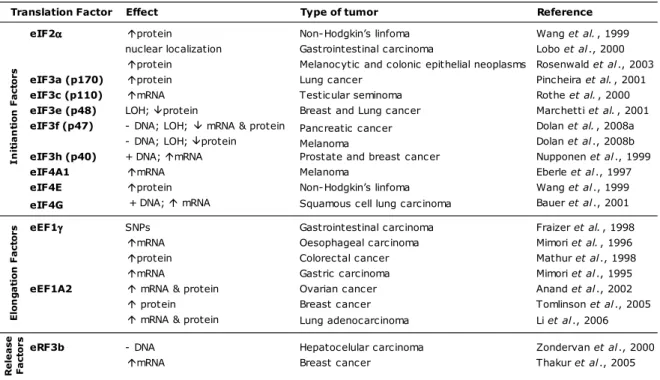
Regulation of translation plays a major role in the control of eukaryotic gene expression (Gebauer & Hentze, 2004). Altering the rate of protein synthesis at the level of translation enables cells to respond rapidly to changes in the intra or extracellular conditions. There are several lines of evidence that support the involvement of translation in the regulation of cell proliferation and cancer development (Clemens & Bommer, 1999; Watkins & Norbury, 2002; Rajasekhar & Holland, 2004; Pandolfi 2004; Bilanges & Stokoe, 2007). Protein synthesis is coupled with cell cycle progression and is regulated in response to nutrient availability, hormones, mitogenic and growth factor stimulation(Caraglia et al., 2000; Meyuhas 2000; Proud 2002). However, the transduction pathways activated do not stimulate the translation of all the mRNAs equally(Holland et al., 2004). The expression of the components of the translation machinery might be selectively regulated by effector proteins, increasing the translation rate of specific oncogenic transcripts(Dua et al., 2001; Meric & Hunt, 2002; Holland et al., 2004).
The transcriptome of a cell is a pool of mRNAs with different efficiencies. Several genes present typical structural features that allow a regulation of their expression at the translation level (Kochetov et al., 1998; Clemens & Bommer, 1999; Meyuhas, 2000). Differential recruitment of mRNA populations to the polisomes might be a rapid response of a cell to the signal transduction pathways activated, resulting in the alterations of the proteome that characterize a cancer cell (Clemens & Bommer, 1999; Watkins & Norbury, 2002; Rajasekhar & Holland, 2004). According to the ribosome filter hypothesis(Mauro & Edelman, 2002), the ribosomal subunits are also considered regulatory elements in translation. Specific sequences at the mRNA molecules are sites of interaction with either rRNAs or ribosomal proteins, especially in the 40S subunit, affecting translation efficiency (Tranque et al., 1998). These interactions are modulated by ribosomal heterogeneity, as a consequence of variation in rRNA or protein composition.
Three main alterations at the translational level can occur in cancer cells: (i) variations in mRNA sequences that increase or decrease the translation efficiency of the transcript (mutations, alternative splicing, alternate polyadenylation sites); (ii) changes in the expression or availability of the components of the translation machinery and (iii) activation of translation through aberrantly activated signal transduction pathways.
Translation of mRNA can be divided in three stages: initiation, elongation and termination. Each stage involves multiple protein factors, though the initiation step is considered the most complex and most tightly regulated (Gebauer & Hentze, 2004; Sonenberg & Hinnebusch, 2009). Therefore, it has been suggested that the initiation step represents the most suitable target for cancer therapy. Accordingly, several studies can be found in literature reporting that the genes involved in the regulation of cell proliferation, growth and death are subject to control at the initiation of translation level (Meric & Hunt, 2002; Stoneley & Willis, 2003; Sonenberg & Dever, 2003; Fingar et al., 2004; Pickering & Willis, 2005; Bilanges & Stokoe, 2007). Effectively, several initiation and also elongation translation factors have been described to be overexpressed in