Rev Odontol UNESP. 2018 Jan-Feb; 47(1): 1-6 © 2018 - ISSN 1807-2577
ORIGINAL ARTICLE
Doi: http://dx.doi.org/10.1590/1807-2577.06717
This is an Open Access article distributed under the terms of the Creative Commons Attribution License, which permits unrestricted use, distribution, and reproduction in any medium, provided the original work is properly cited.
Fluoride release and surface roughness of a new glass ionomer
cement: glass carbomer
Liberação de flúor e rugosidade superficial de um novo cimento de ionômero de vidro: glass carbomer
Célia Maria Condeixa de França LOPES
a*, Jessica GALVAN
b, Ana Claudia Rodrigues CHIBINSKI
b,
Denise Stadler WAMBIER
baDepartamento de Odontologia, UNIVILLE – Universidade da Região de Joinville, Joinville, SC, Brasil
bUEPG – Universidade Estadual de Ponta Grossa, Ponta Grossa, PR, Brasil
Resumo
Objetivo: Este estudo analisou a liberação/recarga de flúor e a rugosidade superficial do carbômero de vidro em comparação a outros cimentos de ionômero vidro (CIVs) encapsulados. Material e método: Os CIVs testados foram o Glass Fill (GC-GCP Dental), Riva Self Cure (RS-SDI), Riva Light Cure (RL-SDI), Equia Fil (EF-GC Europe). A resina composta Luna (LU-SDI) foi empregada como controle. Cinco amostras de cada material foram confeccionadas e mantidas em um umidificador durante 24h (37 °C, 100% de umidade relativa). A liberação de flúor foi aferida em dois tempos: antes (T1: dias 1, 2, 7 e 14) e após aplicação tópica de flúor (T2: dias 15, 16, 21 e 28). A rugosidade superficial também foi aferida nos dois tempos (T1: dias 1 e14; T2: dias 15 e 28). Todas as amostras foram submetidas a uma única aplicação tópica de flúor fosfato acidulado (Flúor Care - FGM). ANOVA dois fatores com medidas repetidas e pós-teste de Tukey (p<0,05) foram empregados na analise estatística. Resultado: O Equia Fil apresentou a maior liberação de flúor em ambos os períodos de avaliação, com liberação maior no T1 (p<0,05). Os demais materiais testados, incluindo o carbômero de vidro, apresentaram liberação semelhante em ambos os períodos (T1 e T2). Em relação à rugosidade superficial não foram observadas diferenças significativas na interação entre os fatores material × tempo (T1 e T2) (p=0,966). Conclusão: Os CIVs testados apresentaram capacidade de liberação e recarga de flúor e não mostraram aumento de rugosidade superfícial pela aplicação tópica de flúor.
Descritores: Cimento de ionômero de vidro; materiais dentários; flúor.
Abstract
Objective: This study analyzed the fluoride release/recharge and surface roughness of glass carbomer compared to other encapsulated glass ionomer cements (GICs). Material and method: The GICs tested were Glass Fill (GC-GCP Dental), Riva Self Cure (RS-SDI), Riva Light Cure (RL-SDI), Equia Fil (EF-GC Europe). The composite resin Luna (LU-SDI) was used as control. Five samples of each material were prepared and kept in a humidifier for
24 hours (37 °C, 100% relative humidity). Fluoride release was measured in two times: before (T1: days 1, 2, 7, 14) and after topical application of fluoride (T2: days 15, 16, 21 and 28). The surface roughness was also measured in both times (T1: days 1 and 14; T2: days 15 and 28). All samples were submitted to a single topical application of acidulated fluoride phosphate (Fluor Care - FGM). Two-way ANOVA with repeated measures and Tukey’s post-test (p <0.05) were used in the statistical analysis. Result: Equia Fil presented the highest fluoride release in both evaluation periods, with a higher release in T1 (p <0.05). The other materials tested, including glass carbomer presented similar release in both periods (T1 and T2). Regarding surface roughness, no significant differences were observed in the interaction between the material × time factors (T1 and T2) (p=0.966). Conclusion: The GICs tested presented fluoride release and recharge ability and showed no surface roughness increase by topical application of fluoride.
Descriptors: Glass ionomer cements; dental materials; fluoride.
INTRODUCTION
The recognized anticariogenic potential of fluoride1 is the main
reason why this ion has been incorporated into several materials used in dentistry. Amongst the fluoride releasing restorative materials, glass ionomer cements (GIC) are the most studied because they
may prevent carie lesions in the tooth/restoration interface and inhibit secondary caries2-4. Fluoride is released from the GIC and
participates in the cycles of des/remineralization1, during clinical
The supply of fluoride for replacement in glass ionomer cements can either originate from daily low concentration sources like fluoride dentifrices and mouth rinses or professional topical applications. Thus the material acts as a fluoride reservoir5.
However, professional topical applications, particulary when acidulated fluoride gel is used, may produce changes on the material, increasing the surface roughness6 and the dental biofilm
accumulation. Consequently, the risk of secondary caries, surface discoloration and fatigue failure of the restoration is enhanced7.
Therefore, there is a practical dilemma: although it is important to provide continuous supply of fluoride to GIC sealants or restorantions, but professional application may alter the surface properties of the material.
The fluoride release/recharge and the surface modifications after fluoride topical application are dependent on several factors like GIC organic matrices, setting mechanisms, fluoride content and environmental conditions6. All these characteristics can vary
between different types of GIC and also withim different brands. That is why the continuous research about this subject is fundamental to support clinical application when new GICs are released.
Glass Carbomer is a new ionomeric material. Its manufacturer states that it differs from conventional GICs because its organic matrix is composed by nanoparticles of glass enriched with fluor/hydroxyapatite8. There are reports in the literature on the
physical and mechanical properties of glass carbomer9-13.
Therefore, the aim of this study was to evaluate the fluoride release and recharge of different types of glass ionomer cements submitted to topical application of acidulated fluoride in vitro, as well as the surface roughness of these materials.
The null hypotheses tested were: (1) that all GICs would have the ability to release/recharge fluoride; (2) that glass carbomer would have a greater release of fluoride and (3) that an application of topical fluoride would increase the surface roughness of the GICs.
MATERIAL AND METHOD
Four encapsulated glass ionomer cements were tested: Riva Self Cure (SDI, Victoria, Australia), Riva Light Cure (SDI, Victoria, Australia), Equia Fil (GC Corporation, Tokyo, Japan) and the new
material Glass Fill (GCP-Dental, Vianen, Netherlands). A composite resin, Luna (SDI, Victoria, Australia) was used as control (Table 1).
Preparation of Test Specimens
Five specimens of each material were made according to the respective manufacturer’s instructions. On a glass plate, a metallic matrix (diameter=5mm and thickness=2mm) lubrified with petroleum jelly (Petrolatum, Quimidrol Joinville, Brazil) was placed over a polyester strip (TDV Dental Ltda., Pomerode, Brazil). The capsules of the materials were homogenized for 10s in a high power mixer (Ultramat 2, SDI, Victoria, Australia) and adapted in the Riva Applicator 2 (SDI, Victoria, Australia), after the rupture of the internal sealing of the material’s capsule with manual pressure. The material was inserted in the metallic matrix. After this procedure, another polyester strip (TDV Dental Ltda.) was placed on top of the specimen and with a glass plate, the material was compressed to spill the excess and result in a smooth surface. For the Glass Fill (GCP-Dental) specimens, the manufacturer recommends to apply a LED light curing lamp (CarboLED Lamp, GCProducts), for 60s, as a heat treatment. Riva Light Cure (SDI) and the composite resin Luna (SDI) were light cured with the same LED lamp for 20s, according to the respective manufacturers’ information, soon after removal of excess material.
The specimens were kept in a humidifier (Kottermann Labortechnick, Uetze, Germany) for 24 h (37C, 100% relative humidity) to complete the glass ionomer cement gelling reaction. After that, the specimens were stored in identified plastic vials containing 20 mL distilled water that was changed daily for 28 days and kept at 37C.
Fluoride Release and Recharge Evaluation
Fluoride release was measured on day 1, 2, 7 and 14 (T1: before fluoride application). On day 15, the specimens were removed from their plastic vials and the moisture excess was removed with absorbent paper. All specimens were immersed in an acidulated phosphate fluoride (Flúor Care-FGM, Joinville, Brazil) in the form of foam for 60s and after that time the excess was removed with absorbent paper and the specimens were immersed again in 20 mL distilled water in their respective plastic vials. New measures of fluoride release were achieved on day 15, 16, 21 and 28 (T2: after fluoride application).
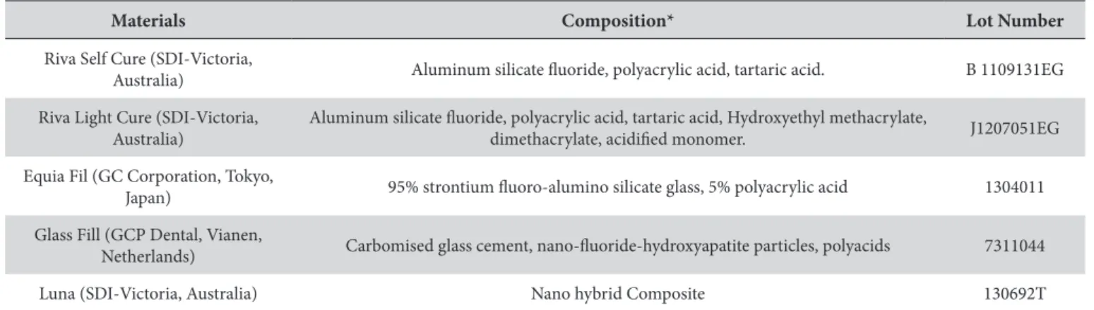
Table 1. Descriptions of the materials used in this study
Materials Composition* Lot Number
Riva Self Cure (SDI-Victoria,
Australia) Aluminum silicate fluoride, polyacrylic acid, tartaric acid. B 1109131EG
Riva Light Cure (SDI-Victoria, Australia)
Aluminum silicate fluoride, polyacrylic acid, tartaric acid, Hydroxyethyl methacrylate,
dimethacrylate, acidified monomer. J1207051EG
Equia Fil (GC Corporation, Tokyo,
Japan) 95% strontium fluoro-alumino silicate glass, 5% polyacrylic acid 1304011
Glass Fill (GCP Dental, Vianen,
Netherlands) Carbomised glass cement, nano-fluoride-hydroxyapatite particles, polyacids 7311044
Luna (SDI-Victoria, Australia) Nano hybrid Composite 130692T
All measures were carried out using a previously calibrated spectrophotometer (Hach DR 4000, Loveland, CO, USA) and the SPADNS colorimetric method. The protocol consisted of retrieving 10 mL distilled water of the vial containing the specimen followed by the addition of 2 mL of the SPADNS 2 fluoride reagente (Hach Company World Headquarters, Loveland, CO, USA). The solution was shaken and after 60s of reaction, it was placed in a 25 mL quartz cuvette for reading and the result in mg/L of fluoride displayed. This procedure was repeated for all samples and the readings regarding the fluoride contents released from each material were recorded.
Surface Roughness Evaluation
The surface roughness was measured with a rugosimeter (Surftest-301 serie 15700438 - Mitutoyo, Suzano, Brazil) on the top surface of the specimen. The rugosimeter was calibrated by the result of the standard plate: 2.95µm for medium roughness, and regulated with a cut-off 0.25mm. Five standard readings (one central point and another four points, north, east, south and west) were recorded; the final reading was the arithmetic mean of these readings (Ra). Surface roughness was measured on day 1 (Ra1 - inicial); day 14 (Ra2 - with 14 days of immersion of the specimens in distilled water); day 15 (Ra3 - soon after application of fluoride); and on day 28 (Ra4 - final roughness).
Statistical Analysis
The data were evaluated using the two-way ANOVA, with repeated measures and Tukey post-test at a significance level of 5% (BIOESTAT 5.0 Program, GraphPad Software, San Diego, California, USA).
RESULT
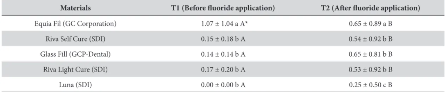
The means of fluoride release before and after topical application are reported in Table 2 and the fluoride release patterns of the tested materials are shown in Figure 1.
The highest fluoride release was observed for Equia Fil (GC Corporation) when compared to the others GICs (p<0.05) in both evaluation periods (before and after the topical application of fluoride). The other products tested, Riva Self Cure (SDI), Riva Light Cure (SDI) and Glass Fill (GCP-Dental), presented similar fluoride release in (T1 and T2) (Table 2), with the highest fluoride release peaks observed within the first 24 hours after topical application (Figure 1). There was no fluoride release by the composite resin
Luna (SDI) at baseline; but after fluoride application, it absorbed fluoride from the medium and released it for a short period of time (Figure 1). The GlassFill (GCP-Dental) showed a peak of fluoride release on the seventh day after topical application (Figure 1), but its mean fluoride release was not different from those of Riva Self Cure (SDI) and Riva Light Cure (SDI) (Table 2).
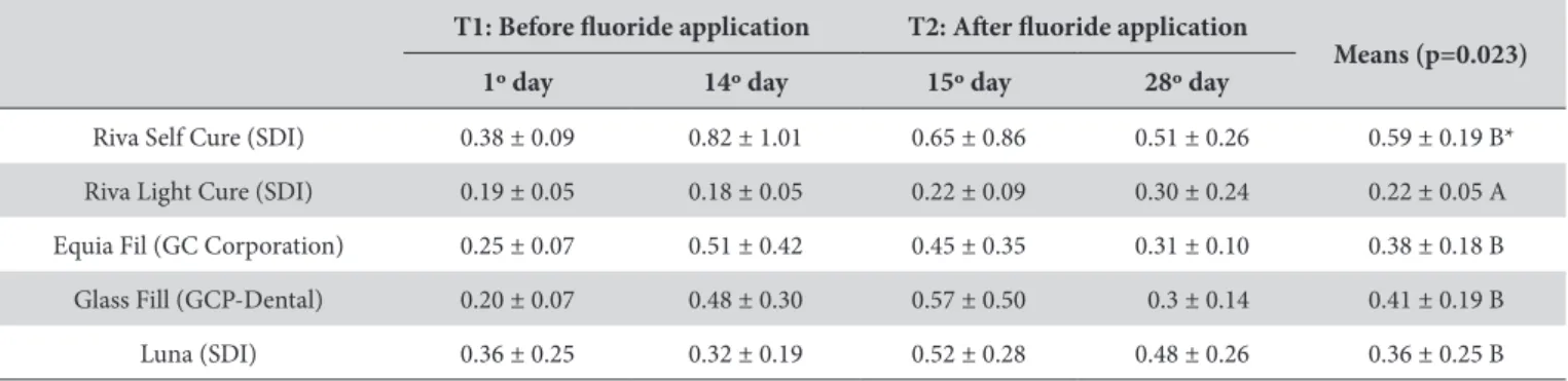
Regarding the surface roughness of the materials tested, no significant difference in the interaction material × time (T1 and T2) (p = 0.966) was observed (Table 3). Riva Light Cure (SDI) presented the lowest roughness mean compared to the other materials tested (p<0.05). Significant differences were not found between the other materials.
The topical application of acidulated phosphate fluoride did not interfere in the roughness of the tested materials.
DISCUSSION
All the tested glass ionomer cements, including the new released glass carbomer cement, showed the ability to release fluoride and to be recharged by topical fluoride applications.
The inicial fluoride release from glass ionomer cement is due to an acid-base reaction and the amount of fluoride released is proportional to the concentration of this ion in the material6. This is
responsible for the “burst effect” phenomenon observed for all tested GICs in this study, which is the release of high amounts of fluoride in the first 24 hours6. After the initial burst, fluoride release slows
down and is followed by a prolonged long-term fluoride release, which occurs when the glass dissolves in the acidified water of the
Table 2. Means and standard deviations of fluoride released before and after topical application of fluoride by the different materials tested
Materials T1 (Before fluoride application) T2 (After fluoride application)
Equia Fil (GC Corporation) 1.07 ± 1.04 a A* 0.65 ± 0.89 a B
Riva Self Cure (SDI) 0.15 ± 0.18 b A 0.54 ± 0.92 b B
Glass Fill (GCP-Dental) 0.14 ± 0.14 b A 0.65 ± 0.81 b B
Riva Light Cure (SDI) 0.17 ± 0.20 b A 0.53 ± 0.92 b B
Luna (SDI) 0.00 ± 0.00 b A 0.25 ± 0.50 c B
*Equal capital letters in the same column indicate absence of statistically significant differences.
hydrogel matrix14. This pattern of fluoride release of the GICs was
observed in the present study and it is in agrement with the literature reports15,16. The combination of different mechanisms, such as
superficial rinse, diffusion through pores and micro fractures and mass diffusion can explain the fluoride release process17. Out of these
mechanisms, the initial superficial rinsing effect contributes for the high level of fluoride release within the first 24 hours, whereas the diffusion through cement pores and fractures promotes the constant release in the following days17. In general, materials with less resin
content have higher porosity, so they exhibit higher initial fluoride release and higher recharge capability15,16, which is in agreement
to what was observed in this study.
The restorative material permeability is fundamental for this process. Thus a completely permeable substance could absorb the ions deep into its bulk; while a relatively impermeable material can only absorb fluoride into the immediate subsurface5. In GIC,
the permeability allows the loosely bound water and the solutes in the porosities to be exchanged with an external medium by passive diffusion16. Since composite resins are not permeable
materials, the fluoride release only occurs after topical fluoride application and for a very short period of time. This release is the result of the washout of fluoride ions that are retained on the surface or in the pores of the composite resin. Filler composition and particle size also have significant influence on the fluoride release16. Fluoroaluminosilicate glass is the key component of the
majority of the GICs fillers used in this study and the main source of fluoride. This component in glass ionomers and resin-modified glass ionomers is soluble and thus releases more fluoride16. One of
the studied materials in this research is a carbomised glass cement, which has nano-fluoride-hydroxyapatite particles in its composition. We hypothesized that this filler particle could originate a different pattern of fluoride release/recharge over time, but this hypothesis was rejected, since the behavior of the glass carbomer was similar to the other GICs. Another significant variation in glass carbomer is that the manufacturer suggests a heat treatment after the material insertion in the cavity; this procedure is claimed to improve its properties. However, radiant heat applied to glass ionomer cements has been shown to have no effect on fluoride release18.
It has been suggested that the recharging ability of glass ionomer cements is dependent on the glass component, particularly upon the structure of the hydrogel layer around glass filler particles, which is formed due to reactions between fluoridated glass particles and
polyacrylic acids19. The pattern of fluoride release after refluoridation
from the materials tested in this study agreed with the findings of other in vitro study15, in which the fluoride release increased within
the first 24 h followed by a rapid return to near pre-exposure levels for several days.
In this present study, protective agents were not used on the surface of the GICs tested to favor fluoride release. According to the literature17, surface protection of GICs definitely prevents the
fluoride release, which might be due to the reduction in the water movement. The surface coating possibly occlude the mechanism of superficial rinse and diffusion through pores17,20. In clinical situations,
the use of a petroleum jelly coat may protect the GICs during the first hours of setting reaction, without hindering fluoride release17.
While the high porosity of the GICs is beneficial for the fluoride release, it also presents adverse effects on the mechanical properties. Regarding the surface roughness, the average range values were 0.22-0.59μm, being the resin modified glass ionomer the one with the lowest value, in the present study. This suggests that the composition of the materials, as the size of its particles21, may be
responsible for these diferences. Therefore, materials with small particles do not invariably show a smoother surface22.
The critical surface roughness for bacterial colonization is 0.2μm23.
All GICs tested in this study presented higher surface roughness than this value after fluoride application. Surface roughness higher than 0.2μm is likely to increase significantly bacterial adhesion, dental plaque maturation and acidity, which increase carie risk22.
But this fact alone does not predispose to the development of new carious lesions, since the disease is a resul of an imbalance in the oral environment and other factors are associated.
Surface disintegration is caused by a selective attack to the polysalt matrix, which is the result of the formation of contact cation-anion ion pairs or complexes between the carboxylic groups of the polyalkenoic acid and metallic ions24. When GIC
is in contact with sodium fluoride, the fluoride ion can compete with the carboxylate groups causing gradual disintegration of the polysalt matrix. The chemical erosion extention then depends not only on the concentration of the fluoride solution but also on the time and frequency of immersion25. Therefore the change in the
surface roughness is material dependent and shorter application times might be preferred to reduce surface alterations of restorative materials21.
Table 3. Means and standard deviations of surface roughness before and after topical application of fluoride by the different materials tested
T1: Before fluoride application T2: After fluoride application
Means (p=0.023)
1º day 14º day 15º day 28º day
Riva Self Cure (SDI) 0.38 ± 0.09 0.82 ± 1.01 0.65 ± 0.86 0.51 ± 0.26 0.59 ± 0.19 B*
Riva Light Cure (SDI) 0.19 ± 0.05 0.18 ± 0.05 0.22 ± 0.09 0.30 ± 0.24 0.22 ± 0.05 A
Equia Fil (GC Corporation) 0.25 ± 0.07 0.51 ± 0.42 0.45 ± 0.35 0.31 ± 0.10 0.38 ± 0.18 B
Glass Fill (GCP-Dental) 0.20 ± 0.07 0.48 ± 0.30 0.57 ± 0.50 0.3 ± 0.14 0.41 ± 0.19 B
Luna (SDI) 0.36 ± 0.25 0.32 ± 0.19 0.52 ± 0.28 0.48 ± 0.26 0.36 ± 0.25 B
Due to different methodologies found in the literature, long-term studies and clinical trials are necessary to clarify the results of this study.
CONCLUSION
Within the limitations of the presente study, it can be concluded that:
• All glass ionomer cements presented fluoride release and re-charge ability. This release of fluoride was more pronounced within the first few days, being reduced over time;
• Glass carbomer showed similar fluoride release compared to the other glass ionomer cements;
• Topical application of acidified fluoride did not interfere with the roughness of the materials.
REFERENCES
1. Lippert F, Hara AT, Martinez-Mier EA, Zero DT. In vitro caries lesion rehardening and enamel fluoride uptake from fluoride varnishes as a function of application mode. Am J Dent. 2013 Apr;26(2):81-5. PMid:24073530.
2. Smales RJ, Gao W. In vitro caries inhibition at the enamel margins of glass ionomer restoratives developed for the ART approach. J Dent. 2000 May;28(4):249-56. PMid:10722898. http://dx.doi.org/10.1016/S0300-5712(99)00071-8.
3. Markovic DL, Petrovic BB, Peric TO. Fluoride content and recharge ability of five glassionomer dental materials. BMC Oral Health. 2008 Jul;8(1):21. PMid:18655734. http://dx.doi.org/10.1186/1472-6831-8-21.
4. Bertolini MJ, Zaghete MA, Gimenes R, Padovani GC, Cruz CA. Preparation and evaluation of an experimental luting glass ionomer cement to be used in dentistry. J Mater Sci Mater Med. 2009 Sep;20(9):1781-5. PMid:19415231. http://dx.doi.org/10.1007/s10856-009-3748-7.
5. Preston AJ, Higham SM, Agalamanyi EA, Mair LH. Fluoride recharge of aesthetic dental materials. J Oral Rehabil. 1999 Dec;26(12):936-40. PMid:10620157. http://dx.doi.org/10.1046/j.1365-2842.1999.00502.x.
6. Wiegand A, Buchalla W, Attin T. Review on fluoride-releasing restorative materials-fluoride release and uptake characteristics, antibacterial activity and influence on caries formation. Dent Mater. 2007 Mar;23(3):343-62. PMid:16616773. http://dx.doi.org/10.1016/j.dental.2006.01.022.
7. Cehreli ZC, Yazici R, García-Godoy F. Effect of 1.23 percent APF gel on fluoride-releasing restorative materials. ASDC J Dent Child. 2000 Sep-Oct;67(5):330-7, 302. PMid:11068665.
8. Arslanoglu Z, Altan H, Sahin O, Tekin MG, Adigüzel M. Evaluation of surface properties of four tooth-colored restorative materials. Acta Phys Pol A. 2015;128(2B):310-3. http://dx.doi.org/10.12693/APhysPolA.128.B-310.
9. Cehreli SB, Tirali RE, Yalcinkaya Z, Cehreli ZC. Microleakage of newly developed glass carbomer cement in primary teeth. Eur J Dent. 2013 Jan;7(1):15-21. PMid:23408469.
10. Lopes CMCF, Schubert EW, Reis A, Wambier DS. Análise da dureza de um novo material restaurador para ART: Glass Carbomer. Rev Odontol UNESP. 2016 Abr;45(2):65-70. http://dx.doi.org/10.1590/1807-2577.10915.
11. Menne-Happ U, Ilie N. Effect of gloss and heat on the mechanical behaviour of a glass carbomer cement. J Dent. 2013 Mar;41(3):223-30. PMid:23174652. http://dx.doi.org/10.1016/j.jdent.2012.11.005.
12. Chen X, Du MQ, Fan MW, Mulder J, Huysmans MCDNJM, Frencken JE. Caries preventive effect of sealants produced with altered glass-ionomer materials, after 2 years. Dent Mater. 2012 May;28(5):554-60. PMid:22300651. http://dx.doi.org/10.1016/j.dental.2012.01.001.
13. Koenraads H, Van der Kroon G, Frencken JE. Compressive strength of two newly developed glass-ionomer materials for use with the Atraumatic Restorative Treatment (ART) approach in class II cavities. Dent Mater. 2009 Apr;25(4):551-6. PMid:19211138. http://dx.doi. org/10.1016/j.dental.2008.12.008.
14. Woolford MJ, Grieve AR. Release of fluoride from glass polyalkenoate (ionomer) cement subjected to radiante heat. J Dent. 1995 Aug;23(4):233-7. PMid:7629328. http://dx.doi.org/10.1016/0300-5712(95)91188-S.
15. Dionysopoulos D, Koliniotou-Koumpia E, Helvatzoglou-Antoniades M, Kotsanos N. Fluoride release and recharge abilities of contemporary fluoride-containing restorative materials and dental adhesives. Dent Mater J. 2013;32(2):296-304. PMid:23538766. http://dx.doi.org/10.4012/ dmj.2012-144.
16. Xu X, Burgess JO. Compressive strength, fluoride release and recharge of fluoride-releasing materials. Biomaterials. 2003 Jun;24(14):2451-61. PMid:12695072. http://dx.doi.org/10.1016/S0142-9612(02)00638-5.
17. Kamatham R, Reddy SJ. Surface coatings on glass ionomer restorations in Pediatric dentistry-Worthy or not? J Indian Soc Pedod Prev Dent. 2013 Oct-Dec;31(4):229-33. PMid:24262395. http://dx.doi.org/10.4103/0970-4388.121818.
18. De Moor RJ, Verbeeck RM, De Maeyer EA. Fluoride release profiles of restorative glass ionomer formulations. Dent Mater. 1996 Mar;12(2):88-95. PMid:9002849. http://dx.doi.org/10.1016/S0109-5641(96)80074-1.
19. Han L, Edward C, Okamoto A, Iwaku M. A comparative study of fluoride-releasing adhesive resin materials. Dent Mater J. 2002 Mar;21(1):9-19. PMid:12046524. http://dx.doi.org/10.4012/dmj.21.9.
20. Tiwari S, Nandlal B. Effect of nano- filled surface coating agent on fluoride release from conventional glass ionomer cement: an in vitro trial. J Indian Soc Pedod Prev Dent. 2013 Apr-Jun;31(2):91-5. PMid:23886719. http://dx.doi.org/10.4103/0970-4388.115703.
22. Bala O, Arisu HD, Yikilgan I, Arslan S, Gullu A. Evaluation of surface roughness and hardness of different glass ionomer cements. Eur J Dent. 2012 Jan;6(1):79-86. PMid:22229011.
23. Bollenl CML, Lambrechts P, Quirynen M. Comparison of surface roughness of oral hard materials to the threshold surface roughness of bacterial plaque retention: a review of the literature. Dent Mater. 1997 Jul;13(4):258-69. PMid:11696906. http://dx.doi.org/10.1016/S0109-5641(97)80038-3.
24. Hadley PC, Billington RW, Pearson GJ, Williams JA. Effect of monovalente ions in glass ionomer cements on their interaction with sodium fluoride solution. Biomaterials. 2000 Jan;21(1):97-102. PMid:10619683. http://dx.doi.org/10.1016/S0142-9612(99)00149-0.
25. De Witte AM, De Maeyer EA, Verbeeck RM. Surface roughening of glass ionomer cements by neutral NaF solutions. Biomaterials. 2003 May;24(11):1995-2000. PMid:12615490. http://dx.doi.org/10.1016/S0142-9612(02)00617-8.
CONFLICTS OF INTERESTS
The authors declare no conflicts of interest.
*CORRESPONDING AUTHOR
Célia Maria Condeixa de França Lopes, Departamento de Odontologia, UNIVILLE – Universidade da Região de Joinville, Rua Rio Grande do Sul, 261/404, 89203-570 Joinville - SC, Brasil, e-mail: [email protected]