Impact of the invasive macroalgae Asparagopsis armata -
– an ecotoxicological assessment
Marta Sofia da Cruz Jacinto
Impact of the invasive macroalgae Asparagopsis armata
-– an ecotoxicological assessment
Marta Sofia da Cruz Jacinto
Dissertação para obtenção do Grau de Mestre em Biotecnologia dos Recursos Marinhos
Dissertação de Mestrado realizada sob a orientação do Doutor Marco Lemos e co-orientação da Doutora Sara Novais e da Doutora Melissa Faria
ii
Title: Impact of the Invasive macroalgae Asparagopsis armata – an ecotoxicological assessment
Título: Impacto da macroalga invasora Asparagopsis armata – uma avaliação ecotoxicológica
Copyright © Marta Sofia da Cruz Jacinto
A Escola Superior de Turismo e Tecnologia do Mar e o Instituto Politécnico de Leiria têm o direito, perpétuo e sem limites geográficos, de arquivar e publicar este trabalho de projeto através de exemplares impressos reproduzidos em papel ou de forma digital, ou por qualquer outro meio conhecido ou que venha a ser inventado, e de a divulgar através de repositórios científicos e de admitir a sua cópia e distribuição com objetivos educacionais ou de investigação, não comerciais, desde que seja dado crédito ao autor e editor.
iii À minha mãe e ao meu pai pela confiança que sempre depositaram em mim, aos meus irmãos por todo o apoio sempre que necessário, aos meus avós
iv
v
Agradecimentos
Agora que esta dissertação está finalmente concluída, não podia deixar de mostrar a minha gratidão a todas as pessoas que de alguma forma contribuíram para a mesma, quer direta ou indiretamente.
Em primeiro lugar tenho de agradecer à minha família, principalmente aos meus pais e aos meus irmãos por todo o apoio incondicional que sempre demonstraram e pelos momentos em que me ajudaram a suportar as coisas menos boas. Nunca deixaram de acreditar em mim e nas minhas capacidades. Estiveram sempre presentes!
Aos meus três orientadores Doutor Marco Lemos, Doutora Sara Novais e Doutora Melissa Faria agradeço de forma sentida todo o acompanhamento e toda a disponibilidade demonstrada ao longo desta etapa. Todos me transmitiram conhecimentos valiosos.
Ao Professor Marco pela mão estendida a quem procurava trabalhar e evoluir no meio laboratorial.
À Melissa Faria, por todo o apoio e ajuda durante a estadia em Barcelona, por todas as palavras reconfortantes e por tão bem me receber numa nova instituição e me transmitir o máximo de conhecimento no mínimo tempo possível.
À Sara Novais, por todas as horas passadas no laboratório, que foram uma ajuda indispensável, e pelas palavras encorajadoras que sempre me prestou.
Aos meus colegas da segunda edição do Mestrado em Biotecnologia dos Recursos Marinhos da ESTM por todos os momentos bem passados e companheirismo, pois com pessoas como vocês por perto o trabalho torna-se mais fácil. “MBRM – Isto é o Futuro!”.
Um obrigado especial à Isa Gomes, à Catarina Correia e ao André Horta que sempre estiveram presentes e disponíveis para me ajudar, mesmo quando passou a ser virtualmente. Apesar de distantes nunca deixaram de ser essenciais na minha vida, pois amigos como vocês são difíceis de encontrar.
Ao Hugo Morais por toda a amizade, por me ajudares, incentivares, aturares e suportares os meus maus momentos e mesmo assim estares sempre ao meu lado com um sorriso na cara. À Tânia Serreira por me mostrar que com calma e dedicação tudo é possível e por mais que as pessoas sejam diferentes em algum ponto elas podem conjugar e formar boas duplas. Ao João Chambel, por todas as palavras amigas nas horas de aflição e por deixar que o laboratório Ornamental se torna-se um refúgio para onde podia sempre fugir.
À Catarina Almeida por ser o meu porto de abrigo que me ajudou a manter a manter a sanidade mental durante estes tempos e por me mostrar que muitas vezes é mais fácil deixar acontecer! Também agradeço ao Manuel Seixas por toda essa maneira de ser que
vi
acalma quem te rodeia e por ouvires muitas vezes os meus delírios. E por me terem ajudado a sorrir nos tempos difíceis.
Ao José Pedro Marques pois fizeste parte da minha vida durante grande parte do tempo desta jornada e foste um dos meus portos seguros. Obrigada por todas as vezes que me acalmaste, aguentaste o meu mau feitio e estiveste do meu lado.
Um agradecimento mais geral para quem de alguma maneira se cruzou comigo nesta etapa e permitiu que a mesma fosse concluída com sucesso.
E um reconhecimento ao Luís Vala, colega e amigo, com muita saudade, pela inspiração e teres sido o pensamento que nunca me permitiu desistir.
vii
Resumo
Os Oceanos representam o maior sistema de suporte de vida sendo a uma grande fonte de riqueza, oportunidade e abundância. No entanto, a humanidade tem levado este ecossistema ao seu limite com crescentes níveis de poluição e outras pressões antropogénicas. A introdução de espécies não-nativas é reconhecida como uma das maiores ameaças à biodiversidade e a segunda maior causa de extinção das espécies. A macroalga vermelha Asparagopsis armata é uma espécie invasora originária da Austrália e que atualmente apresenta uma ampla distribuição em todo o globo devido à sua estratégia oportunista, ausência de predadores e altas taxas de crescimento. Uma questão emergente está relacionada com a capacidade destas espécies invasoras produzirem grandes quantidades de metabolitos halogenados potencialmente tóxicos. Esta característica pode representar um perigo adicional para o equilíbrio ecológico da comunidade invadida.
O presente trabalho teve como objetivo avaliar o potencial ecotoxicológico dos exsudatos de A. armata usando um gastrópode, Gibbula umbilicalis, como organismo modelo. A macroalga recolhida na costa de Peniche (Portugal) foi colocada em tanques no laboratório, durante 12 h, sendo depois o meio recolhido e filtrado para ensaios posteriores com os exsudatos da alga. No ensaio agudo, observou-se a mortalidade de G. umbilicalis que foi exposta a crescentes diluições do exsudato durante 96 h. Adicionalmente, os gastrópodes foram expostos a concentrações não letais do exsudato e analisou-se as respostas bioquímicas recorrendo a biomarcadores relacionados com destoxificação, defesas antioxidantes, danos oxidativos, danos neurotóxicos e metabolismo energético.
Os resultados revelaram que os exsudatos de A. armata afetaram significativamente a sobrevivência dos organismos expostos com uma CL50 96h de 5.03% de exsudato da alga. A exposição aos exsudatos da alga também resultou em efeitos bioquímicos e metabólicos ao nível subcelular com resultados significativos na inibição da glutationa-S-transferase (GST), perda de integridade do ADN e níveis crescentes de atividade da lactato desidrogenase (LDH), dando uma indicação dos mecanismos de toxicidade desta alga marinha. Os níveis mais elevados de danos no ADN ocorreram quando a GST apresentou os níveis mais baixos de atividade e esta mesma atividade aumentou quando os danos no ADN diminuíram, em simultâneo com o aumento dos níveis de atividade da LDH, indicando que as necessidades energéticas aumentam devido à necessidade de sintetizar mais enzima.
Conclui-se que a A. armata tem capacidade de libertar substâncias tóxicas que podem ter potenciais impactos no ambiente envolvente. Adicionalmente, as respostas
viii
bioquímicas estudadas em G. umbilicalis têm potencial para serem usadas como sinais de aviso na determinação dos efeitos provocados pelos compostos libertados por esta macroalga vermelha.
Palavras-chave: Ambientes costeiros, Biomarcadores, Ecotoxicologia, Espécies invasoras, Gibbula
ix
Abstract
The Oceans represent the largest life support system being a major source of wealth, opportunity and abundance. However, mankind has pushed this ecosystem to its limit with increasing pollution levels and other anthropogenic pressures. The introduction of non-native species is recognized as one of the major threats to biodiversity and the second leading cause of species extinction. The red macroalgae Asparagopsis armata is an invasive species native from Australia and currently has a wide distribution across the globe due to its opportunistic strategy, absence of predators, and rapid growth rates. One problematic issue emerging from the invasion of this species is related to its capacity to produce large amounts of halogen metabolites potentially toxic. This characteristic may represent an additional danger to the ecological balance of the invaded community.
This study aimed to assess the ecotoxicological potential of exudates of A. armata using a gastropod, Gibbula umbilicalis, as test organism. The seaweed collected at the coast of Peniche (Portugal) was left in laboratory tanks, for 12 hours, and afterwards the media was collected and filtered for further testing with algae exudates. In the acute test,
G. umbilicalis mortality was observed with exposure to increasing dilutions of exudate for
96 h. Additionally, gastropods were exposed to non-lethal concentrations of exudate and analyzed using biochemical biomarkers responses associated with detoxification, antioxidant defenses, oxidative damage, neurotoxicity and energy metabolism.
The results showed that A. armata exudates significantly affected the survival of exposed organisms and a 96h LC50 of 5.03% of algae exudates as found. Exposure to the algae exudate also disturbed the biochemical and metabolic responses at the subcellular level with significant inhibitions of glutathione-S-transferase (GST), DNA integrity loss and increasing levels of lactate dehydrogenase (LDH) activity, giving an indication of the mechanism of toxicity of this seaweed. The highest levels of DNA damage occurred when the GST had the lowest levels of activity and DNA damages decreased with the increase of GST and LDH activities, indicating that the energy requirements increase due to the necessity to synthesize more enzyme.
In conclusion, A. armata is capable of releasing toxic substances with potential severe impacts to its surrounding environment. Also, the biochemical responses studied in
G. umbilicalis have the potential to be used as early-warning signals to assess effects of
the compounds released by this red seaweed.
Keywords: Biomarkers, Coastal Environments, Ecotoxicology, Gibbula umbilicalis, Invasive
x
xi
Table of Contents
Resumo ... vii
Abstract ... ix
List of Figures ... xiii
List of Tables ... xv
List of Abbreviations ... xvii
Chapter I.General Introduction ...19
1. Pollution on marine ecosystems ...21
2. Invasive Alien Species ...22
2.1. Introduction and Establishment ...22
2.2. Impacts and Management ...24
2.3. Examples of successful invasions ...25
2.4. Macroalgae as invasive ...26
2.5. Asparagopsis armata ...27
3. Sentinel species ...30
3.1. Gibbula umbilicalis as a test organism ...31
4. Ecotoxicological bioassays ...32
4.1. Classical endpoints ...33
4.2. Biochemical Biomarkers ...33
4.3. Connecting different levels of biological organization – ecological relevance .38 5. Aims of the study ...40
Chapter II. Survival and biochemical responses in Gibbula umbilicalis exposed to exudates of the red macroalgae Asparagopsis armata ...41
1. Introduction ...43
2. Material and Methods ...46
2.1. Test Organisms ...46
xii
2.1.2. Gibbula umbilicalis – Collection and culture conditions ... 46
2.2. Exposures design ... 46
2.3. Acute assay - Lethality ... 47
2.4. Subcellular level responses – biochemical biomarkers ... 47
2.4.1. Tissue preparation ... 47
2.4.2. Protein quantification ... 48
2.4.3. Phase II detoxification enzyme activity ... 48
2.4.4. Oxidative stress parameters ... 48
2.4.5. Esterases determination ... 49
2.4.6. Lactate dehydrogenase activity ... 50
2.5. Data analysis ... 50
3. Results ... 50
3.1. Acute assay - Lethality ... 50
3.2. Subcellular level responses – biochemical biomarkers ... 51
3.2.1. Antioxidant and detoxification enzymes ... 51
3.2.2. Oxidative damage ... 53
3.2.3. Esterases and lactate dehydrogenase activities ... 54
3.2.4. Correlation between biochemical biomarkers ... 55
4. Discussion and conclusions ... 56
Chapter III.General Discussion and Concluding remarks ... 61
xiii
List of Figures
Chapter I. General Introduction
Figure 1 – Schematic representation of the life cycle of Asparagopsis armata. ...28
Figure 2 – Photograph of a) tertrasporophyte generation, Falkenbergia rufolanosa (Núcleo de Ambiente da Universidade do Algarve, NAMB) and b) gametophyte generation,
Asparagopsis armata (Fiona Crouch). ...29
Figure 3 – Enzymatic mechanism involved in xenobiotic biotransformation and antioxidant responses (adapted from Howcroft et al., 2009). GST – Glutathione-S-transferase; SOD – Superoxide Dismutase; CAT – Catalase; LPO – Lipid Peroxidation; GPx – Glutathione Peroxidase; GR – Glutathione Reductase; GSH – Reduced Glutathione; GSSG – Oxidized Glutathione. ...36
Figure 4 – Mechanism of action of Actetylcholinesterase (adapted from Soreq and Seidman, 2001) ...37
Figure 5 – Relationship between ecological relevance, time of response and the different levels of biological organization after stress exposure (adapted from Lemos et al., 2010). ...39
Chapter III. Survival and biochemical responses in Gibbula umbilicalis exposed to exudates of the red macroalgae Asparagopsis armata
Figure 6 – Mortality of Gibbula umbilicalis caused by exposure to Asparagopsis armata exudates for 96 h. Bars represents Mean + standard error (SEM), n=8. ...51
Figure 7 – Enzymatic activities of antioxidants (A - CAT and B – SOD) and phase II detoxification (C – GST) defenses of Gibbula umbilicalis when exposed to different percentages of Asparagopsis armata exudate for 96 h. Bars represent Mean + standard error (SEM). n=8, each replicate consisting of a pool of 2 organisms. (*) indicates statistically
xiv
significant differences between exposed groups and control (ANOVA, Dunnett’s post hoc multiple comparison test, p<0.05). ... 52
Figure 8 – Oxidative damage measured in Gibbula umbilicalis exposed for 96h to different percentages of Asparagopsis armata exudates by means of A) Lipid peroxidation (LPO) levels and B) DNA damage levels. Results are presented as Mean + standard error (SEM). n=8, each replicate consisting of a pool of 2 organisms. (*) indicates statistically significant differences between exposed groups and control (ANOVA, Dunnett’s post hoc multiple comparison test, p<0.05). ... 53
Figure 9 – Esterases activities of Gibbula umbilicalis exposed for 96h to different percentages of Asparagopsis armata exudates. A) Acetylcholinesterase (AChE) and B) Carboxylesterase (CbE). Bars are Mean + standard error (SEM). n=8, each replicate consisting of a pool of 2 organisms... 54
Figure 10 – Lactate dehydrogenase (LDH) activity of Gibbula umbilicalis exposed to
Asparagopsis armata exudate for 96 h. Results are presented as Mean + standard error
(SEM). n=8, each replicate consisting of a pool of 2 organisms. (*) indicates statistically significant differences between exposed groups and control (ANOVA, Dunnett’s post hoc multiple comparison test, p<0.05). ... 55
xv List of Tables
Chapter I. General Introduction
Table I – Anthropogenic vectors for accidental marine introductions (Adapted from Bax et al., 2003). ...23
Table II – Taxonomic classification of Asparagopsis armata. ...27
Chapter III. Survival and biochemical responses in Gibbula umbilicalis exposed to exudates of the red macroalgae Asparagopsis armata
Table III – Pearson correlation between biochemical responses that presented statistically significant differences in this study. The correlation coefficient, positive or negative, of the significant correlations (p<0.05) is underlined and emphasized in bold. ...55
xvi
xvii List of Abbreviations
ACh – acetylcholine AChE – acetylcholinesterase CAT – catalase CbE – Carboxylesterase CDNB – 1-chloro-2,4-dinitrobenzene DBA – dibromoacetic acid
DNA – deoxyribonucleic acid
DTNB – 5,5-dithiobis-(2-nitrobenzoic acid) EDTA – ethylenediamine tetracetic acid GPx – glutathione peroxidase
GR – glutathione reductase GSH – glutathione
GSSG – glutathione disulfide GST – glutathione-S-transferase Gww – grams of tissue wet weight H2O2 – hydrogen peroxide
IAS – invasive alien species
IOC – Intergovernmental Oceanographic Commission LC50 – medial lethal concentration
LDH – lactate dehydrogenase LPO – lipid peroxidation MDA – malondialdehyde
NADH – nicotinamide adenine dinucleotic O2- - superoxide radical
OH- - hydroxyl radical
POPs – persistent organic pollutants ppm – parts per million
ROS – reactive oxygen species SOD – superoxide dismutase TMP – 1,1,3,3-tetramethoxypropan
xviii
19
Chapter I.
20
Chapter I. General Introduction
21
1. Pollution on marine ecosystems
The oceans cover more than 70% of the Earth’s surface and contain around 99% of the living space on earth. Subsequently, oceans are home to a great source of the World’s biological biodiversity (Targett et al., 2002; IOC/UNESCO et al., 2011). Moreover, the oceans play an important role in the regulation of the planet’s climate and are responsible for over 35% of the primary production. The significance of marine waters is also economical, with 60% of the total economic value of the biosphere being attributed to the oceans (Costanza and Folke, 1997).
However, there is evidence that the oceans have suffered at the hands of mankind for millennia, with the history of marine environmental pollution dating back to the very beginning of the history of human civilization (Islam and Tanaka, 2004). Human impacts have increased with rapid population growth and substantial developments in technology. However, aquatic pollution did not receive much attention until a threshold level was reached, with possible adverse consequences on the organisms and their ecosystems (Jenssen, 2003). At the present, it has become a major global concern and one of the greatest challenges of the century, and identified by the European Union as one of the main threats to the normal function of ecosystems (Rodrigues et al., 2009; Nota et al., 2010). Many organizations have made several efforts to promote awareness and taken actions to stop biodiversity loss (Islam and Tanaka, 2004).
The United Nations Convention on the Law of the Sea defined pollution as “the
introduction by man, directly or indirectly, of substances or energy into the marine environment, including estuaries, which results or is likely to result in such deleterious effects as harm to living resources and marine life, hazards to human health, hindrance to marine activities, including fishing and other legitimate uses of the sea, impairment of quality for use of the sea water” (Islam and Tanaka, 2004).
Knowledge of the pollution sources and impacts on ecosystems is important not only for a better understanding on the ecosystem responses to stressors but also to formulate prevention measures. Many of these impacts are generally well known and include a range of threats like oil spills, untreated sewage, heavy siltation, eutrophication, persistent organic pollutants (POP’s), heavy metals, acidification, radioactive substances, marine litter, overfishing, and destruction of coastal and marine habitats (McCook, 1999, Nyström et al., 2000; Bellwood et al., 2004). Besides this, new concepts on environmental pollution are emerging, for example biological pollution (e.g. invasive species) (Islam and Tanaka, 2004).
The term “biological pollution” has been growing in recent years with identified impacts of introduction and invasion of species throughout the world (Boudouresque and
Chapter I. General Introduction
22
Verlaque, 2002). An ever-increasing number of articles, journals and books bearing “invasion” in their titles document that it is an extremely active research area, integrating a diversity of fields such as biogeography, ecology, evolutionary biology, biosecurity, conservation practice, and applied management (Heger et al., 2013).
2. Invasive Alien Species
The European Commission (2002) defined Invasive alien species (IAS) as “a
species whose introduction and/or spread outside their natural past or present distribution threatens biological diversity”. As a result, an introduced species is a species that fulfills the
four following criteria: 1) it colonizes a new area where it was not previously present; 2) the extension of its range is linked, directly or indirectly, to human activity; 3) there is a geographical discontinuity between its native area and the new area; and 4) new generations of the non-native species are born in situ without human assistance, consequently constituting self-sustaining populations (Boudouresque and Verlaque, 2002).
2.1. Introduction and Establishment
Globalization has combined widely dispersed human communities into a worldwide economy. This process provides many benefits through the movement of people and goods, but also commonly leads to the transfer of organisms among ecosystems that were previously separate (Keller et al., 2011). Species have always used the oceans to move around the planet, but until recently, this process has been moderated, limited by the currents and the winds. However, since people began travelling they have inadvertently carried “pests” with them, including unnoticed marine organisms (Bax et al., 2003).
Invasions usually occur in two major phases: Introduction and Establishment. The first step in the process is the transfer of a species from its native range into a new place. Natural transport, or dispersal, is not considered invasion, only Human-mediated transportation is considered true invasion, which can be intentional or accidental. Intentional transport involves introduction of new species for a purpose, for example as pets, for hunting, or as ornamental species. Accidental transport is where organisms are unintentionally moved out of their home range. However, only a few species manage to survive transit (Namboothri et al., 2012).
The major vectors responsible for the global movement of aquatic organisms include ships’ ballast, aquarium industry, aquaculture, the bait industry, and fouling of ships’ hulls (Bax et al., 2001). In Table I a more complete source of marine species introduction is presented with the main target associated to each vector.
Chapter I. General Introduction
23
Table I – Anthropogenic vectors for accidental marine introductions (Adapted from Bax et al., 2003).
Source Vector Target
Commercial shipping
Ballast water Plankton, nekton, benthos in sediment
Hull fouling Encrusting, nestling and some mobile species Solid ballast (rocks, sand, etc.) Encrusting, benthos, meiofauna and flora
Aquaculture and fisheries
Intentional release for stock
enhancement Single species
Gear, stock or food movement Various Discarded nets, floats, traps,
trawls, etc. Various
Discarded live packing
material Various
Release of transgenic species Single species Drilling
platforms
Ballast water Plankton, nekton, benthos in sediment
Hull fouling Encrusting, nestling and some mobile species Canals Movement of species through locks due to water motion or
active swimming
Various Aquarium
Industry Accidental or intentional release Aquarium fauna and flora Recreational
boating Hull fouling Encrusting, nestling and some mobile species
Dive practices Snorkeling and scuba gear Algal spores, bacteria, some small mobile species Floating debris Discarded plastic debris Encrusting and some mobile species
Once introduced into a different system, the establishment of species in its new habitat depends on several factors, including the richness of the habitat and it ecological quality. Consequently, when an alien species arrives at a new location, several things can happen: 1) it can find its new habitat unwelcoming and die; 2) it can survive with little environmental impact; or 3) it can take over, harming the naturally existing wildlife in a variety of ways. Factors intrinsic to the species that aid in their establishment and spread comprise: high environmental tolerance, short generation times, rapid growth, a broad diet, early sexual maturity, high reproductive output, and rapid dispersal (Namboothri et al., 2012).
However, the establishment of a naturalized population of a non-native species does not imply that the species has become invasive. To be considered invasive, a species has to establish large populations and spread in its new system, causing damages (Namboothri et al., 2012).
Chapter I. General Introduction
24
2.2. Impacts and Management
Invasive species are causing increasing concern, since they are almost impossible to eradicate, once becoming established (Wallentinus and Nyberg, 2007).
Their impact can be divided into three major areas of influence: environmental, economic, and social (Bax et al., 2003; Zenetos et al., 2005).
These organisms can act as ecosystem engineers, influencing the habitat itself, positively or negatively, directly or indirectly, and physically or chemically. At a first glance they may have a positive impact, by providing places for shelter in previously barren areas or increasing habitat diversity and special heterogeneity, which would increase species richness. However, the opposite effect is most often observed (Wallentinus and Nyberg, 2007).
On a global level, invasive alien species are considered as one of the major threats to biodiversity, both in terrestrial and marine environments (Zenetos et al., 2005; Altamirano et al., 2008; Regulation (EU) No 1143/2014), and the second leading cause of species extinction, along with habitat destruction (Zenetos et al., 2005). According to the National Wildlife Federation approximately 42% of endangered species are at risk owing to invasive species (Pimentel et al., 2005).
The consequences are due to ecological interactions with biota in a variety of ways. For example by competition for resources (including place to settle and spawning grounds, grazing or predation on native organisms, trophic cascading effects, or filling up empty niches), being a reservoir for parasites or a vector for pathogens, by hybridizing with a related species or varieties, by altering the local food web, disrupting pollination services, changing habitat structure, or even being toxic (Zenetos et al., 2005; Wallentinus and Nyberg, 2007). Thereby, the threat extends also to ecosystem functions and services.
Biotic communities throughout the world are being homogenized and restructured through biological invasions. This has the potential to cause large economic losses, particularly in countries that rely on natural and primarily resources of production like agriculture, forestry and fisheries for their development (Namboothri et al., 2012). Human health and economies are also at risk. The impacts on our natural ecosystems and economy cost billions each year.
Approximately 12 000 species in the environment of the European Union (EU) and in other European countries are alien, of which roughly 10 to 15% are estimated to be invasive (Regulation (EU) No 1143/2014). Invasive alien species are estimated to have cost at least €12 billion/year over the past 20 years, and the figure is growing. The risk such
Chapter I. General Introduction
25 invasive species pose may intensify due to increased global trade, transport, tourism, and climate change (Regulation (EU) No 1143/2014).
In Portugal, over the last two centuries, and especially in more recent decades, the number of exotic species has increased significantly, currently amounting to 670 species, which corresponds to approximately 18% of the total native. Several of the exotic species listed in Portugal are considered invasive and about 8% have invasive behavior (Almeida and Freitas, 2012).
Many organizations around the world are creating laws, legislations and regulations to control and eventually eradicate the invasive species. The most recent is the Regulation (EU) No 1143/2014 on the prevention and management of the introduction and spread of invasive alien species of the European Parliament, which began to be applied in the present year of 2015.
2.3. Examples of successful invasions
The studies about aquatic invasive organisms are numerous, some examples are: the green algae Caulerpa taxifolia in the Mediterranean (Meinesz and Hess, 1991) and the zebra mussel (Dreissena polymorpha) in the North American Great Lakes and Europe (Carlton, 1996).
The green alga Caulerpa taxifolia (Vahl) C. Agardh is one of the most publicized introduced marine species (Thibaut et al., 2004; Theil et al., 2007; Burfeind and Udy, 2009). This seaweed is successful at colonizing low light and nutrient enriched areas, so it can live in lower water quality conditions and assimilate nutrients from the water column and sediment (Burfeind and Udy, 2009). It forms dense meadows from few meters under the surface to over 40 m (Thibaut et al., 2004). It is native to tropical and subtropical areas of Australia, with Moreton Bay being the southernmost extent of its native range (Burfeind and Udy, 2009). Caulerpa taxifolia has been recorded from Mediterranean Sea (Croatia, France, Italy, Monaco, Spain and Tunisia), along the coast of California, as well as Japan and eastern Australia (Thibaut et al., 2004; Theil et al., 2007; Burfeind and Udy, 2009). Indeed the invasion in the Mediterranean Sea is a long and well-documented history where its rapid expansion has been observed competing with native sea grass (Burfeind and Udy, 2009).
Zebra mussel is a good example of a successful invasive species. Its rapid expansion and its important ecological and socio-economic effects, have led to numerous studies with this species (Wolfram, 1994; Miller and Watzin, 2007; Costa et al., 2008; Evans et al., 2011; Strayer, 2012; Colomer et al., 2014; Faria et al., 2014; Pain-Devin et al., 2014; Lindim, 2015)
Chapter I. General Introduction
26
Zebra mussel are freshwater bivalves native from the Caspian and Black Sea basins but expanded along European waters courses in the 19th and 20th centuries and reached the Great Lakes and other water bodies in North America during the last decades of the 20th century. In Europe, zebra mussels are reported to exist in Germany, Great Britain, The Netherlands, Czech Republic, Sweden, France, Italy, and Spain, being the last European country to report sightings of this invasive bivalve (Faria et al., 2014; Colomer et al., 2014; Lindim, 2015). It is considered an opportunistic species with the ability to settle in a wide variety of aquatic habitats, essentially because it has a rapid life cycle featuring a massive reproductive potential, along with a high larval mobility, thus implying great dispersal capability. (Colomer et al., 2014).
Dreissena polymorpha is thus a highly successful colonizer able to influence the
new aquatic ecosystem causing several ecological changes (Colomer et al., 2014). Because of their fast growth and high filtration rates, they are able to remove a significant portion of primary production, induce trophic shift, starving out many of the Great Lakes’ native mussel populations, and reduce water turbidity (Lindim, 2015). It also impacts water use significantly increasing operating costs and maintenance of hydraulic works, such as in the case of collapsed drains and water pipes, loss of attraction and rejection by tourists of recreation zones associated with fishing, sailing or swimming (Colomer et al., 2014). Hundreds of millions of dollars are spent annually to control their densities in America and just in Ebro River basin in Spain it causes damage amounting to €2 million per year.
2.4. Macroalgae as invasive
Marine macroalgae are considered autogenic ecosystem engineers because they control resource availability to other species through their physical structure (Crooks, 2002). Seaweeds are organisms that largely influence the architecture on both rocky and sediment bottoms (Wallentinus and Nyberg, 2007).
In coastal habitats, macroalgae constitute an important component of introduced biota, ranging from 8 to 38% of the total number of the recorded non-indigenous species (Altamirano et. al., 2008). The Mediterranean and the NE Atlantic support the highest number of seaweed introductions (Pacios et. al., 2011).
Williams and Smith (2007) reviewed the impacts of introduced seaweeds and pointed out that in the majority of studies (55%) there was a negative effect on native species, although in some cases the effect was not detectable (30%) or even positive (enhancement) (15%) (Pacios et. al., 2011; Guerra-García et. al., 2012).
Chapter I. General Introduction
27
2.5. Asparagopsis armata
Asparagopsis armata is a red algae (Rodophyta), belonging to the Order
Bonnemaisoniales and Family Bonnemaisoniaceae (Table II). Two species of this Genus are currently known: Asparagopsis armata Harvey (1855) and Asparagopsis taxiformis (Delile) Trevisan (1845) (Andreakis et. al., 2004; Guerra-García et. al., 2012). Their characteristic aspect of "asparagus" (Asparagus in Latin) gives them the name, but they differ in the presence of conspicuous harpoon-like barbed spines of A. armata that are very distinctive and give it the common name of harpoon weed. The word “armata” means “armed” (Chualáin et al., 2004).
Table II – Taxonomic classification of Asparagopsis armata.
Kingdom: Plantae Phylum: Rhodophyta Class: Florideophyceae Subclasse: Rhodymeniophycidae Order: Bonnemaisoniales Family: Bonnemaisoniaceae Genus: Asparagopsis
Species: Asparagopsis armata
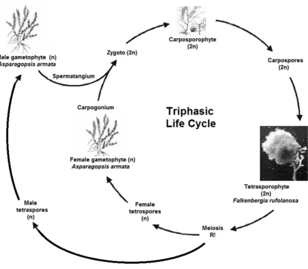
This red seaweed has the most elaborate life-cycle of all marine algae. It has a triphasic life cycle, indicating three distinct phases: the gametophyte, the carposporophyte, and the tetrasporophyte (Figure 1). Besides, the life-cycle is diplohaplontic with a haploid gametophyte and a diploid tetrasporophyte (Chualáin et al., 2004; Pacios et. al., 2011).
There are separate male and female gamethophytes that produce the corresponding gametes, which have half the genetic code necessary for the species (n), in specific reproductive organs, spermatangium and carpogonium, respectively. There are no flagellated cells, thus when the spermatia are released they are carried passively by water currents to the trichogyne located on the carpogonium. Once the gametes are fertilized, they growth into a carposporophyte (2n), a separate generation. The carpospores are eventually released into the water column, where they can settle, germinate, and grow into another generation known as the tetrasporophyte (2n). These tetrasporophyte produce four
Chapter I. General Introduction
28
tetraspores (n), by meiosis that are then released into the water and germinate and grow into more male and female gametophytes. In fact this is a heteromorphic life cycle, that means it has two distinct forms and they are so different that at one time it was considered to be two different species - the gametophyte generation A. armata and the tetrasporophyte generation Falkenbergia rufolanosa (Figure 2) (Chualáin et al., 2004; Garon-Lardiere, 2004; Kraan and Barrington, 2005). Both phases are capable of vegetative reproduction and fragmentation too.
Figure 1 – Schematic representation of the life cycle of Asparagopsis armata.
Asparagopsis armata is found in the lower intertidal to shallow subtidal and
occasionally in deeper pools, up to 15 meters deep, forming natural vegetation belts on exposed coasts (Altamirano et. al., 2008; Soler-Hurtado and García, 2011; Guerra-García et. al., 2012). This species are often found growing on other algae (epiphytically) or, otherwise, can attach to rocky substrates or float freely (most often seen in tetrasporophyte
Chapter I. General Introduction
29 phase). The gametophyte phase is found mainly from March until August in the Atlantic coast of Iberian Peninsula.
Falkenbergia rufolanosa, tetrasporophyte, is a rosy-pink collection of fine, irregularly
branched filaments which appear as small (1 to 3 centimeters in diameter) fluffy pom-pom balls underwater. The sexual gametophyte generation, however, is much different.
Asparagopsis armata are composed of bushy, paired, spirally arranged branchlets and
erect shoots from which numerous side branches develop in all directions. The latter ramify over and over again giving a plumose appearance and branch tips taper into harpoon-like barbs, the characteristic feature of the specie. Its color can range from pale pink through to red and bright purple, but quickly degenerating when removed from the water and becoming distinctly orange (Figure 2) (Andreakis et. al., 2004; Pacios et. al., 2011).
Figure 2 – Photograph of a) tertrasporophyte generation, Falkenbergia rufolanosa (Núcleo de
Ambiente da Universidade do Algarve, NAMB) and b) gametophyte generation, Asparagopsis
armata (Fiona Crouch).
Species of the family Bonnemaisoniaceae are well known as sources of halogenated compounds with strong antibacterial, antifungal and antibiotic activity that can be used for cosmetic and pharmaceutical purposes (Genovese et. al., 2009). The halogenated compounds from Asparagopsis have a wide range of volatility and solubility. This species also produces sulphated galactans with promising therapeutic applications. Asparagopsis
armata is also harvested or grown for the production of agar, a firm gel-like substance, that
is used as a thickener and stabilizer in many products, for example in food industry, as well as in laboratory work as growth medium for bacterial cultures. In general, the production of biologically-active metabolites is inherently linked to an ability to storage compounds into specialized structures (gland cells) in order to avoid autotoxicity (Mata et al., 2006; Genovese et. al., 2009). The gland cell is internal to the parent cell, but maintains a physical connection with the outer cell wall. This connection appears to be a means for the release of metabolites to the algal surface (Paul, 2006). The pungent aroma of these algae is due
Chapter I. General Introduction
30
to an essential oil that is composed mainly of bromoform with smaller amounts of other bromine, chlorine, and iodine-containing methane, ethane, ethanol, acetaldehydes, acetones, 2-acetoxypropanes, propenes, epoxypropanes, acroleins and butenones (Genovese et. al., 2009).
Asparagopsis armata has a strong invasive behavior, and is included in the list of
the “Worst invasive alien species threatening biodiversity in Europe” and also in the list of the “100 Worst Invasives in the Mediterranean Sea” (Altamirano et. al., 2008). It is native of the Southern Hemisphere, from Australia and New Zealand, where it was discovered by Harvey in 1885 (Chualáin et al., 2004; Soler-Hurtado & Guerra-García, 2011; Pacios et. al., 2011; Guerra-García et. al., 2012). This species was introduced to the Northern Hemisphere and, the first appearances in the Atlantic Oceans and Mediterranean Sea date from 1920’s. The first record of its appearance in Europe dates from 1923 (Garon-Lardiere, 2004). Since the spreading, A. armata is now found globally, from Western shores of England and Ireland, the Atlantic coast of France, Spain and Portugal down to the Canary Islands and throughout the Mediterranean Sea, Pacific and Indian Oceans. (Garon-Lardiere, 2004; Kraan and Barrington, 2005).
This seaweed is regarded as invasive because it spreads naturally in natural habitats in short time, colonizing a wide area, displacing native species and produces a significant change in terms of community composition (Chualáin et al., 2004; Soler-Hurtado & Guerra-García, 2011). Prolific vegetative reproduction may explain the fast dispersal rates of the species, which also exhibits an attaching system consisting in basal stolons and rhizoids able to facilitate the establishment of the propagules (Altamirano et. al., 2008).
Additionally, A. armata can be distributed on tide pools during the low tide and these pools may be roughly seen as a microenvironment because they have approximately six hours between low tide and high tide, they constitute an isolated system without water input or output and are exposed to extreme biotic and abiotic factors. These conditions can get adverse for this seaweed and consequently lead to stress disorders and severe consequences for littoral animals, such as invertebrates and fish larvae (Engström-Öst and Isaksson, 2006). Furthermore, A. armata exudates toxicity has been reported in the amphipod Hyale nigra (Paul et. al., 2006).
3. Sentinel species
The selection of sentinel species susceptible to study is an equally important part of any environmental assessment study and responds to five recommendations:
Chapter I. General Introduction
31 − Ecological and economical importance;
− Abundance and susceptibility to study; − The existent of previous works;
− Sensibility;
Sentinel species are used to assess the pollution in a habitat through space and time. Thus, research conducted in aquatic ecotoxicology is mainly based on the use of sentinel species (Beeby and Richmond, 2011).
In field studies, these species can be used for rapid risk assessments to provide information on the environmental conditions of an area. So, they are selected for their capability to reveal ecological perturbations and provide insight about environmental changes, centered on their life history and physiological characteristics (Zettler et al., 2013). A sentinel organism can indicate the presence of toxic substances by the manifestation of unusual symptoms or measureable responses that are not associated to their basal activities. The information about the presence of environmental stressors can be assessed through the study of: the content of certain elements or compounds; the changes on morphological or cellular structure; metabolic/biochemical responses; and behavior or population structure (Beeby and Richmond, 2011). In this line, animal sentinels must have measurable responses to the toxic substance in study, whether that is due to the animal’s death, disappearance, behavioral changes, alterations on biochemical processes, among others determinable aspects.
These organisms are ideally un-endangered species and are usually easy to find and handle. Further, the aptitude of a species to function as an indicator might depend on the position and role inside the ecosystem. (Zettler et al., 2013)
3.1. Gibbula umbilicalis as a test organism
Over the years, benthic organisms have been especially useful in applied research and in ecotoxicology in particular given that they tend to be good indicators of environmental stress.
Gibbula umbilicalis (Costa, 1778) is a sea snail, marine gastropods of the family
Trochidae. It is an eastern Atlantic species with a wide geographical distribution inhabiting temperate waters in the upper intertidal zone on rocky shores where wave energy is low. The preferential habitat is rocky platforms with a dense algal cover but also can be found in pools, under stones, and on upper surfaces of rocks. Their morphologic characteristics are a small top shell and a large round umbilicus, a deep hole on the underside of the shell.
Chapter I. General Introduction
32
The shell color is cream or greenish in a background with broad stripes of red or reddish-purple. This organism becomes sexually mature at about 18 months with a shell size of 8 to 9 mm. (Williams, 1964; Gaudèncio and Guerra, 1986; Underwood, 1972).
In terms of research, they provide excellent opportunities because they are easily found, collected and maintained. Besides, the facility to identify and count along the relative size and low mobility make this organism an appropriate species for ecotoxicological experiments (Cabecinhas et al., 2015). Previous studies revealed the high tolerance of this organism to sewage discharges (Ali and Bream, 2010), and heavy metals (Cabecinhas et al., 2015). Moreover, Cabecinhas and colleagues (2015) confirmed the potential of G.
umbilicalis as a marine model to ecotoxicological tests, along with the use of various
endpoints such survival, behaviour and enzymatic biomarkers.
Regarding the present study, the fact that the G. umbilicalis live around high tide mark and can be distributed by tide pools along the presence of A.armata provides an opportunity to test the exposure of this sea snail to the algal exudates and observe its effects.
4. Ecotoxicological bioassays
Ecotoxicological laboratory tests are important bioassays for a first toxicity assessment and as a first tier for Environmental Risk Assessment (ERA) purposes. ERA is a process of predicting whether there may be a risk of adverse effects of chemical substances in the environment, which can be - either synthetic or natural (e.g. human hormones, toxins produced by algae) - or also the effects of other abiotic stressors such as temperature, U.V. light, predation, etc. (Lemos et al., 2010). It is generally based in information derived from research on physical-chemical characteristics of xenobiotic and from laboratory toxicity tests (Moore et al., 2004) focusing on the relationship between toxic substances in the environment and the potential hazards of these if they exceed certain threshold levels (Binelli et al. 2005). One of the core missions of ecotoxicology is to find concentrations of environmental contaminants that exert an adverse effect on the organisms and to understand the mechanisms by which these contaminants perturb normal biological performance in order to develop appropriate measures to prevent adverse outcomes (Connon et al., 2012).
The biological assessments can be divided into three categories: 1) Acute/lethal tests; 2) Chronic/sublethal responses; 3) Biomarkers of biochemical/cellular/molecular responses (Widdows, 1998).
Chapter I. General Introduction
33
4.1. Classical endpoints
Traditionally, toxic effects are measured using standardized methods, based mainly on acute (e.g. mortality) and chronic responses (e.g. reproduction) of a sensitive biological indicator (Amorim et al. 2005).
Acute tests are short-term exposure experiments (hours or few days) and generally use mortality as endpoint. In acute exposures, organisms come into contact with higher doses of the toxicant in a single event or in multiple events over a short period of time and usually produce immediate effects, depending on absorption time of the toxicant (Widdows, 1998; Connon et al., 2012). The United Nations (2006) define acute tests as those which determine an LC50, Median Lethal Concentration, i.e. concentration causing mortality in fifty percent of exposed organisms. Acute tests are robust and very frequently used given their short time duration, simplicity and unambiguity of the endpoint measured. The results can provide meaningful comparisons of lethality between organisms, toxicants or test conditions (Orchard, 2000).
Avoidance behavior, energy budget, reproduction, feeding and growth rates are also among the most common endpoints used to assess toxicity at an individual level, concerning sublethal effects (Moreira et al., 2006), since reductions in such parameters have been correlated with the presence of toxicants (McLoughlin et al., 2000). However, acute tests are usually employed as a “screening tool” with a broad range of toxic concentrations.
4.2. Biochemical Biomarkers
The biomarkers approach has been incorporated in several pollution monitoring programs in Europe, such as the Water Framework Directive (2000/06/EC). Biomarkers were originally defined, by Depledge in 1994, as any biochemical, histological or physiological alterations or manifestations of environmental stress. Later in 1996, Gestel and Brummelen defined biomarkers in more detail as “any biological response to an
environmental chemical below-individual level, measured inside an organism or in its products (urine, faeces, hairs, feathers, etc.), indicating a departure from the normal status, that cannot be detected from the intact organism”. So, the term is now used in a more
restrictive sense, as biological responses at the sub-individual level resulting from exposure to xenobiotics (foreign toxic compound), and many authors follow this approach.
Given that biomarkers are measured at the molecular or cellular level, they may act as “early warning” signals of biological stress and as a result may anticipate changes at higher levels of biological organization (Figure 5, Lemos et al., 2010). In an environmental
Chapter I. General Introduction
34
context, ideally these early-warning signals could help scientists to better understand to which class of xenobiotics the organisms have been exposed to and if they are causing a toxic effect at critical targets (McCarthy and Shugart, 1990).
Generally, xenobiotic compounds existing in the bodies are lipophilic in nature and, to be efficiently eliminated and excreted, they need to be converted into more water-soluble compounds. There is an extraordinary range of enzymes and biotransformation pathways involved in their detoxification and removal (Chen, 2012). Two main phases in the detoxification process are considered: Phase I and Phase II.
Phase I is usually the first enzymatic defense against foreign compounds, where a non-synthetic alteration occurs, with a functional group being introduced to the chemical structure of the lipophilic compound. It includes reactions such as oxidation, hydrolysis or reduction and produces more water-soluble metabolites by increasing the polarity, making it ready for the next phase. Consequently, this step is defined as “functionalization” (Chen, 2012). For example, the cytochrome P450 enzyme uses oxygen, and as a cofactor NADH, to add a reactive group, being a typical Phase I reaction (Liska, 1998).
Phase II corresponds to a conjugation reaction, where the functional group of the xenobiotic is combined with a chemical group of a small molecule, normally the cofactor of an enzyme (Liska, 1998). Conjugation reactions can be glucuronidation, sulfation, glutathione or aminoacid conjugation and this step increases the solubility of the foreign compound and the excretory potential simplifying its removal from the body (Liska, 1998). Glutathione-S-transferase (GST, EC 2.5.1.18.) is a multigene superfamily of dimeric, belonging to Phase II detoxification enzymes. GST catalyzes the conjugation reaction between xenobiotic substrates (Phase I metabolic products) and sulphydryl group of reduced glutathione (GSH) (Figure 3) (Hayes and Pulford, 1995; Vidal-Liñán et al., 2015). Several studies have been developed on GST activity to assess the toxicity of pollutants (Geracitano et al., 2004; Woo et al., 2009; Faria et al., 2010; Hernández et al., 2013; Cabecinhas et al., 2015; Vidal-Liñán et al., 2015).
In the normal and healthy cell, reactive molecules such as reactive oxygen species (ROS) are produced, including hydrogen peroxide (H2O2), the free superoxide anion (O2-) and hydroxyl (OH∙) radicals (Wright and Welbourn, 2002), although usually there is a balance between the generation of ROS and antioxidant defense mechanisms (Moreira et al., 2006). However, as a consequence of stress conditions, an excess production of ROS can occur and overcome antioxidant defenses. These ROS are extremely potent oxidants capable of reacting with critical cellular macromolecules, causing oxidative stress which eventually leads to oxidative damage such as enzyme inactivation, lipid peroxidation (LPO),
Chapter I. General Introduction
35 DNA damage, protein damage, and ultimately may lead to cell death (Winston and Di Giulio, 1991; Martín-Diaz et al., 2004).
The excess production of superoxide radicals like O2- can reduce the cellular antioxidant capacity (Freitas et al., 2012), or cause peroxidation of membrane lipids, resulting in a deterioration of antioxidant enzyme activities by loss of membrane integrity and the inactivation of membrane-bound enzymes (Wright and Welbourn, 2002). The hydrogen peroxide H2O2 even though not highly reactive can also inhibit some antioxidant enzymes, mainly creating toxic effects at several different subcellular locations due to its fast capacity to penetrate cell membranes. Additionally, H2O2 can be the responsible to produce OH∙ radicals that can extensively attack every type of macromolecule in living cells comprising lipids, proteins and DNA (Wright and Welbourn, 2002), and can lead to several disease conditions such as cancer, cardiovascular disease and neurological disorders (Chen, 2012).
Antioxidant defense mechanisms against xenobiotics are greater in tissues with functions related to food processing like liver or digestive glands (Livingstone, 2001). The main antioxidant enzymes include: the superoxide dismutase (SOD, EC 1.15.1.1.), catalase (CAT, EC 1.11.1.6.), glutathione peroxidase (GPx, EC 1.11.1.9.), among others (Vicente et al., 2012). These defenses are among the most frequently used subcellular biomarkers.
Superoxide dismutase (SOD) is the primary defense against oxygen toxicity (Ameur et al., 2012). SOD is a group of metalloenzymes responsible to catalyze the conversion of superoxide free radical (O2-) into H2O2, which is formed by the transfer of a single electron to oxygen. This H2O2 consequently needs to be detoxified by CAT or GPx (Figure 3) (Stegman et al., 1992; Wright and Welbourn, 2002; Leslie et al., 2013). Catalase (CAT) is a hematin-containing enzyme capable of metabolizing H2O2 into oxygen (O2) and water (H2O). Glutathione Peroxidase (GPx) catalyzes the metabolism of H2O2 into water, involving the oxidation of a cofactor, reduced glutathione (GSH) into its oxidized form (GSSG) (Stegman et al., 1992; Apel and Hirt, 2004). This system acts as the key part to fight against oxygen damage and excess of free radicals produced during Phase I of xenobiotic detoxification (Faria et al., 2009; Yang et al., 2012). There are many studies that establish the toxicity of pesticides and metals based on antioxidant defenses (Geracitano et al., 2004; Richardson et al., 2008; Woo et al., 2009; Douhri and Sayah, 2009; Faria et al., 2010; Comoglio et al., 2011; Costa et al., 2012).
In theory, it is expected, that under oxidative stress the antioxidant enzymes increase their activities. But in practice and due to these enzyme’s transient nature, the results can vary between increase (Moreira et al., 2006; Bouraoui et al., 2010; Faria et al., 2010; Benedetti et al., 2012), decrease (Gravato et al., 2010; Ameur et al., 2012, Oliva et
Chapter I. General Introduction
36
al., 2012) and biphasic responses (Sun and Zhou, 2008; Won et al., 2012). The responses can be influenced by the vulnerability of the organisms exposed, and by the type, number and concentration of chemicals of which the organism was exposed to (Faria et al., 2009). As mentioned above, a failure of the antioxidant defenses to remove excess of ROS leads to oxidative stress, which may cause significant damage to important macromolecules. Consequences of oxidative stress have been measured biochemically as perturbed redox status, LPO, DNA damage and protein damage.
Figure 3 – Enzymatic mechanism involved in xenobiotic biotransformation and antioxidant
responses (adapted from Howcroft et al., 2009). GST – Glutathione-S-transferase; SOD – Superoxide Dismutase; CAT – Catalase; LPO – Lipid Peroxidation; GPx – Glutathione Peroxidase;
GR – Glutathione Reductase; GSH – Reduced Glutathione; GSSG – Oxidized Glutathione.
Lipid Peroxidation occurs with a chain reaction caused by the presence of a single radical that captures membrane lipids electrons forming an instable fatty acid peroxyl radical, reacting with itself or other fatty acids. LPO breakdown products such as epoxides, ketones, aldehydes, and more importantly, malondialdehyde (MDA) may also produce DNA adducts (Lieibovitz and Siegel, 1980). DNA damage can also be a consequence of the ROS accumulation. Structural damage of DNA, if not repaired, could impair the capability of cells to transcribe their own genes, consequently improving gene mutations, cancer and other diseases (Acharya, 1971). Experiments centered on damage to cells have been widely developed (Livingstone, 2001; Jebali et al., 2007; Richardson et al., 2008; Faria et al., 2009; Faria et al., 2010; Comoglio et al., 2011; Velma and Tchounwou, 2013).
Chapter I. General Introduction
37 Besides the above mentioned enzymes, there are others not directly related to the detoxification systems but can also play very important roles in the organisms’ physiology and be used as biomarkers, to indicate other forms of effects.
Cholinesterases represent a well-known group of serine hydrolases. They are considered ubiquitous enzymes which physiological function is to remove acetylcholine (ACh) from synaptic clefts (Strum et al., 1999). Among them, acetylcholinesterase (AChE, EC 3.1.1.7.) function involved in the regulation of the transmission of nerve impulses becoming one of the most central enzymes for nerve response (Bouraoui et al., 2009). It hydrolyzes ACh, in the post-synaptic membrane of cholinergic synapses where the nerve impulse is conducted to the next axon, producing choline and acetate and interrupting thus the nerve impulse (Figure 4) (Payne et al., 1996). When AChE is inhibited the nerve impulse is not interrupted. ACh will accumulate in the synaptic cleft, because it is not hydrolyzed, leading to an overstimulation of cholinergic receptors, neuromuscular paralysis and uncoordinated movements which could end in the organism’s death (Chen, 2012; Pereira et al., 2013). This enzyme is one of the most used biomarker in environmental studies, due to the ability of certain environmental stressors have to inhibit its activity. For example, some pesticides are well-known to inhibit AChE (Nachmansohn and Wilson, 1951). The inhibition of this enzyme by pesticides and metals have been extensively studied (Day and Scott, 1990; Boone et al., 1997; Amitai et al., 1998; Barata et al., 2004; Brown et al., 2004; Frasco et al., 2005; Arufe et al., 2007; Botté et al., 2012; Leomanni et al., 2015).
Chapter I. General Introduction
38
Carboxylesterases (CbE, EC 3.1.1.1.) are another class of serine dependent esterases which hydrolyze a wide range of endogenous and exogenous esters xenobiotics such as organophosphorus and carbamate insecticides, transforming carboxylic acid esters into their corresponding acid and alcohol (Maxwell, 1992; Parkinson, 1996). The mechanisms of action of this enzyme can result in the reduction of the amount of pesticides available for AChE inhibition (Ochoa et al., 2013). It is then possible by combining both carboxylesterase and acetylcholinesterase activities, one may have a more valuable suggestion of exposure.
Other important group of biomarkers is of those related to the energetic budget of an organism. Under normal conditions aerobic organisms use oxygen to generate energy for growth, reproduction and basal metabolism, but under situations of stress a large amount of the energy is used to detoxify and to maintain or compensate basal metabolism activity, leaving less energy available. It is known that xenobiotics are regarded as having effect on the energy consumption of organisms (Calow, 1991; de Coon and Janssen, 2003). Lactate dehydrogenase (LDH, EC 1.1.1.27.) is a cytoplasmic enzyme present in almost all of the body tissues. When oxygen is absent or in short supply, this enzyme catalyzes the reduction of pyruvate to lactate, with the concomitant oxidation of NADH to NAD+. Alterations in the activity of LDH have been used as indicative of changes in the processes of energy production under stressful conditions (Koenig and Solé, 2013). Moreover, LDH is released from the cytoplasm in response to cell damage. Given its stability in the extracellular environment, LDH quantification has been widely used to evaluate the presence of damage and toxicity in tissues and cells (Cook, et al., 2015; Kaja et al., 2015). A number of studies have been developed (Koening and Solé, 2013; Cabecinhas et al., 2015; Kaja et al., 2015), regarding the application of this biomarker to assess the impact of xenobiotics in the environment. Principally using LDH as a variable evaluation in toxicity tests (Diamantino et al., 2001) and correlating with oxygen levels (Wu and Lam, 1997).
4.3. Connecting different levels of biological organization – ecological relevance
The great challenge of ecotoxicology is to link the responses at low levels of biological organization (molecular, cellular or individual) to effects at higher levels (population, community or ecosystem), to be able to make predictions on effects before irreversible damages occur (Kammenga et al., 2000; Scott-Fordmand and Weeks, 2000; Vasseur and Cossu-Leguille, 2003; Gravato and Guilhermino, 2009).
The effects comprise all the abiotic and biotic changes which exceed the natural level or frequency, and in extreme circumstances can affect the functioning of entire
Chapter I. General Introduction
39 ecosystem. In the perspective of ecosystem functioning the most important feature is the interaction between stressors and biodiversity (Connon et al., 2012). To protect the environment and prevent the effects of xenobiotics in it is necessary to understand how different foreign compounds can disturb the ecosystems. However, there are a large number of xenobiotics that coexist in the environment and can have combined and complex effects, along with biotic and abiotic factors (Lemos et al., 2010)
Chemical characterization alone does not provide detailed biological information about possible effects on organisms. In an ideal methodology, cellular level responses linked with responses at whole organism, population, community and ecosystem would be required for a comprehensive assessment of ecotoxicological risk, but such methodologies are difficult and costly. Therefore, the identification of important links between responses at different levels of biological organization with ecosystem functioning can be a suitable step to building more fundamental and effective approaches on ecotoxicology (Connon et al., 2012). In this line, a selection of biological indicators, bioassays of standard whole organisms, biomarkers and in vitro tests is needed to assess the toxic effects of pollution to ecosystems (Valavanidis and Vlachogianni, 2014), because a pollutant stress situation usually provokes a cascade of biological responses.
Figure 5 – Relationship between ecological relevance, time of response and the different levels of
Chapter I. General Introduction
40
Most of the studies integrate at least one endpoint at higher levels of biological organization with lower levels, for example associating mechanistic responses with survival. Since effects at higher hierarchical levels are preceded by variations in biological processes at lower levels of organization, the “early warning signals” biomarkers or the individual approaches can be used in a predictive way in order to detect contamination before it hinders a community or even an entire ecosystem, allowing thus the initiation of management strategies of toxicants (Figure 5) (Lemos et al., 2010).
These early responding endpoints at lower biological levels have the potential to become powerful tools for rapid diagnoses of toxicity, and greater the integration of the above endpoints is, the greater will be our understanding of the adverse effects caused by foreign compounds.
5. Aims of the study
The main purpose of this work was to assess the potential ecotoxicological impact of exudates from red macroalgae Asparagopsis armata in the marine gastropod Gibbula
umbilicalis, under laboratory conditions, to evaluate the hazard of these invasive species
for the ecosystem they are invading.
To fulfill this objective, the following steps were developed:
(a) Development of a protocol for obtaining exudates from A. armata.
(b) Assessment of survival effects of the algae exudates on Gibbula umbilicalis performing acute ecotoxicological tests: lethal concentrations (LCX) were calculated to evaluate the effects of the individual biological level.
(c) Assessment of the effects of non-lethal concentrations of A. armata exudates over biochemical responses of G. umbilicalis. For this, optimization and evaluation of the potential of biomarkers involved in detoxification, oxidative stress responses, neurotoxicity and energetic functions were performed, since these biomarkers have already been considered in other studies as suitable tools for ecological effects assessment.
In sum, the outcome of this study was expected to become a first step tool to understand the toxicological capacity of A. armata, mainly for intertidal organisms, to unravel its mechanisms of toxic action, identify potential impacts and recognize the general potential toxicity of this macroalgae exudates.
41
Chapter II.
Survival and biochemical responses in
Gibbula umbilicalis exposed to exudates of
42
Chapter II. Survival and biochemical responses in Gibbula umbilicalis exposed to exudates of the red macroalgae Asparagopsis armata
43
Survival and biochemical responses in Gibbula umbilicalis exposed to exudates of the red macroalgae Asparagopsis armata
1. Introduction
The oceans are the largest life support systems responsible for sources of wealth, opportunity, and abundance. However, the interaction between mankind and this ecosystem has pushed oceans to their limit, for example with overfishing and pollution (Islam and Tanaka, 2004). Biological invasions are a form of pollution that involves two main issues: the human-mediated transport of a species to an area where it does not naturally occur, and its influence on the economy and the environment (Namboothri et al., 2012). These species are designated invasive alien species (IAS) and are considered as one of the major threats to biodiversity (Altamirano et al., 2008; Regulation No 1143/2014), and the second leading cause of species extinction (Zenetos et al., 2005). This biodiversity loss is caused mainly by the fact that these IAS can compete with native species for available resources. In some cases, such as the case of algal exudates, the competitive behavior is through the release of toxic substances, which are harmful to the surrounding species (Paul, 2006). Until date, little is known about the impact that invasive species have over native ones, and thus deserve further attention.
Asparagopsis armata is a red macroalgae (Rodophyta) globally recognized as an
invasive species. It is found in the intertidal to shallow subtidal areas, forming natural vegetation belts on exposed coasts (Altamirano et al., 2008), mainly from March until August in the Atlantic coast of Iberian Peninsula. Intertidal ecosystems represent areas where various parameters are subjected to unexpected changes, in a small spatial and temporal scale. These factors are further modified by regular events, such as tides (Soler-Hurtado and Guerra-García, 2011).
This red macroalgae produces secondary compounds that are stored in and released from specialized gland cells (Paul, 2006). For example, the pungent aroma of these algae is due to an essential oil that is composed mainly of bromoform with smaller amounts of other bromine, chlorine, and iodine-containing methane, ethane, ethanol, acetaldehydes, acetones, 2-acetoxypropanes, propenes, epoxypropanes, acroleins, and butenones (Genovese et. al., 2009). Over 100 halogenated compounds are found in the genus Asparagopsis alone (Paul, 2006). Considering the fact that this seaweed lives in the intertidal zone, it is distributed along tidal pools during low-tides. Generally, these pools may be seen as microenvironments, since they may last for proximately six hours, between low