A Critical Review on Obstetric Follow-up of Women
Affected by Systemic Lupus Erythematosus
Uma Revisão Crítica Sobre o Acompanhamento Obstétrico
de Mulheres com Lúpus Eritematoso Sistêmico
Danilo Eduardo Abib Pastore
1Maria Laura Costa
1Mary Angela Parpinelli
1Fernanda Garanhani Surita
11Department of Obstetrics and Gynecology, Universidade Estadual de Campinas (Unicamp), Campinas, SP, Brazil
Rev Bras Ginecol Obstet 2018;40:209–224.
Address for correspondence Fernanda Garanhani Surita, MD, PhD, Associate Professor, Department of Obstetrics and Gynecology, Universidade Estadual de Campinas - Unicamp, Rua Alexander Fleming, 101, Campinas, SP, 13083-881, Brazil
(e-mail: surita@unicamp.br).
Keywords
►
systemic lupus
erythematosus
►
pregnancy
►
prenatal care
►
maternal outcomes
►
fetal outcomes
Abstract
Objective
To review the existing recommendations on the prenatal care of women
with systemic lupus erythematosus (SLE), based on currently available scienti
fi
c
evidence.
Methods
An integrative review was performed by two independent researchers,
based on the literature available in the MEDLINE (via PubMed), EMBASE and The
Cochrane Library databases, using the medical subject headings (MeSH) terms
“
systemic lupus erythematosus
”
AND
“
high-risk pregnancy
”
OR
“
prenatal care.
”
Studies published in English between 2007 and 2017 were included; experimental
studies and case reports were excluded. In cases of disagreement regarding the
inclusion of studies, a third senior researcher was consulted. Forty titles were initially
identi
fi
ed; four duplicates were excluded. After reading the abstracts, 7 were further
excluded and 29 were selected for a full-text evaluation.
Results
Systemic lupus erythematosus
fl
ares, preeclampsia, gestation loss, preterm
birth, fetal growth restriction and neonatal lupus syndromes (mainly congenital
heart-block) were the major complications described. The multidisciplinary team should
adopt a speci
fi
c monitoring, with particular therapeutic protocols. There are safe and
effective drug options that should be prescribed for a good control of SLE activity.
Conclusion
Pregnant women with SLE present an increased risk for maternal
complications, pregnancy loss and other adverse outcomes. The disease activity
may worsen and, thereby, increase the risk of other maternal-fetal complications.
Thus, maintaining an adequate control of disease activity and treating
fl
ares quickly
should be a central goal during prenatal care.
Resumo
Objetivo
Revisar as recomendações existentes sobre o cuidado pré-natal às mulheres
com lúpus eritematoso sistêmico (LES), com base em evidências cientí
fi
cas atualmente
disponíveis.
received
November 1, 2017 accepted
December 20, 2017
DOIhttps://doi.org/ 10.1055/s-0038-1625951. ISSN 0100-7203.
Copyright © 2018 by Thieme Revinter Publicações Ltda, Rio de Janeiro, Brazil THIEME
Introduction
General Aspects
Systemic lupus erythematosus (SLE) is an autoimmune and multisystemic disorder of the connective tissue that mainly affects women of childbearing age (about nine women for each man). Immune anomalies, particularly the production of a series of antinuclear antibodies, are another prominent feature of the disease.1
The SLE prevalence varies from 40 to 200 cases per 100,000 inhabitants, more common among Africans and Asians descendants. In Brazil, its prevalence is around 8.7 per 100,000 inhabitants.1,2
The broad spectrum of clinical presentations includes mu-cous-cutaneous, muscle-skeletal, hematological, cardiopulmo-nary, renal and central nervous system manifestations. The most severe forms of organ involvement are lupus nephritis and neuropsychiatric lupus, and these conditions may result in a significant reduction in life expectancy.1Lupus nephritis is one of the leading causes of death along with infections.3
The most common general symptoms are weight loss, anemia, arthralgia and/or arthritis, being the involvement of the osteoarticular system the most frequent clinical mani-festation.1Antiphospholipid syndrome can occur in associa-tion with SLE, and it is characterized by arterial and venous thromboses, as well as recurrent morbidity in pregnancy.4
The American College of Rheumatology (ACR) proposed the criteria for the diagnosis of SLE (►Table 1).5 To be
classified as SLE, at least four criteria should occur in series or simultaneously.1,4,6
A consensus group of experts on SLE, the Systemic Lupus International Collaborating Clinics (SLICC), has proposed revised criteria for the diagnosis of SLE (►Table 2). It requires
either that a patient satisfies at least 4 out of 17 criteria,
including at least one of the 11 clinical criteria and one of the 6 immunologic criteria, or that the patient has biopsy-proven nephritis compatible with SLE and positivity to antinuclear antibodies (ANA) or anti-double-stranded DNA (dsDNA) antibodies.7
Systemic Lupus Erythematosus and Pregnancy
Considering the predilection of SLE in affecting women of childbearing age, pregnancy is of particular importance, with
Métodos
Revisão integrativa realizada por dois pesquisadores independentes, com
base na literatura disponível nos bancos de dados MEDLINE (via PubMed), EMBASE e
The Cochrane Library, usando os cabeçalhos de assuntos médicos, ou termos MeSH,
“
systemic lupus erythematosus
”
E
“
high-risk pregnancy
”
OU
“
prenatal care.
”
Estudos
publicados em inglês entre 2007 e 2017 foram incluídos; estudos experimentais e
relatos de caso foram excluídos. Em caso de desacordo, um terceiro pesquisador sênior
foi consultado. Quarenta títulos foram inicialmente identi
fi
cados; quatro duplicatas
foram excluídas. Após leitura dos resumos, mais 7 artigos foram excluídos e 29 foram
selecionados para uma avaliação de texto completo.
Resultados
Surtos de LES, pré-eclâmpsia, perda de gestação, parto prematuro,
restrição de crescimento fetal e síndromes de lúpus neonatal foram as principais
complicações descritas. A equipe multidisciplinar deve adotar um monitoramento
especí
fi
co, com protocolos terapêuticos apropriados. Há drogas seguras e e
fi
cazes que
devem ser prescritas para um bom controle do LES.
Conclusão
Gestantes com LES apresentam risco aumentado de complicações
mater-nas, perda de gravidez e outros desfechos adversos. A atividade da doença pode piorar
e, assim, aumentar o risco de outras complicações. Assim, manter um controle
adequado da atividade da doença e tratar rapidamente os surtos deve ser um objetivo
central durante o pré-natal.
Palavras-chave
►
lúpus
eritematoso
sistêmico
►
gravidez
►
cuidado
pré-natal
►
resultados
maternos
►
resultados fetais
Table 1 American College of Rheumatology (ACR) criteria for the classification of systemic lupus erythematosus
1. Erythema malar.
2. Discoid lupus.
3. Photosensitivity.
4. Oral ulcers.
5. Arthritis.
6. Serositis (pleuritis or pericarditis).
7. Nephropathy (persistent proteinuria greater than 0.5 g/day and/or glomerular hematuria).
8. Neurological disorders (convulsion or psychosis).
9. Hematologic disorders (hemolytic anemia, leucopenia, thrombocytopenia).
10. Immune disorder (presence of LE cells, anti-DNA or anti-Sm antibodies, false positive VDRL test, anticardiolipin IgG or IgM antibodies,
lupus anticoagulant).
11. Antinuclear antibody (ANA).
Abbreviations: IgG, immunoglobulin G; IgM, immunoglobulin M; LE, lupus erythematosus; Sm, smith; VDRL, venereal disease research laboratory.
Rev Bras Ginecol Obstet Vol. 40 No. 4/2018
relevant impact in maternal and perinatal health.8 The incidence of SLE among pregnant women ranges from 1:660 to 1:2.952; therefore, an understanding on how to manage these patients is essential.9
Although advances in the treatment of obstetric complica-tions and improvements in neonatal care have enabled lupus women to have pregnancies with better outcomes, SLE persists associated with significant fetal and maternal morbidity.8
Conditions with elevated levels of estrogen, such as pregnancy, have the potential to exacerbate SLE. The incidence of disease outbreaks during pregnancy varies between 15 and 63%.10
The impact of pregnancy in the course of lupus remains controversial, especially in relation to the incidence offlares. In contrast, the impact of lupus on gestation is more clearly understood. Women with lupus are no less fertile; outcomes are characterized by higher rates of fetal loss, preterm birth,
Table 2 Systemic lupus international collaborating clinics (SLICC) criteria for the classification of systemic lupus erythematosus
(4 of 17 criteria, including at least one clinical criterion and one immunologic criterion; OR biopsy-proven lupus nephritis7
Criterion Clinical criteria
Acute cutaneous lupus Lupus malar rash (do not count if malar discoid); bullous lupus; toxic epidermal necrolysis variant of SLE; maculopapular lupus rash; photosensitive lupus rash (in the absence of dermatomyositis);
ORsubacute cutaneous lupus (nonindurated psoriasiform and/or annular polycyclic lesions that resolve without scarring, although occasionally with postinflammatory dyspigmentation or telangiectasias)
Chronic cutaneous lupus Classic discoid rash; localized (above the neck); generalized (above and below the neck); hypertrophic (verrucous) lupus; lupus panniculitis (profundus); mucosal lupus; lupus erythematosus tumidus; chilblains lupus;ORdiscoid lupus/lichen planus overlap
Nonscarring alopecia Diffuse thinning or hair fragility with visible broken hairs (in the absence of other causes, such as alopecia areata, drugs, iron deficiency, and androgenic alopecia)
Oral or nasal ulcers Palate, buccal, tongue,ORnasal ulcers (in the absence of other causes, such as vasculitis, Behçet disease, infection [herpesvirus], inflammatory bowel disease, reactive arthritis, and acidic foods)
Joint disease Synovitis involving two or more joints, characterized by swelling or effusionORtenderness in two or more joints and at least 30 minutes of morning stiffness
Serositis Typical pleurisy for more than one day, pleural effusions, or pleural rub,OR
typical pericardial pain (pain with recumbency improved by sitting forward) for more than one day, pericardial effusion, pericardial rub, or pericarditis by electrocardiography in the absence of other causes, such as infection, uremia, and Dressler's syndrome
Renal Urine protein-to-creatinine ratio (or 24-hour urine protein) representing 500 mg protein/ 24 hours,ORred blood cell casts
Neurologic Seizures; psychosis; mononeuritis multiplex (in the absence of other known causes, such as primary vasculitis); myelitis; peripheral or cranial neuropathy (in the absence of other known causes, such as primary vasculitis, infection, and diabetes mellitus);ORacute confusional state (in the absence of other causes, including toxic/metabolic, uremia, drugs)
Hemolytic anemia Hemolytic anemia
Leukopenia or lymphopenia Leukopenia (<4,000/mm3at least once) (in the absence of other known causes, such as Felty syndrome, drugs, and portal hypertension),ORlymphopenia (<1,000/mm3at least once) (in the absence of other known causes, such as glucocorticoids, drugs, and infection)
Thrombocytopenia Thrombocytopenia (<100,000/mm3) at least once in the absence of other known causes, such as drugs, portal hypertension, and thrombotic thrombocytopenic purpura
Immunologic criteria
ANA ANA level above laboratory reference range
Anti-dsDNA Anti-dsDNA antibody level above laboratory reference range (or>2-fold the reference range if tested by ELISA)
Anti-Sm Presence of antibody to Sm nuclear antigen
Antiphospholipid Antiphospholipid antibody positivity as determined by any of the following: Positive test result for lupus anticoagulant; false-positive test result for rapid plasma reagin; medium- or high-titer anticardiolipin antibody level (IgA, IgG, or IgM); or positive test result for anti-β2-glycoprotein I (IgA, IgG, or IgM)
Low complement Low C3; low C4;ORlow CH50
Direct Coombs test Direct Coombs test in the absence of hemolytic anemia
Abbreviations: ANA, antinuclear antibodies; ELISA, enzyme-linked immunosorbent assay; SLE, systemic lupus erythematosus.
and fetal growth restriction (FGR), higher incidence of hyper-tensive disorders and maternal inhyper-tensive care admission. Multiple factors have been identified in association with adverse outcomes, such as lupus activity during pregnancy, previous nephropathy, maternal hypertension, and positivity for anti-phospholipid antibodies.8
Thus, adopting a specific protocol of care for pregnant women with lupus should contribute to reduce the frequency of maternal and fetal adverse outcomes, directly or indirectly related to SLE, improving care standards and ensuring success-ful pregnancies. This review aims to disclose the existing recommendations on prenatal care among health professio-nals attending pregnant women affected by SLE, based on currently available scientific evidence.
Methods
Integrative reviews were conducted to develop an evidence-based context in relation to different perspectives of clinical science studies. The following medical subject headings (MeSH) terms were used for research:“systemic lupus erythematosus”
AND“high-risk pregnancy”OR“prenatal care.”Different scien-tific databases were analyzed: MEDLINE (via PubMed), EMBASE and The Cochrane Library.
The inclusion criteria comprised studies published in English language, between 2007 and 2017 Experimental articles and case reports were excluded. Two independent researchers performed the search strategy in the scientific databases and, if there were disagreements regarding the
final inclusion, a third senior researcher was consulted. We found a total of 40 articles; 29 were accessed in full-text and selected for a qualitative synthesis (►Fig. 1).►Table 3
summarizes their methodologies, results and conclusions.
Results
Preconception Orientation
Adequate counselling, planning and care before, during and after the pregnancy must be the goal of health professionals who look after women with SLE. Luckily, multidisciplinary units are increasingly integrating different medical special-ists (including obstetricians, immunologspecial-ists, rheumatolo-gists, hematologists and nephrologists), which may allow for a more coordinated management of pregnancy along with disease activity.35
The care of pregnant women with SLE must focus on three mainstays: a coordinated medical-obstetrical care, a well-defined management protocol and a well-structured
Records identified through COCHRANE database
(n = 1)
Screening
Included
Eligibility
Identification
Records identified through EMBASE database
(n = 14)
Records after duplicates removed (n = 36)
Records screened (n = 36)
Records excluded (n = 7)
Full-text articles assessed for eligibility
(n =29)
Studies included in qualitative synthesis
(n =29)
Records identified through PUBMED database
(n = 25)
Original articles (n =13)
Review articles (n =16)
Fig. 1 PRISMA 2009flow diagram for article’s inclusion on obstetric follow-up of women affected by systemic lupus erythematosus.
Rev Bras Ginecol Obstet Vol. 40 No. 4/2018
Table 3 Original articles and review articles included on this integrative review
Original Articles
Author, Year (Country)
Study Design Population Description Main Results
Zhan et al.
(2017)11(China)
Retrospective observational study
251 SLE patients with 263 pregnancies assisted at the First
Affiliated Hospital of Sun Yat-Sen University, from 2001 to 2015.
APOs occurred in 70.0% of pregnancies, in which pregnancy loss in 28.5%; PTB in 21.3%; IUGR in 12.2%; and fetal distress in 8.0%. The use of antimalarial medications was associated with lower
risk for APOs (OR 0.3, 95% CI 0.1–0.7,p¼0.01). Fetal umbilical
artery Doppler in the third trimester showed higher resistance among SLE patients with APOs than the ones without APOs
(2.90.9 versus 2.40.5,p¼0.001).
Simard et al.
(2017)12
(Sweden)
Retrospective observational study
742 births to women with SLE and 10,484 births to non-SLE women from the Swedish Lupus Linkage (SLINK) cohort, with at least one pregnancy/birth in the Medical Birth Register, from 2001 to 2012.
Among births to women with SLE, there were 32 (4.3%) diag-noses of early-onset PE, and among births to non-SLE women, there were 55 (0.5%). SLE was associated with an increased risk
of early onset PE (RR 7.8, 95% CI 4.8–12.9, all pregnancies). The
association remained similar upon restriction to women without pregestational hypertension.
Chiu et al.
(2016)13(Taiwan)
Retrospective cohort study
Records of pregnant (1,526) and non-pregnant (2,932) women with SLE, and pregnant (3,052) and non-pregnant (3,052) women without SLE obtained from the Taiwan National Health Insurance Research Dataset, from 1997 to 2010.
Pregnant patients with SLE exhibited significantly increased risk
of ESRD after adjusting for other confounders, like
immuno-suppressant and parity (HR¼3.19, 95% CI: 1.357.52 for
pregnant non-SLE; and HR¼2.77, 95% CI: 1.246.15 for
nonpregnant non-SLE patients). No significant differences in the
ESRD incidence were observed in pregnant and nonpregnant SLE patients. Pregnant SLE patients exhibited better clinical
condi-tion at the baseline and a significantly lower risk of overall
mortality than nonpregnant SLE patients.
Hussein Aly et al.
(2016)14(Egypt)
Prospective observational study
91 pregnancies (84 women) with SLE attending the antenatal clinic at the high-risk pregnancy unit at Cairo University Hospi-tals, from 2010 to 2015.
The most common manifestations of SLE were cutaneous (93%), articular (92%), lupus nephritis (53%), hypertension (39%) and secondary APS (38%). Incidence rates: abortion 15%, FGR 32%, PTB 13%, PE 12%, fetal death 8%, neonatal admission ICU
15%, LBW 22%, SLE antenatalflares 44%. There was association
between hypertension and abortion (p¼0.04), PE
(p¼0.0001) and SLEflares (p¼0.0001). Lupus nephritis and
hypertension were predictors of PE (p¼0.01 andp¼0.002
respectively) and SLEflares (p¼0.048 andp¼0.003
respectively).
Tedeschi et al.
(2016)15(USA)
Retrospective observational study
114 SLE pregnant women referred to Brigham and Women’s
Hospital Lupus Center (Harvard Medical School), from 1990 to 2013.
Most pregnancies resulted in a live term delivery (76.5%).
Factors significantly associated with adverse pregnancy
out-comes were Nephritis (OR 3.6, 95% CI 1.0–12.8), cytopenias (OR
3.9, 95% CI 1.3–11.4), and serositis (OR 5.9, 95% CI 1.0–34.0).
Buyon et al.
(2015)16(USA,
Canada)
Prospective cohort study
385 patients (49% non-Hispanic white; 31% with prior nephritis) with SLE in the PROMISSE study, from 2003 to 2012
APOs occurred in 19.0% (95% CI, 15.2% to 23.2%) of pregnancies; fetal death in 4%, neonatal death in 1%, PTB in 9%, and SGA
neonate in 10%. Severeflares in the second and third trimesters
occurred in 2.5% and 3.0%, respectively. Baseline predictors of
(Continued)
R
e
v
Bra
s
G
ine
col
Obste
t
V
o
l.
40
N
o
.
4
/2018
A
C
ritical
R
e
v
iew
o
n
O
bstetric
Fo
llow
-up
Pa
st
o
re
e
t
a
l.
Table 3 (Continued)
Original Articles
Author, Year (Country)
Study Design Population Description Main Results
APOs included presence of LAC (OR 8.32; CI 3.59 to 19.26), antihypertensive use (OR 7.05; CI 3.05 to 16.31]), and platelet
count (OR 1.33; CI, 1.09 to 1.63 per decrease of 50109 cells/
L). Non-Hispanic white race was protective (OR 0.45; CI, 0.24 to
0.84). Maternalflares, higher disease activity, and smaller
increases in C3 level later in pregnancy also predicted APOs. Among women without baseline risk factors, the APO rate was 7.8%. For those who were either positive or were LAC-negative but nonwhite or Hispanic and using antihypertensives, the APO rate was 58.0% and the fetal or neonatal mortality was 22.0%.
Chen et al.
(2015)17(China)
Retrospective observational study
83 pregnancies in 80 women with SLE attended at the
Zhangzhou Affiliated Hospital of Fujian Medical University, from
2008 to 2013.
The sample was divided into three groups: group A (patients in
remission for>6 months before pregnancy, proteinuria
<0.5 g per day, without renal failure and discontinuation of
cytotoxic drugs for>one year); group B (patients with SLE
disease activity in the six months before pregnancy); group C (patients with new onset SLE during pregnancy). In group A, 76.47% pregnancies achieved full-term deliveries and 80.39% achieved live born infants. In group B and C, the outcome was poor. Among 62 patients (64 pregnancies) diagnosed as SLE
before pregnancy, SLEflares occurred in 27 (42.19%)
pregnan-cies. SLE disease activity in the six months before pregnancy was
significantly associated with lupusflare (OR 5.00, 95%
CI 1.14–21.87,p¼0.03) and fetal loss. New onset lupus during
pregnancy was independently associated with obstetric
com-plications (OR 7.22, 95% CI 2.14–24.38,p¼0.001).
Jakobsen et al.
(2015)18
(Denmark)
Retrospective observational study
84 pregnancies in 39 women diagnosed with SLE referred to a
Danish University Hospital during 1990–2010 (registered at the
Danish National Registry)
SLEflares developed in 46.4%, PE in 8.3%, and HELLP syndrome in
4.8% of cases. Significantly higher rates of premature delivery
(p¼0.0032), C-section (p¼0.015), hypertension (p¼0.025),
and IUGR (p¼0.003) were found. Disease activity (p¼0.021)
increased the risk of prematurity 3-fold. Two NLS and one congenital heart block were described. Birth weight and length
were significantly lower in the SLE cohort.
Tedeschi et al.
(2015)19(USA)
Retrospective observational study
147 pregnancies among 113 women followed at the Brigham
and Women’s Lupus Center (Harvard Medical School), between
1990 and 2013.
Among women with organ-specific lupus activity during the
6 months before conception, the crude odds for the same type of activity during pregnancy was 7.7- to 32.5-fold higher compared with women without that type of activity immedi-ately before conception.
R
e
v
B
ra
s
G
inec
o
l
Obst
e
t
V
o
l.
4
0
No.
4
/2018
A
C
rit
ic
a
l
R
e
v
ie
w
o
n
O
bs
te
tr
ic
Fo
llow
-u
p
Pastore
e
t
a
l.
Table 3 (Continued)
Original Articles
Author, Year (Country)
Study Design Population Description Main Results
Madazli et al.
(2014)20(Turkey)
Retrospective observational study
65 consecutive cases of SLE and pregnancy referred to a Uni-versity Hospital, from 2002 to 2011.
Diseaseflare-up occurred in 7.7% of patients. Mean GA at
delivery was 36.64.2 and mean birth weight was
2,706927 g. Stillbirth, FGR, PE and PTB rates were 4.6, 18.5,
9.2 and 27.6%, respectively. Cases with uterine artery
Doppler abnormalities had significantly poorer obstetric
outcomes.
Fatemi et al.
(2013)21(Iran)
Retrospective observational study
72 pregnancies in 65 patients attending at Lupus Clinic in Isfahan University of Medical Sciences between 1998 and 2012.
No woman with LN experienced preterm Labor or stillbirth. 16 pregnancies either ended in abortion or experienced PE of which seven had LN. Lupus nephritis and positive ANA were related to PE, whereas age of SLE development was associated with preterm labor. LN was associated with PE
and SLEflare.
Gaballa et al.
(2012)22(Egypt)
Case-control study
40 SLE pregnant women from inpatient and outpatient clinics of the rheumatology & rehabilitation and Ob&Gyn Departments of Zagazig University Hospitals; another 35 non-pregnant SLE patients attending rheumatology outpatient clinics were taken as a control group. The study was conducted from 2008 to 2010
Pregnant women comprised group A and non-pregnant women comprised group B. SLEDAI was increased in both groups, more
in group A. Lupusflares were increased during pregnancy as it
occurred in 62.5% of group A compared with 37.14% in group B.
Severeflares were more frequent in group A. Gestational
hypertension and SLEDAI showing disease activity were risk factors for poor maternal outcome. Fetal outcome included full term 37.5%, PTB 25%, FGR 22.5%, stillbirth 12.5%, abortion 7.5% and congenital heart block 2.5%. Factors associated with
poor fetal outcome were severeflares and active renal
disease. Full term pregnancy was more common in patients
with noflares.
Surita et al.
(2007)23(Brazil)
Observational cohort study
67 women with lupus (76 pregnancies) who received care at a tertiary clinic for high-risk pregnancies, at Universidade do Estado de Campinas, Brazil, between 1995 and 2002.
Flare-ups occurred in 85.3% of cases, especially when there was
renal involvement (being the most significant). This was related
to greater numbers of women with PE and poor perinatal outcome. In cases when there was active disease, IUGR was more
common. The placental weight was significantly lower in the
women with renal involvement. Flare-ups and renal involvement in lupus patients during pregnancy are associated with increased maternal and perinatal complications.
R
e
v
Bra
s
G
ine
col
Obste
t
V
o
l.
40
N
o
.
4
/2018
A
C
ritical
R
e
v
iew
o
n
O
bstetric
Fo
llow
-up
Pa
st
o
re
e
t
a
l.
Review Articles
Author, Year Type of Review Main Results Conclusions and Recommendations
Knight and
Nelson-Piercy (2017)24
Narrative review SLE provides challenges in prepregnancy, antenatal,
intrapartum, and postpartum periods for the medical, obstetric, and midwifery teams. Women are at risk of
lupusflares, worsening renal impairment, onset of or
worsening hypertension, PE, miscarriage, FGR, PTB, and/ or neonatal lupus syndrome.
In pregnancy, early referral for hospital-coordinated care, involvement of obstetricians and rheumatologists, an individual management plan, regular reviews, and early
recognition offlares and complications are all important.
A C-section may be required in certain obstetric contexts (e.g., preterm delivery for maternal and/or fetal well-being), but vaginal birth should be the aim for the majority of women. Postnatally, an ongoing individual management plan remains important.
Lateef and Petri (2017)25 Narrative review Outcomes for pregnancy in the setting of SLE have
considerably improved but the maternal and fetal risks
remain high. Diseaseflares, PE, pregnancy loss, PTB, FGR
and neonatal lupus syndromes (especially heart block) remain the main complications.
Specific monitoring and treatment protocols need to be
used for situations such as presence of specific
antibo-dies (antiphospholipid antiboantibo-dies and anti-SSA/SSB). Safe and effective treatment options exist and should be used to control disease activity during pregnancy. Close monitoring and judicious use of medications are the key to achieve optimal outcomes.
Ostensen (2017)26 Narrative review Ideal conditions for pregnancy are conception at a stage
of remission or minimal disease activity while on stable, pregnancy-compatible medication.
Points discussed during preconception counseling should be shared with all doctors and health profes-sionals involved in the care of a pregnant patient. Address family planning in all patients of fertile age. Physicians should actively offer information on repro-duction issues to all patients. Address medication
con-cerns and the benefits of optimal disease control in
pregnancy with all patients.
Moroni and Ponticelli
(2016)27
Narrative review Pregnancy is not contraindicated in women with SLE.
However, pregnant patients with lupus nephritis may run increased risk of PE and PTB. The maternal and fetal outcome are strongly correlated with lupus activity, kidney function and the presence of aPL antibodies.
Ideally, a woman should plan a pregnancy only until her lupus has been under control for at least 6 months.
Yamamoto and Aoki
(2016)28
Narrative review Maternal and fetal risks are higher in females with SLE
than in the general population. However, with appro-priate management of the disease, sufferers may have a relatively uncomplicated pregnancy course.
Factors such as appropriate preconception counseling and medication adjustment, strict disease control prior to pregnancy, intensive surveillance during and after pregnancy by both the obstetrician and rheumatologist, and appropriate interventions play a key role.
Jesus et al. (2015)29 Narrative review The risk offlares depends on the level of maternal
disease activity in the 6–12 months before conception
and is higher in women with repeatedflares before
conception, in those who discontinue useful medica-tions and in women with active glomerulonephritis at conception.
It is a challenge to differentiate lupus nephritis from PE and, in this context, the angiogenic and antiangiogenic cytokines are promising. Prenatal care of pregnant patients with SLE requires close collaboration between rheumatologist and obstetrician. Planning pregnancy is essential to increase the probability of success.
R
e
v
B
ra
s
G
inec
o
l
Obst
e
t
V
o
l.
4
0
No.
4
/2018
A
C
rit
ic
a
l
R
e
v
ie
w
o
n
O
bs
te
tr
ic
Fo
llow
-u
p
Pastore
e
t
a
l.
Table 3 (Continued)
Review Articles
Author, Year Type of Review Main Results Conclusions and Recommendations
Singh and Chowdhary
(2015)30
Narrative review Established risk factors for adverse pregnancy outcomes
include active disease within 6 months prior to con-ception and during pregnancy, active nephritis, maternal hypertension, antiphospholipid antibodies and hypocomplementemia.
Certain aspects such as prevention of PTB, treatment of congenital heart block due to neonatal lupus and recurrent pregnancy loss despite best management, remains challenging. Pregnant patients with SLE should be followed in a high-risk obstetric clinic, and care should be closely coordinated between the obstetrician and rheumatologist.
Lateef and Petri (2013)31 Narrative review Although live births are achieved in the majority of the
pregnancies in women with SLE, active disease and major organ involvement can negatively affect the
out-comes. Diseaseflares during SLE pregnancy pose the
unique issue of recognition and differentiation between physiologic changes and disease state. Similarly, PE and lupus nephritis may lead to diagnostic confusion.
A multidisciplinary approach, with close monitoring, is essential for optimal outcomes. Safe treatment options exist and should be appropriately used for disease activity during pregnancy.
Lateef and Petri (2012)32 Narrative review Maternal and fetal mortality and morbidity are
consid-erably increased among pregnancies with SLE, compared with the general population. Active maternal disease,
renal involvement, specific autoantibody subsets and
advanced organ damage are predictors of poor outcome.
Multidisciplinary care, close monitoring, high-risk sur-veillance, and judicious use of medications are essential to achieve good outcomes.
Stanhope et al. (2012)33 Narrative review Renal involvement in the form of either active LN at the
time of conception, or a LN new onset orflare during
pregnancy increases the risks of PTD, PE, maternal mortality, fetal/neonatal demise, and FGR.
The major goal of immunosuppressive therapy in preg-nancy is control of disease activity with medications that are relatively safe for a growing fetus. Therefore, the use of mycophenolate mofetil, due to increasing evidence supporting its teratogenicity, is contraindicated during pregnancy.
Baer et al. (2011)34 Narrative review The frequency of pregnancy loss in lupus has dropped to
a level commensurate with that of the general popula-tion. The outcomes of lupus pregnancies are better if conception is delayed until the disease has been inactive for at least 6 months, and the medication regimen has been adjusted in advance.
Monitoring should include baseline and monthly laboratory tests, serial ultrasonography, fetal surveil-lance tests, and fetal m-mode echocardiography for mothers with SSA (Ro) or SSB (La) antibodies. If hydro-xychloroquine was in use before conception, it should be maintained throughout pregnancy. If a woman with SLE has antiphospholipid antibodies, prophylactic treatment with aspirin and/or low-molecular weight heparin is
indicated to prevent fetal loss. Lupusflares during
pregnancy are generally treated with hydroxychloro-quine, low-dose prednisone, pulse intravenous methyl-prednisolone, and azathioprine. High-dose prednisone and cyclophosphamide are reserved for severe lupus complications.
(Continued)
R
e
v
Bra
s
G
ine
col
Obste
t
V
o
l.
40
N
o
.
4
/2018
A
C
ritical
R
e
v
iew
o
n
O
bstetric
Fo
llow
-up
Pa
st
o
re
e
t
a
l.
Table 3 (Continued)
Review Articles
Author, Year Type of Review Main Results Conclusions and Recommendations
Ruiz-Irastorza and
Khamashta (2011)35
Narrative review Women with severe active disease or a high degree of
irreversible damage, such as those with symptomatic pulmonary hypertension, heart failure, severe restrictive pulmonary disease or severe chronic renal failure should best avoid pregnancy.
Adequate pregnancy care of women with SLE rests on three pillars: a coordinated medical-obstetrical care, an
agreed and well-defined management protocol and a
good neonatal unit. Pregnancy should be planned following a preconceptional visit for counselling.
Buyon (2009)36 Narrative review Flare rates are generally low for patients who are
clini-cally stable at conception. For patients who have never
had renal disease, there is nofirm evidence that they will
develop active renal disease simply due to being preg-nant. For women with anti-SSA antibodies, the risk of having a child with congenital heart block is 2%, which rises to a recurrence rate of 18%.
For patients with aPL antibodies detected in thefirst
trimester of pregnancy, the lupus anticoagulant is the strongest predictor of serious pregnancy complications.
Doria et al. (2008)37 Narrative review Most SLE patients experience uncomplicated
pregnan-cies. One of the major risks for SLE mothers is the
occurrence of a diseaseflare during pregnancy. Another
major risk of SLE relapse during pregnancy is glomer-ulonephritis, especially if active at the time of conception.
To reduce the risk of maternal and fetal complications, pregnancies must be planned when SLE is inactive and
must be closely and appropriately monitored. Specific
blood tests predict some pregnancy complications.
Clowse (2007)38 Narrative review Pregnancy in a woman with SLE can be complicated by
lupus activity and pregnancy mishaps. Recent studies found an increase in lupus activity during pregnancy, possibly worsened by hormonal shifts required to maintain pregnancy. An elevated risk for poor pregnancy outcomes, such as stillbirth, preterm birth, low birth weight and preeclampsia, is related to an increased lupus activity.
A rheumatologist and a high-risk obstetrician are best equipped to care for women with lupus who become pregnant. Careful planning and treatment may be required to achieve success of gestation.
Witter (2007)39 Narrative review Interactions between SLE and pregnancy include the
overall activity of lupus and pregnancy outcome, the effect of lupus nephritis on pregnancy, the effect of pregnancy on the progression of lupus nephritis, and the differentiation of hypertension related to lupus nephritis from PE.
A live birth can be achieved by close coordination of care
between the patient’s rheumatologist, obstetrician,
and, in the case of renal involvement, her nephrologist.
Abbreviations: ANA, antinuclear antibodies; aPL, antiphospholipid; APOs, adverse pregnancy outcomes; APS, antiphospholipid syndrome; CI, confidence interval; ESRD, end-stage renal disease; FGR, fetal growth restriction; GA, gestational age; HR, hazard ratio; ICU, intensive care unit; IUGR, intrauterine growth retardation; LAC, lupus anticoagulant; LBW, low birth weight; LN, lupus nephritis; NLS, neonatal lupus syndrome; OR, odds ratio; PE, preeclampsia; PTB, preterm birth;; PTD, preterm delivery; RR, relative risk; SGA, small for gestational age; SLE, systemic lupus erythematosus; SLEDAI, Systemic Lupus Erythematosus Disease Activity Index.
R
e
v
B
ra
s
G
inec
o
l
Obst
e
t
V
o
l.
4
0
No.
4
/2018
A
C
rit
ic
a
l
R
e
v
ie
w
o
n
O
bs
te
tr
ic
Fo
llow
-u
p
Pastore
e
t
a
l.
neonatal unit. Preconception counselling is vital to assess the chance of both potential fetal and maternal complications; that way, consistent information regarding specific risk for complications and the expected management plan should be provided (►Table 3).35
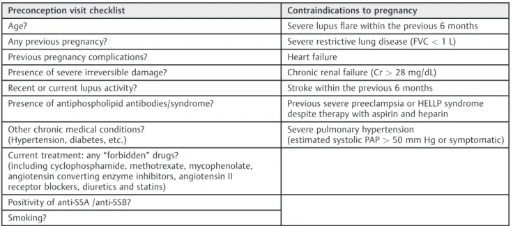
Pregnancy planning is a key-point for women with SLE. Postponing conception until the disease is considered inactive for at least six months significantly improves the outcomes of these pregnancies.26,31,34,35Women who present some form of irreparable organ injury are more likely to undergo com-plications and even additional damage during and after preg-nancy. Some conditions should advise to delay pregnancy, such as severe diseaseflare within the previous six months, recent stroke and active lupus nephritis.32In some situations, preg-nancy may be contraindicated (►Table 4).26,34,35
At the preconception visit, obtaining a complete set of autoantibody profile is recommended, including antiphos-pholipid (aPL) antibodies (anticardiolipin and lupus antico-agulant), complement serum levels, anti-SSA and anti-SSB antibodies. Evaluating the pregnancy risk and assessing the SLE activity and the organ function is important to maintain disease control only in safe medications.32
A higher risk of complications is found among women with severe impairment of organ function, with or without preex-isting severe organ damage.24Besides, the diagnosis of SLE during pregnancy is also related to the occurrence of compli-cations, significantly affecting maternal and fetal outcomes.30
Prenatal Follow-up
General Findings
The prenatal care of a woman with SLE requires close collaboration between the obstetrician and the clinicians (rheumatologist, nephrologist or hematologist), and man-agement in a high-risk referral center. An evaluation by the clinician should occur every 4–6 weeks, whereas the
obstet-ric visit should be every 4 weeks until 20 weeks of gestation; then, every 2 weeks until 28 weeks, and then, weekly until the expected delivery date.34
At every prenatal visit, blood pressure, weight gain, uterine size, fetal heart rate and urinalysis (through a quick outpatient analysis with the dipstick testing) should be assessed, as well as inquiring about symptoms related to lupusflares.34
The differential diagnosis of complications that may arise during pregnancy is not easy. Signs and symptoms of lupus
flares often mimic the ones of normal pregnancy. Those
flares are less frequent in the third trimester, although they may occur at any time during pregnancy or in the immediate postpartum period.34
Laboratory Evaluation during Prenatal Care
In addition to routine pregnancy booking, blood tests (which include a full blood count), baseline tests of renal and hepatic function and baseline urinary protein quantified by a 24-hour collection should be obtained.24Complement studies should comprise further tests (C3, C4, CH5O), anticardiolipin antibodies, anti-dsDNA, lupus anticoagulant and anti-SSA and SSB.
Disease Activity during Prenatal Care (Flares)
Changes in hormonal levels through pregnancy prompt to a shift from Th1 to Th2 lymphocyte dominance; consequently, autoimmune disorders involving Th2-response, such as SLE, are expected toflare.24
It is generally agreed that pregnancy may lead to higher rates of disease flares, with rates from 25 to 65% being reported.18,31,37Different organ systems may have variable response to pregnancy; musculoskeletalflares are less com-mon, whereas renal and hematologicalflares are more com-mon.31The risk offlare seems to be related to the occurrence of disease activity 6–12 months before conception.15,19,22,24,30
Table 4 Preconception visit checklist and contraindications to pregnancy in women with SLE35
Preconception visit checklist Contraindications to pregnancy
Age? Severe lupusflare within the previous 6 months
Any previous pregnancy? Severe restrictive lung disease (FVC<1 L)
Previous pregnancy complications? Heart failure
Presence of severe irreversible damage? Chronic renal failure (Cr>28 mg/dL)
Recent or current lupus activity? Stroke within the previous 6 months
Presence of antiphospholipid antibodies/syndrome? Previous severe preeclampsia or HELLP syndrome despite therapy with aspirin and heparin
Other chronic medical conditions? (Hypertension, diabetes, etc.)
Severe pulmonary hypertension
(estimated systolic PAP>50 mm Hg or symptomatic)
Current treatment: any“forbidden”drugs?
(including cyclophosphamide, methotrexate, mycophenolate, angiotensin converting enzyme inhibitors, angiotensin II receptor blockers, diuretics and statins)
Positivity of anti-SSA /anti-SSB?
Smoking?
Abbreviations: FVC, forced vital capacity; PAP, pulmonary arterial pressure.
A higher risk offlare during pregnancy is noticed when lupus nephritis occurs at conception, even in women in remission.13,21,24,30 One study showed a 30% flare rate during pregnancy or postpartum among 113 pregnancies in women with preexisting lupus nephritis evaluated; other studies suggest a likelihood of up to 60%.24Besides, different reports in the literature indicate lupus nephritis as a predic-tive of poor prognosis for pregnancy.23
It may be difficult to distinguish pregnancy-related signs and symptoms from those of SLE. Therefore, an appropriate assessment by experienced physicians is important.24,37,38 Ambiguous manifestations include fatigue, headaches, arthralgia, edema, hair loss, dyspnea, malar and palmar erythema, anemia and thrombocytopenia. Hence, baseline blood counts and urinalysis with measurement of protein-uria assessed early in gestation are helpful to monitor disease status and identifyflares.24
During pregnancy, liver production of serum C3 and C4 increases, so their levels may persist within the range of normality in cases of active SLE. Relative variations are more important, rather than absolute levels, with a drop of25% in serum complement levels suggesting lupusflare.24
Pregnancy-specific disease activity scales (such as systemic lupus erythematosus pregnancy disease activity index [SLEP-DAI] and lupus activity index in pregnancy [LAI-P]) have been developed, but mostly remain as research tools. In practice, the clinical judgment of an experienced clinician is still considered the gold standard.25,31,32The SLEPDAI scale is a similar instru-ment to the systemic lupus erythematosus disease activity index (SLEDAI) for assessment of lupus activity, assigning different scores for the various clinical and laboratory mani-festations of lupus activity, however taking into account the physiological changes of gestation and the main pathologies of the pregnancy-puerperal cycle that can mimic SLE in activity. Its score ranges from zero to 105 and stratifies the disease activity: absent (up to 4 points), mild to moderate (5–12 points) and severe (up to 12 points) (►Table 5).36
A recent meta-analysis reported rates ranging from 1.5 to 83% for a lupus nephritesflare during pregnancy,33 corrobo-rating with data from previous studies.38Thus, it is strongly recommended a close monitoring, with monthly assessments of disease activity (with special attention to renal function). Besides, the risk of hypertensive disorders of pregnancy increases in the setting of active lupus nephritis.14,33
The frequency of preeclampsia varies from 7.5 to 22.5% for all women with SLE.12,15,18,20,39Lupus renal involvement is often associated with hypertension, and the preeclampsia diagnosis is difficult, since it may be superimposed on chronic hypertension.39
Likewise, in cases of SLE women with glomerular lesions, increased proteinuria may be observed, due to the enlarged glomerularfiltration rate during pregnancy, with this fact not being related to preeclampsia. Thus, the diagnosis of preeclampsia can get more difficult because of increasing blood pressure and proteinuria at term.38,39
The differential diagnosis of preeclampsia in lupus patients may be facilitated by changes in the measures of C3, C4 and CH50, since a reduction in those levels is expected
during lupus activity.39Other laboratory testfindings may be helpful to successfully perform a differential diagnosis: abnormal urinary sedimentation with the presence of eryth-rocyte dysmorphism or cell casts and increased anti-DNA antibody titers (all found in lupus nephritis).23
New onset SLE during pregnancy can be considered as SLE activity and might be associated with worse outcome. Dif-ferentiating the diagnosis of preeclampsia from new onset SLE during pregnancy is a challenge and frequently delays the diagnosis of SLE. However, a Chinese study indicated that new onset SLE during the third trimester of pregnancy might have a better outcome.17
Among patients with stable condition at the time of con-ception, it is expected that disease activity will not worsen, and even if so, theflare is usually mild and occasionally involves some kind of treatment modification.28
Evaluation of Fetal Growth and Vitality
Fetal complications are frequently observed in patients with SLE. Overall, miscarriages and stillbirth may occur in20% of pregnancies in SLE patients.11,15,26,30Patients with a history of nephritis, in special, have an increased risk for such adverse outcomes.14,16
The rate of FGR is estimated to be near of 30%, observed even in mild disease, with an increased risk if there is renal involvement. Small-for-gestational-age is a more common outcome in those born prematurely, but can occur at all gestational ages.20,22,37,39Several studies concluded that the small-for-gestational-age rate outcome among SLE women tends to be higher, condition strongly associated to the presence of diseaseflare-ups during pregnancy.23
Serial obstetric sonography is the most important method to guide surveillance for fetal growth. Crown–rump length measurement in the first trimester presents as the most precise measurement. At 16 to 22 weeks of gestation, an anatomic survey considering diagnosis of fetal anomalies should be followed, also serving to allow thefirst monitoring of growth. At each 4-week periods, new scans should take place, with measurement of amnioticfluid volume. If pre-eclampsia or FGR are diagnosed, the interval can be reduced to 3 weeks.39
Fetal vitality surveillance is an important part of the prenatal care of SLE patients. This should include the nonstress test (NST), the biophysical profile (BPP), and fetal umbilical artery Doppler velocimetry, starting at 26 to 28 weeks and continuing weekly until birth.39
In patients with SLE, alterations of umbilical artery Dopp-ler velocimetry should be managed similarly to those with-out the condition. Normal evaluation of these tests has a high negative predictive value for fetal death.29Association be-tween abnormal uterine artery Doppler and later fetal loss, preeclampsia, FGR and preterm labor were also described.29 Because of the risk of fetal congenital heart block, for women with anti-SSA/SSB antibodies, a fetal echocardiogra-phy should be performed at 18–20 weeks and 26–28 weeks to exclude fetal congenital heart block. An urgent referral to a tertiary care center should be prompted in case of any fetal heart rate abnormality, mostly a slow heart rate.24
Rev Bras Ginecol Obstet Vol. 40 No. 4/2018
Recommended SLE Treatment during Pregnancy
Considering the harmful effects of active disease on both mother and fetus, an appropriate reflection between the risks and benefits of this treatment must take place.26,30
In practice, it is frequent that SLE women to discontinue their medication before conception, due to fear of fetotox-icity, without proper doctor counseling. However, discontin-uation of the medication may lead to active SLE and unfavorable pregnancy outcomes.28
Usually, the immunosuppressive treatment in pregnant women with quiescent lupus should not be changed. The most frequently used agents in lupus patients are glucocorti-coids and hydroxychloroquine, which should be maintained.27 Prednisone at a dosage of 5–10 mg per day is usually considered safe.27Lupusflares thatfit into mild activity can be treated with low-dose prednisone (less than 20 mg/d). Higher doses of corticosteroids, including pulse dose steroids, are options to treat moderate to severe lupus activity.37,38
Hydroxychloroquine is not a teratogenic drug. Its use is recommended to prevent disease activity and reduce the risk of cardiac-neonatal lupus in patients who are carriers of anti-SSA/-antibody.11,27,28In addition, it improves the prognosis of SLE nephritis and prevents death.38
Azathioprine is considered safe, especially if compared with other immunosuppressive drugs. Many studies sustain a transition to this option if the patient wishes to conceive. However, some other reports recently pointed out concerns about late developmental delays in children who were exposed to azathioprine during pregnancy,28,38as well as neonatal leucopenia and/or thrombocytopenia.27
Regarding cyclosporine and tacrolimus, the FDA classifies as category C; however, some meta-analysis studies did not
find significant differences related to birth defects when pregnant women were exposed to them.27,37
Cyclophosphamide should not be prescribed during the
first trimester, because of its association to chromosomal
Table 5 Systemic lupus erythematosus pregnancy disease activity index (SLEPDAI) instrument to stratify SLE activity during pregnancy36
Score Descriptor Modified for
pregnancy
Considerations
8 Seizure Yes (r/o eclampsia)
8 Psychosis No
8 Organic brain syndrome No
8 Visual disturbance No (hypertension is already considered an exclusion in SELENA-SLEDAI and SLEDAI)
8 Cranial nerve disorder Yes (r/o Bell palsy)
8 Lupus headache Yes (r/o Bell palsy)
8 CVA Yes (r/o eclampsia)
8 Vasculitis Yes (consider palmar erythema)
4 Arthritis Yes (consider bland knee effusions)
4 Myositis No
4 Urinary casts No
4 Hematuria Yes (r/o cystitis and vaginal RBC reflective of placental problems)
4 Proteinuria Yes (r/o eclampsia)
4 Pyuria Yes (r/o infection)
2 Rash Yes (consider chloasma)
2 Alopecia Yes (consider normal postpartum alopecia)
2 Mucosal Ulcers No
2 Pleurisy Yes (hyperventilation may be secondary to progesterone, dyspnea secondary to enlarging uterus)
2 Pericarditis No
2 Low complement Yes (complements normally rise during pregnancy)
2 Increased DNA binding No
1 Thrombocytopenia Yes (r/o preeclampsia, HELLP syndrome, incidental thrombocytopenia of pregnancy)
1 Leukopenia Yes (consider normal rise of leukocyte count during pregnancy)
1 Fever No
Abbreviation: CVA, cerebrovascular accident; RBC, red blood cell; r/o: rule out, SELENA-SLEDAI, safety of estrogens in lupus erythematosus national assessment- systemic lupus erythematosus pregnancy activity index; SLEDAI, systemic lupus erythematosus pregnancy activity index.
impairment. During the second or third trimester, it should be reserved only to severeflares unamenable with methylpred-nisolone pulses or other drugs. The use of cyclophosphamide during the second and third trimesters does not seem to increase the risk for congenital abnormalities. Nevertheless, miscarriages and preterm birth may be more frequent.27,37
Leflunomide is associated to teratogenic and fetotoxic effects in animals, and its metabolite is detectable in plasma up to 2 years after discontinuation. Thus, in pregnant wom-en, it is formally contraindicated; and pregnancy must be excluded before starting it.27
Methotrexate is another teratogenic drug, classified by the FDA as X (contraindicated in pregnancy). If used in thefirst trimester, it is associated to FGR and some major malforma-tions, such as absence or hypoplasia of the frontal bones, craniosynostosis, large fontanelle and ocular hypertelorism.27 During thefirst trimester, rituximab has very low trans-placental transfer, with some studies reporting safe preg-nancies and deliveries in those cases of exposure. However, during the second or third trimester, it can cross the placenta and induce severe neonatal lymphopenia.27,37 Hence, in these cases, live vaccines should be avoided in those children during thefirst 6 months of life.27
Handling some complications that often affect pregnant women with SLE justifies a short statement. Since arterial hypertension is a common condition among patients with lupus nephritis, an appropriate management of blood pres-sure in pregnancy may reduce the progression of the disease and avoid several adverse pregnancy outcomes. Labetalol, nifedipine or methyldopa are safe drugs for treating hyper-tension. Angiotensin-converting-enzyme inhibitors should be avoided due to their association to multiple congenital abnormalities.27
Low-dose aspirin is recommended, since it reduces the risk of preeclampsia and perinatal death; besides, it is associated with an increase in the birth weight of those with risk factors, including renal disease. Full anticoagulation with low-molec-ular weight heparin (LMWH) is recommended if there has been a previous thromboembolic event.27
Calcium supplementation is required, mainly for those women in use of corticosteroids and heparin. Supple-mental vitamin D does not reduce the risks of unfavorable outcomes.25
Delivery Assistance
Women with SLE have an increased risk of preterm delivery. This may occur spontaneously or because of maternal and/or fetal complications (such as severe lupusflare, preeclampsia and FGR).24
In gestational age between 24 weeks and 34 weeks and 6 days, accelerating of fetal lung maturation is essential, with two intramuscular steroid injections (preferably, betametha-sone), independently of any maternal steroids administrated before.24
Magnesium sulfate should be considered when gestation-al age is<32 weeks, due to its neuroprotective benefits to
the fetus. As it is well known, it ought to be administrated in cases of severe preeclampsia.24
The aim in a pregnant SLE patient should be to accomplish a spontaneous labor at term with vaginal delivery. However, available data have revealed that women with SLE are more expected to undergo a cesarean section (>33%; odds ratio
[OR] 1.7; confidence interval [CI] 95% 1.6–1.9). In spite of that, it is recommended that C-sections should be reserved only for obstetric indications, due to its extra risk factor for venous thromboembolism (VTE), blood loss and infection, as well as repercussions for future gestations.24
Adjusting maternal medication for labor may be required. Intravenous hydrocortisone may be necessary to overcome the physiological stress of labor if long-term oral steroids have been taken. If a woman receives standard prophylactic LMWH, it should be discontinued at the onset of spontaneous labor, as well as on the night before induced labor or elective cesarean section. Regional anesthesia (epidural or spinal) can be per-formed 12 hours after the last LMWH dose.24
Postpartum Care
Rigorous monitoring for severe maternal exacerbations is strongly recommended for those who had anticipated delivery because of a SLEflare or coexisting preeclampsia. The treat-ment for postpartum active SLE is similar to that for non-pregnant women. Nonetheless, it should be noticed that several medications for aggressive therapy are not recommended during breastfeeding. Thus, the risks and benefits of continuing breastfeeding must be clarified to the lactating mother.28
All women who received antenatal LMWH should contin-ue its use for 6 weeks postpartum, in a prophylactic dosage, since puerperium is also a period of increased VTE risk. Afterward, the postpartum VTE risk should be assessed.24
In patients with SLE, postpartum counseling to offer safe contraception is particularly important. Good choices are long-acting reversible contraception (LARC) methods. They are considered reliable and less dependent on patient com-mitment.24 Progestogen-only methods are safe and may become a suitable option.24
Estrogen-containing contraceptives must not be used by women with aPL antibodies or antiphospholipid syndrome (APS), moderate to severe active SLE (including lupus nephri-tis) and some other conditions, such as hypertension, smok-ing, obesity or previous VTE, since they increase a woman’s VTE risk. In cases of well-defined SLE with stable and/or low-active disease, the use of combined oral contraceptive may be suitable if wished.24Barrier methods present a high failure rate (15–32%); thus, they should not be used as single methods.24
Discussion
It is well established that pregnant women with SLE present a higher risk for maternal complications and pregnancy wastage, in spite of significant progress con-cerning success rates lately. During pregnancy, the disease activity may worsen and consequently rise the risk of other maternal and fetal complications. Therefore, holding an adequate control of disease activity and treating flares quickly must be a core-objective during prenatal care.
Rev Bras Ginecol Obstet Vol. 40 No. 4/2018
Multidisciplinary care, coordinated by obstetricians and clinicians, with close monitoring, should allow for early diagnosis of complications.
Considering the data obtained on this review, the disease activity should be systematically evaluated by SLEP-DAI,25,31,32,36 since it presents as the factor that guides adjustment or change in medication. All pregnant women with clinical suspicion of active or poorly controlled disease should be hospitalized due to the severity of the maternal condition and fast deterioration of fetal vitality conditions that may be associated with this event.24,25
Regarding the appropriate treatment, prednisone is an immunosuppressant that can be safely used during pregnan-cy. The association with gestational diabetes in lupus is low and is not a limiting factor for the use of medication. However, pregnant women using high doses should be screened for gestational diabetes.24 Hydroxychloroquine may be used during gestation, since it is associated with reduced disease activity.27,40,41
Azathioprine, tacrolimus and cyclosporine could be used as a therapeutic option in cases resistant to prednisone. Non-steroidal anti-inflammatory drugs, leflunomide, cyclophos-phamide, methotrexate and mycophenolate mofetil should not be prescribed.27,28,37,40,41
Furthermore, prophylaxis of preeclampsia should be per-formed with AAS 100mg/d between 12 and34 weeks of gestation and calcium carbonate 1.5 g/d throughout the entire gestational period.24,40
We strongly recommend follow-up of fetal growth and vitality with serial sonography (at least one per trimester), Doppler velocimetry assessment from 26 weeks (repeated every 2 weeks if normal and weekly if altered), NST from 28 weeks and fetal echocardiography between 24 and 30 weeks for patients with anti-SSA.29,39,42
Labor delivery must be determined according to obstetric indication and should occur no later than full-term. In the cases of patients taking corticosteroids at immunosuppres-sive dose (1 mg/kg), we recommend prophylactic antibiotics due to the risk of infections and sepsis.27,31
Contraceptive counseling may include LARC or progesto-gen-only methods and surgical sterilization (with social or medical indication). Combined oral contraceptives present relative contraindication, considering the risk of VTE.24,40
After all, it is important to notice that the present study had some limitations: randomized trials did not integrate this review, which would certainly increase its degree of evidence. However, it should be emphasized that SLE in pregnancy is a condition whose incidence is not so high, which could justify the lack of these trials. In addition, one cannot deny the existence of a publication bias, with often the best results disclosed to the scientific community.
On the other hand, there are strengths of this study that should be underlined: a wide variety of studies performed in different countries, with the opinions of several experts, each with varied backgrounds, were part of this integrative re-view. Besides, the lack of available meta-analysis reinforces the importance of including other reviews made by these specialists.
Conclusion
In conclusion, SLE pregnant women present an increased risk for maternal complications, pregnancy loss and other ad-verse perinatal outcomes. The diagnosis of the disease during pregnancy may be highly difficult, as well as the identifi ca-tion of worsening disease activity. These condica-tions, there-fore, increase the risk of other maternal-fetal complications. Thus, close prenatal care, multidisciplinary team, adequate control of disease activity and treatingflares quickly should be a central goal for better results
Conflicts of Interest
The authors have stated explicitly that there are no conflicts of interest in connection with this article.
References
1 Shaikh MF, Jordan N, D’Cruz DP. Systemic lupus erythematosus. Clin Med (Lond) 2017;17(01):78–83. Doi: 10.7861/clinmedi-cine.17-1-78
2 Sato EI. Lúpus eritematoso sistêmico. In: Borges DR, Rothschild HA, eds.Atualização Terapêutica: Manual Prático de Diagnóstico e Tratamento. 21ª ed. São Paulo, SP: Artes Médicas; 2003 3 Gómez-Puerta JA, Cervera R. Lupus eritematoso sistémico.
Med-icina & Laboratorio 2008;14:221–223
4 Santamaria JR, Badziak D, Barros MF, Mandelli FL, Cavalin LC, Sato M. Síndrome antifosfolípide. An Bras Dermatol 2005;80:225–239. Doi: 10.1590/S0365-05962005000300002
5 American College of Rheumatology Ad Hoc Committee on Sys-temic Lupus Erythematosus Guidelines. Guidelines for referral and management of systemic lupus erythematosus in adults. Arthritis Rheum 1999;42(09):1785–1796. Doi: 10.1002/1529-0131(199909)42:9<1785:AID-ANR1>3.0.CO;2-#
6 Borba EF, Latorre LC, Brenol JCT, et al. [Consensus of systemic lupus erythematosus]. Rev Bras Reumatol 2008;48:196–207. Doi: 10.1590/S0482-50042008000400002
7 Petri M, Orbai AM, Alarcón GS, et al. Derivation and validation of the Systemic Lupus International Collaborating Clinics classifica-tion criteria for systemic lupus erythematosus. Arthritis Rheum 2012;64(08):2677–2686. Doi: 10.1002/art.34473
8 Cortés-Hernández J, Ordi-Ros J, Paredes F, Casellas M, Castillo F, Vilardell-Tarres M. Clinical predictors of fetal and maternal out-come in systemic lupus erythematosus: a prospective study of 103 pregnancies. Rheumatology (Oxford) 2002;41(06):643–650. Doi: 10.1093/rheumatology/41.6.643
9 Rahman FZ, Rahman J, Al-Suleiman SA, Rahman MS. Pregnancy outcome in lupus nephropathy. Arch Gynecol Obstet 2005;271 (03):222–226. Doi: 10.1007/s00404-003-0574-x
10 Warren JB, Silver RM. Autoimmune disease in pregnancy: sys-temic lupus erythematosus and antiphospholipid syndrome. Obstet Gynecol Clin North Am 2004;31(02):345–372, vi–vii 11 Zhan Z, Yang Y, Zhan Y, Chen D, Liang L, Yang X. Fetal outcomes and
associated factors of adverse outcomes of pregnancy in southern Chinese women with systemic lupus erythematosus. PLoS One 2017;12(04):e0176457. Doi: 10.1371/journal.pone.0176457 12 Simard JF, Arkema EV, Nguyen C, et al. Early-onset preeclampsia in
lupus pregnancy. Paediatr Perinat Epidemiol 2017;31(01):29–36. Doi: 10.1111/ppe.12332
13 Chiu TF, Chuang YW, Lin CL, et al. Long-term outcomes of systemic lupus erythematous patients after pregnancy: a Nationwide Popu-lation-Based Cohort Study. PLoS One 2016;11(12):e0167946. Doi: 10.1371/journal.pone.0167946
14 Hussein Aly EA, Riyad RM, Mokbel AN. Pregnancy outcome in patients with systemic lupus erythematosus: a single center
study in the High Risk Pregnancy unit. Middle East Fertil Soc J 2016;21:168–174. Doi: 10.1016/j.mefs.2015.12.003
15 Tedeschi SK, Guan H, Fine A, Costenbader KH, Bermas B. Organ-specific systemic lupus erythematosus activity during pregnancy is associated with adverse pregnancy outcomes. Clin Rheumatol 2016;35(07):1725–1732. Doi: 10.1007/s10067-016-3270-5 16 Buyon JP, Kim MY, Guerra MM, et al. Predictors of pregnancy
outcomes in patients with lupus: a Cohort Study. Ann Intern Med 2015;163(03):153–163. Doi: 10.7326/M14-2235
17 Chen S, Sun X, Wu B, Lian X. Pregnancy in women with systemic lupus erythematosus: a retrospective study of 83 pregnancies at a single centre. Int J Environ Res Public Health 2015;12(08): 9876–9888. Doi: 10.3390/ijerph120809876
18 Jakobsen IM, Helmig RB, Stengaard-Pedersen K. Maternal and foetal outcomes in pregnant systemic lupus erythematosus patients: an incident cohort from a stable referral population followed during 1990-2010. Scand J Rheumatol 2015;44(05): 377–384. Doi: 10.3109/03009742.2015.1013982
19 Tedeschi SK, Massarotti E, Guan H, Fine A, Bermas BL, Costenbader KH. Specific systemic lupus erythematosus disease manifesta-tions in the six months prior to conception are associated with similar disease manifestations during pregnancy. Lupus 2015;24 (12):1283–1292. Doi: 10.1177/0961203315586455
20 Madazli R, Yuksel MA, Oncul M, Imamoglu M, Yilmaz H. Obstetric outcomes and prognostic factors of lupus pregnancies. Arch Gynecol Obstet 2014;289(01):49–53. Doi: 10.1007/s00404-013-2935-4
21 Fatemi A, Fard RM, Sayedbonakdar Z, Farajzadegan Z, Saber M. The role of lupus nephritis in development of adverse maternal and fetal outcomes during pregnancy. Int J Prev Med 2013;4(09): 1004–1010
22 Gaballa HA, El-Shahawy EED, Atta DS, Gerbash EF. Clinical and serological risk factors of systemic lupus erythematosus out-comes during pregnancy. Egyp Rheumatol. 2012;34:159–165. Doi: 10.1016/j.ejr.2012.04.004
23 Surita FG, Parpinelli MA, Yonehara E, Krupa F, Cecatti JG. Systemic lupus erythematosus and pregnancy: clinical evolution, maternal and perinatal outcomes and placentalfindings. Sao Paulo Med J 2007;125(02):91–95. Doi: 10.1590/S1516-31802007000200005 24 Knight CL, Nelson-Piercy C. Management of systemic lupus erythe-matosus during pregnancy: challenges and solutions. Open Access Rheumatol 2017;9:37–53. Doi: 10.2147/OARRR.S87828
25 Lateef A, Petri M. Systemic lupus erythematosus and pregnancy. Rheum Dis Clin North Am 2017;43(02):215–226. Doi: 10.1016/j. rdc.2016.12.009
26 Østensen M. Preconception Counseling. Rheum Dis Clin North Am 2017;43(02):189–199. Doi: 10.1016/j.rdc.2016.12.003
27 Moroni G, Ponticelli C. Pregnancy in women with systemic lupus erythematosus (SLE). Eur J Intern Med 2016;32:7–12. Doi: 10.1016/ j.ejim.2016.04.005
28 Yamamoto Y, Aoki S. Systemic lupus erythematosus: strategies to improve pregnancy outcomes. Int J Womens Health 2016; 8:265–272. Doi: 10.2147/IJWH.S9015
29 de Jesus GR, Mendoza-Pinto C, de Jesus NR, et al. Understanding and managing pregnancy in patients with lupus. Autoimmune Dis 2015;2015:943490
30 Singh AG, Chowdhary VR. Pregnancy-related issues in women with systemic lupus erythematosus. Int J Rheum Dis 2015;18(02): 172–181. Doi: 10.1111/1756-185X.12524
31 Lateef A, Petri M. Managing lupus patients during pregnancy. Best Pract Res Clin Rheumatol 2013;27(03):435–447. Doi: 10.1016/j. berh.2013.07.005
32 Lateef A, Petri M. Management of pregnancy in systemic lupus erythematosus. Nat Rev Rheumatol 2012;8(12):710–718. Doi: 10.1038/nrrheum.2012.133
33 Stanhope TJ, White WM, Moder KG, Smyth A, Garovic VD. Obstetric nephrology: lupus and lupus nephritis in pregnancy. Clin J Am Soc Nephrol 2012;7(12):2089–2099. Doi: 10.2215/CJN.12441211 34 Baer AN, Witter FR, Petri M. Lupus and pregnancy. Obstet Gynecol
Surv 2011;66(10):639–653. Doi: 10.1097/OGX.0b013e318239e1ee 35 Ruiz-Irastorza G, Khamashta MA. Lupus and pregnancy: integrat-ing clues from the bench and bedside. Eur J Clin Invest 2011;41 (06):672–678. Doi: 10.1111/j.1365-2362.2010.02443.x 36 Buyon JP. Updates on lupus and pregnancy. Bull NYU Hosp Jt Dis
2009;67(03):271–275
37 Doria A, Tincani A, Lockshin M. Challenges of lupus pregnancies. Rheumatology (Oxford) 2008;47(Suppl 3):iii9–iii12. Doi: 10.1093/ rheumatology/ken151
38 Clowse ME. Lupus activity in pregnancy. Rheum Dis Clin North Am 2007;33(02):237–252, v
39 Witter FR. Management of the high-risk lupus pregnant patient. Rheum Dis Clin North Am 2007;33(02):253–265, v–vi
40 Andreoli L, Crisafulli F, Tincani A. Pregnancy and reproductive aspects of systemic lupus erythematosus. Curr Opin Rheumatol 2017;29(05):473–479. Doi: 10.1097/BOR.0000000000000415 41 Keeling SO, Oswald AE. Pregnancy and rheumatic disease:“by the
book”or“by the doc”. Clin Rheumatol 2009;28(01):1–9. Doi: 10.1007/s10067-008-1031-9
42 Andreoli L, Bertsias GK, Agmon-Levin N, et al. EULAR recommenda-tions for women’s health and the management of family planning, assisted reproduction, pregnancy and menopause in patients with systemic lupus erythematosus and/or antiphospholipid syndrome. Ann Rheum Dis 2017;76(03):476–485. Doi: 10.1136/annrheumdis-2016-209770
Rev Bras Ginecol Obstet Vol. 40 No. 4/2018