Marta Alexandra Rodrigues Casanova
Development of an improved propionibacterium
for potential use as a nutraceutical towards
the prevention/treatment of colorectal cancer
UMinho|20 15 Mar ta Ale xandr a Rodrigues Casano va De velopment of an impro
ved propionibacterium for po
tential use as a nutraceutical tow
ards t
he pre
Marta Alexandra Rodrigues Casanova
Dissertação de Mestrado
Mestrado Genética Molecular
Development of an improved propionibacterium
for potential use as a nutraceutical towards
the prevention/treatment of colorectal cancer
Trabalho efetuado sob a orientação da
Professora Doutora Ana Arminda Lopes Preto Almeida
a da
Agradecimentos
A ajuda e todo o apoio prestados durante a realização desta dissertação foram cruciais para o sucesso deste trabalho restando-me assim expressar os meus sinceros agradecimentos.
As orientadoras, Professora Doutora Ana Preto e Professora Doutora Lígia Rodrigues, por terem depositado confiança em mim e me terem dado a liberdade e à-vontade de conduzir este projeto, pela simpatia e amizade, pela coordenação e ajuda ao longo de todo o trabalho laboratorial, assim como muita paciência na redação desta dissertação.
À Professora Doutora Madalena Alves pela oportunidade de trabalhar no LBA, usufruindo de um laboratório qualificado e com uma equipa de investigadores fantásticos.
Á Engª. Madalena Vieira, á Aline e ao Sr. Santos, pela simpatia, disponibilidade e conhecimentos técnicos.
Agradeço a todos os docentes e não-docentes do CBMA e CEB pelo bom ambiente de trabalho proporcionado no decurso desta dissertação.
Ao grupo BRIDGE pelo apoio, troca de conhecimentos e pelo espirito de interajuda. Um agradecimento especial a Joana Alves e Ana Guedes que prestaram auxílio laboratorial no início deste trabalho, a Carla Magalhães por toda a sua dedicação e auxilio, assim como outros membros do BRIDGE que foram cruciais no desenvolvimento deste trabalho pela ajuda e apoio prestados nas horas de maior aperto mas principalmente pela amizade (Raquel, Maura, Sónia, Ana Luisa, Salomé, Rita, Andreia João, Sérgio, Ariane, Daniela, Joaquim, Jorge e Sara).
Ao grupo da Ana Preto pela ajuda, apoio e troca de conhecimentos, principalmente à Suellen e ao João que me auxiliaram no início do trabalho laboratorial com as linhas celulares.
Aos meus patrões agradeço essencialmente pela compreensão e pelo apoio.
Á minha família, pais, irmão, avós e tia que sempre me apoiaram e desempenharam um papel importantíssimo ao longo da minha formação cívica e académica.
Ao Rui Matos pela sua amizade e apoio.
Por fim ao meu namorado, Jorge Ferreira, que nos últimos anos tem sido o meu grande apoio e orgulho. Agradeço por todo apoio prestado e essencialmente por toda a sua paciência, incentivo e motivação que me ajudaram a ultrapassar as dificuldades que foram surgindo no decurso desta dissertação.
Dedico esta Tese á minha madrinha Maria Arminda Gomes Rodrigues
Development of an improved propionibacterium for potential use as a nutraceutical towards the prevention/treatment of colorectal cancer
Abstract
Propionibacterium freudenreichii is a commercially important bacterium that is well-known for its role as ripening starter in the cheese industry and its probiotic potential. These bacteria may beneficially modulate the intestinal ecosystem and can exert anti-neoplastic effects, particularly against colorectal cancer (CRC), via the production of short chain fatty acids (SCFAs), namely acetate and propionate. Thus, propionibacteria can be envisaged as a potential nutraceutical towards the prevention/treatment of CRC. In that sense, the aim of this thesis was to develop strategies to enhance the production of SCFA by P. freudenreichii under the simulation of human colon environment, as well as to evaluate its effects on CRC cells. In order to optimize and characterize the production of SCFAs, acetate and propionate by P. freudenreichii, different culture media and different fermentation conditions were evaluated. Moreover, the SCFAs toxic concentrations for the bacterium were determined. Additionally, a digestive stress challenge and random mutagenesis of P. freudenreichii DSM 20271 were performed. Finally, the effect of the propionibacteria fermentation broth on CRC cells and the CRC cells conditioned medium on the growth and biotransformation performance of the bacteria were studied.
The basal medium (BM) was found to be the best to produce SCFA by P. freudenreichii with high amounts of acetate and propionate being produced, mainly when supplemented with glycerol. However, the results obtained with the medium “mimicking the content of the human colon” (MCHC) were not favorable regarding SCFAs production. The adapted P. freudenreichii to digestive stress lost the ability to produce high amounts of SCFAs in yeast extract-lactate (YEL) and BM media, in particular propionate. Moreover, partial inhibition of the bacteria growth and SCFAs production occurred at the following concentrations of pure SCFAs: 4 g L-1 acetate; 3 g L-1 propionate; 6 g L-1
propionate; 1 g L-1 acetate and 3 g L-1 propionate. Pure acetate and propionate, as well as the bacterial
fermentation broth inhibited the CRC cells RKO proliferation and promoted their accumulation in the sub-G1 phase of the cell cycle. In conclusion, the results gathered in this work suggest that the co-culture of P. freudenreichii and CRC cells was found to be possible and favorable for the bacteria and that P. freudenreichii could potentially be used in the CRC prevention/treatment via their ability to produce SCFAs.
Desenvolvimento de uma Propionibacterium melhorada para potencial uso como um nutracêutico para a prevenção / tratamento do cancro colorectal
Resumo
A Propionibacterium freudenreichii é uma bactéria comercialmente importante, conhecida pela sua utilização como cultura de arranque na produção de queijo, bem como pelo seu potencial probiótico. Estas bactérias podem modular beneficamente o ecossistema intestinal e exercer os efeitos antineoplásicos, em particular contra o cancro colorectal (CRC), através da produção de ácidos gordos de cadeia curta (AGCC), nomeadamente acetato e propionato. Assim, as propionibactérias podem ser vistas como potenciais nutracêuticos para a prevenção/tratamento do CRC. Nesse sentido, o objectivo desta tese foi desenvolver estratégias para aumentar a produção de AGCC pela P. freudenreichii em condições que simulam o cólon humano, bem como avaliar o seu efeito nas células de CRC. A fim de otimizar e caracterizar a produção dos AGCC, acetato e propionato, diferentes meios de cultura e diferentes condições foram avaliados. Além disso, as concentrações de AGCC tóxicas para a bactéria foram determinadas. Adicionalmente, realizaram-se ensaios de adaptação ao stresse digestivo e de mutação aleatória na P. freudenreichii DSM 20271. Finalmente, foi estudado o efeito do meio de fermentação da bactéria nas células de CRC, bem como o efeito do meio condicionado das células de CRC no crescimento e produção de AGCC pela bactéria.
O meio basal (BM) demonstrou ser o melhor para produzir AGCC pela P. freudenreichii, obtendo-se grandes quantidades de acetato e propionato, principalmente no meio BM suplementado com glicerol. No entanto, os resultados obtidos com o meio que "imita o conteúdo do cólon humano" (MCHC) não foram favoráveis relativamente à produção de AGCC. A P. freudenreichii adaptada ao stresse digestivo perdeu a capacidade de produzir grandes quantidades de AGCC, particularmente propionato, nos meios de extrato de levedura-lactato (YEL) e BM. Adicionalmente, observou-se uma inibição parcial do crescimento bacteriano e produção de AGCC para as seguintes concentrações de AGCC puros: 4 g L-1 acetato; 3 g L-1 propionato; 6 g L-1 propionato; 1 g L-1 acetato e 3 g L-1
propionato. O acetato e o propionato, bem como o meio de fermentação da bactéria inibiram a proliferação das células de CRC RKO e induziram um aumento de células na fase sub-G1 do ciclo celular. Em conclusão, os resultados deste trabalho sugerem que a co-cultura entre células de CRC e P. freudenreichii é possível e favorável para a bactéria e que a P. freudenreichii poderá potencialmente ser usada na prevenção/tratamento de CRC através da sua capacidade de produzir AGCC.
Scientific output
Poster presentation in conferences:
- Casanova M., Preto A., Rodrigues LR. “Improved production of acetate and propionate by Propionibacterium freudenreichii”. II Symposium of the PhD Programme on Molecular and Environmental Biology, Braga, November 2014.
- Casanova M., Preto A., Rodrigues LR. “Improved production of acetate and propionate by Propionibacterium freudenreichii”. XVIII Congress of the Portuguese Biochemical Society, Coimbra, December 2014.
Table of contents
Agradecimentos ... iii
Abstract...v
Resumo... vii
Scientific output ... ix
Poster presentation in conferences: ... ix
Table of contents ... xi
List of tables ... xv
List of figures ... xvii
List of symbols and abbreviations ... xxiii
Chapter 1.| Literature review ... 1
1.1. Cancer ... 3
1.1.1. Colorectal cancer ... 5
1.2. Digestive system ... 8
1.2.1. Colon ... 9
1.3. Gastrointestinal microbiota... 10
1.3.1. Composition of gastrointestinal microbiota ... 10
1.1.1.1. Colorectal cancer and gastrointestinal microbiota ... 12
1.3.2. Microbiota modulation of gastrointestinal functions... 13
1.4. Nutraceuticals... 14
1.4.1. Nutraceuticals role in colorectal cancer ... 15
1.5. Propionibacteria as a probiotic ... 16
1.6. Short chain fatty acids... 18
1.6.1. Short chain fatty acids antitumor activity ... 19
Chapter 2.|
Rationale
and aims ... 21
2.1. Rationale of the thesis project ... 23
2.2. Aims ... 24
2.3. Thesis project outline ... 24
Chapter 3.| Strategies to improve acetate and propionate production by
Propionibacterium
and enhance resistance to digestive stress ... 27
3.1. Background ... 29
3.2. Materials and methods ... 31
3.2.1. Culture media... 31
3.2.1.1. Stock solutions of carbon source ... 33
3.2.2. Strain maintenance and reactivation ... 33
3.2.3. Analytical methods ... 34
3.2.3.2. Short-chain Fatty Acids Quantification ... 34
3.2.4. Monitoring bacterial growth and SCFAs production ... 35
3.2.5. Digestive Stress Challenge ... 36
3.2.6. Toxicity assay of Acetate and Propionate ... 37
3.2.7. Random Mutagenesis ... 38
3.2.8. Statistical analysis ... 39
3.3. Results and Discussion ... 40
3.3.1. Optimization and Characterization of bacterial growth and SCFAs production in different media ... 40
3.3.1.1. Yeast Extract-Lactate medium (YEL) / Basal Medium (BM) ... 40
3.3.1.2. Medium mimicking the content of the human colon ... 47
3.3.1.3. Dulbecco‘s Modified Eagle‘s Medium ... 50
3.3.2. Digestive Stress Challenge ... 52
3.3.2.1. Propionibacterium freudenreichii DSM 20271 – Adapted Propionibacterium freudenreichii to digestive stress ... 56
3.3.3. Toxicity assay using acetate and propionate ... 58
3.3.4. Random mutagenesis ... 63
3.4 Conclusion ... 64
Chapter 4.| Effect of
Propionibacterium
fermentation broths on colorectal cancer cells
... 65
4.1. Background ... 67
4.2. Materials and methods ... 69
4.2.1. Bacterial cultures ... 69
4.2.1.1. Bacterial strains ... 69
4.2.1.2. Supernatants preparation ... 69
4.2.1.3. Sterile bacterial media preparation ... 70
4.2.2. Cell culture ... 70
4.2.3. DMEM medium “consumed” ... 70
4.2.4. Preparation of carboxylic acid solution ... 71
4.2.5. Cell treatment conditions ... 71
4.2.6. Cell proliferation assay ... 72
4.2.7. Cell cycle analysis ... 72
4.2.8. Statistical analysis ... 73
4.3. Results and discussion ... 74
4.3.1. Acetate and propionate inhibits cell proliferation in CRC cell line ... 74
4.3.2. Effect of P. freudenreichii media in colorectal cancer cells proliferation ... 75
4.3.3. Fermentation broth of Propionibacterium freudenreichii inhibits colorectal cancer cells proliferation ... 77
4.3.4. DMEM fermentation broth of Propionibacterium freudenreichii induce cell cycle arrest of CRC cells ... 80
Chapter 5.| Effect of colorectal cancer cells conditioned medium in growth and
biotransformation performance of
Propionibacterium
... 85
5.1. Background ... 87
5.2. Materials and methods ... 89
5.2.1. Cell Culture ... 89
5.2.2. Conditioned medium by RKO cells ... 89
5.2.2.1. Quantification of glucose concentration in the medium ... 89
5.2.3. Bacteria Culture ... 89
5.2.4. Biomass and short-chain fatty acids production assays ... 90
5.2.4.1. Bacteria biomass determination ... 90
5.2.4.2. Short-chain fatty acids quantification ... 90
5.2.5. Statistical analysis ... 90
5.3. Results and discussion ... 91
5.4. Conclusion ... 95
Chapter 6.| General conclusions and future perspectives ... 97
6.1. Main conclusions ... 99
6.2. Suggestions for future work ... 100
List of tables
Table 3.1. Composition of YEL and BM media ... 31
Table 3.2. Stock solution used in YEL and BM media ... 31
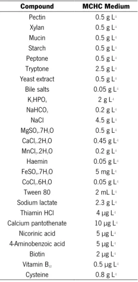
Table 3.3. Composition of MCHC Medium ... 32
Table 3.4. Equations for quantifications of SCFAs ... 35
Table 3.5. Conditions used to perform the digestive stress challenges ... 36
Table 4.1. Amounts of SCFAs concentration present in fermentation broth of P. freudenreichii .. 69
Table 4.2. Assays and treatments performed in RKO cell line ... 71
Table 5.1. Conditions used to evaluate the effect of conditioned medium in growth and SCFAs production by Propionibacterium freudenreichii. ... 90
List of figures
Figure 1.1. Estimated cancer incidence and mortality rates Portugal in 2012 (Adapted from (Ferlay
et al. 2013)). ... 4
Figure 1.2. The Hallmarks of Cancer originally proposed by Hanahan and Weinberg (Adapted from
(Hanahan and Weinberg 2011)). ... 4
Figure 1.3. The role of genes and environment in the development of cancer (Adapted from (Anand
et al. 2008)). ... 5
Figure 1.4. Histological steps of the colorectal cancer, polyp to adenocarcinoma sequence,
associated with commonly methylated genes (and loci) involving in this process (Lao and Grady 2011). ... 7
Figure 1.5. The Human large intestine anatomy (Adapted from (WebMD 2014))... 9 Figure 1.6. Representation of the diversity of bacteria in the human intestine. Wedges represent
division: Those numerically abundant in the human gut are red, rare divisions are green, and undetected are black. Wedge length is a measure of evolutionary distance from the common ancestor (Adapted from (Backhed et al. 2005)). ... 12
Figure 1.7. Therapeutic areas covered by nutraceutical products (Das et al. 2012). ... 15 Figure 1.8. Schematic representation of main fermentation end products in propionibacteria
(Zarate 2012). ... 18
Figure 2.1. Thesis project outline ... 25 Figure 3.1. Monitoring of growth and SCFAs production by P. freudenreichii. ... 36
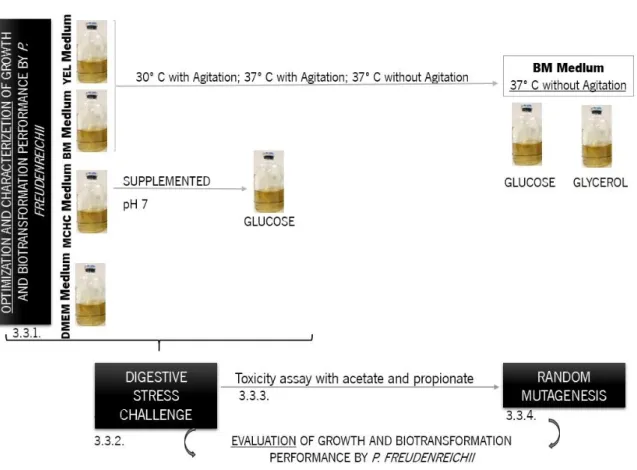
Figure 3.2. Assays performed to optimize and characterize P. freudenreichii and to study digestive stress challenge as well as random mutagenesis. ... 40
Figure 3.3. Biomass, acetate and propionate production by P. freudenreichii in YEL medium (A) and BM medium (B) at pH 7, 30 ºC with agitation ( ), 37 ºC with agitation ( ) and 37 ºC without agitation ( ). Concentration in gram per liter of biomass, acetate and propionate over time in hours. Each condition was run in triplicate and the mean ± SEM are represented. ... 42
Figure 3.4. Bach fermentations kinetic parameters for P. freudenreichii in YEL medium and BM medium at pH 7, 30 ºC with agitation ( ), 37 ºC with agitation ( ) and 37 ºC without agitation (
). Concentration in gram per liter per hour of growth rate and productivity; in gram per gram of yield. Each condition was run in triplicate and the mean ± SEM are represented. Values significantly different from 30 ºC with agitation of YEL medium (a); 37 ºC with agitation of YEL medium (b); 37 ºC without agitation of YEL medium (c): * p<0.05; ** p<0.01; *** p<0.001. One-way ANOVA and Tukey᾽s Test were used. ... 43
Figure 3.5. Biomass, acetate and propionate production by P. freudenreichii in BM medium ( ) and supplemented with 3.7 g L-1 of glucose ( ) and glycerol ( ) at pH 7, 37 ºC without agitation
Concentration in gram per liter of biomass, acetate and propionate over time in hours. Each condition was run in triplicate and mean ± SEM are represented. ... 45
Figure 3.6. Bach fermentations kinetic of P. freudenreichii in BM medium as control and supplemented with 3.7 g L-1 of glucose and glycerol, 37 ºC without agitation. Concentration in gram
per liter per hour of growth rate and productivity; in gram per gram of yield. Each condition was run in triplicate and the mean ± SEM are represented. Values significantly different from control and compare all with all conditions: * p<0.05; ** p<0.01; *** p<0.001. One-way ANOVA and Tukey᾽s Test were used. ... 46
Figure 3.7. Biomass, acetate and propionate production by P. freudenreichii in MCHC medium at pH 6 ( ); MCHC medium at pH 7 ( ) and MCHC medium (pH 6) supplemented with glucose (
) at pH 6, 37 ºC without agitation. Concentration in gram per liter of biomass, acetate and propionate over time in hours. Each condition was run in triplicate and the mean ± SEM are represented. ... 48
Figure 3.8. Bach fermentations kinetic of P. freudenreichii in MCHC medium (pH 6) as control, MCHC medium at pH 7 and MCHC medium supplemented with glucose at pH 6, 37 ºC without agitation. Concentration in gram per liter per hour of growth rate and productivity; in gram per gram of yield. Each condition was run in triplicate and the mean ± SEM are represented. Values significantly different from control and compare all with all conditions: * p<0.05; ** p<0.01; *** p<0.001. One-way ANOVA and Tukey᾽s Test were used. ... 49
Figure 3.9. Biomass, acetate and propionate production by P. freudenreichii in YEL medium at pH 7 ( ); BM medium at pH 7 ( ); MCHC medium at pH 6 ( ); DMEM medium at pH 7.4 ( ), cultivated at 37 ºC without agitation. Concentration in gram per liter of biomass, acetate and propionate over time in hours. Each condition was run in triplicate, excluding DMEM medium (duplicate), the mean ± SEM are represented. ... 51
Figure 3.10. Bach fermentations kinetic of P. freudenreichii in YEL medium at pH 7; BM medium at pH 7; MCHC medium at pH 6; DMEM medium at pH 7.4, cultivated at 37 ºC without agitation. Concentration in gram per liter per hour of growth rate and productivity; in gram per gram of yield. Each condition was run in triplicate, excluding DMEM medium (duplicated), the average and standard error of the mean are represented. Values significantly different: * p<0.05; ** p<0.01; *** p<0.001. One-way ANOVA and Tukey Test. ... 52
Figure 3.11. Biomass, acetate and propionate production by adapted P. freudenreichii recovered in liquid media (A) and solid media (B). Bacteria was cultivated in YEL medium ( ), BM medium (
) MCHC medium ( ) at 37º C without agitation. Concentration in gram per liter of biomass, acetate and propionate over time in hours. Each condition was run in nine independent experiments and the mean ± SEM are represented. ... 54
Figure 3.12. Bach fermentations kinetic of adapted P. freudenreichii recovered in liquid media and solid media. Bacteria was cultivated in YEL medium ( ), BM medium ( ) MCHC medium ( ) at 37º C without agitation. Concentration in gram per liter per hour of growth rate and productivity; in gram per gram of yield. Each condition was run nine times in independent experiments and the average and standard error of the mean are represented. Values significantly different from liquid recovery of YEL medium (a), BM medium (b) MCHC medium (c): * p<0.05; ** p<0.01; *** p<0.001. One-way ANOVA and Tukey Test. ... 55
Figure 3.13. Biomass ( ), acetate ( ) and propionate ( ) production by adapted P. freudenreichii recovered in solid media. Bacteria was cultivated in DMEM medium at 37º C without agitation. Concentration in gram per liter of biomass, acetate and propionate over time in hours. Each condition was run in duplicated bottles and the average and standard error of the mean (SEM) are represented. ... 56
Figure 3.14. Bach fermentations kinetic of P. freudenreichii DSM 20271 and adapted P. freudenreichii cultivated in YEL medium ( ), BM medium ( ) MCHC medium ( ) and DMEM medium ( ) at 37º C without agitation. Concentration in gram per liter of biomass, acetate and propionate; in gram per liter per hour of growth rate and productivity; in gram per gram of yield. Each condition was run in triplicate, excluding DMEM medium (duplicate), the mean ± SEM are represented. Values significantly different between the same medium: * p<0.05; ** p<0.01; *** p<0.001. One-way ANOVA and Tukey᾽s Test were used. ... 58
Figure 3.15. Batch fermentation kinetics of biomass, acetate and propionate production by
-1 of chloramine as positive control ( ) and supplemented with an initial acetate concentration of 1
g L-1 ( ), 2 g L-1 ( ), 4 g L-1 ( ) and 8 g L-1 ( ) at 37º C without agitation. Concentration in gram
per liter of biomass, acetate and propionate over time in hours. Each condition was run in triplicate and mean ± SEM are represented. ... 59
Figure 3.16. Batch fermentation kinetics of biomass, acetate and propionate production by
adapted P. freudenreichii in YEL medium used as negative control ( ), supplemented with 10 g L -1 of chloramine as positive control ( ) and supplemented with an initial propionate concentration
of 3 g L-1 ( ), 6 g L-1 ( ), 12 g L-1 ( ) and 24 g L-1 ( ) at 37º C without agitation. Concentration
in gram per liter of biomass, acetate and propionate over time in hours. Each condition was run in triplicate and the mean ± SEM are represented. ... 60
Figure 3.17. Batch fermentation kinetics of biomass, acetate and propionate production by
adapted P. freudenreichii in YEL medium used as negative control ( ), supplemented with 10 g L -1 of chloramine as positive control ( ) and supplemented with an initial propionate concentration
of 3 g L-1 ( ), 6 g L-1 ( ), 12 g L-1 ( ) and 24 g L-1 ( ) at 37º C without agitation. Concentration
in gram per liter of biomass, acetate and propionate over time in hours. Each condition was run in triplicate and mean ± SEM are represented. ... 61
Figure 3.18. Bach fermentations kinetic of adapted P. freudenreichii cultivated in YEL medium used as control ( ) and supplemented with an initial concentration of acetate ( ) propionate ( ) and 1 g L-1 acetate and 3 g L-1 propionate ( ) at 37º C without agitation. Concentration in gram per
liter of biomass, acetate and propionate; in gram per liter per hour of growth rate and productivity; in gram per gram of yield. Each condition was run in triplicate, the mean ± SEM are represented. Values significantly different between same medium: * p<0.05; ** p<0.01; *** p<0.001. One-way ANOVA and Dunett`s test were used. ... 62
Figure 4.1. Cell proliferation analysis by SRB in CRC-derived cell line RKO treated with acetate and
propionate, separately and together. Cells were incubated with IC50 ( ), IC30 ( ) and concentration
of acetate and propionate detected in fermentation broth by P. freudenreichii ( ) for 48 h. Cells were incubated with fresh complete medium or hydrogen peroxide (1mM) as a negative ( ) and positive ( ) control, respectively. Values represent mean ± S.E.M. of at least three independent experiments. *** p<0.001, compared with negative control cells. ### p<0.001, compared with IC50
p<0.001 compared with Acetate & Propionate detected in fermentation broth of P. freudenreichii. One-way ANOVA and Tukey`s Test were used. ... 75
Figure 4.2. Cell proliferation analysis by SRB in CRC-derived cell line RKO treated with
Propionibacterium media: BM medium ( ), YEL medium ( ), MCHC medium with pH 6 ( ) and pH 7 ( ). A) Cells were incubated with a total volume of different sterile media from Propionibacterium and with fresh DMEM complete medium as negative control ( A). B) Cells were incubated with a different sterile media of Propionibacterium diluted (50% v/v) in fresh DMEM complete medium. Fresh DMEM complete medium diluted (50% v/v) in HEPES solution (20 mM; pH 7.4) was used as negative control ( B). As a positive control was used hydrogen peroxide (1mM) (
). Values represent mean ± S.E.M. of at least three independent experiments. *** p<0.001, compared with negative control cells. One-way ANOVA and Dunnett`s Test were used. ... 76
Figure 4.3. Cell proliferation analysis by SRB in CRC-derived cell line RKO treated with pure acetate
and propionate ( ), BM supernatant ( ), BM supernatant deproteinized ( ) by P. freudenreichii DSM 20271 and adapted P. freudenreichii. Cells were incubated with a supernatants diluted (50% v/v) in fresh DMEM complete medium and with fresh DMEM complete medium diluted (50% v/v) in sterile BM medium as negative control ( ). Concentration presented corresponding the dilution being, in condition of P. freudenreichii DSM 20271 and adapted P. freudenreichii, 14 mM – 10 mM of acetate and 32 mM – 20 mM propionate, respectively. As a positive control was used hydrogen peroxide (1mM) ( ). Values represent mean ± S.E.M. of at least three independent experiments. ** p<0.01, compared with negative control cells. No significant difference between different conditions. One-way ANOVA and Tukey`s Test were used. ... 78
Figure 4.4. Cell proliferation analysis by SRB in CRC-derived cell line RKO treated with pure acetate
and propionate ( ), DMEM supernatant ( ), DMEM supernatant deproteinized ( ) by P. freudenreichii and adapted P. freudenreichii. A) Cells were incubated with a total volume of fermentation broth of P.freudenreichii and with DMEM medium “consumed” by RKO cell line as negative control ( A). Concentration presented in condition of P. freudenreichii DSM 20271 and adapted P. freudenreichii were 13 mM – 12 mM of acetate and 35 mM – 36 mM propionate, respectively. B) Cells were incubated with a supernatants diluted (50% v/v) in fresh DMEM complete medium and with fresh DMEM complete medium diluted (50% v/v) in DMEM medium “consumed” by RKO cell line as negative control ( B). Concentration presented corresponding the dilution being, in condition of P. freudenreichii DSM 20271 and adapted P. freudenreichii, 6.5 mM – 6 mM of acetate and 17.5 mM – 18 mM propionate, respectively. As a positive control was used hydrogen
peroxide (1mM) ( ). Values represent mean ± S.E.M. of at least three independent experiments. * p<0.05; ** p<0.01*** p<0.001, compared with negative control cells and between some conditions. One-way ANOVA and Tukey`s Test. ... 79
Figure 4.5. Impact of fermentation broth on cell cycle distribution. (A) Analysis of the treatment
effect on the sub-G1 subpopulation of RKO cells by flow cytometry. Percentage of sub-G1 cells are shown. (B) Analysis of the distribution of cell-cycle phases: G0/G1( ); S( ); G2/M( ) and SUB-G1( ) in CRC-derived cell line RKO treated with pure acetate and propionate, DMEM supernatant, DMEM supernatant deproteinized by P. freudenreichii DSM 20271 and adapted P. freudenreichii, these conditions were diluted (50% v/v) in fresh DMEM complete medium. Fresh DMEM complete medium diluted (50% v/v) in DMEM medium “consumed” by RKO cell line and hydrogen peroxide (1mM) were used as negative and positive control, respectively. Values represent mean ± S.E.M. of at least two independent experiments. * p<0.05; *** p<0.001, comparing the percentage of sub-G1 population of treated cells with negative control and respective control of pure SCFAs. One-way ANOVA and Tukey`s Test were used. ... 81
Figure 5.1. Biomass, acetate and propionate production by P. freudenreichii DSM 20271 (A) and adapted P. freudenreichii (B) at pH 7, 37 ºC without agitation. Conditioned medium ( ); Control of conditioned medium ( ); Conditioned medium diluted 50% (v/v) in fresh DMEM medium ( ) and Control of conditioned medium diluted ( ). Concentration in gram per liter of biomass, acetate and propionate over time in hours. Each condition was run in duplicate and the mean ± SEM are represented. ... 92
Figure 5.2. Bach fermentations kinetic of P. freudenreichii DSM 20271 and adapted P. freudenreichii at pH 7, 37 ºC without agitation. Conditioned medium ( ); Control of conditioned medium ( ); Conditioned medium diluted 50% (v/v) in fresh DMEM medium ( ) and Control of conditioned medium diluted ( ). Concentration in gram per liter per hour of growth rate and productivity; in gram per gram of yield. Each condition was run in duplicate and the mean ± SEM are represented. Values significantly different between conditioned medium conditions and respective controls as well as conditioned medium diluted 50% (v/v) in fresh DMEM medium (a); control of conditioned medium diluted (b); conditioned medium (c) and control of conditioned medium (d): * p<0.05; ** p<0.01; *** p<0.001. “ns” report that no significant different. One-way ANOVA and Tukey᾽s Test were used. ... 93
List of symbols and abbreviations
[A], Concentration of AcidAA, Area Acid
Adapted P. freudenreichii, Propionibacterium freudenreichii subsp. freudenreichii DSM 20271 tolerant to digestive stress established by us in the laboratory (chapter 3)
AIS, Area Internal Standard APC, Adenomatous polyposis coli BM, Basal médium
Cat-D, Cathepsin D
CCM, Control with DMEM medium diluted in HEPES solution CFB, Cytophaga-Flavobacterium-Bacteroides phylum
CLA, Conjugated linoleic acid CM, ‘Conditioned medium’
CM½, Conditioned medium diluted in fresh DMEM medium (50% v/v); CRC, Colorectal cancer
DCW, Dry cell weight
DMEM, Dulbecco‘s Modified Eagle‘s Medium
ENU, N-nitroso-N-ethylurea; N-ethyl-N-nitrosourea or Ethylnitrosourea FAO, Food and Agriculture Organization
FAP, Familial adenomatous polyposis FBS, Fetal bovine serum
FOBT, Fecal occult blood testing GERD, Gastroesophageal reflux disease GI, Gastrointestinal
GRAS, Generally regarded as safe HMP, Human microbiome project HNPCC, Lynch syndrome
HPLC, High performance liquid chromatography IC30, 30 % maximal inhibitory concentration
IC50, Concentrations of half maximal inhibitory concentration
LMP, Lysosomal membrane permeabilization
MCHC, “Mimic the content of the human colon” medium NTG, N-methyl-N'-nitro-N-nitrosoguanidine
OD, Optical density
P. freudenreichii, Propionibacterium freudenreichii subsp. freudenreichii DSM 20271 PI, Propidium iodide
QPS, Qualified presumption of safety R, Correlation Coefficient
RT, Retention Time
SCFA, Short chain fatty acid SEM, Standard error of the mean
VSL#3, Commercially available probiotic formulation WHO, World health organization
Chapter 1.|
1.1. Cancer
Formerly humans died by natural causes or through violence, accidents and by an amazing variety of infectious diseases but with the years the main causes of death vary. In the past decade, the leading causes of death worldwide were ischaemic heart disease, stroke, chronic obstructive lung disease and lower respiratory infections. Actually, the leading causes of death are heart disease and cancer (World Health Organization 2014). Although changes in living conditions during the 20th
century and advances in medicine have contributed to change morbidity and mortality standards, there is still a shadow over humanity, namely cancer, once that although it is the second leading cause of death after heart disease (World Health Organization 2014), the death rates have remained constant compared with death rates for heart disease which have been decreasing (Siegel et al. 2014).
Cancer is a public health problem worldwide that induce death every day and its incidence is increasing. The results of GLOBOCAN 2012 are worrying as according to these results, an estimated 14.1 million new cancer cases and 8.2 million cancer related deaths occurred in 2012 (Ferlay et al. 2013), compared with 12.7 million and 7.6 million respectively, in 2008 and predict a substantive increase to 19.3 million new cancer cases per year by 2025, due to growth and ageing of the global population (Ferlay et al. 2010). In 2012, lung (1.8 million, 13.0% of the total), breast (1.7 million, 11.9%), and colorectal (1.4 million, 9.7%) cancers represented the most common diagnosed cancer worldwide and lung (1.6 million, 19.4% of the total), liver (0.8 million, 9.1%), and stomach (0.7 million, 8.8%) cancers were the most common causes of cancer death (Ferlay et al. 2013).
In Portugal, cancer is also a serious health problem. In 2012, there was an estimated 49 thousands new cancer cases and 24 thousands cancer related deaths being colorectal, prostate and breast cancers the most diagnosed but also the most common causes of cancer death (See Figure 1.1)(Ferlay et al. 2013).
Figure 1.1. Estimated cancer incidence and mortality rates Portugal in 2012 (Adapted from (Ferlay et al.
2013)).
Cancer is a disease normally associated to a rapid cell proliferation and uncontrolled cell growth. However cancer is more complex than this simply characterization evolving survival or ‘hardiness’ strategies that allows the existence of this disease (Aktipis et al. 2013). Cancer is a multistep process that leads normal cells to acquire biological capabilities becoming malignant cells. These biological capabilities are designated by “hallmarks of cancer” which were proposed by Hanahan and Weinberg in 2000. Together constitute an organizing principle that provides a logical framework for understanding the remarkable diversity of neoplastic diseases. The hallmarks of cancer include sustaining proliferative signaling, evading growth suppressors, resisting cell death, enabling replicative immortality, inducing angiogenesis, and activating invasion and metastasis (Hanahan and Weinberg 2000). Conceptual progress in the last decade has added two emerging hallmarks of potential generality to this list: reprogramming of energy metabolism and evading immune destruction (See Figure 1.2)(Hanahan and Weinberg 2011).
Figure 1.2. The Hallmarks of Cancer originally proposed by Hanahan and Weinberg (Adapted from (Hanahan
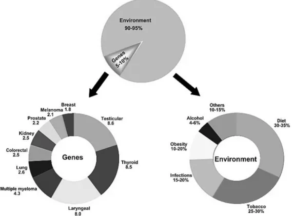
All cancer are a result of multiple mutations (Loeb and Loeb 2000; Hahn and Weinberg 2002), but only 5-10% of all cancer cases can be attributed to genetic defects, whereas the remaining 90-95% have their roots in the environment and lifestyle (See Figure 1.3) (Anand et al. 2008). This data shown that the majority of cancers are not of hereditary origin and that lifestyle factors, such as dietary habits, smoking, alcohol consumption, and infection, have a profound influence on their development (Irigaray et al. 2007; Mucci et al. 2001). The hereditary factors cannot be modified, but we can prevent cancer by manipulating and modifying the lifestyle and environmental factors.
Figure 1.3. The role of genes and environment in the development of cancer (Adapted from (Anand et al.
2008)).
1.1.1. Colorectal cancer
Colorectal cancer (CRC) is a malignancy that affects the large intestine wall, and depending on the degree of invasion can compromise other organs, either directly or through metastasis (World Health Organization 2014).
In Portugal, CRC is a growing healthy problem, as it is incidence and mortality rates have been increasing since the 1980s (Portuguese society for digestive endoscopy 2012). According to the most recent estimates by the International Agency for Research on Cancer (Ferlay et al. 2013) CRC, in Portugal, is the most commonly diagnosed cancer with 7129 new cases and it is the most
common cause of cancer deaths with 3797 deaths reported in 2012 (See Figure 1.1)(Ferlay et al. 2013).
In the development of CRC, a strong genetic component but also environmental factors, including diet and lifestyle, have a major impact on its risk. These factors are strongly supported by epidemiological studies, which have shown that environmental factors have the potential to influence the normal structure and function of the colon, and various dietary components have been proposed as being either beneficial or potentially harmful to colonic health (Lipkin et al. 1999). For example, Azcarate-Peril and co-workers reviewed this issue and an increasing number of studies demonstrated that lifestyle-associated factors that increase the risks of CRC include elevated body mass index, obesity, and reduced physical activity (Azcarate-Peril et al. 2011). However, it has been suggested that diet regimens rich in fruit, vegetables and poor in meat might have a protective effect, reducing the incidence of colorectal adenomas by modulating the composition of the normal nonpathogenic commensal microbiota (Azcarate-Peril et al. 2011; Davis and Milner 2009; Hakansson et al. 2012). CRC is thought to develop over many years in a multistep process that transform normal glandular epithelium into invasive adenocarcinoma as a result the accumulation of both acquired genetic and epigenetic changes (Vogelstein et al. 1988). The steps involved in this process were first described by Fearon and Vogelstein (1990) in the classic adenoma to cancer progression, based on the concept that progression is accompanied by the accumulation of molecular alterations in which adenomatous polyposis coli (APC), K-RAS, and p53 genes play a central role (Fearon and Vogelstein 1990). This model allows to understand the molecular pathogenesis of CRC and is based on the premise that tubular and tubulovillous adenomas are premalignant neoplasms that will progress to invasive adenocarcinoma (See Figure 1.4) (Lao and Grady 2011; Fearon and Vogelstein 1990). However it is known that there are more classes of premalignant polyps with potential for malignant transformation and there are multiple molecular pathways to CRC and it is important to distinguished well-defined inherited syndromes as Lynch syndrome (HNPCC) and familial adenomatous polyposis (FAP), which are unusual conditions that have a substantial cancer risk with a percentage of 2% to 5% of all CRCs (Lichtenstein et al. 2000).
Figure 1.4. Histological steps of the colorectal cancer, polyp to adenocarcinoma sequence, associated with
commonly methylated genes (and loci) involving in this process (Lao and Grady 2011).
Assessment of the CRC epigenome has revealed that virtually all CRCs have aberrantly methylated genes and that the average CRC methylome has hundreds to thousands of abnormally methylated genes (Ahnen 2011). These genes are implicated in either the initiation or progression of CRC (Barrow and Michels 2014). We can see in Figure 4 the common methylated genes (and loci) identified at the histological steps of CRC polyp to adenocarcinoma sequence. The genes listed between normal epithelium and aberrant crypt focus as well as those genes listed between aberrant crypt focus and polyp/adenoma might be involved in the initiation of colorectal cancer, as well as those genes listed between polyp/adenoma and adenocarcinoma could have a role in the progression and metastasis of CRC (Lao and Grady 2011).
APC, K-RAS and p53 genes play a critical role in development of CRC. APC regulate the entry of epithelial cells into adenoma-carcinoma progression, being proposed as a ‘‘gatekeeper’’ gene (Kinzler and Vogelstein 1996). A mutation of the gatekeeper leads to a permanent imbalance of cell division over cell death. The RAS oncogene promotes tumor formation through stimulation of cell proliferation, motility and regulation of apoptosis. Activating mutations in the RAS oncogenes mediates deregulated cell growth, evasion of apoptosis and malignant transformation (Shaw et al. 2011; Smith et al. 2002). The TP53 gene product, p53, functions as a transcription factor, exerting cell cycle control by binding to specific recognition sequences in a variety of genes including p21, Bax, and Bcl-2 in response to DNA damage or other cellular stress (Levine 1997). Mutations in p53 render the cells susceptible to failure of apoptosis and increased accumulation of DNA damage, allowing unregulated growth (el-Deiry et al. 1993).
In the onset of the disease, when CRC is early and highly curable, symptoms and signs are less common and less obvious being most common and prominent late in colon cancer. Advanced CRC is likely to be incurable, what difficult an early diagnostic and affects the effectiveness of treatment (Cappell 2005), thus, symptoms depend on cancer location, cancer size and presence of metastases.
There are a range of screening and diagnostic tests for CRC as fecal occult blood testing (FOBT), barium enema, flexible sigmoidoscopy, colonoscopy (Helm et al. 2003; Selby et al. 1992; Detsky 2001; Spinzi and Minoli 2001; Cappell 2005), which the general asymptomatic population should perform regularly for early detection and prevention of CRC.
The treatment depends on the staging of CRC being the surgery the main curative treatment. Chemotherapy and radiotherapy may be recommended depending on the staging of cancer and other medical factors. The drugs must often use for CRC include 5-Fluorouracil, capecitabine, irinotecan and oxaliplatin (American Cancer Society 2015). However, there are national CRC treatment guidelines that helps the physicians in the choice of treatment. This guidelines are annually adapted to take into account Portuguese clinical practice (Pinto et al. 2010).
It is generally agreed that, given the significant negative effect of CRC on people’s quality of life and the associated high mortality rates, it is essential to develop primary and secondary strategies for the prevention of CRC. As a primary prevention strategy is very important to educate the general population to modify dietary risk factors and adopt healthy habits. As a secondary prevention strategy is essential the detection and reduction of premalignant adenomatous lesion and early detection of cancer in a curable stage should be the primary goal.
1.2. Digestive system
The digestive system is a group of organs working together to convert food into energy and basic nutrients to feed the entire body. Food passes through a long tube inside the body known as the gastrointestinal (GI) tract suffering mechanical and chemical action that allows the degradation of food (Seeley 2011). GI tract is made up of the oral cavity, pharynx, esophagus, stomach, small intestines and large intestines. There are others organs that help your body to digest food but do not have food pass through them as teeth, tongue, salivary glands, liver, gallbladder and pancreas (Gray and Lewis 1918; Seeley 2011).
The intake of food, its breakdown into nutrients and their absorption and elimination of the indigestible waste is essential for a healthy body. Actually, there are a variety of digestive problems such as gastroesophageal reflux disease (GERD), irritable bowel syndrome, celiac disease, food allergies, diverticulitis, ulcerative colitis and Crohn’s disease (Cencic and Chingwaru 2010). Thus, it is essential that each organ of the digestive system preform their function correctly.
1.2.1. Colon
The large intestine or colon allows the lower GI tract and it is the portion of GI which extends from the end of the ileum to anus. It is about 1.5 meters long, being one fifth of the whole extent of the GI tract and their contents take about 18-24 hours to cover its entire extension (Seeley 2011; Gray and Lewis 1918). The large intestine is divided into the cecum, colon, rectum and anal canal (See Figure 1.5). The colon corresponding the major part of the large intestine being constituted by the ascending, transverse, descending and sigmoid colon (Seeley 2011).
Figure 1.5. The Human large intestine anatomy (Adapted from (WebMD 2014)).
The main function of the large intestine are: reabsorption of water and mineral ions; formation and temporary storage of feces; maintaining a resident of population of over 500 species of bacteria and bacterial fermentation of indigestible materials (Seeley 2011).
The waste left over from the digestive process, namely chime, arrive to large intestine by cecum and it is passed through the colon by means of peristalsis, first in a liquid state and ultimately in a solid form. The feces passes through the colon, water and salts is absorbed as well occurs the
secretion of mucus by mucus glands present in the mucosa layer. During this transit the feces suffer the intensive action of microorganisms present in the lumen of colon. These bacteria perform several useful functions such as synthesizing various vitamins, processing waste products and food particles and protecting against harmful bacteria (Cencic and Chingwaru 2010).
1.3. Gastrointestinal microbiota
In the human GI tract resides the microbiota, a large and diverse community of microorganism which have a critical role in the evolution of the intestinal functions and in overall health of the host (Saavedra and Dattilo 2012).
The total amount of genes in the various species represented in our indigenous microbial communities is estimated about 2-4 million, exceeding the number of our human genes >100-fold (Backhed et al. 2005). The collective genomes of microbiota is termed “microbiome” and it was introduced in 2001 by Hooper and Gordon. Microbiome provide us genetic and metabolic attributes which we not have, including the ability to harvest otherwise inaccessible nutrients (Di Mauro et al. 2013).
In order to collect and integrate the genomic information from many diverse human microbiomes an international collaborative project, “the human microbiome project (HMP)”, was launched in 2007 (Turnbaugh et al. 2007). Thus the Human Microbiome Project Consortium provided the first reliable estimates of the structure, function, and diversity of the healthy (“reference”) human microbiome (Human Microbiome Project 2012b, 2012a) and all data are available at http://commonfund.nih.gov/hmp/publications.aspx.
Actually, microbiota is considered as an “organ within an organ” with its own functions: modulates expression of genes involved in mucosal barrier fortification, angiogenesis, postnatal intestinal maturation and a critical role in supporting normal digestions as well as affects energy harvest from the diet and energy storage in the host, fermenting unused energy substrates to short chain fatty acids (SCFAs) (Hooper et al. 2001; Saavedra and Dattilo 2012). These functions are intimately strain-related.
1.3.1. Composition of gastrointestinal microbiota
The composition of GI microbiota undergoes major modifications during our life being influenced by the host genotype and physiology, the colonization history, environmental factors, food and drugs (e.g. antibiotics) (Sharma et al. 2010).
Infants are born without GI microbiota, but rapidly after birth the infants GI is colonized by bacteria coming from the maternal vaginal and intestinal flora, and it is an important stage of development of intestinal functions (Hattori and Taylor 2009; Aagaard et al. 2013). The composition of the microbiota stays unstable until the age of approximately 3-4 years, when it becomes mature (den Besten et al. 2013).
The adult human GI tract contains all three domains of life: Bacteria, Archaea, and Eukarya, being the Bacteria the most dominant and most diverse group of microorganisms present in the human colon with 1014 citizens (98% of all species ) (Hattori and Taylor 2009). Bacteria is usualy
separated into two broad categories, namely as beneficial (e.g., Bifidobacterium and Lactobacillus) or harmful (e.g., Enterobacteriaceae and Clostridium spp.) which compete with each other to colonized the GI tract (Backhed et al. 2005).
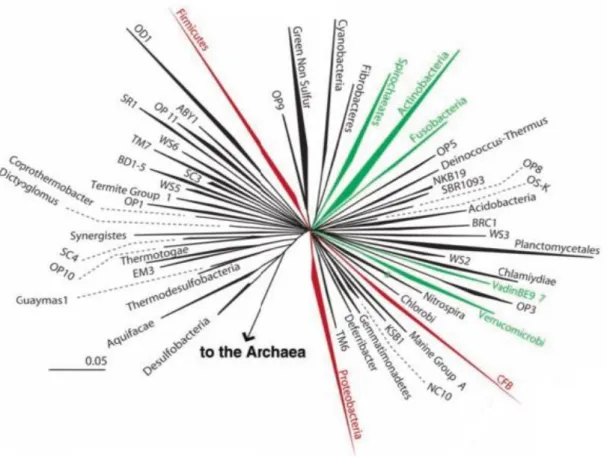
In the composition of GI microbiota there may be between 500-1000 different species present, based on variation in 16S ribosomal RNA genes, which belong to more than 70 genera. The most abundant phyla in the gut is Firmicutes, Bacteroidetes, Proteobacteria and Actinobacteria (Hattori and Taylor 2009; Abubucker et al. 2012). The Figure 1.6 represent the diversity of bacteria in GI tract being CFB the designation of Cytophaga-Flavobacterium-Bacteroides phylum (Backhed et al. 2005).
The Bacteroidetes phylum manly produces acetate and propionate, whereas the Firmicutes phylum has butyrate as its primary metabolic end product. Most bacterial activity occurs in the proximal colon where substrate availability is highest, once the availability of substrate declines, extraction of free water reduces diffusion of substrates and thus microbial products (Di Mauro et al. 2013).
Figure 1.6. Representation of the diversity of bacteria in the human intestine. Wedges represent division:
Those numerically abundant in the human gut are red, rare divisions are green, and undetected are black. Wedge length is a measure of evolutionary distance from the common ancestor (Adapted from (Backhed et al. 2005)).
The Human Microbiome Project (HMP) provided a reference collection of 16S ribosomal RNA gene sequences collected from the human gut that allow us to better associate changes in the microbiome with changes in health, but it is important to highlight that there is no single healthy microbiome. Each person harbors a unique and varied collection of bacteria that is the result of a life history as well as their interactions with the environment, diet and medication used (Ding and Schloss 2014). Thus it is important to understand the microbiome diversity and the mechanisms that are associated with this diversity to assess disease risk and personalize therapies. The second phase of the HMP, which is ongoing (2013-2016), will be a powerful feature to understand the role of the microbiome in human health and disease (Integrative 2014)
1.1.1.1. Colorectal cancer and gastrointestinal microbiota
The association between CRC and GI microbiota has been studied for many years and several of these studies have shown that patients with CRC have significant changes in GI microbiota, significant decreases in GI SCFAs concentrations and a significant increase in GI pH compared with
the healthy individuals (Scanlan et al. 2008; Sobhani et al. 2011; Ohara et al. 2010; Ohigashi et al. 2013).
These studies suggested that the changes in microbial community correspond to a decrease in bacteria species of Firmicutes and Bacteroidetes, demonstrating that microbial community composition changes collectively in CRC (Scanlan et al. 2008; Ohigashi et al. 2013; Ohara et al. 2010). It has been previously reported that specific bacterial species, such as Streptococcus bovis (Tjalsma et al. 2006; Ellmerich et al. 2000) or Bacteroides (Aries et al. 1969; Hill et al. 1971) are involved in CRC.
However some people have doubts if the change in microbiota in CRC patients is a result or a cause for the initiation of colon carcinogenesis. CRC itself, the symptoms of the disease, and its therapy have the potential to alter the GI microbial composition. Changes in the microbiome may precede or contribute to the development of factors related to cancer susceptibility (Lampe 2008). The results of Ohigashi and co-workers suggest that it is not the progression of CRC that causes changes in the GI environment but rather that cancer initiates and progresses in a GI environment that has changed (Ohigashi et al. 2013).
1.3.2. Microbiota modulation of gastrointestinal functions
The human GI microbiota is intrinsically linked with the organism health, since this endogenous microbiota form a symbiotic relationship with their eukaryotic host and this close partnership helps maintaining homeostasis by performing essential and non-redundant tasks (Zhu et al. 2011). This symbiotic relationship comprises a diverse communities of microbes, which include mutualists (symbiotically beneficial microbes), commensals (microbes that are neither harmful nor beneficial to the host), and pathogens (microbes that are detrimental to the host) (Aagaard et al. 2013).
The host has evolved to establish many processes that sustain unresponsiveness toward the commensal bacteria while at the same time maintaining responsiveness toward pathogens. These processes include the production of IgA and various antimicrobial peptides and epigenetic control of pro-inflammatory responses, all of which separate routes leading to excessive inflammatory response (Hattori and Taylor 2009). On the other hand, pathogens also have evolved to equip various virulence factors, including effectors that confer additional abilities for evading the host defense system, eventually inducing pro-inflammatory responses via change of the microbiota composition in favor of the pathogens (Raskin et al. 2006; Stecher et al. 2007). In contrast, commensal bacteria is evolved
in carbohydrate metabolism, energy production, cell maturation and proliferation toward intestinal homeostasis (Rawls et al. 2007).
Mutualist bacteria as Propionibacterium freudenreichii has a symbiotic beneficial relationship with host due to their end products of fermentation, which are essential mucosal nutrients including amino acids and SCFAs (O'Sullivan et al. 2005). Besides fermentation, the metabolic products of microflora includes vitamins K and B complex, secondary bile acid production, neutralization of dietary carcinogens such as nitrosamines, and conversion to active metabolites of some prodrugs (Cencic and Chingwaru 2010).
1.4. Nutraceuticals
The term "nutraceutical" was created from "nutrition" and "pharmaceutical" in 1989 by Stephen DeFelice, MD, founder and chairman of the Foundation for Innovation in Medicine (FIM) (Cranford, NJ). According to DeFelice, nutraceutical can be defined as “a food (or a part of food) that provides medical or health benefits, including the prevention and or treatment of a disease” (Brower 1998). The food sources used as nutraceuticals are all natural and can be categorized as dietary fiber, probiotics, prebiotics, polyunsaturated fatty acids, antioxidant, vitamins, polyphenols or spices (Das et al. 2012).
According to a consensus definition, a probiotic is “a live microbial food ingredient that is beneficial to health” (Salminen et al. 1998) and a prebiotic is "a selectively fermented ingredient, or a fiber that allows specific changes, both in the composition and/or activity of the gastrointestinal microflora, conferring benefits on the well-being and health of host " (de Vrese and Schrezenmeir 2008; Douglas and Sanders 2008).
Probiotics claimed benefits that include the improvement of lactose digestion, increased resistance to invasion by pathogenic bacteria in the gut, prevention of intestinal disturbances, treatment and prevention of antibiotic treatment and acute diarrhea, alleviation of irritable bowel syndrome and of inflammatory bowel disease, and possibly protection against cancer (Salminen et al. 1998; Kumar et al. 2010). In contrast, the specific effects of prebiotics on health are indirect (Cencic and Chingwaru 2010). However, probiotics and prebiotics share unique roles in human nutrition, largely centered on manipulation of populations or activities of the microbiota that colonize the human GI tract.
The regular consumption of probiotics or prebiotics has a positive impact in health that include enhanced immune function, improved colonic integrity, decreased incidence and duration of intestinal infections, down-regulated allergic response, and improved digestion and elimination (Douglas and Sanders 2008).
The Food and Agriculture Organization (FAO) of the United Nations and World Health Organization (WHO) Expert Consultation develop guidelines, recommend criteria, and define the methodology for evaluation of probiotics, besides identifying which data are required to accurately substantiate health claims. The selection of probiotics must respect these standards that include safety, technological properties, survival to digestive stresses and at least one property that benefits human health (Sanders and Marco 2010).
Nutraceuticals can cover most of the therapeutic areas such as anti-arthritic, cold and cough, sleeping disorders, digestion and prevention of certain cancers, osteoporosis, blood pressure, cholesterol control, pain killers, depression and diabetes (See Figure 1.7) (Das et al. 2012).
Figure 1.7. Therapeutic areas covered by nutraceutical products (Das et al. 2012).
1.4.1. Nutraceuticals role in colorectal cancer
Currently, there is a general consensus that probiotic, and particularly propionibacteria, play a role in the CRC development. This role has been experimentally demonstrated in the recent years. Bassaganya-Riera and co-workers demonstrated the efficacy of VSL#3 (commercially available probiotic formulation) and conjugated linoleic acid (CLA) in suppressing colon carcinogenesis and also showed that CLA has a more pronounced carcinogenic and anti-inflammatory activity than VSL#3 (Bassaganya-Riera et al. 2012).
Hakansson and co-workers demonstrated using a rat model that the consumption of blueberry husks and probiotics promotes a delay of colonic carcinogenesis and hepatic injuries (Hakansson et al. 2012). Others studies showed that propionibacteria possesses mechanisms of cancer prevention at a cellular level including the promotion of differentiation, induction of apoptosis and inhibition of proliferation of colon tumor cell lines but not on normal epithelial cells, via their metabolites, namely SCFAs (Jan et al. 2002; Marques et al. 2013; Bindels et al. 2012; Abrahamse et al. 1999; Emenaker et al. 2001).
Regarding tumor prevention, there are some nutraceuticals that have been well studied such as phytochemicals, including curcumin (turmeric), capsaicin (green chilies), epigallocatechin gallate (green tea), gingerol (ginger), genistein (soya beans), sulforaphane (cruciferous vegetables), tangeretin (citrus species), allicin (garlic), diallyl sulfide (garlic), anethole (fennel, camphor), and ß-carotene (Tripathi et al. 2005; Wargovich et al. 2010; Xavier et al. 2009; Ramos et al. 2013). Some of these nutraceuticals have been sold as food supplement, herbal products and processed foods that promise benefits health.
1.5. Propionibacteria as a probiotic
Various microorganisms have traditionally been employed for the manufacture of fermented milks and cheeses. Dairy propionibacteria have a long history of safe use in fermented food products and possess the Generally Regarded as Safe (GRAS) and Qualified Presumption of Safety (QPS) status (Thierry et al. 2011).
A number of publications have reviewed the probiotic properties of propionibacteria (Azcarate-Peril et al. 2011; Thierry et al. 2011; Salminen et al. 1998; Sanders and Marco 2010; Saikali et al. 2004). This health-beneficial properties is a result of the broad variety of functional metabolites which they produce, such as acetate, propionate, vitamins type B, CLA, trehalose, bifidogenic factors, among others (Cousin et al. 2012a; Dalmasso et al. 2011; Borowicki et al. 2011; Thierry et al. 2011; Ammar et al. 2013; Chen et al. 2012; Lan et al. 2007a). Although, many of the properties above-mentioned that have been reported for propionibacteria are highly strain-dependent (Cousin et al. 2012a; Lan et al. 2007a).
Dairy propionibacteria has low nutritional requirements and is able to survive and remain active in various environments, including cheese, but also in the GI tract (Herve et al. 2007). During the cheese manufacturing, Propionibacterium has to stand different successive stresses including
heating over 50 °C, acidification of the curd to pH 5.2, osmotic stress due the NaCl addition in the brining step, and low temperature (4 to 12 °C) during cheese ripening. Therefore, it also bears the acid- and bile-related stresses encountered in the digestive tract, which is a prerequisite for its use as probiotic (Lan et al. 2007a).
At present, the genus Propionibacterium is classified as Actinobacteria with a high G+C content, that make them more related to corynebacteria and mycobacteria than lactic acid bacteria. The current taxonomic position of propionibacteria is the following: Phylum Actinobacteria; Class Actinobacteria; Subclass Actinobacteridae; Order Actinomycetales; Suborder Propionibacterineae; Family Propionibacteriaceae; Genus Propionibacterium, which comprises 14 species such as freudenreichii, acidipropionici, acnes and others. In the more conventional and general way, propionibacteria are divided based on habitat of origin, in two main groups “dairy or classical propionibacteria” and “cutaneous propionibacteria” (Thierry et al. 2011; Zarate 2012).
Propionibacteria are Gram positive, catalase positive, high G+C%, non-spore forming and non-motile pleomorphic bacteria. In general, microorganisms of the genus Propionibacterium are anaerobic to slightly aerotolerant and morphologically heterogeneous including rod-shaped and filamentous branched cells that may occur singly, in pairs forming a V or a Y shape, or arranged in “Chinese characters” (Falentin et al. 2010; Thierry et al. 2011).
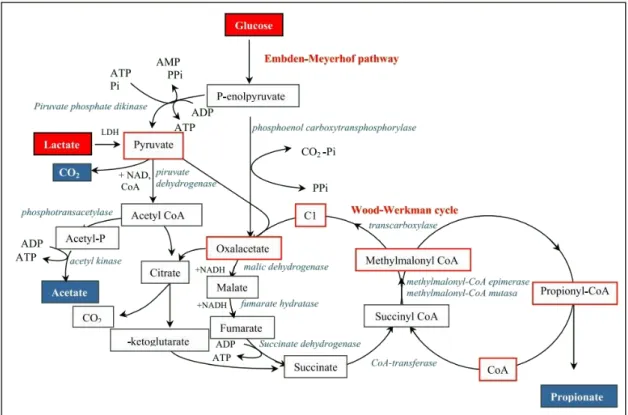
Propionibacteria has a peculiar metabolism characterized by the formation of acetate and propionate as the main fermentation end products, and also by several interconnected pathways that are used simultaneously (Poonam et al. 2012). This bacterium can metabolize a variety of substrates like carbohydrates, polyols (glycerol; erythritol; adonitol) and organic acids (lactic and gluconic acids) (Feng et al. 2011).
The production of acetate and propionate by these bacteria involves a complex metabolic cycle with several reactions in which substrates are metabolized to pyruvate via glycolysis, pentose phosphate or the Entner-Doudoroff pathways, generating ATP and reduced co-enzymes. Pyruvate is then oxidized to acetate and carbon dioxide or reduced to propionate (See Figure 1.8) (Zarate 2012). The latter transformation occurs via the Wood-Werkman cycle or transcarboxilase cycle which represents the key component of the central carbon metabolic pathway in propionibacteria (Crow 1987; Thierry et al. 2011). The Wood-Werkman cycle in propionibacteria is unique, function as a cyclic process, because the propionate production is coupled to oxidative phosphorylation and yields more ATP than in the other propionate-producing bacteria (Falentin et al. 2010).
Figure 1.8. Schematic representation of main fermentation end products in propionibacteria (Zarate 2012).
Depending on the strains, the substrate used, and the environmental conditions, propionibacteria modulate the proportions of pyruvate either reduced to propionate, or oxidized to acetate and carbon dioxide, to maintain the redox balance (Thierry et al. 2011). For example, the disruption of acetate kinase gene in P. acidipropionici led to a 14% decrease of acetate production and a 13% increase of propionate yield using glucose as substrate (Suwannakham et al. 2006).
1.6. Short chain fatty acids
SCFAs are saturated aliphatic organic acids that consist of 1-6 carbons of which acetate (C2), propionate (C3) and butyrate (C4) are the most abundant (95%) in the human gut (Cook and Sellin 1998). Acetate, propionate and butyrate are present in an approximate molar ration of 60:20:20 in the GI tract (Hijova and Chmelarova 2007; Binder 2010; Cummings et al. 1987). Depending on the diet, the total concentration of SCFAs decreases from 70-140 mM in the proximal colon to 20-70 Mm in the distal colon (Topping and Clifton 2001). In the cecum and colon 95% of the produced SCFAs are rapidly absorbed by colonocytes while the remaining 5% is secreted in feces (Topping and Clifton 2001; Ruppin et al. 1980; Hoverstad et al. 1982)
To the microbial community, SCFAs are a necessary waste product, required to balance redox equivalent production in the anaerobic environment of the gut. SCFAs also have many other activities including the regulation of host genes involved in maintenance of intestinal homeostasis (Comalada et al. 2006).
The amount and type of fiber consumed has dramatic effects on the composition of the gut microbiota and consequently on the type and amount of SCFAs produced (den Besten et al. 2013). The luminal pH in the colon is the result of the microbial SCFA production and the neutralizing capacity of bicarbonate, once most SCFAs are absorbed by the host in exchange for bicarbonate. As the concentration of SCFAs decline from proximal to the distal colon, the pH increases from cecum to rectum (Cummings et al. 1987; Annison et al. 2003; Ward and Coates 1987). This drop in pH from the ileum to the cecum is very important. Since the lower pH values change gut microbiota composition and it prevents overgrowth by pH-sensitive pathogenic bacteria (Duncan et al. 2009). Studies of human fecal microbial communities showed that at pH 5.5 the butyrate-producing Firmicutes-related bacteria comprised 20% of total population and when the luminal pH increases to 6.5 the butyrate-producing bacteria almost completely disappear and the acetate- and propionate-producing Bacteroides-related bacteria become dominant (Walker et al. 2005).
SCFAs produced by the microbiota in the human gut can be found in hepatic, portal and peripheral blood (Cummings et al. 1987; Murase et al. 1995). These SCFAs affect lipid, glucose and cholesterol metabolism in various tissues. These results indicate that SCFAs are transported from the intestinal lumen into the blood compartment of the host and are taken up by organs where they act as substrates or signal molecules (Gao et al. 2009; Fushimi et al. 2006; Demigne et al. 1995; Todesco et al. 1991)
It became apparent that SCFAs might play a key role in the prevention and treatment of the metabolic syndrome, bowel disorders and certain types of cancer (Donohoe et al. 2011; Blouin et al. 2011; Tang et al. 2011b). Some studies have demonstrated that an improvement of SCFA synthesis by colonic microbiota positively influenced the treatment of ulcerative colitis, Crohn᾽s disease and antibiotic-associated diarrhea (Binder 2010; Cummings et al. 1987; Di Sabatino et al. 2005)
1.6.1. Short chain fatty acids antitumor activity
In the recent years, some authors have been demonstrated the antitumor activity of SCFAs. Butyrate in particular is the preferred energy source for the cells in the colonic mucosa and has been
demonstrated to induce apoptosis in CRC cells and its production is safe and without consequences for the normal epithelium growth (Fung et al. 2011). The mechanisms by which butyrate and others SCFAs regulate cell proliferation/differentiation and apoptosis are still unclear. In this sense, it has been described that butyrate is able to block cell proliferation, mainly in the G0-G1 and G2-M phases of cell cycle (Darzynkiewicz et al. 1981; Heerdt et al. 1997). This effect could be mediated by inhibition of the histone deacetylase activity (Fung et al. 2011), an increased cyclin D and p21Waf1
expression (Hinnebusch et al. 2002; Hu et al. 2011) or a decreased expression of the proto-oncogenes c-src and c-myc (Foss et al. 1989; Souleimani and Asselin 1993). On the other hand, butyrate has also been described to reduce the levels of apoptosis inhibitors such as Bcl-2 and Bc-XL together with the upregulation of pro-apoptotic Bak and Bax, or the induction of caspase-3 protease activity in some tumor cell lines (Mandal et al. 2001). Butyrate and propionate can induce autophagy in cancer cells by inhibiting apoptosis, whereas inhibition of autophagy enhances the induction of apoptosis by SCFA (Donohoe et al. 2011; Tang et al. 2011a; Lee et al. 2012; Lee and Lee 2012).
Although the effect of acetate and propionate is less studied and their implications on the physiology of the colonocytes are not well documented, these are also known to regulate cell proliferation/differentiation and apoptosis (Lan et al. 2007b; Jan et al. 2002). Similarly to butyrate, propionate and acetate induce apoptosis through analogous events, including cell cycle arrest in the G2/M phase, mitochondrial depolarization, generation of reactive oxygen species, Bax translocation, caspase activation, chromatin condensation and nuclear degradation (Jan et al. 2002; Lan et al. 2008; Lan et al. 2007a; Lan et al. 2007b). Our group showed that in CRC-derived cell lines acetate per se inhibits proliferation and induces apoptosis, lysosomal membrane permeabilization (LMP) and cathepsin D (Cat-D) release from the lysosome (Marques et al. 2013). Our results led us to conclude that acetate induces LMP and subsequent release of Cat-D in CRC cells undergoing apoptosis, and suggest exploiting novel strategies using acetate as a prevention/therapeutic agent in CRC, through simultaneous treatment with Cat-D inhibitors.