Research Article
Combining Hyaluronic Acid with Chitosan
Enhances Gene Delivery
Ana V. Oliveira,
1,2Diogo B. Bitoque,
1,3and Gabriela A. Silva
1,41Centre for Molecular and Structural Biomedicine, Institute for Biotechnology and Bioengineering (CBME/IBB, LA),
University of Algarve, 8005-139 Faro, Portugal
2Doctoral Program in Biomedical Sciences, Department of Biomedical Sciences and Medicine, University of Algarve,
8005-139 Faro, Portugal
3ProRegeM Doctoral Program, PhD Program in Mechanisms of Disease and Regenerative Medicine, University of Algarve,
8005-139 Faro, Portugal
4Department of Biomedical Sciences and Medicine, University of Algarve, 8005-139 Faro, Portugal
Correspondence should be addressed to Gabriela A. Silva; gasilva@ualg.pt
Received 12 September 2014; Revised 28 October 2014; Accepted 29 October 2014; Published 18 November 2014 Academic Editor: Xuping Sun
Copyright © 2014 Ana V. Oliveira et al. This is an open access article distributed under the Creative Commons Attribution License, which permits unrestricted use, distribution, and reproduction in any medium, provided the original work is properly cited. The low gene transfer efficiency of chitosan-DNA polyplexes is a consequence of their high stability and consequent slow DNA release. The incorporation of an anionic polymer is believed to loosen chitosan interactions with DNA and thus promote higher transfection efficiencies. In this work, several formulations of chitosan-DNA polyplexes incorporating hyaluronic acid were prepared and characterized for their gene transfection efficiency on both HEK293 and retinal pigment epithelial cells. The different polyplex formulations showed morphology, size, and charge compatible with a role in gene delivery. The incorporation of hyaluronic acid rendered the formulations less stable, as was the goal, but it did not affect the loading and protection of the DNA. Compared with chitosan alone, the transfection efficiency had a 4-fold improvement, which was attributed to the presence of hyaluronic acid. Overall, our hybrid chitosan-hyaluronic acid polyplexes showed a significant improvement of the efficiency of chitosan-based nonviral vectors in vitro, suggesting that this strategy can further improve the transfection efficiency of nonviral vectors.
1. Introduction
Nonviral gene therapy is currently limited by the lack of vectors with gene transfer efficiency similar to viral vectors. Chitosan is one of the most studied cationic polymers for
nonviral gene therapy, both in vitro and in vivo [1–3].
Although based polyplexes (complexes of chitosan-nucleic acids) have desirable characteristics for gene therapy such as efficient nucleic acid encapsulation and protection against degradation, they show low gene transfer efficiency, which is the major obstacle to its use as a gene therapy vector
[1,4]. Several studies suggest a direct correlation between the
stability of polyplexes and transfection efficiency, proposing that the high stability and strong interactions between chi-tosan and DNA are the cause for the low transfection results
[2,5,6]. Polyplex stability is thus a crucial parameter when
designing a polymer based gene delivery vector; it should be stable enough to withstand the cellular internalization process, but not too stable that once inside the cell it will not
release its therapeutic load [7,8].
Approaches for improving the efficiency of chitosan-mediated gene transfer focus on chemical modification of chitosan and the incorporation of anionic biopolymers. We have previously modified chitosan to incorporate disulfide bonds that could be cleaved intracellularly, but the increase
in transfection efficiency was moderate [9]. The
incorpora-tion of anionic polymers, which destabilize polyplexes and
hence facilitate DNA release [4, 7, 8, 10, 11], is another
strategy that has shown promising results. Competition-binding assays showed that the addition of alginate effectively reduces the interaction strength between CS and DNA, which improved DNA release and consequently transfection
Volume 2014, Article ID 246347, 9 pages http://dx.doi.org/10.1155/2014/246347
[10]. Other studies have shown that coating polyplexes with hyaluronic acid (HA) enhanced internalization and trans-fection in association with other cationic polymers, such as
polyethyleneimine [12,13]. It has also been previously shown
that the incorporation of HA into the polyplex formulation increased green fluorescent protein (GFP) expression and the authors suggest this increase to be related to (1) improved internalization due to interactions with the cellular surface, (2) HA function as a transcription activator, and (3) HA
accumulation in the perinuclear region and cell nuclei [14].
Furthermore, several studies hypothesized that HA could be used to improve targeting through the specific HA/CD44 receptor interaction and could be of value to both gene and
drug delivery strategies [14,15].
In this work, we designed CS polyplexes incorporating HA (CSHA) with two different molecular weights (MW) in several CS to HA ratios and evaluated their potential in
vitro for gene delivery. While other studies have used CSHA
polyplexes targeting the anterior part of the eye [12,14,16],
this is, to our knowledge, the first report in the posterior part of the eye. We have tested these formulations in cells of the retinal pigment epithelium, a cell layer of the retina
that supports the overall health of the retina [17] and whose
function, when compromised, is implicated in several retinal pathologies such as age-related macular degeneration, among others.
2. Materials and Methods
2.1. Materials. Chitosan (CS) with a MW of 15 kDa and a
degree of deacetylation of 84% was purchased from Poly-sciences Inc., USA. Hyaluronic acid, with 132 or 214 kDa, from here on designated HA132 or HA214, respectively, was purchased from Lifecore Biomedical Inc., USA. All other reagents were of analytical grade and were used without further purification.
2.1.1. Plasmid and Cell Lines. A plasmid expressing enhanced
green fluorescent protein (GFP) driven by the cytomega-lovirus promoter (kindly provided by Dr. Jean Bennett, University of Pennsylvania, USA) was amplified in Top 10 bacteria and purified using a Plasmid Maxi kit (Qiagen, USA) according to the manufacturer’s instructions. Plasmid DNA (pDNA) was dissolved in TE buffer, and the concentration was determined at 260 nm using a NanoDrop 2000c spec-trophotometer (Thermo Scientific, USA).
Two cell lines were used in the cytotoxicity and transfec-tion studies: human embryonic kidney 293 (HEK293) cells, a cell line generally used to assess transfection efficiency, and human retinal pigment epithelial cells (ARPE-19), a cell line derived from normal human eyes. All cell culture reagents were purchased from Sigma-Aldrich (USA).
2.2. Methods
2.2.1. Preparation of CS and CSHA Polyplexes. CS polyplexes
were prepared as previously described by our lab [9]. Briefly,
a CS solution (0.02% (W/V) in 0.1 M acetic acid, pH 3) and
a 5 mM of sodium sulphate solution were separately heated
to 55∘C. Equal volumes of both solutions were quickly mixed
together, vortexed for 30 s, placed on ice, and stored at 4∘C.
CSHA polyplexes were prepared using a HA132 or HA214 solution (0.1% (W/V) in MilliQ water). The following CS : HA weight ratios: 3 : 1, 4 : 1, 5 : 1, 7 : 1, and 10 : 1, were used, keeping
constant the CS amount (50𝜇g) and varying the HA amount.
In order to use equal volumes of both solutions, HA was diluted in 5 mM sodium sulphate and then mixed with the CS solution, as described above.
To prepare CS and CSHA polyplexes loaded with pDNA
at a NH3+: PO4− ratio of 5 : 1, 50𝜇g of CS and 16.1 𝜇g
of pDNA were used. pDNA was mixed with the sodium sulfate solution and this solution was mixed with the CS solution, as described above, thus producing CSpDNA 5 : 1 or CSHApDNA 5 : 1 polyplexes. The 5 : 1 ratio was chosen based on previous results for CS-pDNA, which showed this ratio to
be the most appropriate for cell transfection [1,9].
2.2.2. Polyplex Characterization. Size analysis was performed
using dynamic light scattering (DLS) and noninvasive
back-scatter technology with a detection angle of 173∘, and zeta
potential (ZP) was measured using laser Doppler velocimetry and phase analysis light scattering technology (Zetasizer Nano ZS, Malvern instruments, UK). All polyplexes were
analyzed in ddH2O at 25∘C. The polydispersity index (PdI)
was calculated based on DLS measurements using the Zeta-sizer Nano Series software v 6.20.
Transmission electron microscopy (TEM, JEOL JEM-1011 electron microscope, Tokyo, Japan) was used to evaluate the morphology of the polyplexes. Prior to analysis, samples were stained with 2% (w/v) phosphotungstic acid and placed on copper grids with Formvar films.
2.2.3. Evaluation of pDNA Encapsulation Efficiency and Pro-tection from Degradation. The pDNA complexation,
reten-tion, and integrity in the polyplexes were assessed by gel electrophoresis. Free pDNA and polyplexes were separately incubated with 1 unit of DNAse I (Sigma-Aldrich, USA) for
15 min at 37∘C. The reaction was stopped by 1𝜇L of a 50 mM
EDTA solution and heated at 70∘C for 10 min. The integrity of
the pDNA was then analyzed by agarose gel electrophoresis in 1% (W/V) agarose in TAE buffer with ethidium bromide. Gels were subjected to a 70 mV voltage for 1.5 h and further visualized under UV light (AlphaImager, Alpha Innotech, USA).
2.2.4. Evaluation of Polyplex Stability. Polyplex stability was
evaluated in two different ways: stability at physiological
temperature (37∘C) and pH (7.4) and also long-term stability
(4∘C, pH 7.4). Briefly, polyplexes were incubated in equal
volumes of either PBS or DMEM (with 10% FBS [fetal bovine
serum]) at 37∘C for 1 to 3 days. Polyplex stability, evaluated by
pDNA retention, was performed as described in the previous section. For evaluation of long-term stability, polyplexes were
incubated at 4 or 37∘C and their size and polydispersity
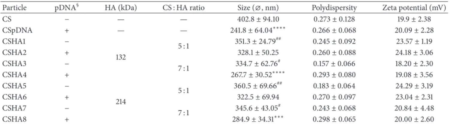
Table 1: Composition, size, polydispersity, and zeta potential of CS and CSHA polyplexes.
Particle pDNA§ HA (kDa) CS : HA ratio Size (⌀, nm) Polydispersity Zeta potential (mV)
CS − — — 402.8± 94.10 0.273± 0.128 19.9± 2.38 CSpDNA + — — 241.8± 64.04∗∗∗∗ 0.266± 0.068 20.09± 2.28 CSHA1 − 132 5 : 1 351.3± 24.79 ## 0.245± 0.092 23.57± 1.19 CSHA2 + 328.1± 50.25 0.260± 0.088 24.18± 3.06 CSHA3 − 7 : 1 334.7± 62.76 # 0.157± 0.066 18.20± 2.30 CSHA4 + 267.7± 30.52∗∗∗∗ 0.293± 0.080 19.08± 3.56 CSHA5 − 214 5 : 1 360.5± 69.66 ## 0.183± 0.064 24.29± 3.19 CSHA6 + 322.5± 69.94 0.270± 0.097 23.04± 2.31 CSHA7 − 7 : 1 345.6± 43.05 # 0.243± 0.068 20.84± 4.48 CSHA8 + 284.9± 34.31∗∗∗ 0.298± 0.065 20.00± 2.60
§Presence or absence of pDNA in the formulation is indicated by + or−, respectively. Values marked with asterisks are statistically different to CS value,
∗∗∗𝑃 < 0.001,∗∗∗∗𝑃 < 0.0001. Values marked with cardinals are statistically different to CSDNA value,#𝑃 < 0.05,##𝑃 < 0.01 (statistical differences
determined by Tukey’s multiple comparisons test).
2.2.5. Cell Culture. Cells were cultured at 37∘C, under a
5% CO2 atmosphere. Different culture media were used,
according to each cell’s specifications: HEK293 in Dulbecco’s Modified Eagle’s Medium (DMEM) and ARPE-19 in DMEM mixture with F-12 HAM, both supplemented with 10% of fetal bovine serum, 1% penicillin/streptomycin, and 1% Glu-tamine.
2.2.6. Cytotoxicity Evaluation. An MTT assay was performed
to evaluate the cytotoxicity of the polyplexes. Cells were
plated at a density of 1.5 × 10⁴ cells/well in 48-well flat
bottom tissue culture plates and the assay was carried out
as described previously [9]. Cells were incubated in culture
medium containing different amounts of polyplexes (from
0.667 up to 13.3𝜇g of CS per cm2of growth area) for 72 h. As
positive and negative controls of cell viability, cells cultured in standard cell culture conditions and cells incubated with
a latex extract in culture medium (1.5 cm2/mL) were used,
respectively. Absorbance was measured using a microplate reader (Tecan Infinite 2000, USA), at 570 and 630 nm, for cell viability/formazan formation and background, respec-tively. After subtracting the background (OD = OD570 nm – OD630 nm), cell viability was calculated as follows: cell
viabil-ity (%) = (ODsample)/(ODcontrol)× 100, where ODcontrol
and ODsample are cells not challenged and challenged by polyplexes, respectively. Each value was averaged from triplicates and each experiment was carried out thrice.
2.2.7. Transfection Studies. For the transfection studies, cells
were plated at 1× 10⁵cells/well in 6-well tissue culture plates.
FuGENE HD (Promega, USA) was used as positive trans-fection control according to the manufacturer’s instructions. CS and CSHA polyplexes were added to plated cells at a
ratio of 1𝜇g of pDNA per well and further incubated for
72 h. Nontransfected cells were used as negative transfection control.
Transfection efficiency was evaluated quantitatively by flow cytometry by scoring GFP positive cells (FACScalibur, BD Biosciences, USA) using FL-1H, green channel. A total of
1× 10⁵events were counted for each sample. The percentage
of positive events corresponds to the gated events minus the nontransfected cells.
2.2.8. Statistical Analysis. Results presented are mean ±
standard deviation of at least three independent experiments. Statistical analysis was performed with GraphPad Prism 5 software. Data were subjected to analysis of variance (one-and two-way ANOVA) (one-and multiple comparisons tests using a confidence interval of 95%.
3. Results
3.1. Size, Polydispersity, Surface Charge, and Morphology. The
initial step in polyplex characterization was the determi-nation of polyplexes size, polydispersity (PdI), and surface charge (by zeta potential, ZP), and the results are presented
in Table1. For some of the tested CS : HA ratios (3 : 1, 4 : 1, and
10 : 1) we observed aggregation and consequently large sizes and high PdI. These ratios were not tested further (data not shown).
Polyplexes formulated without pDNA had sizes between
334.7± 62.76 nm and 402.8 ± 94.10 nm while those
formu-lated with pDNA revealed sizes between 241.8 ± 64.04 nm
and 328.1± 50.25 nm. The observed decrease in size is
sta-tistically different between CS polyplexes and pDNA loaded polyplexes CSDNA, CSHA4, and CSHA8; differences were also found between CSDNA polyplexes and pDNA unloaded CSHA formulations.
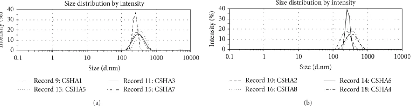
All mean PdI values are under 0.300, reflecting homo-geneous preparations. The only statistically significant dif-ferences found were between formulations CSHA3 and CSHA4 and CSHA3 and CSHA8 (𝑃 ≤ 0.05). These might reflect minor differences in the homogeneity of the polyplex suspensions mainly due to the presence of an additional anionic compound in the mixture. Polyplexes were produced as a single homogeneous preparation as illustrated by the
DSL size distribution graphs (Figure1), where no secondary
peaks were observed. Regarding the surface charge of the polyplexes, all displayed a positive charge with ZP values
0 10 20 30 40 0.1 1 10 100 1000 10000 In te n si ty (%)
Size distribution by intensity
Record9: CSHA1 Record11: CSHA3
Record13: CSHA5 Record15: CSHA7
Size (d.nm) (a) In te n si ty (%)
Size distribution by intensity
0 10 20 30 40 0.1 1 10 100 1000 10000
Record10: CSHA2 Record14: CSHA6
Record16: CSHA8 Record18: CSHA4
Size (d.nm)
(b)
Figure 1: Dynamic light scattering raw data representative graphs for CSHA polyplexes: (a) without pDNA and (b) with pDNA.
CSHA1 CSHA3 CSHA5 CSHA7
CSHA4
CSHA2 CSHA6 CSHA8
Figure 2: TEM microphotographs of CSHA polyplexes showing that different formulations have a regular, close to spherical morphology (amplification: 100,000x, scale bar represents 2𝜇m).
found to be statistically different were only for CSHA2 versus CSHA3 and CSHA3 versus CSHA5 (𝑃 ≤ 0.05).
The morphology of the polyplexes was also analyzed by TEM and the microphotographs revealed near spherical polyplexes with sizes consistent to the ones determined by
DSL (Figure2).
3.2. pDNA Complexation and Protection from DNase Degra-dation. The second step in the characterization of the
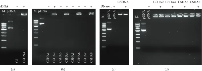
poly-plexes was to evaluate if the presence of HA in the different formulations affected pDNA complexation and protection against nuclease degradation. All formulations complexed pDNA successfully, as observed by the absence of pDNA
migration in an agarose gel (Figures3(a)and3(b)). Polyplexes
without pDNA (CS and CSHA) were used as controls and, as expected, did not produce any detectable signal. These results were further confirmed by performing the same experiment
in a 0.3% agarose gel (Figure4).
Regarding pDNA protection against nuclease degra-dation, all formulations showed a similar behavior, with
no detectable pDNA degradation (Figures 3(c) and 3(d)).
Uncomplexed pDNA was used as a control and, as expected, was completely degraded by DNase I.
3.3. Polyplex Stability. Stability at physiological conditions is
an important factor since polyplexes should be stable enough to protect pDNA and only release it once it has reached the intracellular milieu. We therefore tested the stability of the polyplexes at physiological pH and in the presence of serum, to mimic in vivo conditions, by incubating the polyplexes with PBS (pH 7.4) or DMEM with 10% FBS, respectively. No detectable pDNA release was observed regardless of the
formulation and period of incubation (Figure5).
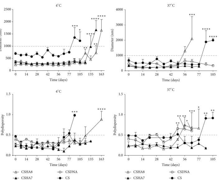
The shelf life of the produced polyplexes is also an important factor to consider. Long-term stability of the
polyplexes was evaluated at 4 and 37∘C by analysis of their
size and PdI (Figure6). Results depicted in Figure 6 were
evaluated by variance analysis (1-way ANOVA) and Dunnett’s post hoc test to compare values to the starting time point (𝑡 = 0). All CSHA formulations performed very similarly and therefore the results shown for formulations CSHA7 and 8 are representative of the other CSHA formulations.
The results showed that both size and PdI directly increased with temperature and that all tested formulations
remained stable at 4∘C for an extended period of time (up to
13 weeks). The size of CSDNA polyplexes remained constant throughout time. The presence of pDNA in the formulation seemed to increase stability in CS formulations. Although
CS CS D N A M pDNA pDNA − + (a) CS H A 1 CS H A 2 CS H A 3 CS H A 4 CS H A 5 CS H A 6 CS H A 7 CS H A 8 M pDNA + + − − − + − + (b) M pDN A pD N A DNase I CSDNA + + (c)
CSHA2 CSHA4 CSHA6 CSHA8
M pDN A pD N A + + + + + (d)
Figure 3: Polyplexes encapsulation efficiency and nuclease protection analyzed by 1% agarose gel electrophoresis, DNA visualized with ethidium bromide, (a) and (b) pDNA encapsulation in CSpDNA and CSHApDNA polyplexes, respectively, and lanes positive for polyplexes but negative for pDNA represent unloaded polyplexes. DNA is protected against DNase I digestion: (c) CSpDNA and (d) CSHApDNA polyplexes after incubation with DNase I.
CS CS D N A CS H A 1 CS H A 2 CS H A 3 CS H A 4 CS H A 5 CS H A 6 CS H A 7 CS H A 8 M pDNA pDNA − + − + − + − + − +
Figure 4: Polyplexes encapsulation efficiency analyzed by 0.3% agarose gel electrophoresis, DNA visualized with ethidium bromide, and lanes positive for polyplexes but negative for pDNA represent unloaded polyplexes.
there were no statistical differences in the size of CSDNA
polyplexes, their PdI at 37∘C started to increase, reflecting
their decreasing stability at this temperature, after 49 days
(Figure6). Also, measurements of some formulations were
discontinued due to visible aggregation and/or insufficient volume, hence the difference in the depicted time points.
As aimed, formulations containing HA and pDNA were less stable probably due to increased repulsion between charged groups. The stability of CSHA8 formulation
decreased after 119 days at 4∘C and much earlier at 37∘C (63
days), as can be observed by the increase in size and PdI
(Figure6).
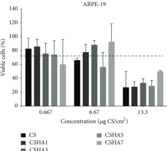
3.4. In Vitro Studies. Cytotoxicity of the developed polyplexes
was evaluated using an MTT assay in the retinal cell line
(ARPE-19, Figure7). For HEK293 cells, our previous studies
have indicated absence of cytotoxicity [9]. Statistical analysis
revealed no differences between CS and CSHA formulations. Cell viability was above 75% regardless of formulation and concentration, except for the highest concentration tested, which caused a decrease in cellular viability, with values below 50% for all formulations. However, this concentration is above the range to be used in vivo.
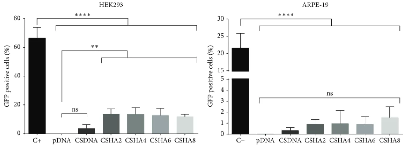
One of the key points of our study was to evaluate if the introduction of HA would affect the stability of the polyplexes to facilitate DNA release and hence increase their transfection efficiency. CS and CSHA formulations with pDNA coding for GFP were tested both in the retinal cell line ARPE-19 and in HEK293 cells. The latter were used as a transfection control, as their permissibility in terms of transfection is widely accepted. As a control of the GFP expression system we used FuGene HD, a commercial reagent. The percentage of GFP
positive cells 72 h after transfection is presented in Figure8.
Results showed that the transfection efficiency varied with cell line and type of formulation. HEK293 cells displayed higher levels of GFP expression when compared with the RPE cell line, as expected, based on their different mitotic rates. All formulations incorporating HA present increased GFP expression when compared to CS polyplexes, with CSHA polyplexes showing a near 4-fold increase in transfection efficiency in HEK293 cells.
4. Discussion
4.1. Polyplexes. This work aims to evaluate the effect that
incorporating HA into CS polyplexes would have in the stability of the polyplexes and in their transfection efficiency. In summary, the characterization of the polyplexes revealed differences in size for pDNA loaded and unloaded polyplexes, but not between CS and CSHA polyplexes. This size difference
Incubation PBS DMEM M DNase P Free DNA + + + + + + P+ 0 d 1 d 2 d 3 d 1 d 2 d 3 d (a) M Free DNA + + + + + + 1 d 2 d 3 d 1 d 2 d 3 d (b)
Figure 5: Polyplexes remain stable at physiological conditions, (a) CSpDNA and (b) CSHApDNA polyplexes after incubation in PBS or DMEM with 10% FBS at 37∘C. Polyplex pDNA retention analyzed by 1% agarose gel electrophoresis and pDNA visualized with ethidium bromide (P-polyplexes, P+DNase-polyplexes incubated with DNase I, data shown only for CSHA8 but representative of all CSHA polyplexes).
Time (days) Dia m et er (nm) 0 500 1000 1500 2000 2500 Time (days) Dia m et er (nm) 0 1000 2000 3000 4000 Po ly d is p er si ty 0.0 0.5 1.0 1.5 Po ly d is p er si ty 0.0 0.5 1.0 1.5 CS CSDNA CSHA7 CSHA8 CS CSDNA CSHA7 CSHA8 * 0 14 28 42 56 77 105 135 163 Time (days) 0 14 28 42 56 77 105 135 163 4∘C 4∘C ∗∗∗ ∗∗∗∗ ∗∗∗ ∗∗ ∗∗ ∗∗ ∗∗ ∗∗ ∗ ∗ ∗∗∗ ∗∗∗∗ ∗∗∗∗∗∗∗∗ ∗∗∗∗ ∗∗∗∗ ∗∗∗ ∗∗ ∗∗ ∗ 0 14 28 42 56 77 105 Time (days) 0 14 28 42 56 77 105 37∘C 37∘C
Figure 6: Particle stability at 4∘C (left column) and 37∘C (right column) as verified by continued monitoring of their size (upper row) and polydispersity (lower row). Significant differences to initial value were determined by Dunnett’s multiple comparisons test (∗∗∗∗𝑃 < 0.0001, ∗∗∗𝑃 < 0.001,∗∗𝑃 < 0.01,∗𝑃 < 0.05).
ARPE-19 V ia b le cells (%) 0.667 6.67 13.3 0 20 40 60 80 100 120 140 CS CSHA1 CSHA3 CSHA5 CSHA7 Concentration (𝜇g CS/cm2)
Figure 7: Cytotoxicity in RPE cell line (ARPE-19), given as percent-age of viable cells after 72 h incubation with polyplexes at increasing concentrations. Polyplex concentration is given in total CS𝜇g in polyplexes per cm2of growth area.
between polyplexes with and without pDNA has been previ-ously observed and is in accordance with the literature, since CS establishes strong interactions with negatively charged pDNA that contribute to a higher chain entanglement, thus
producing smaller polyplexes [9].
Variations in size and surface charge were expected after the incorporation of HA into the formulation, since it has been shown previously that an increase in size and decrease
in surface charge associated with HA content [12,14]. In our
study no effect of HA was observed in either size or surface charge. When addressing the effect of the MW of HA on the size of polyplexes, studies have shown the formation of smaller polyplexes with HA of increasing MW and also with
the use of higher CS : HA ratios [11]. In this aspect, our results
are well correlated with the literature, since using HA with a high MW produced polyplexes with smaller size and positive surface charge, whereas the use of low ratios increased sizes and decreased surface charge, causing aggregation (data not
shown) as shown by Duceppe and Tabrizian (2009) [11].
It has also been suggested that the MW of the anionic
polymer can influence the morphology of the polyplexes [11].
This was not observed in our study, since no morphological differences were noticeable either among CSHA formulations or between CS and CSHA polyplexes. We attribute this dissimilarity of our results with the literature to differences in the characteristics of the polymers and polyplexes such as MW and CS : HA ratios used in our study. Also, this difference in polyplex behavior might be related to differences in the localization and degree of incorporation of HA in the
polyplexes [14]. During the chain entanglement process HA
chains may have been trapped in the interior of the polyplex, thus not contributing to a significant decrease in surface charge.
4.2. Stability. Previous studies have shown that HA can
influence the stability of polyplexes without affecting pDNA
binding to CS [8]. Our results support these findings and
also show that polyplexes remain stable at physiological conditions and are able to protect pDNA from nuclease degradation. Only in the long-term stability assay a decrease in polyplex stability was observed in CSHA polyplexes, more
pronounced at 37∘C.
4.3. Cytotoxicity. Polyplex charge is also an important
param-eter due to its relation to toxicity and transfection efficiency, as some authors have shown that a reduction in the positive charge results in an increase in transfection efficiency
asso-ciated with a decrease in toxicity [18,19]. A decrease in CS
polyplex cytotoxicity with increasing amounts of HA in their formulations has been reported by De La Fuente et al. (2008), but our results show no such differences when either different
CS : HA ratios or MW were used [14]. Since no decrease in
surface charge was observed, we expected the incorporation of HA into the polyplexes to have no significant effect in the cytotoxicity. Nevertheless, considering the concentrations to be used in vivo our polyplexes can be considered a safe option for gene delivery.
4.4. Transfection. It is believed that one of the major causes
of low transfection with polyplexes is their high stability and
consequently inefficient and slow DNA delivery [8,11].
Con-sidering that all formulations displayed similar size, surface charge, and cytotoxicity, this suggests that the introduction of HA affected the interactions between CS and pDNA and modulated the release behavior, thereby resulting in a more efficient transfection. The observed transfection efficiency improvement over CS formulations may also be related to increased cellular internalization since it has been shown previously by de la Fuente that HA containing polyplexes can be internalized via interactions with the membrane CD44
receptor [14]. Recent studies with the same cell lines but using
a lipid-based delivery system also containing HA corroborate our results and also support the idea that HA enhances the transfection efficiency by modulating the DNA condensation degree in the vector. The same study also states that the participation of the CD44 receptor in the internalization of the vectors is an important factor for increased transfection
[20].
Similar to the results observed for HEK293 cells, a trend of higher GFP expression levels was observed when ARPE-19 were transfected with CSHA polyplexes, but it is not statistically significant. One possible explanation for this is their dividing rate, which is considerably lower than the one of HEK293 cells. The fact that cell lines with different mitotic rates were transfected demonstrates the versatility of our vectors and paves the way to possible future applications in other tissues. Additionally, it is known that the expression of CD44, the receptor for HA, in RPE is affected both by their proliferative state and by confluence, which might explain
these results [21]. It has also been described that CD44 is
increased in pathological conditions such as RPE wound
HEK293 GFP p o si ti ve cells (%) GFP p o si ti ve cells (%) 0 20 40 60 80 ns ARPE-19 0 1 2 3 4 5 15 20 25 30 ns
C+ pDNA CSDNA CSHA2 CSHA4 CSHA6 CSHA8 C+ pDNA CSDNA CSHA2 CSHA4 CSHA6 CSHA8
∗∗∗∗ ∗∗∗∗
∗∗
Figure 8: Transfection efficiency 72 h after transfection as percentage of GFP positive cells. Statistical differences were calculated using Tukey and Dunnett’s multiple comparisons test (∗∗∗∗𝑃 < 0.0001,∗∗𝑃 < 0.01, ns—not significant).
suggest that polyplex CD44-dependent internalization in
vivo and in a pathological situation could be greatly increased
when compared with in vitro results. Based on this, our results lay the foundation for the use of CSHA polyplexes for gene delivery in animal models of retinal diseases.
5. Conclusions
In our study we describe the characterization of a hybrid polyplex for retinal nonviral gene therapy. The polyplexes containing both cationic (CS) and anionic (HA) polymers display an improved performance over the previously devel-oped chitosan-based polyplexes. The incorporation of HA into the polyplexes resulted in a decrease in polyplex stability that most likely enabled a more rapid pDNA release whilst still protecting pDNA from degradation.
CSHA formulations showed an increase in in vitro cell transfection, which suggests that this strategy might be very effective in vivo, especially in retinal cells expressing CD44 and in pathological situations where CD44 is known to be involved, allowing cellular targeting by HA-containing formulations through CD44 interaction.
Conflict of Interests
The authors declare that there is no conflict of interests regarding the publication of this paper.
Acknowledgments
The authors acknowledge the financial support of Fundac¸˜ao para a Ciˆencia e Tecnologia (PTDC/SAU-BEB/098475/2008 to Gabriela A. Silva, SFRH/BD/70318/2010 individual fel-lowship to Ana V. Oliveira, and SFRH/BD/52424/2013 doc-toral fellowship to Diogo B. Bitoque under the ProRegeM Ph.D. program) and IBB/LA under the project PEst-OE/EQB/LA0023/2013 and the Marie Curie Reintegration Grant (PIRG-GA-2009-249314 to Gabriela A. Silva) under the FP7 program.
References
[1] H.-Q. Mao, K. Roy, V. L. Troung-Le et al., “Chitosan-DNA nanoparticles as gene carriers: synthesis, characterization and transfection efficiency,” Journal of Controlled Release, vol. 70, no. 3, pp. 399–421, 2001.
[2] X. Zhao, S.-B. Yu, F.-L. Wu, Z.-B. Mao, and C.-L. Yu, “Transfec-tion of primary chondrocytes using chitosan-pEGFP nanopar-ticles,” Journal of Controlled Release, vol. 112, no. 2, pp. 223–228, 2006.
[3] M. K¨oping-H¨ogg˚ard, I. Tubulekas, H. Guan et al., “Chitosan as a nonviral gene delivery system. Structure-property relationships and characteristics compared with polyethylenimine in vitro and after lung administration in vivo,” Gene Therapy, vol. 8, no. 14, pp. 1108–1121, 2001.
[4] S.-F. Peng, M.-J. Yang, C.-J. Su et al., “Effects of incorporation of poly(𝛾-glutamic acid) in chitosan/DNA complex nanoparticles on cellular uptake and transfection efficiency,” Biomaterials, vol. 30, no. 9, pp. 1797–1808, 2009.
[5] F. C. MacLaughlin, R. J. Mumper, J. Wang et al., “Chitosan and depolymerized chitosan oligomers as condensing carriers for in vivo plasmid delivery,” Journal of Controlled Release, vol. 56, no. 1–3, pp. 259–272, 1998.
[6] T. Kiang, J. Wen, H. W. Lim, and K. W. Leong, “The effect of the degree of chitosan deacetylation on the efficiency of gene transfection,” Biomaterials, vol. 25, no. 22, pp. 5293–5301, 2004. [7] S. Danielsen, G. Maurstad, and B. T. Stokke, “DNA-polycation complexation polyplex stability in the presence of competing polyanions,” Biopolymers, vol. 77, no. 2, pp. 86–97, 2005. [8] S. Danielsen, S. Strand, C. de Lange Davies, and B. T. Stokke,
“Glycosaminoglycan destabilization of DNA-chitosan poly-plexes for gene delivery depends on chitosan chain length and GAG properties,” Biochimica et Biophysica Acta: General
Subjects, vol. 1721, no. 1–3, pp. 44–54, 2005.
[9] A. V. Oliveira, A. P. Silva, D. B. Bitoque, G. A. Silva, and A. M. Rosa da Costa, “Transfection efficiency of chitosan and thiolated chitosan in retinal pigment epithelium cells: a comparative study,” Journal of Pharmacy and Bioallied Sciences, vol. 5, no. 2, pp. 111–118, 2013.
[10] K. L. Douglas, C. A. Piccirillo, and M. Tabrizian, “Effects of alginate inclusion on the vector properties of chitosan-based
nanoparticles,” Journal of Controlled Release, vol. 115, no. 3, pp. 354–361, 2006.
[11] N. Duceppe and M. Tabrizian, “Factors influencing the transfection efficiency of ultra low molecular weight chi-tosan/hyaluronic acid nanoparticles,” Biomaterials, vol. 30, no. 13, pp. 2625–2631, 2009.
[12] M. de La Fuente, B. Seijo, and M. J. Alonso, “Design of novel polysaccharidic nanostructures for gene delivery,”
Nanotechnol-ogy, vol. 19, no. 7, Article ID 075105, 2008.
[13] T. Ito, N. Iida-Tanaka, T. Niidome et al., “Hyaluronic acid and its derivative as a multi-functional gene expression enhancer: protection from non-specific interactions, adhesion to targeted cells, and transcriptional activation,” Journal of Controlled
Release, vol. 112, no. 3, pp. 382–388, 2006.
[14] M. De La Fuente, B. Seijo, and M. J. Alonso, “Novel hyaluronic acid-chitosan nanoparticles for ocular gene therapy,”
Investiga-tive Ophthalmology and Visual Science, vol. 49, no. 5, pp. 2016–
2024, 2008.
[15] S. Jaracz, J. Chen, L. V. Kuznetsova, and I. Ojima, “Recent advances in tumor-targeting anticancer drug conjugates,”
Bioor-ganic and Medicinal Chemistry, vol. 13, no. 17, pp. 5043–5054,
2005.
[16] M. de la Fuente, B. Seijo, and M. J. Alonso, “Bioadhesive hyaluronan-chitosan nanoparticles can transport genes across the ocular mucosa and transfect ocular tissue,” Gene Therapy, vol. 15, no. 9, pp. 668–676, 2008.
[17] O. Strauss, “The retinal pigment epithelium in visual function,”
Physiological Reviews, vol. 85, no. 3, pp. 845–881, 2005.
[18] P. Erbacher, S. Zou, T. Bettinger, A. M. Steffan, and J. S. Remy, “Chitosan-based vector/DNA complexes for gene delivery: biophysical characteristics and transfection ability,”
Pharmaceu-tical Research, vol. 15, no. 9, pp. 1332–1339, 1998.
[19] D. Putnam, C. A. Gentry, D. W. Pack, and R. Langer, “Polymer-based gene delivery with low cytotoxicity by a unique balance of side-chain termini,” Proceedings of the National Academy of
Sciences of the United States of America, vol. 98, no. 3, pp. 1200–
1205, 2001.
[20] P. S. Apaolaza, D. Delgado, A. D. Pozo-Rodr´ıguez, A. R. Gasc´on, and M. ´A. Solin´ıs, “A novel gene therapy vector based on hyaluronic acid and solid lipid nanoparticles for ocular diseases,” International Journal of Pharmaceutics, vol. 465, no. 1-2, pp. 413–426, 2014.
[21] N.-P. Liu, W. L. Roberts, L. P. Hale et al., “Expression of CD44 and variant isoforms in cultured human retinal pigment epithelial cells,” Investigative Ophthalmology & Visual Science, vol. 38, no. 10, pp. 2027–2037, 1997.
[22] S. Singh, J. Zheng, S. C. Peiper, and B. J. Mclaughlin, “Gene expression profile of ARPE-19 during repair of the monolayer,”
Graefe’s Archive for Clinical and Experimental Ophthalmology,
vol. 239, no. 12, pp. 946–951, 2001.
[23] H. Mochimaru, E. Takahashi, N. Tsukamoto et al., “Involvement of hyaluronan and its receptor CD44 with choroidal neovascu-larization,” Investigative Ophthalmology and Visual Science, vol. 50, no. 9, pp. 4410–4415, 2009.
Submit your manuscripts at
http://www.hindawi.com
Scientifica
Hindawi Publishing Corporation
http://www.hindawi.com Volume 2014
Hindawi Publishing Corporation
http://www.hindawi.com Volume 2014 Hindawi Publishing Corporation
http://www.hindawi.com Volume 2014
Hindawi Publishing Corporation
http://www.hindawi.com Volume 2014
Ceramics
Journal ofHindawi Publishing Corporation
http://www.hindawi.com Volume 2014
Nanoparticles
Journal of Hindawi Publishing Corporationhttp://www.hindawi.com Volume 2014
Hindawi Publishing Corporation
http://www.hindawi.com Volume 2014
International Journal of
Biomaterials
Hindawi Publishing Corporation
http://www.hindawi.com Volume 2014
Nanoscience
Journal ofTextiles
Hindawi Publishing Corporation
http://www.hindawi.com Volume 2014
Journal of
Hindawi Publishing Corporation
http://www.hindawi.com Volume 2014
Crystallography
Journal of Hindawi Publishing Corporationhttp://www.hindawi.com Volume 2014
The Scientific
World Journal
Hindawi Publishing Corporation
http://www.hindawi.com Volume 2014
Hindawi Publishing Corporation
http://www.hindawi.com Volume 2014
Coatings
Journal ofAdvances in
Materials Science and Engineering
Hindawi Publishing Corporation
http://www.hindawi.com Volume 2014
Hindawi Publishing Corporation
http://www.hindawi.com Volume 2014
Hindawi Publishing Corporation
http://www.hindawi.com Volume 2014
Metallurgy
Journal ofHindawi Publishing Corporation
http://www.hindawi.com Volume 2014
BioMed
Research International
Materials
Journal of Hindawi Publishing Corporationhttp://www.hindawi.com Volume 2014
N
a
no
ma
te
ria
ls
Hindawi Publishing Corporation
http://www.hindawi.com Volume 2014
Journal of