Contents lists available atScienceDirect
Stem Cell Research
journal homepage:www.elsevier.com/locate/scr
Lab Resource: Stem Cell Line
Generation and characterization of a human iPS cell line from a
patient-related control to study disease mechanisms associated with DAND5
p.R152H alteration
Selin Pars
a,1, Fernando Cristo
a,1, José M. Inácio
a,1, Graça Rosas
a, Isabel Marques Carreira
b,c,d,
Joana Barbosa Melo
b,c,d, Patrícia Mendes
e, Duarte Saraiva Martins
f, Luís Pereira de Almeida
g,
José Maio
e, Rui Anjos
f, José A. Belo
a,⁎aStem Cells and Development Laboratory, CEDOC, NOVA Medical School/Faculdade de Ciências Médicas, Universidade Nova de Lisboa, Lisbon, Portugal bCytogenetics and Genomics Laboratory, Faculty of Medicine, University of Coimbra, Coimbra, Portugal
cCNC.IBILI Consortium, University of Coimbra, Coimbra, Portugal
dCIMAGO - Center of Investigation on Environment Genetics and Oncobiology, Faculty of Medicine, University of Coimbra, Portugal eDepartamento Materno-Infantil, Centro Hospital do Algarve, EPE, Faro, Portugal
fHospital de Santa Cruz, Centro Hospitalar Lisboa Ocidental, Lisbon, Portugal
gCNC - Center for Neurosciences & Cell Biology, University of Coimbra, Coimbra, Portugal
A B S T R A C T
A DAND5-control human iPSC line was generated from the urinary cells of a phenotypically normal donor. Exfoliated renal epithelial (RE) cells were collected and reprogrammed into iPSCs using Sendai virus repro-gramming system. The pluripotency, in vitro differentiation potential, karyotype stability, and the transgene-free status of generated iPSC line were analyzed and confirmed. This cell line can be exploited as a control iPSC line to better understand the mechanisms involved in DAND5-associated cardiac disease.
Resource table.
Unique stem cell line identifier
NMSUNLi002 Alternative name(s) of
stem cell line
iUC-DAND5_455/control Institution CEDOC, NOVA Medical School Contact information of
distributor
José A. Belo,[email protected] Type of cell line iPSC
Origin Human
Additional origin info Sex: male
Ethnicity: Caucasian
Cell Source Exfoliated renal epithelial cells isolated from urine
Clonality Clonal
Method of reprogramming
Transgene-free (Sendai virus vector) Genetic Modification NO
Type of Modification N/A Associated disease N/A
Gene/locus NM_152654.2:c.455G; DAND5 c.G455G; p.R152R Method of modification N/A Name of transgene or resistance N/A Inducible/constitutive system N/A Date archived/stock date January 2018 Cell line repository/
bank
N/A
Ethical approval Approved by the Ethics Committee of NOVA Medical School (Protocol N° 13/2016/ CEFCM) and by the National Committee for Data Protection (CNPD, Permit N° 8694/ 2016).
https://doi.org/10.1016/j.scr.2018.04.015
Received 6 April 2018; Received in revised form 19 April 2018; Accepted 26 April 2018 ⁎Corresponding author.
1Equal authors.
E-mail address:[email protected](J.A. Belo).
Available online 28 April 2018
1873-5061/ © 2018 Published by Elsevier B.V. This is an open access article under the CC BY-NC-ND license (http://creativecommons.org/licenses/BY-NC-ND/4.0/).
Resource utility
This DAND5-control iPSC line is essential in studying the disease related impairment in heart formation of DAND5 found in a previous study. This control cell line will allow to uncover the role of DAND5 c.455 G > A variant in the molecular mechanisms of cardiomyocyte proliferation along with the previously established line.
Resource details
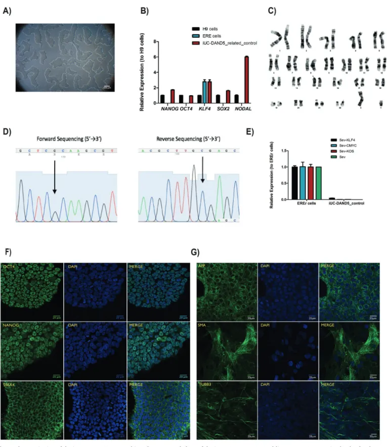
DAND5 is a Nodal antagonist that is involved in the correct left/ right body axis establishment during gastrulation (Belo et al., 2017). The heart is thefirst organ to be formed and the first organ to be af-fected by improper levels of Nodal signaling throughout embryo de-velopment. Several variants of different genes involved in this signaling pathway have been associated with Congenital Heart Disease (Deng et al., 2015). Recently, we have identified and functionally character-ized a c.455G > A DAND5-variant in a patient diagnosed with ven-tricular septal defect with overriding aorta, right venven-tricular hyper-trophy, and pulmonary atresia (a case of extreme tetralogy of Fallot phenotype) (Cristo et al., 2017a). The variant was inherited from the apparently healthy mother. To further study the impact of altered DAND5 proteins in molecular pathways that are involved in cardiac development and on cardiomyocyte behavior, we have generated and characterized the NMSUNLi001-A variant cell line, which has a het-erozygous non-synonymous variant in exon 2 of DAND5 gene (c.455G > A), causing an amino acid change of p.R152H in the func-tional domain of the DAND5 protein (Cristo et al., 2017b). However, to unveil the precise mechanism of how this variant affects early heart development, the generation of control iPSC lines is essential. Here, we generated and characterized a DAND5 patient-related (father) control iPSC line that does not carry any alteration in the locus of the variant described in the NMSUNLi001-A cell line. This cell line will be utilized for disease modeling purpose. Hence, exfoliated renal epithelial (RE) cells were collected from urine sample collected from a healthy donor. After being grown during 7 days in culture, the RE cells were repro-grammed using CytoTune™-iPS 2.0 Reprogramming Kit (Life Technol-ogies, Invitrogen). The kit utilizes non-transmissible, non-integrating form of Sendai Virus (SeV) vectors that deliver four key transcription factors (SOX2, OCT3/4, c-MYC and KFL-4) to reprogram the somatic cells into a pluripotent state, and the iPSC colonies appeared 17 days after the delivery of the reprogramming factors. At this time, we ob-served that cells assumed typical stem cell morphology. To obtain homogeneous and clonal iPSCs lines, we manually picked and expanded several single cell-derived iPSCs colonies. Among those, one sub-clone that best displayed the ESC-like morphology (Fig. 1A) was chosen for further characterization. Firstly, after 27 passages in culture, DNA Sanger sequencing and karyotype analysis proved the genotype 455G in DAND5 exon 2 (Fig. 1D), and the number (46, XY) and arrangement of chromosomes (Fig. 1C). We assessed the transgene-free status of the iPSC line by qPCR (Fig. 1E), confirming the clearance of the viral vectors. Since the cytoplasmic nature of SeV only allows the exogenous reprogramming vectors to be cleared after several passages, we used an early passage of iPSCs as a positive control. The pluripotency of the cells was analyzed by bothfluorescence immunocytochemistry (Fig. 1F) and qPCR (Fig. 1B). We confirmed the expression of the key plur-ipotency factors OCT4, NANOG, and SSEA4 both at protein and mRNA level. At mRNA level, we additionally confirmed the expression of pluripotency markers NODAL and SOX2. Embryoid body (EB) forma-tion assay was performed to assess the spontaneous differentiaforma-tion potential of the iPSCs in vitro. From this assay, we assessed that the EBs cultured for 19 days expressed markers of the three germ layers: en-doderm, mesoderm, ectoderm, i.e., alpha-fetoprotein (AFP), smooth muscle actin (SMA), tubulin beta-3 chain (TUBB3), respectively
Materials and methods Reprogramming of RE cells
RE cells were collected and expanded in culture. After ~4 days in culture, cells started to become evident and the medium was changed to REBM™ supplemented with REGM™ BulletKit (Lonza). When cells reached ~80% confluency, they were seeded on a 6-well plate and reprogrammed using CytoTune™-iPS 2.0 Sendai Reprogramming Kit (Life Technologies). At day 8 post-transduction, cells were passaged onto a 100 mm culture dish coated with Geltrex (Gibco, Thermo Fisher Scientific) and the next day medium was changed to Essential 8™ (E8) Flex medium, replaced until the iPSCs have emerged. 17 days post-transduction, colonies that best display an ESC-like morphology were picked and expanded with daily renewal of the E8 Flex medium. Sequencing
To confirm the absence of the c.455G nucleotide in the established DAND5-control cell line, genomic DNA was extracted using ISOLATE II Genomic DNA kit (Bioline). Then, using the primers indicated in Table 2, exon 2 of DAND5 was amplified by PCR and purified using NZYGelpure kit (NZYTech). Sequencing was conducted by STAB VIDA (http://www.stabvida.com/).
RNA extraction and real time qRT-PCR
The clearance of SeV transgenes and the expression of pluripotency markers OCT4, NANOG, SSEA4 (primers listed inTable 2) were carried out using Direct-zol™ RNA MiniPrep (Zymo Research). Subsequently, reverse transcription and qRT-PCR were performed.
Embryoid body formation assay
Embryoid bodies (EBs) consisting of approximately 2000 iPS cells/ 20μl drop in Essential 8™ (E8) medium with 4 mg/ml polyvinylalcohol and RevitaCell™ Supplement (Thermo Fisher Scientific) were generated using hanging drop method. After 2 days, EBs were suspended in 50% E8 medium and 50% differentiation medium (DMEM with 20% FBS, Pen/Strep, NEAA, 2 mML-glutamine, and 0,1 mMβ-mercaptoethanol) and grown 3 more days. At day 5, EBs were placed on 24-well-plate with differentiation medium changes every other day. By day 19, EBs werefixed with 4% formaldehyde and the immunocytochemistry as-sayed for the three germ layers AFP, SMA and TUBB3.
Fluorescent immunocytochemistry (ICC)
Cells werefixed with 4% paraformaldehyde, permeabilized, blocked and incubated with primary and secondary antibodies (listed in Table 2) overnight at 4 °C. Prior to image acquisition, DAPI was used to stain DNA. Allfluorescent images were acquired with confocal micro-scopy.
Karyotyping
Chromosome analysis was performed using GTG high resolution banding technique, according to standard procedures with a minimum of 10 metaphase spreads analyzed. Analysis of GTG-banded chromo-somes was performed at a resolution of 400 bands per haploid genome and karyotypes were established according to the International System for Human Cytogenetic Nomenclature (ISCN 2016).
Fig. 1. Characterization of the iUC-DAND5_455/control iPSC line. A. Morphology of the iUC-DAND5_455/control line. B. mRNA expression levels of endogenous pluripotency markers in H9 cells (Black - positive control), ERE cells (Blue) and iUC-DAND5_455/control line (Red). CT-values were normalized to the geometric mean of the two housekeeping genes GAPDH andβ-actin and with H9 human embryonic stem cell line as reference (set to 1). C. Karyotype of representative metaphase showing normal 46 chromosomes (XY). D. DNA sequence confirming the normal homozygous c.455G genotype in the iUC-DAND5_455/control line. E. Absolute quantitative real-time PCR showing absence of the vectors and the exogenous reprogramming factor in iPSCs (right) and presence of the reprogramming factors in the EREr control cells (left). F. Immunodetection of pluripotency markers of iUC-DAND5_455/control line. G. Immunofluorescence analyses of in vitro differentiation of EBs using specific antibodies against the endodermal marker α-fetoprotein (AFP), ectodermal marker βIII-tubulin (TUBB3) and mesodermal markersα-smooth muscle actin (SMA). Nuclei were stained with DAPI (scale bars = 20µm).
STR analysis
iUC-DAND5_455/control cells and the corresponding ERE cells were authenticated by STR analysis performed by STAB VIDA (http://www. stabvida.com/).
Supplementary data to this article can be found online athttps:// doi.org/10.1016/j.scr.2018.04.015.
Author contributions
Conceived and designed the experiments: FC, SP, JMI, JB; Diagnosis of patients: PM, JM, RA; Patient recruitment, sample collection and clinical data collection: FC, JMI, PM, JM, RA, DM; Analyzed the data:
writing the manuscript: SP, FC, JMI and JB. All authors read and ap-proved thefinal manuscript.
Ethical statement
All the experimental protocols were approved by the Ethics Committee of the NOVA Medical School (Protocol N.°13/2016/CEFCM) and by the National Committee for Data Protection (CNPD, Permit N.° 8694/2016), according to European Union legislation. Written in-formed consent was obtained from patient guardian prior to sample collection.
Acknowledgements
Table 1
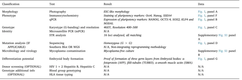
Characterization and validation.
Classification Test Result Data
Morphology Photography ESC-like morphology Fig. 1, panel A
Phenotype Immunocytochemistry Staining of pluripotency markers: Oct4, Nanog, SSEA4 Fig. 1, panel F qPCR Expression of pluripotency markers: NANOG, OCT3/4, SOX2, KLF4 and
NODAL
Fig. 1, panel B
Genotype Karyotype (G-banding) and resolution 46XY, Resolution 400–500 Fig. 1, panel C
Identity Microsatellite PCR (mPCR) N/A
STR analysis 16 loci analyzed, all matching SupplementaryFig. S1panel
A Mutation analysis (IF
APPLICABLE)
Sequencing Homozygous (G > G) Fig. 1, panel D
Southern Blot OR WGS N/A, Non-integrating reprogramming methodology
Microbiology and virology Mycoplasma contamination Mycoplasma-free culture SupplementaryFig. S1panel
B Differentiation potential Embryoid body formation Proof of formation of three germ layers from Embryoid bodies:
α-fetoprotein (AFP),βIII-tubulin (TUBB3), α-smooth muscle actin (SMA).
Fig. 1, panel G
Donor screening (OPTIONAL) HIV 1 + 2 Hepatitis B, Hepatitis C N/A N/A
Genotype additional info (OPTIONAL)
Blood group genotyping N/A N/A
HLA tissue typing N/A N/A
Table 2 Reagents details.
Antibodies used for immunocytochemistry/flow-citometry
Antibody Dilution Company Cat # and RRID
Pluripotency Markers Rabbit anti-NANO 1:200 Abcam Cat# ab21624, RRID:AB_446437
Rabbit anti-OCT4 1:400 Abcam Cat# ab19857, RRID:AB_445175
Mouse anti-SSEA4 1:200 Abcam Cat# ab16287, RRID:AB_778073
Differentiation Markers Mouse anti-Human TUBB3 1:400 Sigma-Aldrich Cat# T8660, RRID:AB_477590
Mouse anti-Human SMA 1:600 Dako Cat# M0851, RRID:AB_2223500
Rabbit anti-Human AFP 1:200 Dako Cat# A0008, RRID:AB_2650473
Secondary antibodies Alexa Fluor 488-conjugated Donkey anti-Mouse IgG (H + L) 1:300 Jackson ImmunoResearch Labs Cat# 715–545-150, RRID:AB_2340846
Alexa Fluor 488-conjugated Donkey anti-Rabbit IgG (H + L) 1:300 Jackson ImmunoResearch Labs Cat# 711–545-152, RRID:AB_2313584
Primers
Target Forward/Reverse primer (5′-3′)
Elimination of Sendai Virus transgenes (qPCR) Sev GGATCACTAGGTGATATCGAGC/ACCAGACAAGAGTTT AAGAGATATGTATC
Sev-KLF4 TTCCTGCATGCCAGAGGAGCCC/AATGTATCGAAGGTG CTCAA
Sev-C-MYC TAACTGACTAGCAGGCTTGTCG/TCCACATACAGTCCT GGATGATGATG
Sev-KOS ATGCACCGCTACGACGTGAGCGC/ACCTTGACAATC CTGATGTGG
Pluripotency Markers (qPCR) NANOG CATGAGTGTGGATCCAGCTTG/CCTGAATAAGCAGATCCATGG
OCT3/4 GACAGGGGGAGGGGAGGAGCTAGG/CTTCCCTCCAACCAGTTGCCCCAAAC
SOX2 GGGAAATGGGAGGGGTGCAAAAGAGG/TTGCGTGAGTGTGGATGGGATTGGTG
KLF4 ACCAGGCACTACCGTAAACACA/GGTCCGACCTGGAAAATGCT
NODAL GGGCAAGAGGCACCGTCGACATCA/GGGACTCGGTGGGGCTGGTAACGTTTC
House-Keeping Genes (qPCR) GAPDH CTGGTAAAGTGGATATTGTTGCCAT/TGGAATCATATTGGAACATGTAAACC
β-actin GCAAAGACCTGTACGCCAAC/AGTACTTGCGCTCAGGAGGA
Mycoplasma detection Pair 1 CTGCAGATTGCAAAGCAAGA/CCTCCTTCTTCACCTGCTTG
Pair 2 GGCGAATGGGTGAGTAACACG/CGGATAACGCTTGCGACCTATG
Targeted mutation analysis/sequencing DAND5 exon 2 GGAAGTGGACAGGTGATTATCC/CAC
generous donation of the urine sample used in this study. We also would like to thank Ana Jardim for technical support in karyotype analysis. This work was supported by Fundação para a Ciência e a Tecnologia (PTDC/BIM-MED/3363/2014). iNOVA4Health - UID/ Multi/04462/2013, a programfinancially supported by Fundação para a Ciência e Tecnologia/Ministério da Educação e Ciência, through na-tional funds and co-funded by FEDER under the PT2020 Partnership Agreement is acknowledged.
References
Belo, J., Marques, S., Inácio, J., 2017. The role of Cerl2 in the establishment of left-right
asymmetries during axis formation and heart development. J. Cardiovasc. Dev. Dis. 4, 13.
Cristo, F., Inácio, J.M., de, Almeida S., Mendes, P., Martins, D.S., Maio, J., Anjos, R., Belo, J.A., 2017a. Functional study of DAND5 variant in patients with congenital heart disease and laterality defects. BMC Med. Genet. 18.
Cristo, F., Inácio, J.M., Rosas, G., Carreira, I.M., Melo, J.B., de, Almeida L.P., Mendes, P., Martins, D.S., Maio, J., Anjos, R., Belo, J.A., 2017b. Generation of human iPSC line from a patient with laterality defects and associated congenital heart anomalies carrying a DAND5 missense alteration. Stem Cell Res. 25, 152–156.
Deng, H., Xia, H., Deng, S., 2015. Genetic basis of human left-right asymmetry disorders. Expert Rev. Mol. Med. 16, e19.