Orientadora de Dissertação:
D
OUTORAC
HRISTAR
HINERProfessor de Seminário de Dissertação:
D
OUTORR
UIO
LIVEIRATese submetida como requisito parcial para a obtenção do grau de:
M
ESTRE EMN
EUROCIÊNCIASC
OGNITIVAS EC
OMPORTAMENTAISR
AMOF
UNDAMENTAL2016/2017
REGENERATIVE NEUROGENESIS IN DROSOPHILA
MELANOGASTER:
The influence of Age and Activity in the adult brain
Dissertação de Mestrado realizada sob a orientação da Doutora Christa Rhiner (Fundação Champalimaud), apresentada no ISPA – Instituto Universitário para obtenção de grau de Mestre na especialidade de Neurociências Cognitivas e Comportamentais
Acknowledgments
To begin with, I do not have enough words to express my gratitude to my advisor, Dr. Christa Rhiner. From accepting me as a member of her laboratory, teaching me everything about this wonderful research field, all the advice, suggestions, enormous understanding, kindness words… Thank you so much for everything!
The Stem Cells & Regeneration Lab is without any doubt the best lab to work! Anabel and Silvia, you were undoubtedly very important in my growth as a scientist, and have helped in everything I needed to accomplish this project during the last year. Thank you for all your support and friendship.
I also have a lot to thank the great team at Cell Fitness Lab, who from the beginning received me so well and helped with everything I needed. We have so many good memories together.
Um grande obrigada aos meus colegas de mestrado mais próximos: Catarina e Ana Filipa, vocês sabem bem como foi o nosso primeiro ano juntas, sem dúvida que fizemos com que tudo fosse mais fácil apenas por estarmos na companhia umas das outras. Ricardo, sem ti esta longa jornada não teria sido igual, fizeste sem dúvida com que tudo se tornasse mais interessante, divertido e descontraído.
Aos meus companheiros diários nesta aventura de tese, Catarina, Sara, Andreia e Bernardo, o grupo das bolachinhas de chocolate, não houve ninguém que me compreendesse melhor ou ajudasse tanto no dia-a-dia como vocês! Obrigada por tudo, são sem dúvida amigos que levo para a vida...
Um muito obrigada a todos os meus amigos que sempre me apoiaram e motivaram para continuar nesta grande aventura, interessados em saber tudo o que iria descobrir com o projecto. Principalmente à Rita, Ana, Carolina e Carolina (afilhada), as minhas mais que tudo!
E por fim, mas a meu ver os mais fundamentais, a minha família e namorado que me apoiam em tudo incondicionalmente... Por todo o carinho, suporte, amor e paciência nas alturas mais stressantes, um enorme Obrigada não basta!
Resumo
Sendo o Encéfalo um dos órgãos mais importantes do organismo, a descoberta de que o processo de neurogénese continuava presente durante a fase adulta do animal foi uma das grandes revelações científcas da última metade do século XX. Assim, uma das vantagens na investigação deste processo: Neurogénese Adulta, é a sua aplicação em diversos organismos, que posteriormente possibilitará uma compreensão mais completa e aprofundada do mesmo. A Drosophila melanogaster foi recentemente apresentada como um óptimo modelo de estudo devido à sua acessibilidade genética e grande capacidade neuroregenerativa após lesão, contribuindo para o acesso a aspectos até então inexplorados.
O objectivo principal deste trabalho foi o estudo da regulação do processo de Neurogénese regenerativa com foco na acção de dois factores: a Idade e a Actividade. Tal foi possível através da aplicação de um sistema de sensível de “lineage tracing”, o qual permite a visualização e quantificação de neurónios recentemente produzidos após uma lesão.
Nos resultados obtidos, destacou-se a evidência de que o envelhecimento não promove uma diminuição na capacidade regenerativa no cérebro adulto da mosca, e persiste num nível constante até às 6 semanas de idade, o é próximo da vida inteira. Diferentes protocolos foram realizados para estudar o efeito da actividade física na neurogénese regenerativa. As experiências optimizadas ainda estão a decorrer e não poderam ser concluídas, porém dados preliminares indicam, que as moscas que têm um maior espaço envolvente anteriormente à lesão podem regenerar mais facilmente comparado com moscas que estão confinadas num espaço reduzido.
No futuro, será importante será importante investigar quais os genes que regulam a ativação de células progenitoras neurais adultas induzida através de lesão, e ainda quais os fatores que controlam a diferenciação neuronal para obter uma compreensão mais detalhada de como a idade e a atividade influenciam a regeneração no cérebro adulto.
Palavras-Chave: Drosophila melanogaster, neurogénese regenerativa, lóbulo óptico,
“lineage tracing”, regeneração, lesão cerebral, regulação de células estaminais, Idade e Actividade
Abstract
Being the brain one of the most important organs, the discovery that neurogenesis was continuous even in the adult phases was one of the great scientific revelations of the last century. The research of adult neurogenesis in several model systems will allow a better and complete understanding of this process. Drosophila melanogaster has been proposed as a novel model due to its genetic accessibility and its ability to regenerate neurons after injury, thereby opening the way to unexplored aspects.
The main goal of the present project was to study the regulation of regenerative neurogenesis with respect to Age and Activity. This was achieved by applying sensitive lineage tracing, which allowed the visualization and quantification of the newly generated neurons upon injury.
The observed results demonstrate that the regenerative capacity in adult fly brains does not decrease with age and persists on a constant level up to 6 weeks of age, which is close to the entire life span. Different paradigms were tested to study the effect of physical activity on regenerative neurogenesis. Experiments with the optimized set-up are still ongoing and could not be concluded, but preliminary data indicates, that flies that could move in a large compartment before injury may regenerate more favorably compared to flies that were kept in a more confined space.
In the future, it will be important to gain further insight into which genes regulate injury-induced activation of adult neural progenitor cells and which factors control neuronal differentiation to gain a more detailed understanding of how age and activity impinge on regeneration.
Keywords: Drosophila melanogaster, regenerative neurogenesis, optic lobe, lineage
Index
Introduction ... 1
Adult Neurogenesis ... 1
Adult Neurogenesis in Mammals ... 2
Adult Neurogenesis in Drosophila ... 4
Regenerative Neurogenesis ... 7
Regulation of Adult Neurogenesis ... 9
Aims and scope of the Thesis ... 11
Methods and Materials ... 12
Fly husbandry ... 12
Perma-Twin Flies and System functionality ... 12
Perma-Twin System Validation ... 13
Acute brain damage experiment ... 13
Age experiments ... 13
Activity experiment – Food search behavior ... 14
Activity experiment – Space conditioning ... 14
Brain dissection and Staining ... 15
TUNEL Staining ... 15
Mounting of fly brain ... 15
Clone Analysis and Image acquisition ... 16
Results ... 18
Perma-Twin System Validation ... 18
Acute Brain Damage Induces Neurogenesis in the Adult Brain – Protocol validation ... 19
Age effect in the regenerative neurogenesis of the Adult Brain ... 21
Activity experiments - Environmental stimulation ... 23
Discussion ... 25
Conclusion and Future Perspectives ... 28
References ... 31
Appendices ... 35
Protocols ... 35
1- Appendix: Perma-Twin Method ... 35
2- Appendix: Right Optic Lobe Injury experiment ... 36
3- Appendix: Adult Brain dissection ... 37
4- Appendix: Fixation ... 38
5- Appendix: TUNEL-staining Protocol ... 39
6- Appendix: Mounting slides ... 40
8- Appendix: Activity Experiment ... 42
8.1- Food Searching ... 42
8.2- Conditioned space environment ... 44
Figures Index
Figure 1: Adult Neurogenesis in the sub granular zone (SGZ) of the dentate girus (DG) ... 3
Figure 2: Adult Neurogenesis in the sub ventricular zone (SVZ) ... 4
Figure 3: Origin and location of Drosophila larval Neuroblasts ... 5
Figure 4: Adult neurogenesis in the adult optic lobe of Drosophila... 6
Figure 5: Adult neurogenesis in Drosophila ... 6
Figure 6: Regenerative neurogenesis in adult brain of Drosophila ... 8
Figure 7: Adult neurogenesis and brain regeneration in Drosophila ... 8
Figure 8: Hierarchical levels of Regulation, from social context to genes ... 9
Figure 9: Perma-twin labelling system ... 12
Figure 10: Experimental conditions to observe regenerative neurogenesis in the adult brain after different timepoints ... 13
Figure 11: Experimental conditions to observe the impact of conditioned spatial environment on the regenerative neurogenesis of the adult brain ... 14
Figure 12: Zeiss LSM 880 Confocal Microscope ... 17
Figure 13: Validation of Perma-twin system in Drosophila adult brain ... 19
Figure 14: Acute brain damage on a 2 weeks adult brain, Perma-twin fly. ... 19
Figure 15: Perma-twin flies with 2 weeks old, right optic lobe (ROL) in three different conditions. ... 20
Figure 16: TUNEL staining in an injured ROL ... 20
Figure 17: Representative images of right ... 21
Figure 18: Quantification of newly proliferative cells 3 days, 2, 3, 4 and 6 weeks after brain damage ... 22
Figure 19: Perma-twin adult flies with 14 days at the beginning of activity experiment. ... 23
Figure 20: Average number of labeled cells per brain ... 24
Figure A 1: Right OL (optic lobe) injury in the Drosophila’s adult brain. ... 36
Figure A 2: Dissection materials. ... 37
Figure A 3: Mounting materials. ... 40
Figure A 4: Activity experiment Materials 8.1 ... 43
Figure A 5: Conditions of Activity experiment - Conditioned space (8.2) ... 44
Figure A 6: Differences between optic lobes, Perma-twin flies with 4 weeks old... 45
Figure A 7: Average number of the proliferative clones counted with GFP label between the undamaged (Left optic lobes) and damaged (Right optic lobes) conditions. ... 45
Introduction
Adult Neurogenesis
The knowledge of the Nervous System has grown and innovated vastly in recent years through new methods and improved techniques in the field of Research, however until the middle of last century, the brain was viewed as a fixed system:
“Once the development was ended, the founts of growth and regeneration of the axons and dendrites dried up irrevocably. In the adult centers, the nerve paths are something fixed, ended, and immutable. Everything may die, nothing may be regenerated. It is for the science of the future to change, if possible, this harsh decree” (Cajal, 1913).
The process of neurogenesis was conventionally accepted to occur only at embryonic developmental stages in the mammalian central nervous system. The discovery of newborn nerve cells from neural stem cells (NSCs) in the adult brain, a process called adult neurogenesis, has shown us that the nervous system is much more dynamic and has a great plasticity than we had ever thought. The publication of Altman’s team in 1965, revealed the first evidence of such a process in the hippocampus of adult rats, from the labeling of cells by a radioactive compound ([H3]-thymidine) that is integrated into the DNA of dividing cells (Altman & Das, 1965). However, this labeling technique did not allow to prove that the new cells would become integrated neurons into the pre-existing brain circuits, and thus made the discovery been neglected until 1981, when Nottebohm reported the presence of adult-born neurons in the songbird brain (Martinez-Marcos, et al., 2016; Migaud, et al., 2016).
Adult neurogenesis is an intricate process that includes the formation, migration, differentiation, selection, and maintenance of new nerve cells in the adult brain. A subset of newly formed neurons will incorporate into the functional neuronal circuits, thus contributing to the structural and functional plasticity of this great organ. (Gage, 2000; Fernández-Hernández & Rhiner, 2015) The identification of this significant activity in the adult state suggests a contribution to the body's homeostasis. In areas where there is adult neurogenesis, regular production of new cells represents an additional mechanism of brain plasticity. However, the meaning and functional implication of neurogenesis in adult mammals is still an open question. (Migaud, et al., 2016; Zhao, et al., 2008)
Stem cells are defined as an undifferentiated population of cells capable of producing both self-renewing and differentiating daughter cells. Some of them persist throughout life and are involved in the maintenance and repair of the adult tissues, replacing cells in normal homeostasis, injury or disease. (Weissman, 2015) In order to keep up with all the changes and the specific needs of the tissues, stem cells have to adapt their properties throughout the life of the organism. Thus, at an earlier stage, during embryonic development these cells divide at a high rate to accompany the constant growth of tissues, whereas, in the more mature stage, they are in a mostly quiescent state, only maintaining tissue homeostasis. (Signer & Morrison, 2013)
For an area to be characterized as a neurogenic niche it is necessary that a suitable microenvironment allows the maintenance of the cellular renewal and/or multipotency of the neural stem cells (Gage, 2000). These have been extensively studied and exhibit as key features the control of: stem cells proliferation rate, the fate of it is daughter cells, and it dynamically regulate the stem cell pool and protect it from depletion, only by providing the source of growth factors and adhesion molecules that will influence the final properties of the stem cells (Gage & Temple, 2013; Gonen & Toledano, 2014).
Adult Neurogenesis in Mammals
Adult neurogenesis began to be studied in mammals, and these have been the main focus of research for a long time. Thus, with the knowledge acquired over the last decades, it is already possible to highlight some characteristics and defined concepts (Ming & Song, 2005). A very important discovery is that this phenomenon seems to be evolutionarily conserved among Vertebrates, revealing several points in common (Kempermann, 2016). In mammals’ case, new nerve cells are continuously created from neural progenitors present in at least two specific areas of the brain throughout life: the subgranular zone (SGZ) of the dentate gyrus (hippocampus), and the subventricular zone (SVZ) lining the lateral ventricles (Ming & Song, 2011; Zhao, et al., 2008). These two main neurogenic niches differ in their morphology, proliferative behavior and expression markers (Fuentealba, et al., 2012)
The subgranular zone (SGZ) is located between the granular cell layer and hilus of the hippocampal dentate (Figure 1). In the adult human brain, this is the zone that seems to represent greater neurogenic relevance (Spalding, et al., 2013). Our knowledge of this
specific area comes mainly from studies with rats. Here, radial glia-like cells (Cell Type I) proliferate until they reach the state of intermediate progenitor cells, which migrate to the granular cell layer. When they reach this layer, cells undergo several cycles of division and differentiation to form a population of post-mitotic immature granular cells, which in turn differentiate into a neuronal subtype, excitatory glutamatergic granular neurons, and establish new connections in networks (Gonçalves, et al., 2016; Ming, & Song, 2011; Zhao, et al., 2008).
The subventricular zone (SVZ) (figure 2) of the adult brain has ependymal cells, slow cycling radial glial-like cells, also known as B cells, which in turn give rise to fast dividing intermediate progenitor cells (type C cells), with a large proliferative capacity. These cells later differentiate into migrate neuroblasts (type A cells) that go from the rostral migratory stream (RMS) to the olfactory bulb (OB). Once there, the neurogenesis persists as the cells differentiate into several types of interneurons and integrate preexisting olfactory circuit in the OB. (Ming & Song, 2011; Ernst & Frisén, 2015) The extent of this process in the Human brain appears to be insignificant after an early postnatal period. Reporter data about this are controversial, the topic is still an open debate in the scientific community (Bergmann et al., 2012; Curtis et al., 2007).
Figure 1: Adult Neurogenesis in the sub granular zone (SGZ) of the dentate girus (DG). The left part shows a frontal cross-section of an adult mouse brain and the location of the SGZ. The diverse stages of the neural stem cells’ development are represented on the right side. The SGZ have different types of cells (Type I NSCs, and Type II and III progenitor cells), which can be distinguished using distinct morphologic and molecular markers. (Adapted from Migaud, et al., 2016).
In recent years, this research topic – Adult Neurogenesis in Mammals, has presented several advances from a multidisciplinary point of view, including the several signaling pathways regarding the regulation of the proliferation of adult NSCs, the possible functions of new neurons in various contexts (e.g.: diseases, behaviors, etc.), such as the environmental regulation.
Since the beginning of adult neurogenesis research, the identification of newborn neurons has been made by three major approaches: incorporation of exogenous nucleotide analogs, as [H3]-thymidine and Bromodeoxyuridine (BrdU) or Ethynyl-Deoxyuridine (EdU), during DNA replication; genetic marking by stereotaxic injection of retrovirus carrying a reporter gene; the development of transgenic models under specific promoters, in which reporter proteins are expressed under the control of promoters of immature neuron-specific genes. In some cases, a method to measure the birth-dating carbon-14 (C14) has been used to demonstrate adult neurogenesis in the human brain. Even though these techniques have helped the researching of adult neurogenesis, they still display limitations and disadvantages. (Imayoshi, et al., 2009; Kuhn, et al., 2016; Ryu, et al. 2016) Thus, it is important that we acquire more resourceful approaches in a near future.
Adult Neurogenesis in Drosophila
Despite all the new information that we already acquire about adult neurogenesis, there are still several issues that required further study. Consequently, to improve the knowledge Figure 2: Adult Neurogenesis in the sub ventricular zone (SVZ). The left side show a frontal cross-section of an adult mouse brain and the location of the SVZ, in the walls of the lateral ventricle. On the right side of the scheme are the illustration of the stages of morphologic and physiological development of neural stem cells. (Adapted from Migaud, et al., 2016)
about this phenomenon it is necessary to compare the studies of different species, besides mammals.
One of the most versatile model organism that has been used in research over the past century is the Drosophila melanogaster, also known as fruit fly. In the field of genetics, this organism has already offered several insights regarding cellular processes and molecular mechanisms. It thus represents a suitable model for the analysis of adult neurogenesis since it presents characteristics such as a short generation time, a strong genetic toolbox and even a highly conserved genome (Jennings, 2011).
Until recently, it has been long assumed that there were no neural progenitor cells in the adult brain of Drosophila. Nevertheless, the study of neurogenesis in the stages of larvae and pupae had provided many insights about this process, which can help in the search of the proliferation and progenitors in the adult brain.
It is during the embryogenic stages that the neuroblasts form for the first time from the neuroepithelium, located in the ventrolateral region of the embryo. These pass through several self-renewing asymmetric divisions, each of which gives rise to another neuroblast and a ganglionic mother cell (GMC), which in turn divides only once to form two daughter cells, which may be neurons or glia. The larval brain contains different types of neuroblasts (NBs), based on their position and lineage characteristics (See figure 3 for details). (Homem & Knoblich, 2012) In the case of the Optic lobes, the neuroblasts first undergo symmetric divisions to expand the progenitor pool, but the lately generated NBs divide asymmetrically, producing a GMC that further gives rise to terminally differentiated progeny (Weng & Lee, 2011).
Figure 3: Origin and location of Drosophila larval Neuroblasts. VNC- ventral nerve cord with the thoracic NBs (dark brown) and abdominal NBs (light brown); OL- optic lobes in the brain lobes; central brain with type I NBs (yellow), type II NBs (green) and mushroom body NBs (orange). (Adapted from Homem & Knoblich, 2012)
Regarding previous works and the idea that adult neurogenesis is normally based on very slow proliferating progenitors, Fernández-Hernández et al. (2013) apply an improved method, based on the twin-spot MARCM (Yu, et al., 2009), to permanently label newly generated cells in the adult brain of Drosophila, called “Perma-Twin” system. Using this, they identify the generation of new neurons in the medulla cortex of the optic lobes (Figure 4). These represent the possibility to use Drosophila as a new model system to study the regulation of adult neurogenesis. Besides the sustained capacity (Flipase source) of this system, it also represents a beneficial alternative to detect cell proliferation compared to incorporation methods as EdU- and BrdU, which will eventually depend on direct "contact" with the tissues of interest. In this specific case, the access is even harder because of the blood-brain barrier, beyond the negative effects of this kind of reagents during cell-cycle progression.
The labeled generated twins in the medulla cortex contained at least one Elav-positive (neuronal marker) cell and were negative for the glial marker Repo, after the Perma-twin activation. Therefore, these data are the proof that adult neurogenesis really occurs in the adult optic lobes. (Fernández-Hernández et al., 2013)
Besides this, the expression of the larval neuroblast marker – Deadpan, was found in small clusters of dividing cells, which also contained newly formed neurons (Figure 5). Under physiologic conditions, the majority of the neural precursor cells are in a quiescent state. At a normal state, only 4-6 divisions per optic lobe were the estimated events of adult neurogenesis in a week (Fernández-Hernández & Rhiner, 2015).
Figure 4: Adult neurogenesis in the adult optic lobe of Drosophila. When the system is on, clones appear in the medulla cortex (MC) of the optic lobe (OL). Scale bar represents 20 µm. (Adapted from Fernández-Hernández, et al., 2013)
Figure 5: Adult neurogenesis in Drosophila. (Adapted from Fernández-Hernández & Rhiner 2015).
The work of this team had a main effect in this research field, but it is only in the beginning stages, and much more questions need to be addressed.
Regenerative Neurogenesis
Some parts of the nervous system that were damaged or affected somehow can be repaired by new cells formed in the neurogenic regions, in this way, these specific areas will be able to recover. This process is known as Regenerative or injury-induced neurogenesis. (Alunni & Bally-Cuif, 2016)
In addition to physiological adult neurogenesis levels, the injured brain can initiate specific regeneration programs for the recovery of the internal homeostasis state (Yu et al., 2014). NSCs are quiescent most of the time, which prevents the waste of energy. Thus, when these cells are activated for the process of repairing an injury, it is necessary to leave this basal state. In previous studies, in both rat and zebrafish, Notch1 signaling is required to maintain the activation (proliferation) of the NSCs, controlling both cell division and pluripotency (Alunni & Bally-Cuif, 2016), however, it has not been further discoveries of the possible involving factors.
Fernández-Hernández & Rhiner (2015) has referred three major strategies for a successful tissue regeneration: “I- Recruitment and/or activation of adult stem cells, II- de-differentiation to a progenitor-like state, III- trans-de-differentiation of cells close to the injury site”, also a vast number of progenitors near the damage.
As it is already known, in the mammalian brain the regenerative capacity after injury is really low (Fernández-Hernández & Rhiner, 2015). However, due to the various diseases or lesions affecting the brain, the identification of the mechanisms that control the process of regenerative neurogenesis in the context of physiological cell loss or injury is a research field of great interest.
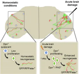
Once Drosophila has demonstrated a robust neurogenesis upon injury, it represents a very suitable model to analyze regenerative neurogenesis in the adult brain. Diverse approaches to induce traumatic brain injury have been shown in the recent years, but the adopted model is based on a mechanic brain injury, where the fruit fly brain is damage with a thin filament in one of the optic lobes (For schematic representation see figure 6) (Fernández-Hernández et al., 2013).
The lesion caused by the filament in the optic lobe of the adult fly leads to the activation of the normally quiescent progenitor cells. The proximity of these to the site of damage promotes the upregulation of dMyc (drosophila myc) and the nuclear transcription of factor Dpn (Deadpan), significant for the proliferation of neuroblasts, as previously mentioned. This leads to the formation and consequent insertion of new neurons around the lesion site (Fernández-Hernández et al., 2013). Such results show a regenerative intervention of quiescent adult neural progenitors in the case of injury (Figure 7).
These new models of acute damage applied to the study of regeneration, in the adult brain, may represent new knowledge based on comparative analysis, in search of new regulatory factors, stem cell activation or regenerative neurogenesis.
Figure 6: Regenerative neurogenesis in adult brain of Drosophila. Traumatic brain injury paradigm. La- Lamina, Med- Medulla; L- Lobula; Lop- Lobular plate. (Adapted from Simões & Rhiner, 2017)
Figure 7: Adult neurogenesis and brain regeneration in Drosophila. (Adapted from Fernández-Hernández, et al., 2013).
Regulation of Adult Neurogenesis
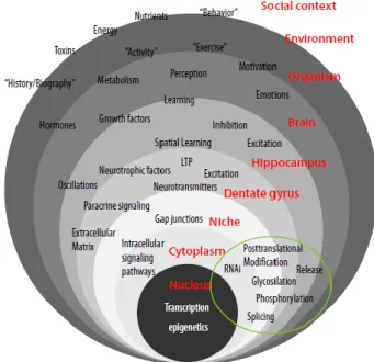
According to Ryu et al., (2016), it seems perfectly normal that adult neurogenesis and its resulting brain plasticity are intimately regulated by external factors and behavioral responses since one of the main functions of the Nervous System is detecting the surrounding environment information and converting it into functional/behavioral outcomes. Regardless the limitations of the methods applied in this field, over the last years, studies have described several intrinsic and extrinsic factors in the regulatory process (Figure 8) (Aimone et al., 2014).
A meticulous description of all the type of factors involved is beyond the scope of this introduction, further details can be found in several detailed reviews (Aimone et al., 2014; Kempermann, 2011; Ming & Song, 2011; Sawada & Sawamoto, 2013; Zhao et al., 2008).
The extrinsic factors can influence both positive and negative the distinct levels of neurogenesis through the life of the organisms. In the context of adult neurogenesis research, the two key regulators that have been considered antagonistic due to their impact are Age and Activity. The second promotes an increase in the number of NSCs in the neurogenic regions while the first tends to be associated with their decline. However, there may be a reciprocal relationship between this two factors, where Age can affect Activity, and Activity influence aging. (Aimone et al., 2014; Kempermann, 2015) Most of the studies reporting the
Figure 8: Hierarchical levels of Regulation, from social context to genes. (LTP- long-term potentiation). (Adapted from Kempermann, 2011)
effects of Age and Activity in Adult neurogenesis has been done in Mammals, namely in rodents.
The aging is the most well-studied negative regulator, displaying significant reductions in diverse levels of neurogenesis, like the number of cells proliferation, differentiation or even, survival. The first studies date from the 90’s, when the adult neurogenesis starts to be seen as a lifelong process. In 1998, Eriksson et al., reported the study of this process in the adult human brain in individuals as old as in their early seventies, yet with very low numbers of adult neurogenesis. The effects of this regulatory factor in the old brain might be caused by a loss of some specific signals provided for the niche, that were required to maintain and control the precursor cells. (Kempermann, 2015; Rando, 2006; Ryu et al., 2016)
Activity may be distinguished on a behavioral level, as physical exercise (e.g. running) and cognitive stimulation (e.g. learning tasks). The first one showed that is important to have a well-defined paradigm, where voluntary wheel running and forced exercise in a treadmill is the best studies paradigms in rodents’ adult brain. The main effects are the increasing number of precursor cells’ proliferation in the hippocampus, which results in the expansion of progenitor cells that are available for future neuronal maturation and functional integration. In contrast, the cognitive stimulation (as environmental enrichment or learning tasks) that theoretically is more specific to hippocampus have shown none or a limited effect on cell proliferation but recruit new neurons for long-term survival. So, this shows that although this both types of Activity can increase the levels of adult neurogenesis, they have different ways of doing it. The physical exercise appears to affect the early phases of neurogenesis, namely the cell proliferation, while the environment enrichment has a larger impact on the cell survival and neuronal differentiation. (Aimone et al., 2014; Kempermann, 2015)
Kempermann (2011) has declared that the regulation of adult neurogenesis occurs to achieve, maintain, modify or improve the function of the new neurons, the neurogenic area, the brain, the individual, etc.
Aims and scope of the Thesis
The main purpose of this project is to apply one of the most versatile model organism in research - the Drosophila melanogaster, as a model system to study the regulation of adult neurogenesis in the context of acute brain damage. Based on a mitotic recombination-dependent lineage-labeling method called Perma-Twin system is possible to permanently label new generating cells and their progeny in the optic lobes of the adult fruit fly (Fernández-Hernández, et al., 2013), and the application of this tool will help us in the search of a new/better understanding of the effects that some regulatory agents may have on the process of regenerative neurogenesis.
Most of the available research on the regulatory effects of extrinsic factors in the adult neurogenesis is based on Mammals evidence, so in order to expand our knowledge in this field we tested the effects that Age and Activity can have in the regenerative capacity of the Drosophila adult brain (region of interest: optic lobe), since these were classified as the antagonistic key regulators by Kempermann (2016).
The recognition of Age and Activity’s effects, in this specific case and model, can contribute with new knowledge to the study of neurogenesis and regeneration in the adult brain, which may in the future be translated into potential evidence in several other species and will eventually be taken as common-sense knowledge.
Methods and Materials
Fly husbandry
Most of the fly stocks were maintained at 25ºC and relative humidity of 70% (12 Light: 12 Dark cycle), and the Perma-twin crosses at 18ºC and relative humidity of 70% (12 Light: 12 Dark cycle). Perma-twin experiments were carried out at 30ºC for optimal Gal4 activity. Flies were raised on the standard food (Vienna recipe) in low-density conditions.
Perma-Twin Flies and System functionality
To generate the Perma-Twin flies, males from w; FRT40A, UAS-CD8-GFP, CD2-mir/ CyO; act-Gal4, flp/ TM6B were crossed to virgin females of the w; FRT40A, UAS-CD2-RFP, UAS-GFP-mir/ CyO; tub-Gal80ts/ TM6B. The crosses were set up at room temperature and, after the first 24 hours, changed to 18ºC during all the development. (See Appendix 1)
The offspring were collected in separate vials and kept at 18ºC after adult eclosion until the desired age had been achieved. With these conditions, the perma-twin system is inactive, and no early divisions will be labeled because Gal80ts represses Gal4. Once the flies achieve the designated age after eclosion, they were selected against the balancers (without Curly O and TM6B), to collect only the final genotype: w; FRT40A, UAS-CD8-GFP, UAS-CD2-mir/ FRT40A, UAS-CD2-RFP, UAS-GFP-mir; act-Gal4, UAS-FIP/ tubGal80ts. These flies have then be shifted to 29º/30ºC – permissive
temperature, for the activation of the Perma-twin system, when the Gal4 became active and can drive the UAS-constructs expression, allowing Flipase-mediated recombination and subsequent labeling. With this method, only the twin cells coming from an adult cell division will be labeled with the fluorescent receptors: GFP and RFP. (Adapted from Fernández-Hernández, et al., 2013)
Figure 9: Perma-twin labelling system. (Adapted from Fernández-Hernández, et al., 2013)
Perma-Twin System Validation:
First, it is important to test the Twin method to validate its functionality. Perma-twin flies were kept at 18ºC during all the development until they were 3 days and 2, 3, 4 and 6 weeks-old adults when their brains were dissected. Samples were analyzed to see if the system was off once the flies were kept at 18ºC all the time.
As a second experiment, to confirm that after the injury there are still no labeled cells when the system is off (18ºC), the exact protocol as previously was performed, while using adult flies with 3 days and 2, 4 weeks old, which ones were injured at 18ºC in the ROL. After 5 days at 18ºC, brains were dissected.
Acute brain damage experiment
The Perma-twin flies were anesthetized with CO2. Posteriorly they were aligned with
the right eye up and the fly was lesioned to the level of the medulla (ROL) by introducing a metal filament (0,1 mm) through the eye. They were allowed to recover at room temperature (approx. 22ºC) for two hours. After 5 days at 30ºC, the brains were dissected to analyze the number of labeled cells. (See Appendix 2 for more details)
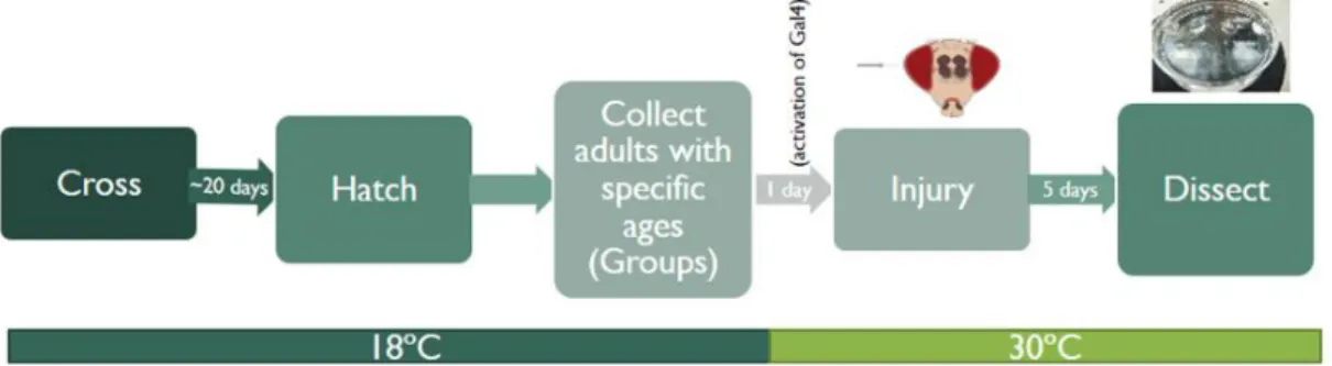
Age experiments
The Perma-Twin flies grew at 18ºC and stay in that conditions until the adults achieved the specific age of 3 days and 2, 3, 4 and 6 weeks (groups made for each age). To allow the activation of Gal4 expression the selected flies were transferred to 30ºC for 24 hours. Next day, flies were damage with a thin filament (0,1 mm) through the right eye into the medulla at room temperature and left to recover for 2 hours. From this time on, flies were kept at 30ºC and adult brains dissected 5 days after the damage. (See Appendix 7 for more details)
Figure 10: Experimental conditions to observe regenerative neurogenesis in the adult brain after different timepoints. Perma-twin flies were kept at 18ºC up to a specific age after adult eclosion (perma-twin system off – Gal80ts is active). After one day at 30ºC, ROL injury was performed, and then flies were shifted to 30ºC again. Brains were dissected 5 days
Activity experiment – Food search behavior
The Perma-Twin flies grew at 18ºC and, 14 days after the adult eclosion they were transferred to 30ºC for 24 hours to allow the activation of Gal4 expression. Next day, only males were selected to the final genotype and were made two distinct groups with the same number of flies. The process of starvation occurs overnight at 30ºC (empty vials only with filter paper and water, to retain the humidity) (See the figure A.4 in appendix 8.1). In the morning, flies were shifted to the specific bottles (I-Control, with normal food and II-Activity, with 7 drops of food; See figure A.4 in appendix 8.1) at 30ºC. In the next 4 days, the bottles were changed for new ones every morning. Brains were dissected, and the samples were analyzed to see if there was any difference between the two groups (Control vs Activity) in the total number of labeled cells in the optic lobes.
Activity experiment – Environmental conditioning
Perma-twin flies grew at 18ºC and when the adults hatched, they were collected and divided between the conditioned bottles (reduced or increased space, see figure A.5 in appendix 8.2). When the flies completed 5 days as adults, the females were selected, and shifted to new bottles at 30ºC for 1 day, in order to activate the perma-twin system. In the next day, acute brain damage protocol in the ROL was performed in all the flies at room temperature (Appendix 2). Then, they were relocated to new bottles (at the respective condition as in the beginning, reduced or increased environment) at 30ºC, for the next 5 days. In the end of that time, brains were dissected and samples analyzed between the two different groups.
Figure 11: Experimental conditions to observe the impact of conditioned spatial environment on the regenerative neurogenesis of the adult brain. Perma-twin flies were kept at 18ºC until 5 days after adult eclosion (perma-twin system off – Gal80ts is active). Then flies were shifted to 30ºC to activate perma-twin labelling. After 24h, mechanical damage in the ROL was performed. Brains were dissected 5 days after damage.
Brain dissection and Staining
Flies were put to sleep with CO2 and then the brains were dissected in ice-cold
Phosphate-buffered saline (PBS), in batches of 30 minutes, with forceps number 5. Dissected brains were immediately fixed in 4% (v/v) Formaldehyde (PFA) in PBS for 20-30 minutes at room temperature in the dark. Then samples were washed 3 times for 15 minutes each with 1X PBS, always at the same temperature. When the samples weren´t mounted on the same day, they were stored in PBS at 4ºC. (See figureA.2 in Appendix 3)
TUNEL Staining
Brains were dissected 5 days after damage and fixed and washed with PBS-T (0,4%) one time quickly and 20 minutes at room temperature. In the next step, samples were incubated during 1 hour in equilibrate TUNEL buffer: 20μL TdT buffer 5x + 4μL CoCl 25mM + 76μL PBS. Then, brains were incubated in TUNEL solution: 100μL TdT buffer + 0,3μL Terminal Transferase + 0,2μL dUTPs, for 2 hours at 37ºC. Following, the previous solution was removed, and the brains were incubated in STOP buffer citrate for 15 minutes. After some washes with PBST at room temperature, samples were incubated overnight with primary antibody (mouse anti-elav). In the next morning, after some washes, incubate the brains for 2 hours at room temperature with streptavidine and a second antibody (anti-elav), in the dark. To finalize, 3 washes of 15 minutes were done in PBS, and at the end, the brains were mounting. (See Appendix 5 for more details)
Mounting of fly brain
The samples were washed one more time with 1X PBS at room temperature and entire brains were transferred to the slide. A step was generated placing two coverslips onto a glass slide and the brains were mounted in between, in order to preserve their morphology. The 1mounting media used was Vectashield with DAPI (VectorLabs). To finalize it was sealed around with nail polish. (See figure A.3 in Appendix 6)
Generation of a new RFP stock – Perma-Twin System
The construction of the Perma-twin stock implies the cross between two lines, being one of them the w; FRT40A, UAS-CD2-RFP, UAS-GFP-mir/ CyO; tub-Gal80ts/ TM6B, which carry the RFP marker and the temperature repressor (tub-Gal80ts). To generate this stock several steps are needed. In the first part, males from the original stock yw; FRT40A, UAS-CD2-RFP, UAS-GFP; TM3/ TM6B were crossed to virgins females from the w; if/ CyO; MKRS/ TM6B line. At the same time, males from ywf; CyO/if; tub-Gal80ts/ TM6B were crossed with virgins of the line w; if/ CyO; MKRS/ TM6B. For the second part, the progeny collected from the first crosses were carefully selected and a new cross between females (virgins): yw; FRT40A, UAS-CD2-RFP, UAS-GFP/if; MKRS/TM6B and males: w; CyO/if; tub-Gal80ts/ TM6B was performed. The offspring were collected and selected according to the desired genotype: w; FRT40A, UAS-CD2-RFP, UAS-GFP-mir/ CyO; tub-Gal80ts/ TM6B. In the end, to verify the veracity of the new stock, we tested with a perma-twin experiment.
The crosses were all set up and kept at 25ºC, and flies were flipped every three days to optimal conditions.
Clone Analysis and Image acquisition
Analysis and quantification of the generated clones were focused generally on the optic lobes, and in age experiments only on the damaged ROL, of the Drosophila melanogaster adult brain. Confocal stacks of all marked cells were acquired, and cells were quantified in maximum projections. Only clones containing the green label (Green Fluorescent Protein – GFP marker) were counted, in order to maintain consistency of the results, since it remained constant (unlike the Red Fluorescent Protein - RFP) throughout the project. Under these circumstances, quantifications could even be considered an underestimation of the real process.
Images were acquired with a Zeiss LSM 880 Confocal Microscope (Figure 12), using the XYZ acquisition mode, 1024x1024 pixels resolution and in one direction scanning mode. For multi-stack images, a step of 0,4 µm was used on 40x objective (only optic lobe) and 1 µm for the 20x objective (entire brain). Images were processed with Zeiss Zen Lite and Image J software for brightness and contrast corrections.
Statistical analysis
Statistical analyses were performed with GraphPad Prism 5. The significance of the Age experiment data - the study of the age effects in the regenerative neurogenesis of the Drosophila adult brain, was assessed with a one-way ANOVA after verification of the normality assumptions with the recommended D’Agostino and Pearson omnibus test (p>0,05 for all the age groups). The differences between the Age groups were analyzed with the Tukey HSD test for α=0,05. p-values are indicated by * p <0,05, ** p<0,01, *** p<0,001 and ns p>0,05. In the graphs, means and standard deviation (SD) are shown. The sample size is indicated in the figure.
Results
The experiments described in this project were planned in collaboration with Christa Rhiner, and all have been executed and analyzed by Carolina Alves. The first section refers to testing the Perma-twin System, which was adapted from the work of Fernández-Hernández, et al. (2013), and show how the system behaves.
The second section is dedicated to the Age effects in the regenerative capacity of the adult neurogenesis on the injured Drosophila’s brain, tested among 5 different timepoints of adulthood (3 days as the youngest age, and 6 weeks [~45 days] as the oldest). Based on previous research from mammals we expected to see a decrease in the number of proliferative cells as the flies age, being seen as a negative regulatory factor.
The last section represents the Activity experiments, where were tested its effects as a positive agent in the regulation of adult neurogenesis. As result, we expected to see an increase in the number of labeled clones in the neurogenic region (medulla cortex of the ROL) due to environmental stimulation (food distribution or surrounding space).
Perma-Twin System Validation
Once all the flies used in the experiments have the Perma-twin system, the first step was to validate the system functionality. For this, it was necessary to prove the veracity of the system when it is in "off" mode through the Gal80 expression at 18ºC, as shown in figure 13A. As can be observed, when the perma-twin flies are always in a low-temperature environment there is no membrane marker GFP or RFP in their cells, only DAPI (nuclei) being observed. Such a result is critical to ensuring that the data found in the system's “on” mode (Gal4 active) does not have any background interference, and it was possible to visualize this scenario for all ages used in the experiment, from the youngest to the oldest.
After this confirmation, it was necessary to perform the same experiment but in a damaged condition, as it would be studied in the project - Regenerative Neurogenesis. In this way, the Perma-twin flies were always kept at 18ºC and when they reached the specific age they were submitted to the injury protocol without changing the surrounding temperature. Based on the results of this experiment (see Fig.13B as an example), it was possible to observe that, although most of the analyzed brains have the same pattern of fluorescent reporters’ absence (GFP and RFP), there may be a 1-3 labeled cells noisiness in the final values (indicated with an arrow in the Figure 13B).
Together these outcomes brought us the foundation for the next trials, representing the basal state of the System.
Acute Brain Damage Induces Neurogenesis in the Adult Brain – Protocol
validation
In this project, the main research focus was on the regenerative function of the Adult Neurogenesis process. The damage protocol used was adapted from the original method of acute brain damage by Fernández-Hernández, et al. (2013), and the outcome was a well-defined visualization of the injury effect on the tissue (Figure 14). As can be seen in figure 15, there is an increase on the number of proliferative cells after damage (Fig.15C) compared to the homeostatic process of neurogenesis in the fruit fly optic lobe (Fig.15B).
Figure 13: Validation of Perma-twin system in Drosophila adult brain. (A) Adult brain uninjured at 18ºC, only show DAPI (blue). Fly with 2 weeks old when dissected. (B) Adult brain injured at 18ºC, show mainly DAPI (blue), and sometimes some background indicated with grey arrow. Fly with 2 weeks old when damaged. Scale bar represents 100 µm.
Figure 14: Acute brain damage on a 2 weeks adult brain, Perma-twin fly. (A) ROL injured at 30ºC, with the area shown in B (grey square). (B) Damage area, show the proliferative cells labeled with GFP (green). Scale bar represents 50 µm and 10 µm.
In order to track the effects of the lesion on the optic lobe tissue, we used the TUNEL staining, that promotes labeling of apoptotic cells resultant from the damage experiment. This protocol allows us to see the affected cells (see Figure 16), whose seem to be very close to one another and in a specific area (localized). The protocol seems to have worked and helps us to define the damaged area (grey arrow in the Fig.16).
Figure 15: Perma-twin flies with 2 weeks old, right optic lobe (ROL) in three different conditions. (A) ROL without injury at 18ºC, show only DAPI (nuclei). (B) ROL uninjured at 30ºC, homeostatic conditions, show labeled cells with GFP (green) and DAPI (blue). (C) ROL with injury at 30ºC, proliferation in regenerative condition, show the GFP marker (green), RFP (red) and DAPI (blue). Scale bar represents 50 µm.
Figure 16: TUNEL staining in an injured ROL, Perma-twin fly with 3 days when damage protocol was performed. Neurons are marked by ELAV (magenta), apoptotic cells by TUNEL (red), and nuclei with DAPI (blue). Scale bar represents 50 µm.
Age effect in the regenerative neurogenesis of the Adult Brain
From previous studies, Age has been pointed as a negative factor in the adult neurogenesis regulation, leading to a decrease of the progenitor cells in older brains (Kempermann, 2015; Rando, 2006; Ryu et al., 2016). In order to test this hypothesis and see if the same situation occurs in the Drosophila model, some different timepoints (3 days, 2, 3, 4 and 6 weeks old) were selected to analyze this effect.
Perma-twin flies were kept at 18ºC until they reached the selected adulthood timepoints, and were transferred to 30ºC. After the activation of the system by the temperature shift, the acute brain damage protocol was performed (Figure 17), and 5 days later the brains were dissected. The Age effect on the brain regeneration process was analyzed taking into account the moment when the lesion was made since it was the only point of the protocol that differed between the different groups. So essentially we observed how the regenerative capacity of the fly adult brain was affected by the age after five days of damage.
A major change observed in comparison with the original work of the Perma-twin system was the loss of the red fluorescent marker, as can be seen in figure 17. This shows the clear absence of RFP-labeled cells. The situation became more visible over time, and since we did not know the specific reason for this, the safest solution was to rebuild the original RFP line. This step is still being done.
Because of this absence, the quantification of the new proliferative cells in the ROL had to be restricted only to the green fluorescent reporter - GFP.
Figure 17: Representative images of right OLs 3 days (A), 2 weeks (B), 3 weeks (C), 4 weeks (D) and 6 weeks (E) after brain damage showing the extent of proliferation (GFP cells), DAPI (blue). Scale bar represents 50 µm.
Figure 18 shows the graphical representation of the age impact. Overall the results showed that in the Drosophila model there is no negative effect of Age on the regenerative process, on the contrary, the highest average is from the oldest flies of the experiment. The significant differences were found between: 3 days and 6 weeks (***), 2 weeks and 6 weeks (**), and 3 weeks and 6 weeks (**), the first being the most significant. Since there were no differences between the two most advanced ages, it will be interesting to see whether the mean of the labeled cells increases or stabilizes at even older ages.
Within the range of ages selected for the experiment, the 2 weeks are the reference timepoint for the adult brain in its functional/mature state, thus the 3 day fly is still seen as in the beginning of maturation, being really close from the larvae/pupae phases, and, consequently the 6 weeks as the elderly.
However, since the perma-twin system requires the flies’ maintenance at 18 ° C until the system can be activated, and knowing that at this temperature there is a delay in the effect of time on the organism aging, it may be necessary to think that the ages presented do not correspond to the actual physical state of the flies. So for an enhanced conclusion of the results, it is necessary to carry out the experiment with more advanced ages that kept at different temperatures might not be achievable.
Figure 18: Quantification of newly proliferative cells 3 days, 2, 3, 4 and 6 weeks after brain damage. The graph represents the number of GFP cells in damaged areas of the right optic lobe (ROL). Means are shown as the columns and error bars are shown as ± SD (sample size shown in each column: n). Significance was determined using One-way ANOVA test, and the differences between the ages from Tukey test. Asterisks indicate significant pairwise differences (* p ≤ 0.05; ** p ≤ 0.01, *** p ≤ 0.001).
Activity experiments -
Environmental stimulation
To study the effects of Activity on the adult neurogenesis of the Drosophila brain, the behavior selected was the food searching, where perma-twin flies were placed in a special bottle with a different food distribution pattern, in opposition to a conventional food distribution (see Appendix 8.1 for more details).
New Perma-twin flies with 14 days of adulthood were tested in the food searching protocol, in order to see if this kind of activity can have an impact on the level of adult neurogenesis in the fruit fly brain. The samples analyzed were from healthy male flies, which have less fat in the body, what means a smaller starvation time. The starvation worked as a motivation to search for the food in the experiment.
By analyzing Figure 19, we can verify that the left brain (Fig.19A) shows only 1 GFP labeled cell (green) comparatively to the right (Fig.19B) that have at least 12. Together with the data from Figure 20, these could translate into a positive effect on the process of adult neurogenesis, these flies were not injured. When the flies have to search for food, they become more active, and it can have an increase in the number of new proliferative cells in the adult brain.
Figure 19: Perma-twin adult flies with 14 days at the beginning of activity experiment. (A) Fly brain from normal food bottles (control); (B) Fly brain from activity bottles (7 drops of food). Scale bar represents 50 µm. Labeled cells (GFP) are indicated by the arrows.
Due to the sample’s small size (6 Perma-Twin males) analyzed, and since it was only applied to males, the results lose their significant value that will allow them to be extrapolated to the entire Drosophila population. Besides that, this task has several variables involved, which were difficult to control, and so, the results cannot be entirely reliable.
To solve this situation and try to simplify the paradigm, we developed a new experiment related to the constraint of the surrounding environment, in which we hypothesized that flies with more space will be theoretically more active than flies that live in a reduced environment. Considering that, we have developed a task where we selected two opposite space conditions: reduced and increased, being the only distinctive variable between the two groups. This time, we chose to have only females because the aggressiveness was one of the issues that could have possibly interfered with the previous experiment results (males in groups show more aggressive behavior than females), besides the available food amount that now is equal for the two conditions.
This time we wanted to study the side effects of activity in the context of brain injury. This experience is still happening, and so, it is not yet possible to show results. We hope to unravel this relationship between Activity and regenerative neurogenesis in the Drosophila adult brain model in a near future.
0 2 4 6 8 10 12 14 Control Activity Av er a g e o f g re en la b eled c lo n es o n th e O L's Condition
(regular - control or distributed - activity)
Activity impact on the process of Adult Neurogenesis
Figure 20: Average number of labeled cells per brain (two optic lobe each) of each condition from 6 flies (3 of each condition – control and activity). Influence of Activity on the process of homeostatic neurogenesis (without damage). Perma-twin flies with 2 weeks old flies.
Discussion
Regenerative Neurogenesis and Perma-twin System
Ever since the first moment that the ultimate brain’s dogma – perceived as a static system, was contested, a new research field emerged, unveiling the required knowledge that would turn possible to understand new aspects of it such as its great plasticity and constant evolution (Altman & Das, 1965; Nottebohm, 1981). So far, there are two major findings: one related to the active process of generating new cells in the adult brain due to the proliferation and differentiation of neural stem cells niches and that seems to be a conservative trace within the animal kingdom (Alunni & Bally-Cuif, 2016; Kempermann, 2016).
Being the brain one of the body’s most important organs it is more passive to major threats, many of which have only been diagnosed currently, and so, the identification of research models becomes a fundamental aspect to the human race. Drosophila melanogaster provides a unique opportunity to study adult neurogenesis through its genetic power and versatility, as proposed by Fernández-Hernández, et al. (2013) (Fernández-Hernández & Rhiner, 2015).
The neurogenesis is already well known in the early development stages (Hartenstein & Wodarz, 2013), whereas in the adult is still in a primordial state of research, and so, when applied the same instruments used upon the first, what is verifiable is incongruity in its results, making, most of them, incompatible for the adult.
The system used in the present work has overcome many of the previous methods disadvantages (Imayoshi, et al., 2009), allowing a permanent labeling of newly generated cells and its progeny due to a sustained functional capacity controlled by the presence of a thermosensitive element. Since it can be expressed throughout all body, Perma-Twin system offers the opportunity to uncover new neurogenic regions. Until now, optic lobes are the only areas that show neurogenesis in the fruit fly. (Fernández-Hernández, et al., 2013)
During the project, we came across a problem regarding the loss of function of the red marker (RFP). We began to have a set of points scattered around the analyzed area without really labeling any membrane cell. The fly stocks used to build the perma-twin genotype were the same from the original lab, and once this situation has been previously observed,
where the RFP lose its function, we can conclude that this transgene seems to have some toxicity and the protein with time will not be expressed at the cell's membrane. A possible solution to this event can be the renewing of the RFP line every 6 months or maybe create a different membrane tagged marker. There is an urgent need to find out what is causing this, so we are trying to rebuild de RFP line and solve the situation.
Although at this time we are only observing the GFP-labeled cells, the study of adult neurogenesis remains achievable. As formerly showed in the adult state, the cell lineage tends to be triggered by symmetric divisions, which means that for now, we are only seeing nearly half of the progenitor’s daughter cells (Fernández-Hernández, et al., 2013).
Age effects in the regenerative capacity of the adult brain
As the organism ages, the body suffers a decline within the functions of its tissues and organs, what usually leads to a decrease in its lifespan. One of the key strategies to keep the organism healthy and under the proper homeostatic conditions is the replacement of old or damaged cells by the production of new ones. Thus, Age is seen as one of the main regulatory factors of the adult neurogenesis, having a negative impact with a clear decrease on is function, in most animals. (Signer & Morrison, 2013)
The Drosophila’s adult brain seems to be an exception to this pattern according to our results. The average number of GFP-labeled cells is bigger in the older timepoint, with 6 weeks, than any other. Until the 6 weeks of adulthood, we can observe an increase of the regenerative neurogenesis level, what in this species could be linked with its short life cycle and rapid aging (Hales, et al., 2015). The production of new neurons throughout the life of the organism entails costs (energetic and time consuming) by the continued maintenance of the quiescent stem cells but also by their activation when necessary to protect the animal, for example in case of injury. Thus, it is expected that there are differences between animals with a short life cycle such as Drosophila and most mammals that have a lifespan much longer, consequently showing a lower abundance or even almost absence of newly born cells in the adult brain (bigger costs). (Amrein, et al., 2011; Simões & Rhiner, 2017)
As has been said above, it will be fundamental to verify more advanced ages in order to observe a possible stabilization of the labeled cell numbers, by what is seen with the nonsignificant difference between the two oldest ages (4 and 6 weeks), or if on the contrary
there is a solid increase in the adult proliferating cells numbers. A similar situation is observed in the Drosophila midgut, where aging disturbs the control mechanisms of signaling networks, which provides the intestinal stem cells homeostasis. Outcomes as the intestinal stem cell hyperproliferation are derived by this specific regulatory agent, which can conduct pathological conditions in the organism. (Gonen & Toledano, 2014; Nászai, et al., 2015)
One of the next steps in this experiment will be the combination of the current protocol with behavioral tasks after the damage, to see if the cellular regeneration translates into a functional feature and integrate into pre-existing circuits.
Influence of Activity in the process of adult neurogenesis
One of the main external regulatory factors presented as a stimulator of the neurogenesis process is Activity, which can be distinguished between physical exercise and cognitive stimulation, being the first chosen form to apply in our experiments (Kempermann, 2015).
From our preliminary results, where we only tested the protocol in undamaged flies with two weeks old, we saw that the males in a standard environment show less GFP-labeled cells than the ones that were exposed to a distributed food environment (the searching behavior made them more active). However, the sample was considerably small, and the experiment had many uncontrolled variables (the food amount between groups, the aggressive behavior shown, etc.), so these results could not be conclusive.
The new protocol was planned more carefully, with only one variation: environmental conditioned (reduced or increased space). This time, we only selected Perma-twin females to avoid the aggressive behavior prior encountered in males, and they were 5 days old. We are currently analyzing the samples, so we cannot reveal or discuss any results. However, we projected that the energetic flies correspond to those that had more available space, thus more freedom of movement.
For the future, we want to apply the Buridan’s paradigm (Götz, 1980), where only the healthy flies move between two stripes in the Buridan’s machine arena, as a new approaching trial testing the activity impact on the regenerative neurogenesis.
Conclusion and Future Perspectives
The current project aimed to investigate the possible effects of two regulatory factors, such as age and activity, in the remarkable capacity of regeneration on the adult brain of the fruit fly, through the use of an innovative genetic tool that allows the detection of cell proliferation by permanently labeling it with the fluorescent reporters GFP and RFP.
Undoubtedly, the discovery of Drosophila as a model system for the analysis of brain regeneration improves this research field, being an organism that presents great advantages due to its plasticity, genetic accessibility and also a behavioral diversity.
The results showed evidence that aging does not decrease the level of adult neurogenesis upon damage in Drosophila. Contrariwise to what is seen in most animals, the inverse effect was observed, the older age was the one with the highest number of labeled new cells. It appears to be due to an increase in tissue proliferative capacity, but it remains to be seen whether this effect will result in a renewal of the function affected by the injury.
The organism is in permanent contact with the surrounding environment and is influenced by several factors. One of the key regulators that seems to affect the extent of adult neurogenesis is Activity (experience). Although our experiment reveals some positive effect on the total amount of proliferative cells in the adult brain over an enriched environment, it is important to improve the task and sample size for clear and meaningful conclusions.
Overall, the use of Drosophila model in the study of regenerative neurogenesis can bring major findings in the genetic mechanisms, neural plasticity, regenerative context and behavioral assays.
In the future, it should be of relevance to carry out some of the following experiences to complement the results of this project.
Regarding the main topic of adult neurogenesis regulation it is essential to investigate the effect of other external or internal factors, such as the learning proccess and neurotransmitters influence, for example.
It is necessary to create new protocols to adapt to the Drosophila model in order to find more information about this significant function of the main mechanism.
Specifically to the regulatory factors selected in this work, it will be important to understand if the tissue regeneration enhanced by it, translates into a gain of the lost abilities (damage context) through behavioral tasks. And in the Age experiment, it will be relevant to find out what time would be considered the oldest in Drosophila life that could be compared to the current data and complements our research topic.
It will be important to gain further insight into which genes regulate injury-induced activation of adult neural progenitor cells and which factors control neuronal differentiation to gain a more detailed understanding of how age and activity impinge on regeneration.
Relatively to the Perma-twin System we need to find a way to look for other possible ways of brain injury so that we can explore new neurogenic areas in the adult brain of the fly, with better specificity. Some of the responses we are observing may be specific to the optic lobes area, as it had been already shown in mammals between the two most significant zones (SubVentricular Zone and SubGranular Zone) (Fuentealba, et al., 2012). Such discovery would not only open up new possible areas of study but would also allow us to visualize different reactions/actions of the brain in a healthy and damaged context.
These may be the next steps on researching a topic so significant nowadays as it is the Adult Neurogenesis.
“Adult neurogenesis is the solution that evolution found to solve a particular functional challenge”. (Kempermann, 2011)
References
Aimone, J. B., Li, Y., Lee, S. W., Clemenson, G. D., Deng, W., & Gage, F. H. (2014). Regulation and function of adult neurogenesis: from genes to cognition. Physiological reviews, 94(4), pp. 991-1026.
Amrein, I., Isler, K., & Lipp, H. P. (2011). Comparing adult hippocampal neurogenesis in mammalian species and orders: influence of chronological age and life history stage. European Journal of Neuroscience, 34(6), pp. 978-987.
Altman, J. O. S. E. P. H., & Das, G. D. (1965). Post-natal origin of microneurones in the rat brain. Nature, 207 (5000), pp. 953-956.
Alunni, A., & Bally-Cuif, L. (2016). A comparative view of regenerative neurogenesis in vertebrates. Development, 143 (5), pp. 741-753.
Bergmann, O., Liebl, J., Bernard, S., Alkass, K., Yeung, M.S., Steier, P., Kutschera, W., Johnson, L., Landen, M., Druid, H., et al., (2012). The age of olfactory bulb neurons in humans. Neuron, 74, pp. 634-639.
Cajal, S.R. (1913). Degeneration and Regeneration of the Nervous System. Oxford University Press.
Curtis, M.A., Kam, M., Nannmark, U., Anderson, M.F., Axell, M.Z., Wikkelso, C., Holtas, S., van Roon-Mom, W.M., Bjork-Eriksson, T., Nordborg, C., et al., (2007). Human neuroblasts migrate to the olfactory bulb via a lateral ventricular extension. Science, 315, pp. 1243-1249.
Eriksson, P. S., Perfilieva, E., Björk-Eriksson, T., Alborn, A. M., Nordborg, C., Peterson, D. A., & Gage, F. H. (1998). Neurogenesis in the adult human hippocampus. Nature medicine, 4 (11), pp. 1313-1317.
Ernst, Aurélie & Frisén, Jonas (2015). Adult Neurogenesis in Humans- Common and Unique Traits in Mammals. PLoS Biology, 13 (1), pp. 1-12.
Fernández-Hernández, I., Rhiner, C., & Moreno, E. (2013). Adult neurogenesis in Drosophila. Cell reports, 3(6), pp. 1857-1865.