Joana Carolina Marinho Cunha
Licenciada em Biologia
Exploring motor neuron degeneration in
ALS - prevention by glycoursodeoxycholic
acid and signaling to microglia
Dissertação para obtenção do Grau de Mestre em Genética Molecular e Biomedicina
Orientador: Dora Maria Tuna de Oliveira Brites
Investigadora Coordenadora e Professora Catedrática Convidada
Co-orientador: Ana Rita Mendonça Vaz
Doutora
Faculdade de Farmácia, Universidade de Lisboa
Júri:
Presidente: Doutora Margarida Casal Ribeiro Castro-Caldas Braga Arguente: Doutora Paula Alexandra Costa Marçal Correia
Vogal: Professora Doutora Dora Maria Tuna de Oliveira Brites
iii Exploring motor neuron degeneration in ALS - prevention by glycoursodeoxycholic acid and signaling to microglia
Copyright Joana Carolina Marinho Cunha, FCT/UNL, UNL
v Vaz AR, Cunha C, Fernandes A, Brites D. Evaluation of NSC-34 suitability for co-culturing with microglia as an in vitro model of ALS. 8th FENS, 14-18 July 2012, Barcelona. [Poster]
Cunha C, Schmucki N, Var AR, Brites D. Glycoursodeoxycholic Acid Prevents and Recovers ALS Motor neurons from Apoptosis, Nitrosative Stress and Metalloproteinase Activation. 26ª Reunião do Grupo de Estudo do Envelhecimento Cerebral e Demências, 29-30 June 2012, Hotel dos Templários, Tomar. [Poster]
Vaz AR, Cunha C, Fernandes A, Brites D. Exploring the interaction of motor-neurons with microglia in ALS and the restoring ability of glycoursodeoxycholic acid. 2012 Champalimaud Neuroscience Symposium. 30 September-3 October 2012. Lisboa. [Poster]
vii
ix O meu primeiro agradecimento é dirigido à Professora Doutora Dora Brites, orientadora desta tese. A Professora foi a principal responsável pela minha vinda para este grupo onde fui tão bem recebida e desde já lhe agradeço muito pela oportunidade que me proporcionou. Aprendi muito durante este ano e sinto que estou mais perto de saber o que é fazer investigação. Tal não seria possível sem a constante ajuda e orientação da Professora. Obrigada pelo exemplo que nos dá de dedicação, empenho e gosto pela Ciência, e por me fazer acreditar que é sempre possível fazer melhor! Obrigada também pela confiança que depositou em mim e pelo rigor e exigência que me ensinou a impor em todos os aspectos do meu trabalho. Tenho a certeza que o que me ensinou vai ser útil durante toda a minha vida, tanto a nível profissional como pessoal. Muito obrigada.
Um grande muito obrigada vai para ti, Rita. Há tanta coisa para dizer e agradecer que se torna difícil por tudo em palavras. Mas tenho a certeza que sabes o quanto agradeço por tudo o que fizeste por mim este ano. Sim, eu sei que é o teu trabalho, não preciso de agradecer, mas este é o espaço ideal para o fazer! Gostei muito de trabalhar contigo e espero que a parceria dure mais uns tempinhos! Passámos por muitos bons momentos no laboratório a discutir ideias, a obter resultados e a tentar arranjar soluções quando as coisas não corriam tão bem. Por isso também muito obrigada. Ensinaste-me a investigar, a ter espírito crítico sobre o que estava a observar e a confiar no fruto do meu trabalho. Um agradecimento também pela pessoa que te tornaste fora das 4 paredes do CPM. A amizade foi crescendo e é algo do qual me orgulho muito! Obrigada por tudo…
Á Professora Doutora Margarida Castro Caldas, orientadora interna desta tese, quero também agradecer a disponibilidade que demonstrou desde início no esclarecimento de qualquer dúvida.
Gostaria também de dar uma palavra de agradecimento ao Professor Rui e à Professora Alexandra. Obrigada pelos conhecimentos que me foram transmitindo ao longo deste ano e do espírito crítico com que avaliaram o meu trabalho, que é tão importante neste mundo da Ciência.
Queria também expressar o meu agradecimento para com a Doutora Júlia Costa por gentilmente me ter cedido a linha celular NSC-34, e com a Doutora Teresa Pais, a linha de microglia N9, colaborações sem as quais o meu trabalho não poderia ter sido realizado.
Adelaide e Sofia, a vocês também quero agradecer muito, por estarem sempre disponíveis para o que precisasse! É bom sentir que estamos rodeados de pessoas dispostas a ajudar e que o fazem
com gosto… Obrigada Adelaide por teres acompanhado a exploração do vasto mundo da microglia
x
O meu próximo agradecimento vai para todas as investigadoras mais pequenas…
Obrigada por me receberem de mãos abertas! Vocês tornaram este ano muito mais fácil porque quando se trabalha num ambiente assim tudo é melhor… Gosto muito de todas vocês.
Filipa, foi contigo que aprendi a primeira técnica, Western Blot. Desde logo te mostraste muito disponível para ajudar sempre com um sorriso nos lábios e, por isso, muito obrigada! És um amor de
pessoa e eu adoro a tua voz de bacon… Por vezes passamos maus momentos que nos tentam
deitar abaixo mas o importante é ter sempre força para continuar. Tenho a certeza que, no fim, tudo vai correr bem!
Muito obrigada Inês por me mostrares o real significado de “trabalho eficaz”. Tens uma capacidade de
trabalho e organização incrível! Espero um dia poder ser como tu… Obrigada por sempre me
lembrares que os horários são para ser cumpridos e por me esclareceres tantas dúvidas existênciais (às vezes as diluições e concentrações conseguem dar-nos a volta à cabeça!). Obrigada também por todos os momentos fora do trabalho e com isto quero dizer, no Siesta!!! Vá, não foram só esses…
Para ti, que juntamente com a Inês, criaste o Gres Siesta, um muito obrigada. Divertimento e gargalhada nunca faltou! Sim, Andreia, confessa lá se eu não sou a pessoa a quem mais gostas de chatear… (brincadeirinha) Embora tenhas passado bastante tempo fora, é como se tivesses estado sempre lá! Obrigada por promoveres o convívio entre as meninas e por fazeres de nós cobaias das
tuas doçarias…
Inês, Andreia e Cátia, MUITO OBRIGADA pela maratona na sexta-feira 13 de Julho! Esta cabecinha às vezes… Obrigada por me terem socorrido tão prontamente. Apesar de tudo fiquei feliz por saber que posso sempre contar com vocês. (ainda bem que me lembrei a tempo…)
Gisela, companheirinha de mestrado, obrigada também por teres percorrido este caminho comigo. Foi um ano atribulado para nós mas a boa disposição constante ajudou a ultrapassar todas as
dificuldades! Obrigada e Boa Sorte para a etapa que se segue…
Claúdia, para ti vai um agradecimento especial. Acompanhaste-me desde o início no laboratório. Também tu estavas a começar uma nova etapa e fui bom partilhá-la contigo! Foi tão bom… Na nossa vasta inexperiência lá fomos descobrindo que células são essas de nome microglia! És uma pessao fantástica, gosto muito de ti. Obrigada por todos os momentos de brincadeira e partilha. Tens uma força de vontade incrível, caso contrário não tinhas vivido este ano com tanta boa disposição! Bem sabemos que correr de um lado para o outro não é fácil… Obrigada por tudo!
Last, but not least… um MUITO OBRIGADA para ti, Cati! A minha mana que gosta tanto de me
chamar os nomes mais agradáveis de sempre! Não vale a pena referir, guardamos só para nós…
Foste uma das pessoas mais importantes neste meu percurso. Sem ti, este ano teria sido bem diferente! Obrigada por todos os momentos… Não apagaria nenhum! Mais houvesse mas o tempo
xi Obrigada pelo companheirismo, amizade e boa disposição. Espero ter correspondido… Obrigada pela companhia até casa, obrigada pelos jantares, obrigada pelos cafés, obrigada pelos serões! Gostei muito de viver este ano contigo e sinto que ganhei uma amiga! Gosto muito de ti…
Quero também agraceder a todos os que partilharam comigo a rotina do CPM… Especialmente na
cave escura! Em particular, queria agradecer aos meninos que tão bem me souberam receber e que depressa me fizeram sentir em casa.
Duarte, obrigada por me lembrares que uma rapariga não pode andar sempre de calças de ganga e por me manteres actualizada em relação à nossa amiga Florence…
André, obrigada por todos os bons momentos! Obrigada pelos truques de magia, pela cultura musical, pelas trocas de palavras em papelinhos amarelos… Muito obrigada também por contribuíres para o meu conhecimento científico (:P)… E tantas vezes te chateei, obrigada!!
Mimi… antes de mais deixa-me agradecer-te por tão gentilmente me teres atribuído essa alcunha que
tanto gosto!! Odeio dizer isto, mas acabei por me habituar! (:P) Obrigada por contribuíres para que este ano corresse tão bem. Obrigada por todos os serões e discussões com e sem sentido. Obrigada pelas gargalhadas e pelas boleias. Obrigada por todas as perguntas e pelas cusquices! Obrigada por
tudo… Gosto de ti pa!
Obrigada às meninas do 58 com quem partilhei tanta coisa este ano… Obrigada pelo convívio, pelos
jantares e pelos serões ao computador. Sim, porque também trabalhámos! Um especial obrigada a ti Inês por todos os momentos que passámos juntas na nossa linda salinha! Primeiro pelos serões a
ver “Friends” e agora nesta fase final pelo trabalho (quase) partilhado… Obrigada! Também vou ter
saudades… Oh, esquece! Este ano há mais… Para ti também, Sofi, que me acompanhaste nesta
caminhada rumo à capital um MUITO OBRIGADA por teres estado por perto.
Quero também agradecer aos meus amigos da terrinha. É bom chegar a casa e estar com as pessoas de sempre... Obrigada por me darem um novo ânimo para cada semana seguinte! Em especial, tenho que agradecer muito às minhas meninas. MUITO OBRIGADA pela vossa amizade incondicional. Por tudo o que vivemos e ainda vamos viver juntas. Ainda bem que fazem parte da
minha vida… Adoro-vos.
Antes de deixar os amigos e passar à família, quero também agradecer às Fenomenais! Porque se o
que nos uniou foi o gosto pelo “pontapé na bola”, agora somos, na verdade, uma grande família…
Vou ter saudades vossas! E do “pontapé na bola” também…
Não posso também deixar de agradecer à minha família por tudo o que fizeram por mim…
xii
Aos meus padrinhos, Rosa e Gabriel, que me acompanham desde sempre em especial ao meu padrinho por toda a disponibilidade e dedicação à “madrinha”!
Um especial obrigada para os meus tios, agora lisboetas, Tujó e Sónia. Obrigada por todo o apoio e por todo o carinho com que sempre me recebem. Sinto-me sempre um pouco mais protegida por saber que estão perto. Também a vocês meus lindos, Gui e Tita, quero agradecer por me transmitirem tantas coisas boas. Espero poder estar sempre pertinho de vocês e ver-vos tornarem-se nas pessoas fantásticas que tenho a certeza que vão ser. Gosto muito de vocês.
xiii Amyotrophic lateral sclerosis (ALS) is a fatal neurodegenerative disease that affects mainly motor neurons. Neuronal pathology involves glial cells, in particular microglia. However it is not known how these cells interact with motor neurons. This is particularly important because till now no therapy has shown efficacy in ALS treatment.
Here, we aim (i) to evaluate the suitability of NSC-34, a hybrid cell line of neuroblastoma and motor neurons, as a model of ALS, (ii) to explore the reactivity of microglia to the neuronal released factors and (iii) to assess the efficacy of glycoursodeoxycholic acid (GUDCA), which already has shown beneficial effects in several neurodevelopmental and neurodegenerative diseases.
For that, we used NSC-34 cells transfected with human superoxide dismutase 1 (hSOD1), either wild type or mutated in G93A and the microglial N9 cell line. We observed mitochondrial dysfunction, energy impairment, NO production and metalloproteinase-9 activation, with consequent apoptosis in NSC-34/hSOD1G93A cells after 4 days of differentiation, in comparison to NSC-34/hSOD1wt cells. In addition, we established GUDCA as an anti-apoptotic and anti-inflammatory agent, able to prevent all the above mentioned features. Finally, released neuronal factors induced N9 microglia apoptosis and decreased their phagocytic ability.
Overall, our results emphasize NSC-34/hSOD1G93A cells as a good ALS model, highlight GUDCA as having beneficial effects and point to microglia neuroprotective failure as a determinant mechanism of ALS pathogenesis.
xv A Esclerose Lateral Amiotrófica (ELA) é uma doença neurodegenerativa que afeta principalmente os neurónios motores da medula espinhal e tronco cerebral. Evidências recentes sugerem o envolvimento de outras células nervosas, em particular, a microglia. No entanto, não é ainda conhecido o modo como estas células interagem com os neurónios motores.
No presente estudo, pretendeu-se (i) avaliar de que forma a linha celular NSC-34, resultante de um híbrido entre neuroblastoma e neurónios motores obtidos da medula espinhal, pode ser usada como modelo in vitro de ELA, após transfecção com a superóxido dismutase-1 humana (hSOD1)
normal ou com mutação G93A, (ii) explorar a reatividade das células da microglia (linha celular N9) para com os factores libertados pelos neurónios motores e (iii) investigar a eficácia do ácido glicoursodesoxicólico (AGUDC), o qual já demonstrou efeitos benéficos em outras doenças quer neurodegenerativas, quer do neurodesenvolvimento.
Os resultados obtidos indicam que aos 4 dias após a indução da diferenciação, as células apresentam disfunção da mitocôndria, falência energética, aumento da produção de óxido nítrico (NO) e morte celular por apoptose. Além disso, verificou-se a activação da metaloproteinase-9 da matriz extracelular (MMP-9), que poderá funcionar como biomarcador da doença. Estabeleceu-se também a eficácia do AGUDC como agente anti-apoptótico e anti-inflamatório uma vez que preveniu a disfunção mitocondrial, apoptose e libertação de MMP-9 e NO nas células que expressavam a hSOD1 mutada. Curiosamente, observou-se que os factores libertados pelas células NSC-34 transfectadas com hSOD1 mutada activam a microglia, reduzem a sua actividade fagocítica e induzem a apoptose nestas células.
Em resumo, os resultados aqui obtidos reforçam o potencial das células NSC-34 que expressam hSOD1 mutada como um bom modelo para o estudo in vitro da ELA, demonstram o efeito protector
do AGUDC e apontam para a perda de mecanismos de neuroprotecção da microglia como determinante na patogenecidade da ELA.
xvii
ABBREVIATIONS ... XXIII
I.INTRODUCTION ...1
1. Amyotrophic Lateral Sclerosis – an overview ...1
1.1 Motor neuron vulnerability in ALS ...2
1.2 Molecular mechanisms of motor neuron degeneration ...3
1.2.1 Protein aggregation: SOD1 mediated toxicity ...3
1.2.2 Oxidative stress ...4
1.2.3 Mitochondrial dysfunction ...5
1.2.4 Excitotoxicity of glutamate ...6
1.2.5 Endoplasmic Reticulum stress ...6
1.2.6 Apoptotic cell death...7
1.3 Non cell-autonomous disease – involvement of glial cell activation ...9
1.3.1 Astrocytes ...9
1.3.2 Oligodendrocytes and Schwann cells ... 10
1.3.3 Microglia ... 11
1.4 Suitable models for ALS pathogenesis studies ... 12
1.5 Is GUDCA a promising therapy? ... 13
2.Neuroprotective
vs
. neurotoxic properties of microglia in ALS ...152.1 Microglia: role in CNS ... 15
2.1.1 Monitoring CNS environment under physiological conditions ... 15
2.1.2 Defining Microglial Activation ... 16
2.1.2.1 Migration ... 18
2.1.2.2 Phagocytosis ... 19
2.1.2.3 Antigen presentation ... 20
2.1.2.4 Production of inflammatory mediators ... 21
2.2 Models for the evaluation of microglial reactivity ... 22
2.3 Microglia function in response to injury: neuroprotection and neurotoxicity ... 23
2.3.1 Microglia as main players in ALS ... 23
xviii
II.MATERIALS AND METHODS ...29
1. Materials ...29
1.1 Chemicals ... 29
1.2 Antibodies ... 29
1.3 Equipment ... 30
2. Methods ...30
2.1 Cell lines ... 30
2.1.1 NSC-34 cell line ... 30
2.1.2 N9 cell line ... 31
2.2 Cell treatments ... 31
2.2.1 NSC-34 cells ... 31
2.2.2 N9 cells... 32
2.3 Evaluation of cell death ... 32
2.3.1 Necrosis ... 32
2.3.2 Apoptosis ... 33
2.4 MTS assay ... 33
2.5 Immunocytochemistry ... 33
2.6 Western Blot assay ... 33
2.7 Evaluation of mitochondria viability by MitoTracker Red® ... 34
2.8 Quantification of extracellular ATP ... 34
2.9 Measurement of release glutamate ... 34
2.10 Quantification of nitrite levels ... 34
2.11 Gelatin zymography ... 35
2.12 Assessment of microglial phagocytic properties ... 35
2.12 Migration assay ... 35
2.13 Statistical analysis ... 36
III.RESULTS ...37
1. Characterization of NSC-34 cell line: evaluation of its suitability to assess molecular mechanisms involved in motor neuron degeneration in ALS ...37
xix 1.3 Mitochondria functional deficits and bioenergetic failure are characteristics of NSC-34 cells
expressing human SOD1 mutated in G93A ... 41
1.4 NSC-34/hSOD1G93A cells manifest increased inflammatory features ... 42
1.5 GUDCA reveals beneficial effects on the NSC-34 function after mutant SOD1 transfection ... 44
2. Evaluation of microglial response to LPS and to soluble factors released by NSC-34/hSOD1G93A ...45
2.1 Characterization of a murine microglia cell line (N9) dynamics when facing an inflammatory stimulus ... 45
2.2 N9 microglia are not attracted by soluble factors released from motor neurons expressing hSOD1G93A ... 47
2.3 N9 microglial cell death by factors released from motor neurons are not different between NSC-34/hSOD1wt and NSC-34/hSOD1G93A in causing cytotoxicity, which only increase by the time of exposure ... 48
2.4 Microglia evidence an increased amoeboid morphology and low phagocytic ability after long exposure to NSC-34/hSOD1G93A conditioned media ... 50
IV.DISCUSSION ...55
Future perspectives ...63
xxi
I.INTRODUCTION ...1
Figure I.1. Corticospinal tract is affected in ALS disease. ...2
Figure I.2. Molecular mechanisms involved in ALS pathogenesis. ...8
Figure I.3. ALS is a non-cell autonomous disease. ... 12
Figure I.4. Metabolism of UDCA after oral administration. ... 14
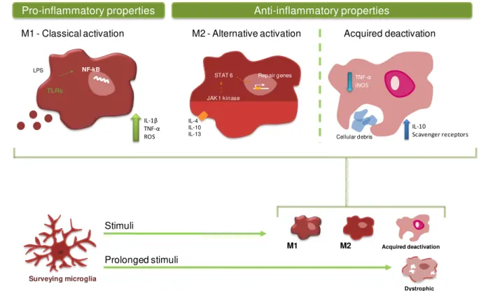
Figure I.5. Diversity of microglia phenotypes... 18
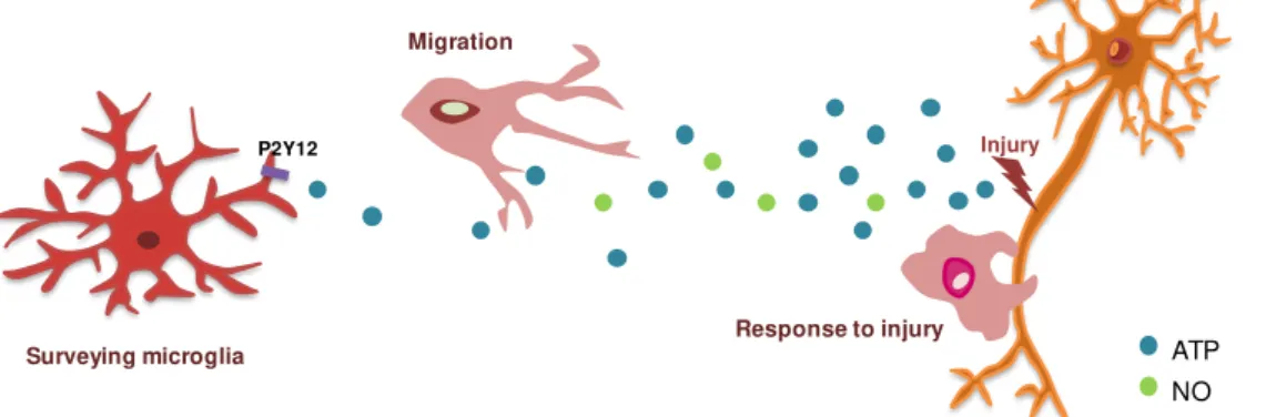
Figure I.6. ATP mediates chemotatic function of microglia ... 19
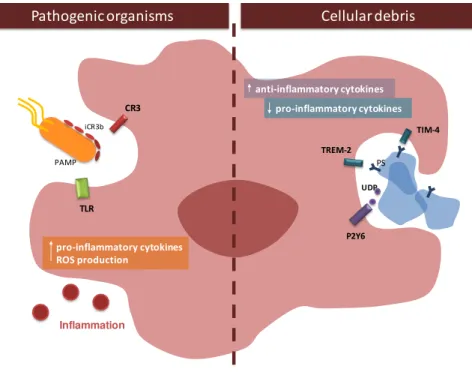
Figure I.7. Microglia phagocytosis and induced responses. ... 20
Figure I.8. Interplay between microglia and neurons in microglia in ALS. ... 26
II.MATERIALS AND METHODS ...29
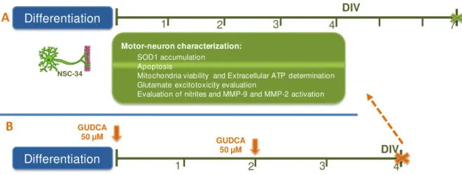
Figure II.1. Experimental procedure used in the characterization of transfected NSC-34 cell line and in the study of the neuroprotective effects of GUDCA... 31
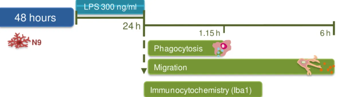
Figure II.2. Experimental procedure of N9 microglial cells characterization. ... 32
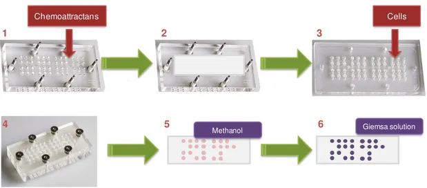
Figure II.3. Chemotaxis assay. ... 36
III.RESULTS ...37
Figure III.1. Differentiation induces neuron-like morphology in NSC-34 cells. ... 38
Figure III.2. NSC-34 cells demonstrate efficient transfected transfection with human SOD1. ... 39
Figure III.3. Accumulation of SOD1 is noticed at 3 days in vitro (DIV) in NSC-34/hSOD1G93A cells. ... 39
Figure III.4. Differentiated NSC-34/hSOD1G93A cells evidence increased apoptosis after 4 days in vitro (DIV) and reduced viability in the first 2 DIV... 40
Figure III.5. Differentiated NSC-34/hSOD1G93A cells show altered energy metabolism evidenced by decreased mitochondria viability and reduced release of ATP and glutamate. ... 42
Figure III.6. Matrix metalloproteinase-9 (MMP-9) activation and NO production are elevated in NSC-34/hSOD1G93A cells at 4 DIV. ... 43
Figure III.7. Beneficial effects of GUDCA on NSC-34/hSOD1G93A cellular stress. ... 44
Figure III.8. Microglia evidence amoeboid morphology when incubated with LPS for 24 hours. ... 45
Figure III.9. Phagocytosis is increased in N9 microglia cells exposed to LPS ... 46
Figure III.10. Microglia stimulated by LPS evidence decreased chemotaxis ability to ATP. ... 47
xxii
Figure III.12. Soluble factors released from NSC-34/hSOD1wt and NSC-34/hSOD1G93A equally enhance necrosis of N9 microglia in a time-dependent manner. ... 49
Figure III.13. Soluble factors released from NSC-34/hSOD1wt and NSC-34/hSOD1G93A equally enhance apoptosis of N9 microglia in a time-dependent manner. ... 50
Figure III.14. Microglia demonstrate amoeboid morphology after 24 hours of incubation with NSC-34/SOD1G93A conditioned media collected at 4 DIV. ... 51
Figure III.15. Conditioned media from NSC-34/hSOD1G93A incubated for 4 DIV reduces microglial phagocytic ability. ... 52
IV.DISCUSSION ...55
xxiii ALDH1L1 Aldehyde dehydrogenase family 1, member L1
ALS Amyotrophic Lateral Sclerosis
AMPA α-amino-3-hydroxy-5-methyl-4-isoxazole propionic acid
ATP Adenosine triphosphate
Aβ Amyloid-beta
CCL2 Chemokine motif ligand 2
CNS Central nervous system
CSF Cerebrospinal fluid
Cyt c Cytochrome c
DMEM-F12 Dulbecco’s modified Eagle’s medium-Ham’s F12 medium
EAAT2 Excitatory aminoacid transporter 2
ER Endoplasmic reticulum
fALS Familial Amyotrophic Lateral Sclerosis
FBS Fetal bovine seruum
GFAP Glial fibrillary acidic protein
GluR2 Glutamate Receptor 2
GUDCA Glycoursodeoxycholic acid
Iba1 Ionized calcium binding adaptor protein
IFN-γ Interferon-gamma
IL Interleukin
iNOS Inducible nitric oxidase synthase
LPS Lipopolysaccharide
MHC Major histocompatibility complex
MMP Matrix metalloproteinase
mSOD1 Mutant Superoxide dismutase 1
NF-κB Nuclear factor - κB
NMDA N-methyl-ᴅ-aspartate
NO Nitric oxide
PI Propidium iodide
PS Phosphatidylserine
RNS Reactive nitrogen species
ROS Reactive oxygen species
xxiv
SOD1 Superoxide dismutase 1
Tc T citotoxic cell
TGF-β Transforming growth factor-β
Th T helper cell
TIM-4 T cell immunoglobulin mucin 4
TLR Toll-like receptor
TNF-α Tumour necrosis factor α
TRAF2 Tumour necrosis factor receptor-associated factor 2
Treg T regulatory cell
TREM-2 Triggering receptor expressed by myeloid cells-2
TUDCA Tauroursodeoxycholic acid
UCB Unconjugated bilirubin
UDCA Ursodeoxycholic acid
UPR Unfolded protein response
1
I. Introduction
I. I
NTRODUCTION
1. Amyotrophic Lateral Sclerosis – an overview
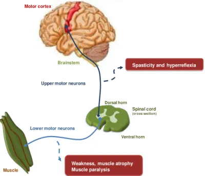
Amyotrophic Lateral Sclerosis (ALS), also known as Lou Gehrig’s disease, is a neurodegenerative disorder characterized by the selective loss of upper and lower motor neurons, and was first described by Jean-Martin Charcot and his colleague Alexis Joffroy in 1869. As represented in Figure I.1, in motor cortex, upper motor neurons degeneration results in spasticity and hyperreflexia, while death of lower motor neurons in brainstem and spinal cord leads to weakness, muscle atrophy and, ultimately, to paralysis of voluntary muscles, culminating in death from respiratory failure, as reviewed in Tripathi and Al-Chalabi (2008). The mean age of onset is 55–60 years, and men are more affected than women. The average survival from symptom onset is approximately 3-5 years but some patients demonstrate a slower disease course (Wood-Allum and Shaw, 2010).
Muscle cramps and fasciculations seem to be the primary clinical features as they seem to precede other symptoms for years. Moreover, degeneration of motor neurons seems to begin focally and disseminates into contiguous groups of motor neurons, which implies that disease is propagated to healthy neurons in neighbouring areas, where glial cells also have a role (Ferraiuolo et al., 2011).
Most of ALS cases have sporadic origin (sALS), however about 10% are inherited, being designated as familial ALS (fALS). Usually, fALS is associated with autossomal dominant inheritance and about 20% of familial cases are due to mutations in superoxide dismutase 1 (SOD1) (Rothstein, 2009). Recent findings have also demonstrated the role of newly identified genes in pathogenesis of ALS (Musaro, 2010), as it will be further discuss. Interestingly, sALS and fALS share similar clinical and pathological features, therefore research regarding fALS can also be applied to sporadic cases (Bruijn
2
Despite the precise molecular pathways involved in ALS are not yet elucidated, accumulating evidence reveals that ALS is a multifactorial disease resulting not only from altered molecular mechanisms within motor neurons but also from the involvement of other cells, such as glia (Ferraiuolo et al., 2011).
Figure I.1. Corticospinal tract is affected in ALS disease. Upper motor neurons prolong their axons from the motor cortex through brainstem to spinal cord. In ALS, degeneration of these neurons results in spasticity and hyperreflexia. On the other hand, degeneration of lower motor neurons leads to weakness and muscle atrophy and, ultimately, to muscle paralysis as dying neurons fail to make connection between spinal cord (or brainstem) and muscles. Adapted from Kiernan et al.(2011).
1.1 Motor neuron vulnerability in ALS
Selective death of motor neurons appears to begin focally and asymmetrically in the upper and lower limb with progressive spreading of injury to contiguous groups of motor neurons. Although the underlying mechanisms are not completely understood, special features of motor neuron physiology could raise the susceptibility to injury. First, the large cell size and long axon processes demand a robust cytoskeleton with high content of neurofilaments. In order to correctly perform neuronal transmission through all the axon, motor neurons need high metabolic rate and optimal mitochondrial function (Ferraiuolo et al., 2011). Due to this intense mitochondria activity, amplified production of
reactive oxygen species (ROS) is observed. High intrinsic oxidative stress is hence a putative mark of specific vulnerability of motor neurons. Moreover, with the accumulation of mutations and oxidative damage characteristic of aging, neurons may undergo special sensitivity to those mechanisms (Shaw and Eggett, 2000).
Motor neurons are extremely sensitive to glutamate accumulation, which can occur following inappropriate neurotransmission or dysfunction of the underlying mechanisms. Expression of α -amino-3-hydroxy-5-methyl-4-isoxazole propionic acid (AMPA)/kainate receptors lacking the GluR2 (Williams
et al., 1997) is one of the causes since this subunit is impermeable to calcium influx, therefore
preventing the accumulation of intracellular calcium. In addition, the low expression of calcium-Spasticity and hyperreflexia
Weakness, muscle atrophy Muscle paralysis Upper motor neurons
Lower motor neurons
Spinal cord
(cross section)
Ventral horn Dorsal horn
Motor cortex
Brainstem
3 buffering proteins aggravates the described calcium homeostasis deregulation. This topic will be further discussed in the section of molecular mechanisms involved in ALS.
Furthermore, motor neurons also evidence reduced capacity for heat shock response and sensitivity to endoplasmic reticulum (ER) stress, which can predispose the cells to oxidative damage and calcium overload (Ferraiuolo et al., 2011). Finally, reduced proteasome function and high
expression of particular proteins, such as SOD1, possibly enhance motor neuron vulnerability to toxicity resulting from mutant proteins accumulation, a key feature of ALS disease.
1.2 Molecular mechanisms of motor neuron degeneration
Atrophy of dying motor neurons is described as the pathological hallmark of ALS (Pasinelli and Brown, 2006; Tovar et al., 2009). Nevertheless, ever more studies have tryed to understand the
molecular mechanisms implicated in the selective loss of motor neurons and recently some of the pathways involved have been discovered. Genetic factors, mitochondrial dysfunction, ER stress, oxidative damage, excitotoxicity, protein aggregation and axonal transport impairment, among others, seem to be interrelated and implicated in the degeneration of motor neurons in ALS.
1.2.1 Protein aggregation: SOD1 mediated toxicity
Protein aggregation has been described as one of the pathological features of ALS. So far, cytoplasmic inclusions of different mutant proteins were found to be related with fALS cases, namely mutations in TAR DNA-binding protein 43 (TDP-43), fused in sarcoma (FUS) and in copper-zinc superoxide dismutase 1 (SOD1) (Ferraiuolo et al., 2011; Ince et al., 2011). Furthermore, these mutant
proteins are also found in sporadic cases reinforcing their role in the pathogenesis of ALS. SOD1 mutations are the most common, being associated to 20% of familial cases, which make them the basis for several in vitro and in vivo models of ALS (Gomes et al., 2008; Van Den Bosch, 2011).
SOD1 is a ubiquitous protein which functions as a homodimer and its normal function is to catalyse the dismutation of superoxide radicals to hydrogen peroxide (Tripathi and Al-Chalabi, 2008). SOD1 is mainly found in cytosolic compartment but it can also be found in mitochondria. Mutations in SOD1 were first identified by Deng and colleagues (1993) and nowadays over then 100 mutations are recognized.
Conformational instability of mutant SOD1 (mSOD1), which induces the formation of harmful aggregates, are observed in SOD1 transgenic mice (Karch et al., 2009; Wang et al., 2003). Therefore,
aggregation of the mutant protein is currently most favored as the main cause of SOD1 mediated toxicity (Tripathi and Al-Chalabi, 2008). Putative mechanisms of toxicity induced by SOD1 aggregation include sequestration of essential cellular components, reduced anti-apoptotic chaperone activity, impaired protein degradation as a cause of overwhelmed proteasome and damage of specific organelles, such as mitochondria and peroxisomes (Ferraiuolo et al., 2011; Rothstein, 2009).
Loss of function is unlikely involved in ALS. In fact, it was reported that mice where SOD1 was homozygously deleted did not develop the disease (Tripathi and Al-Chalabi, 2008; Wong et al., 1995).
However, some ALS familial cases have evidenced reduced SOD1 dismutation activity (Liu et al.,
4
Brown, 2006). Indeed, Liu and colleagues (1999) have already described it, where an oxidative function is acquired by G93A mutation in SOD1.
Finally, SOD1 aggregation may be an early event in the disease as it appears by the time of disease onset and its abundance increases with disease progression. Moreover, Friedlander and colleagues (1997) have demonstrated that the effects of mSOD1 expression seem to appear primarily within motor neurons and, in later stages of the disease, the rate of progression is determined by SOD1 mutations acting on microglia.
1.2.2 Oxidative stress
The classical concept of oxidative stress describes a mechanism resulting from imbalance between generation of ROS and antioxidant activities (Sies, 1997). More recently, this concept is being redefined as “a disruption of redox signaling and control that recognizes the occurrence of
compartmentalized cellular redox circuits”, as cited by Packer and Cadenas (2007), whereby ROS and
reactive nitrogen species (RNS) production are increased. ROS and RNS include nitric oxide (NO), hydroxyl radical and peroxynitrite, among others, and in physiological conditions are involved in cell signaling (Aguiar et al., 2012). However, in cases of excessive production, these species may cause
damage to proteins, lipids, DNA and, in particular, to messenger RNA. The central nervous system (CNS) is especially sensitive to oxidative damage because of the low levels of some antioxidant enzymes expression, high content of easily oxidized substrates and high production of ROS during neurochemical reactions (Carri et al., 2003).
Carri and colleagues (2003) have extensively reviewed the role of oxidative stress in the pathogenesis of ALS, specifying a series of oxidative damage markers such as intracellular levels of ROS, lipid peroxidation or protein nitration, among others. Patients with ALS show elevated markers of free radicals collected from cerebrospinal fluid (CSF), serum and urine (Mitsumoto et al., 2008;
Simpson et al., 2004; Smith et al., 1998). Accumulation of SOD1-related ALS has particular interest in
this mechanism since it is involved in the endogenous anti-oxidant capacity of the cell. Whether mSOD1 mediates oxidative damage through loss or gain of function is still controversial, as mentioned in the previous section. One hypothesis defends that mSOD1 provokes aberrant oxyradical reactions (Pasinelli and Brown, 2006). Indeed, mSOD1 exposes the active copper site to aberrant substrate, developing peroxidative activity. Furthermore, mSOD1 may also cause oxidative damage by mechanisms beyond its catalytic activity. NADPH oxidase-2 (NOX2) expression is increased in SOD1 transgenic mice and in CNS of ALS patients (Wu et al., 2006). Signaling required for the transcription
factor nuclear erytrhroid 2-related factor 2 (NRF-2) activation (a major regulator of the antioxidant response) may be disrupted in ALS mice models and patients (Ferraiuolo et al., 2011).
5 1.2.3 Mitochondrial dysfunction
Mitochondrial dysfunction has been extensively studied as one of the causes of motor neuron degeneration in ALS. Some of the most common features include mitochondrial depolarization, decrease of adenosine triphosphate (ATP) synthesis, impairment of calcium homeostasis and increased production of ROS. Moreover, mitochondrial damage has been attributed to the accumulation of mutant SOD1 in several in vitro and in vivo studies of ALS disease (Ferraiuolo et al.,
2011; Pasinelli and Brown, 2006; Volonte et al., 2011).
As already mentioned, accumulation of SOD1 is implicated in pathological mechanisms of the disease. Besides being a cytosolic protein, SOD1 can be found within mitochondria and the formation of mutant SOD1 aggregates in vacuoles, in the mitochondria intermembrane space has been described in an ALS mice model (Igoudjil et al., 2011). Vacuolation appears as a result of
degenerating mitochondria which, ultimately, leads to cell death, as referred in Tripathi and Al-Chalabi (2008). Indeed, vacuoles are evident at early stages of the disease in transgenic mice expressing human G93A mutation in SOD1 (SOD1G93A) and increase in number and volume along disease progression (Bendotti et al., 2001; Kong and Xu, 1998). In addition, reduced respiratory chain activity
and ATP production is evidenced in these mice. These findings seem to be corroborated by reports on ALS patients presenting clusters of abnormal mitochondria and defects in respiratory chain complexes I and IV (Pasinelli and Brown, 2006). How mutant SOD1 aggregates can induce mitochondrial impairment is still a matter of debate but at least three hypotheses are being tested (Pasinelli and Brown, 2006). First, mSOD1 could be involved in fusing the peroxisomes and the outer membrane opening pores in mitochondria membrane, thus, allowing the release of cytochrome c and triggering
apoptosis. Second, disruption of translocation machinery, in particular of the translocator outer membrane (TOM) complex, may result from aggregation of mutant SOD1 in the outer membrane limiting the import of functional proteins into the mitochondria. At last, abnormal interaction with other mitochondrial proteins can promote mitochondrial damage. Recently, Bcl2, an anti-apoptotic factor, has also been referred to be sequestered by SOD1 aggregates, disrupting its function (Pasinelli et al.,
2004).
Calcium buffering is also impaired in ALS transgenic mice. This deregulation could increase the susceptibility of motor neurons to the altered calcium homeostasis associated with glutamate excitotoxicity (Ferraiuolo et al., 2011). High calcium concentrations induce production of ROS in
mitochondria (Zhou et al. 2010) and contribute to the activation of cell death pathways as
caspase-mediated apoptosis (Hajnoczky et al., 2006; Roy and Hajnoczky, 2008).
6
1.2.4 Excitotoxicity of glutamate
Glutamate is the excitatory neurotransmitter of the corticospinal tracts and certain spinal interneurons. There are three groups of glutamate receptors in postsynaptic neurons essential to a physiological neurotransmission. N-methyl-ᴅ-aspartate (NMDA) receptors stimulation occurs with
calcium and sodium entry while non-NMDA receptors, namely AMPA/kainate and G-protein linked metabotropic receptors, allow mainly the influx of sodium (Tripathi and Al-Chalabi, 2008; Van Den Bosch et al., 2006). GluR2 is a subunit present in AMPA receptors that is responsible for the
resistance to calcium entry being important in glutamate excitotoxicity. During neurotransmission, excitatory signals are ended by the removal of glutamate from the synaptic cleft by glutamate reuptake transporters. When neuronal energy homeostasis or glutamate receptor expression is altered, excessive glutamate is released and accumulates in the synaptic clefts leading to excitotoxicity. Glutamate excitotoxicity is, hence, a well-recognized mechanism of neuronal death resulting from excessive activation of postsynaptic glutamate receptors, and is involved in ALS (Rothstein, 2009). Indeed, a twofold increase in glutamate levels was found in cerebrospinal fluid of ALS patients (Rothstein et al., 1990). In addition, a study with 400 patients with sporadic ALS showed that the
amount of glutamate was correlated with the disease severity (Spreux-Varoquaux et al., 2002).
Furthermore, hyperexcitability of the motor system in the presymptomatic or early stages of the disease were found by electrophysiological studies in humans (Vucic and Kiernan, 2006; Vucic et al.,
2008).
Clearance of glutamate after neurotransmission is critical in preventing excitotoxicity. Excitatory aminoacid transporter 2 (EAAT2), also known as glutamate transporter-1 (GLT1), is the most abundant transporter and is essential for maintaining low levels of extracellular glutamate. It is highly expressed in astrocytes, which are known to be important for rapid removal of synaptic glutamate released by motor neurons. Studies using SOD1 transgenic mice have reported that EAAT2 expression in ventral horn is reduced in presymptomatic stage and completely abolished in end-stage of the disease, as reviewed in Rothstein (2009). Overexpression of EAAT2 delayed the onset of motor neurons injury and decreased the caspase-3 activation and the formation of aggregates in SOD1G93A mice (Guo et al., 2003).
On the other hand, release of glutamate is accompanied by the massive influx of calcium through permissive receptors. Calcium entry induces the production of NO and of other ROS species leading to consequent organelle damage and cell death. Motor neurons are particularly sensitive to calcium accumulation as they have low capacity to buffer calcium and lack the GluR2 subunit of AMPA receptors (Pasinelli and Brown, 2006), which enhances the vulnerability of motor neurons to excessive glutamate stimulation.
1.2.5 Endoplasmic Reticulum stress
7 response (UPR), is elicited in order to rapidly decrease the load of misfolded proteins. UPR involves the recognition of aberrant proteins by ER-chaperones that promotes a correct protein folding (Ferraiuolo et al., 2011; Kanekura et al., 2009; Walker and Atkin, 2011).
Recently, studies in cerebrospinal fluid and in spinal cord from patients that died with ALS, as well as in ALS mice models, have shown that levels of ER stress-related proteins were upregulated evidencing the contribution of ER in the pathogenesis of the disease. ER stress sensors include activating transcription factor 6 (ATF6), inositol-requiring kinase 1 (IRE1) and PKR-like endoplasmic reticulum kinase (PERK), an ER-resident type I transmembrane protein kinase (Walker and Atkin, 2011), which are actively repressed by association with the chaperone immunoglobulin binding protein (BiP) (Kanekura et al., 2009). ATF6 is activated in a motor neuron like cell line (NSC-34) expressing
mutant SOD1 in G93A as showed by Prell and colleagues (2012). Protein disulphide isomerase (PDI), another ER chaperone and marker of UPR, is also activated in SOD1 transgenic mice and biological samples from patients with sporadic ALS and, interestingly, evidenced to be colocalized with SOD1 inclusions (Atkin et al., 2006; Atkin et al., 2008). Furthermore, activation of stress sensors induces
downstream pathways implicated in apoptosis, like caspase-12 activation, leading to motor neuron degeneration (Ferraiuolo et al., 2011).
1.2.6 Apoptotic cell death
Apoptosis is a mechanism of programmed cell death essential for maintaining the homeostasis of different tissues, including the CNS. As previously mentioned, ALS disease can result from the disruption of several interconnected cellular mechanisms and organelles. Apoptosis seems to be also involved in motor neuron degeneration in ALS, as many of the referred mechanisms, such as mitochondrial dysfunction or ER stress, can promote the downstream cascade signaling of apoptotic cell death (Ferraiuolo et al., 2011).
Transgenic ALS mice have been a useful model to explore and understand the mechanisms involved in motor neuron death, particularly mice expressing mutant SOD1. Indeed, biomarkers of apoptosis can be detected in the terminal stages of ALS in transgenic mice as well as in humans. How SOD1 promotes apoptosis, either by aggregates formation or changes in its function (mutations can transform SOD1 from an anti- to a pro-apoptotic protein) is still a matter of debate (Pasinelli and Brown, 2006).
The first evidence of SOD1 mediated apoptosis comes from the impairment of the association of cytochrome c with the inner membrane of the mitochondria. Cytochrome c is translocated from the
mitochondria to the cytosol and promotes the activation of caspase-9 which initiates the apoptotic process in the mitochondria. At the same time that disease progresses in ALS mice, a reduction in intra-mitochondrial cytochrome c is observed (Bacman et al., 2006). In addition, caspase mediated
apoptosis is strictly connected with motor neuron degeneration. Caspase-1 seems to be involved at early stages, even before disease onset, and precedes the activation of caspase-9. Following activation of caspase-9, caspases-7 and -3 are stimulated, as schematically represented in Figure I.2. Caspase-7 activation coincides with the disease onset in SOD1G93A mice (Guegan et al., 2001) and
8
Interestingly, caspase-3 is expressed both in neurons and astrocytes and are responsible for cleavage and inactivation of the glutamate transporter EAAT2 (Pasinelli and Brown, 2006), which leads to glutamate excitotoxicity as the transporter fails to remove it from the synaptic cleft (see section 1.2.4).
Despite the role as an apoptotic inducer, caspase-1 is mainly a player in inflammation (Pasinelli and Brown, 2006). As it will be further analyzed, glial cells are involved in the progression of the disease. This finding demonstrates a relationship between motor neuron degeneration and astrogliosis as well as with microgliosis. Hence, this glial activation can be, not only important for disease progression, but also is an early event in ALS.
Moreover, Bcl2, an anti-apoptotic protein, seems to be related with apoptosis in ALS. In fact, overexpression of Bcl2 preserves motor function and prolonged life span in SOD1G93A mice (Pasinelli et al., 2004). SOD1 can bind to Bcl2 promoting the formation of complex aggregates. When
entrapped in these inclusions, Bcl2 become non-functional and the mechanism of apoptosis repression is disrupted. Also, another suggested feature is that Bcl2 undergoes conformational modification and becomes toxic (Rothstein, 2009).
Figure I.2. Molecular mechanisms involved in ALS pathogenesis. Motor neuron degeneration results from interrelated mechanisms that ultimately induce apoptotic events. Mutant superoxide dismutase 1 (mSOD1) accumulation is the primary event. Production of nitric oxide (NO) and reactive oxygen species (ROS) is induced, and oxidative damage is increased. Mutant SOD1 impairs the proteasome who fails in correct misfolded proteins and disrupt mitochondrial function as suggested by the decrease in ATP production. Mitochondria fusion and fission can also be observed. Nonetheless, sequestration of the anti-apoptotic protein Bcl2 and release of cytochrome c promote caspase activation and consecutive apoptosis. Caspase-9 (Casp9) seems to be early activated followed by activation of caspases-3 (Casp3) and -7 (Casp7). Caspase-1 (Casp1) is also activated and its role is extended also to inflammatory responses. As misfolded proteins are accumulated, unfolded protein response (UPR) promotes the release of ER stress sensors which activate caspase-12 (Casp12). Glutamate excitotoxic accumulation in the extracellular space and massive influx of calcium through N-methyl-ᴅ-aspartate (NMDA) and α-amino-3-hydroxy-5-methyl-4-isoxazole propionic acid (AMPA)/kainate receptors leads to NO and ROS production further increasing oxidative stress.
Ca2+ UPR ATF6 IRE PERK Glutamate ATP Cyt c
9 1.3 Non cell-autonomous disease – involvement of glial cell activation
Initial evidences of pathological mechanisms involved in ALS suggested that the disease is cell autonomous, resulting from different cellular dysfunctions, which ultimately triggers motor neuron death. However, accumulating evidence shows the involvement of other non-neuronal cells in pathogenesis, particular in disease progression. This concept was first proposed by Ince and colleagues in 1996 with the observation of microglia and astroglial cells activation in an in vitro model
of ALS. In addition, activation of microglia, astrocytes and appearance of lymphocytes in post-mortem tissue of ALS patients and in spinal cord of SOD1 transgenic mice was similarly observed (Kawamata
et al., 1992; Philips and Robberecht, 2011). Studies with chimeric mice, where the expression of
mutant SOD1 in G93A was selectively induced on motor neurons, astrocytes or microglia have demonstrated that SOD1G93A-overexpressing neurons surrounded by healthy glia remained relatively intact, although healthy motor neurons presented signs of injury when surrounded by mutant SOD1G93A - overexpressing glia (Clement et al., 2003).
As reviewed in Philips and Robberecht (2011), studies in post-mortem tissues of ALS patients and in spinal cord of SOD1 transgenic mice evidenced changes in the morphology of astrocytes and microglia from a surveying phenotype to an activated state with upregulation of surface markers. CD11b, CD68 and ionized calcium binding adaptor protein (Iba1) expression were elevated in microglial cells, as well as glial fibrillary acidic protein (GFAP) and aldehyde dehydrogenase family 1, member L1 (ALDH1L1) in astrocytes. Taken together, these results evidenced that expression of mutant SOD1 in motor neurons is necessary to incite ALS but also requires toxicity of the mutant protein in the surrounding cells.
In addition, several studies with different approaches have demonstrated that mutant SOD1 expression in motor neuron determines the initial timing of disease onset and early progression in some cases, but does not have a significant contribution to later disease progression, (Ilieva et al.,
2009). Consequently, astrocytes and microglia must have decisive implications in disease progression after its onset. Indeed, mutant SOD1 gene excision from microglia and selective reduction in astrocytes significantly slowed disease progression (Boilleé et al., 2006; Yamanaka et al., 2008).
However, there is still some controversia on whether astrogliosis and microgliosis can be detrimental or beneficial, what surely will depend from the levels that are induced.
1.3.1 Astrocytes
10
neuronal integrity through the release of neurotrophic factors such as glial-cell derived neurotrophic factor (GDNF), brain-derived neurotrophic factor (BDNF), insulin-like growth factor 1 (IGF1) and vascular endothelial growth factor (VEGF). This release seems to be disrupted in ALS, since the administration of some of these compounds or their overexpression in a mouse model expressing mutant SOD1 have demonstrated to increase mice survival (Kaspar et al., 2003; Park et al., 2009). In
addition, activated astrocytes fail to confer protection to glutamate excitotoxicity due to the loss of EAAT2 transporters, as observed in both transgenic mice and in sporadic and familial cases of ALS (Lasiene and Yamanaka, 2011), indicating the importance of astrocytic damage to motor neuron degeneration in ALS. Moreover, astrocytes expressing mutant SOD1 release factors that inhibit the expression of GluR2 in motor neurons (Ilieva et al., 2009). Unlike GluR2, NMDA receptor is permeable
to calcium facilitating its influx and, thus, promotes motor neuron toxicity resulting from intracellular calcium accumulation. On the other hand, upregulation of inducible nitric oxidase synthase (iNOS) and consequently the release of toxic factors, particularly ROS and NO, suggest astrocytic involvement in ALS (Philips and Robberecht, 2011). In vitro studies also evidenced that SOD1 astrocytes release
factors which are capable of inducing motor neurons degeneration (Haidet-Phillips et al., 2011).
Studies in post-mortem tissues of ALS patients have provided clues for the role of non-neuronal cells in the late stage of the disease. In the spinal cord, astrocytosis is observed in ventral and dorsal horn, as well as in the intersection of fibers with the corticospinal tract entrance in the grey matter. Activated astrocytes also appear in cortical grey matter, subcortical white matter and are not restricted to motor cortex (Philips and Robberecht, 2011).
For all that findings, astrocytes have now much interest in the design of therapeutic strategies in ALS. Interestingly, glial-restricted precursor cells have been transplanted in the spinal cord of SOD1G93A rats to evaluate the possible protective role of the wild type SOD1 astrocyte precursor (Lepore et al., 2008). These cells showed to be able to differentiate into GFAP-expressing astrocytes
and were capable to rescue motor neuron injury, in a way that seems dependent of elevated glutamate scavenging activity. Moreover, it was observed that this transplantation led to a significant increase in rat survival.
1.3.2Oligodendrocytes and Schwann cells
Oligodendrocytes in CNS, and Schwann cells in the peripheral nervous system, are responsible for the myelin sheaths surrounding neurons which provide electrical insulation essential for rapid signal conduction. Schwann cells also participate in the clearance of debris and in guiding the axon after neuron damage (Ilieva et al., 2009). Although there is little evidence that these cells might be
involved in ALS pathogenesis, some recent studies have been trying to clarify this concept. As reviewed by Lasiene and Yamanaka (2011), abnormalities in oligodendrocytes related myelin, such as loss of compact myelin and lamellae detachment, are seen in spinal cords of pre-symptomatic SOD1 transgenic rats and aggravated at symptomatic stages. In what concerns to Schwann cells, intriguing findings were recent revealed.
11 elimination of mutant SOD1 specifically in Schwann cells failed to slow disease progression. Instead, a substantial acceleration of the late phase of disease was observed (Lobsiger et al., 2009). Therefore,
the underlying mechanism suggests a protective role to mutant SOD1 possibly due to the dismutation activity that can ameliorate some oxidative damage within the cells (Ilieva et al., 2009). However,
recently, Wang and colleagues (2012) found that knockdown of mutant SOD1 in Schwann cells of SODG85R transgenic mice delayed disease onset and extended survival indicating that SOD1G85R expression is neurotoxic. These results implies that different mutations confer different outcomes to cell toxicity and, in the case of Schwann cells, oxidative damage seems to be an important feature in the context of ALS.
1.3.3 Microglia
Microglia is the most important glial cell in the context of ALS as they are the typical immune cells of the CNS. There is accumulating evidence that neuroinflammation is as an imperative feature in ALS and, thus, seem to be strictly connected with microglia activation (Neusch et al., 2007). Microglia
expressing mutant SOD1 change from their normal surveying state to an activated one, migrate to initial sites of injury and trigger high production of extracellular superoxide and ROS (Ferraiuolo et al.,
2011). Indeed, like astrocytes, microglia damage has been shown to rapidly contribute to disease progression (Ilieva et al., 2009). Furthermore, Boillée and colleagues (2006) have demonstrated that
reduced expression of mutant SOD1 specifically in microglia in a mouse model of ALS significantly slowed disease progression. Also, studies indicated that bone marrow transplantation of SOD1G93A transgenic mice with microglia expressing wild type SOD1 altered the functional properties of microglia and prolonged mice survival (Beers et al., 2006). More recently, it was observed that microglia is
highly reactive in preclinical stages of ALS in the transgenic rat model with mutant SOD1G93A (Graber et al., 2010) however its ablation in spinal cord close to clinical onset has not shown to protect
motor neurons (Gowing et al., 2008). Moreover, replacement of microglia cells expressing mutant
SOD1 using clodronate liposomes significantly slowed disease progression and prolonged survival of the transgenic ALS mice (Lee et al., 2012).
Such observations may underlie a clue in linking microglial activation with ALS progression. The present dissertation focuses on the comprehension of microglia properties in ALS pathogenesis using
in vitro models, therefore, microglia features will be further dissected in the section 2 of this
12
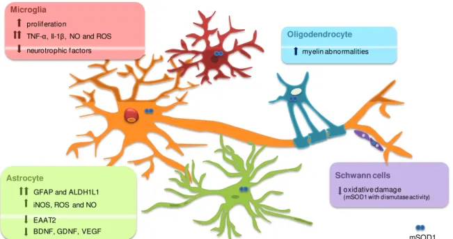
Figure I.3. ALS is a non-cell autonomous disease. Non-neuronal cells in the CNS are also involved in ALS pathogenesis. Microglia shows increase proliferation as well as production of inflammatory cytokines such as tumour necrosis factor alpha (TNF-α) or interleukin-1 beta (IL-1β) and reactive oxygen species (ROS) when mutant SOD1 (mSOD1) accumulates within the cell. In addition, reduction of neurotrophic factors accounts for motor neuron degeneration. Oligodendrocytes seem to be involved just to some extent as myelin abnormalities can be observed. Schwann cells have reduced oxidative damage when express mSOD1 with dismutase activity being able to slow disease progression. Finally, astrocytes express high amounts of reactive oxygen species (ROS) and nitric oxide (NO) and fail to support neuronal function as factors such as BDNF (brain derived neurotrophic factor), GDNF (glial-cell derived growth factor) or vascular endothelial growth factor (VEGF) are reduced. EAAT2 (excitatory aminoacid transporter-2) is inactivated by mutant SOD1 contributing to excitotoxicity due to lack of glutamate clearance from the synaptic cleft. Adapted from Ilieva et al. (2009).
1.4 Suitable models for ALS pathogenesis studies
Studies in biological samples from ALS patients and genetic analysis have provided important clues for the candidate intermediates involved in the pathogenesis of the disease. However, more clear and specific mechanisms have been uncovered by in vitro and in vivo models.
Since the study of the sporadic form of the disease is extremely difficult by the absence of suitable models, most recent research focuses on the genetics of familial ALS that has shown several mutations. Among them, mutations in SOD1 are the most studied. Therefore, mice expressing mutant proteins associated with fALS have been created in order to unravel the mechanisms of motor neuron loss, especially with different forms of SOD1 (Van Den Bosch, 2011). The first transgenic mice created expressed the human protein with substitution of glycine over adenine at position 93 (G93A) by its insertion in the genome (Gurney, 1994). These mice show progressive hind limb weakness leading to paralysis and death, and were able to replicate the disease progression observed in patients. Overexpression of non-mutated protein gives no phenotype, supporting the role of mutant SOD1 as cause of ALS in mice (Van Den Bosch, 2011). Further reports also showed various abnormalities in the mice before disease onset, such as behavioral motor changes (Mead et al., 2011),
electrophysiological dysfunctions (Bories et al., 2007) and mitochondria damage (Bendotti et al., Schwann cells
oxidative damage
(mSOD1 with dismutaseactivity)
Oligodendrocyte
myelin abnormalities
mSOD1
Microglia
proliferation
TNF-α, Il-1β, NO and ROS neurotrophic factors
Astrocyte
GFAP and ALDH1L1
BDNF, GDNF, VEGF iNOS, ROS and NO
13 2001). Therefore, several mice expressing SOD1 mutant were created with other mutations such as G37R, G85R or D90A (Bruijn et al., 1997; Jonsson et al., 2006; Wong et al., 1995) that evidenced
similar phenotype as SOD1G93A transgenic mice.
In vitro studies addressing mechanisms implicated in ALS have also been performed in cultures of
motor neurons isolated from mice spinal cord (Tovar et al., 2009) and in NSC-34, a cell line that
results from hybridization between neuroblastoma cells and motor neurons from mice spinal cord (Cashman et al., 1992). The study of such a complex disease using in vitro models, which represent
such a reduced and limited system, has several inconvenient but, still, can provide important informations on motor neurons physiology at cellular and molecular levels (Tovar et al., 2009).
NSC-34 cells, as reported by Cashman and colleagues (1992), seem to be a good model as they evidence morphological and physiological properties of motor neurons including extension of processes, formation of contacts with cultured myotubes, support of action potentials and expression of neurofilament proteins, among other features. Moreover, these cells may be able to model aspects of neuromuscular synapse formation, as referred by Martinou and colleagues (1991). Recently, NSC-34 cells have been transfected with mutant SOD1 in G93A and some features of ALS pathogenesis are becoming uncovered, such as the formation of mutant SOD1 aggregates and Golgi apparatus disruption (Gomes et al., 2010; Gomes et al., 2008) and mitochondrial impairment (Raimondi et al.,
2006). Also, NSC-34 cells expressing SOD1G93A evidenced less proliferation and differentiation ability (Gomes et al., 2008). However, NSC-34 cells retain some characteristic of neuroblastoma cells
that can interfere with several cellular responses, such as the presence of the gene N-myc, an oncogene involved in cell proliferation. For the study of the pathways involved in neuronal death and its prevention by possible therapeutic agents, the effects of N-myc can disrupt the underlying mechanisms (Tovar et al., 2009). Nonetheless, this in vitro model can be a useful tool to explore early
molecular mechanisms of ALS as well as the features implicated in the disease progression.
1.5 Is GUDCA a promising therapy?
ALS is an extremely severe disease and attempts to find a novel therapeutic agent are imperative since the only drug approved for use in ALS is riluzole which only slightly prolongs survival (Pasinelli and Brown, 2006). Riluzole is a benzothiazole derivative and has been proven to ameliorate glutamate excitotoxicity through different mechanisms including inhibition of pre-synaptic release of glutamate or inactivation of neurons sodium channels that reduces glutamate transmission (Tripathi and Al-Chalabi, 2008). However, a different property seems to be associated with riluzole neuroprotection effects, as other anti-glutamate agents fail to be effective in clinical trials (Ferraiuolo et al., 2011). So far, there is
no successful drug for ALS treatment and search for novel therapeutic agents that could prevent motor neuron degeneration is of a great importance. In this context, we propose to study the efficacy of glycoursodeoxycholic acid (GUDCA), a conjugated species of ursodeoxycholic acid (UDCA) with glycine, on the prevention of neuronal degeneration in the cellular models of ALS since it demonstrates several neuroprotective features, as it will be further discussed.
14
several functions in the liver, reinforcing hepatocyte function. Mechanisms of action include alteration of bile acids pool, mitochondrial integrity, immune modulation and anti-apoptotic properties, as summarized in Lazaridis et al. (2001). Therefore, UDCA is commonly used for the treatment of
hepatobiliary disorders (Rudolph and Link, 2002). Following oral administration, UDCA is consequently submitted to hepatic conjugation either with glycine or taurine, producing, respectively, glycoursodeoxycholic (GUDCA) and tauroursodeoxycholic (TUDCA) acids, as schematically represented in Figure I.4. GUDCA is the major species, accounting to 79.8% of UDCA conjugation and, so, is the form with the highest clinical relevance (Lazaridis et al., 2001). These resulting
conjugated species have shown cytoprotective properties in different CNS cells, such as neurons, astrocytes or microglia (Rodrigues et al., 2000; Silva et al., 2012; Vaz et al., 2010). Interestingly,
TUDCA has already proven beneficial effects in many neurodegenerative diseases, namely in
Alzheimer’s, where it was able to inhibit apoptosis in an in vitro model of AD mutant neuroblastoma
cells (Ramalho et al., 2006; Ramalho et al., 2008).
The ability of GUDCA to prevent the demise of different cells from CNS in conditions mimicking a moderate and severe hyperbilirubinemia have been extensively studied by our group. In this regard, protection against unconjugated bilirubin (UCB)-induced oxidative damage was achieved by pre-incubation of rat cortical neurons with 50 µM of GUDCA (Brito et al., 2008). Later, Vaz and colleagues
(2010) have demonstrated that GUDCA restore cellular antioxidant potential on immature rat neurons after UCB treatment, evidencing the ability of this bile acid to modulate oxidative stress. In the same study, it was reported that GUDCA is able to ameliorate UCB-induced mitochondrial respiratory chain dysfunction and to reverse the inhibition of cytochrome c oxidase activity (Vaz et al., 2010).
Immunosuppressive action was also showed in a study performed by Fernandes and colleagues (2007) with astrocytes exposed to UCB. Here, previous incubation with GUDCA reduced astroglial production and release of tumour necrosis factor-alpha (TNF-α) and interleukin-1 beta (IL-1β). More
recently, neuroprotective effects of GUDCA were extended to the prevention of NO increase and glutamate release as well as to benefits in neuronal outgrowth dynamics and synaptic activity disruption by UCB in cortical neurons (Silva et al., 2012).
15 in the potential therapeutic properties of the bile acid in the treatment of ALS. Indeed, Min and colleagues (2012) have performed a clinical trial using UDCA in 80 ALS patients. Although without conclusive results, the patients were tolerant to oral administration and this pilot study could open a new view in the use of UDCA and its conjugated species in the treatment of ALS. In addition, a promising argument that GUDCA could actually be succeeded relies in the fact that other anti-inflammatory drugs have proven to be efficacious in ALS mice models treatments (Philips and Robberecht, 2011).
2. Neuroprotective vs. neurotoxic properties of microglia in ALS
2.1 Microglia: role in CNS
Microglia constitute about 10-20% of glial cell population and exhibit a small cell soma with fine and long processes with dynamic protrusions (Nimmerjahn et al., 2005). They populate CNS
parenchyma in both white and grey matter, exhibiting a higher density within the hippocampus, basal ganglia and substancia nigra (Lawson et al., 1990; Tambuyzer et al., 2009). Microglia origin is still a
matter of discussion, however some authors believe that microglia consists of a subpopulation generated from bone-marrow cells that migrate to CNS during embryonic development and a second one derived from myeloid progenitor cells that enter the brain after birth (Ginhoux et al., 2010; Ohsawa
and Kohsaka, 2011; Prinz and Mildner, 2011). Microglial cells share many properties of macrophages as they belong to the mononuclear phagocyte lineage (Vilhardt, 2005) and in fact, they are considered the resident immune cells of CNS, as they act as the brain’s innate immune system (Aloisi, 2001; Streit, 2002). However, it was recently shown that, at resting conditions, they differ from macrophages as they express much lower CD45 levels, which represents the only phenotypical mean available to distinguish the two populations (Greter and Merad, 2012).
They are continuously monitoring the surrounding environment with their highly branches and motile cell processes (Nimmerjahn et al., 2005; Ohsawa and Kohsaka, 2011) and are the first line of
defence against pathogenic organisms. When the tightly regulated CNS homeostasis is disturbed, microglia rapidly change their morphology, gene expression profile and functional behaviour (Calvo and Bennett, 2011), which is considered an activated phenotype. In fact, the traditional classification of microglia phenotypes includes a resting state, where microglia have a highly ramified morphology with small cell body in contrast to the amoeboid morphology of activated microglia. This concept implies that microglia are inactive or quiescente before an activation stimulus. However, accumulating evidence indicate that microglia activation should be considered a change in functional phenotypes rather than an awakening (Hanisch and Kettenmann, 2007). Microglia constitute, hence, a unique population and distinct from all other CNS cells and its functions will be described in the next sections.
2.1.1 Monitoring CNS environment under physiological conditions