João Abel Rainho Fonseca
Licenciado em Biologia
Exploring the role of proteolysis in
Extracellular Matrix remodeling: Links to
Chronic Obstructive Pulmonary Disease
and Lung Cancer
Dissertação para obtenção do Grau de Mestre em Genética Molecular e Biomedicina
Orientador: Susana Seixas, PhD, Instituto de Investigação e
Inovação em Saúde, Universidade do Porto (I3S); Instituto de
Patologia e Imunologia Molecular, Universidade do Porto
(IPATIMUP).
i Exploring the role of proteolysis in Extracellular Matrix remodeling: Links to Chronic Obstructi ve Pulmonary Disease and Lung Cancer
Copyright João Abel Rainho Fonseca, FCT/UNL, UNL
iii
AKNOWLEDGMENTS
Começo por agradecer ao diretor do i3S, Professor Doutor Mário Barbosa, bem como ao diretor do IPATIMUP, Professor Doutor Manuel Sobrinho Simões, pela oportunidade de realizar o meu trabalho em ambos os institutos. Agradeço também à Doutora Luísa Pereira, por me ter recebido no seu grupo “Genetic
Diversity”.
À Doutora Susana Seixas, minha orientadora, pela oportunidade de me envolver este projeto, por todo o apoio, compreensão, disponibilidade, e tudo aquilo que me transmitiu ao longo deste ano, essencial para o desenvolvimento do projecto.
À Sílvia e à Patrícia, membros do grupo ao qual pertencia, pela disponibilidade para me ajudarem com qualquer dúvida que me surgisse durante o meu trabalho, pela simpatia e boa disposição, proporcionando sempre um bom ambiente no laboratório e um grande apoio ao longo deste ano.
À Joana, minha colega de mestrado, que em conjunto realizamos várias etapas do nosso trabalho, sendo que este apoio mútuo foi importante ao longo do ano. Ao meu compincha de laboratório, Alex, por todas as asneiras que fizemos juntos no laboratório, e de todas as vezes que nos apoiamos um ao outro para não desanimar.
Ao resto do grupo Genetic Diversity, por toda a ajuda prestada durante o desenvolvimento deste projecto, tendo sido essencial em vários pontos do percurso.
À minha família por todo o apoio ao longo do ano, incentivando-me a continuar a acreditar em mim mesmo quando tive dúvidas. Um grande obrigado mãe e pai, pela dedicação em fazer com que nunca fosse a baixo, a todos os meus tios (Luís, Lucília, Adelaide, Amélia, Francisco, Graça), ao meu avô Joaquim, à minha prima Catarina, à Mi e ao Gavina, à Sandra, por toda a preocupação a tentar saber “como está a correr
essa coisa”, sabem que o vosso apoio foi bastante importante. Um grande obrigado à minha irmã Inês, por me estar sempre a dizer que eu não faço nada, motivando-me assim a mostrar-lhe o contrário, e ao meu pequeno irmão André, por me proporcionar os momentos mais hilariantes deste ano, sendo a melhor
escapatória da “chata vida de adulto”. Por fim, obrigado “big bro” André, por me teres guiado sempre que
precisei, considero-te um exemplo, e espero um dia chegar onde estás, ou pelo menos lá perto, desde que
“seja aquilo que eu me veja a fazer”.
v
ABSTRACT
Chronic obstructive pulmonary disease (COPD) and lung cancer (LC) are two complex disorders, currently representing the 4th cause of death and the most lethal cancer in Western countries, respectively. A mechanistic link between COPD and LC has been proposed due to an overlap of risk factors of both diseases, where uncontrolled proteolysis may be a critical event in their progress and outcomes. The activity of proteases, their substrates and inhibitors have a significant impact in the extracellular matrix (ECM) remodeling, which may ultimately lead to the development of COPD and LC. Despite the identification of several susceptibility factors in both diseases, there is still many aspects of their pathogenesis that require further elucidation. To address this issue, for our study, we selected 73 proteolysis genes, based on their roles in ECM remodeling, lung expression and/or presence in lung samples and former reports by Genome Wide Association Studies. In a first analysis, we took benefit of The Cancer Genome Atlas on-line database regarding clinical, epidemiological and mutational (somatic and germline) information for two common LC subtypes (adenocarcinoma and squamous cell carcinoma). We found that somatic mutability differs from germline trends and between the two LC subtypes, possibly affecting ECM in distinct ways. Then, we screened by means of PCR-based and Sanger sequencing techniques SERPINB3/B4 and CTSG genes, in a
small cohort of COPD and LC patients from which blood and bronchoalveolar lavage fluid samples were collected. Even though, we could not detect any somatic mutation in our sample, for SERPINB3 we detect
a considerable number of low-frequency variants in COPD cases in particular, suggesting a misfunction of this gene as a possible genetic risk factor for lung disease. Additional studies in larger cohorts of patients and controls are necessary to confirm this hypothesis.
vii
RESUMO
A doença pulmonar obstrutiva crónica (DPOC) e o cancro do pulmão (CP) são duas doenças complexas, representando atualmente a quarta maior causa de morte e o cancro mais letal em países ocidentais, respetivamente. Tem sido sugerida uma associação mecanística entre a DPOC e o CP em parte devido à partilha de fatores de risco comuns em que a desregulação da proteólise pode também constituir um acontecimento crítico na sua evolução. A atividade das protéases, seus substratos e inibidores têm um impacto significativo na remodelação da matriz extracelular (MEC) o que em ultima análise pode levar ao desenvolvimento da DPOC e CP. Apesar de alguns fatores de suscetibilidade a ambas as doenças terem sido já reconhecidos, muitos aspetos da sua patogénese requerem um estudo mais aprofundado. Neste trabalho foram selecionados de 73 genes de proteólise, tendo em consideração o papel de cada um na remodelação da MEC, expressão ou presença em tecido pulmonar, e descrição por parte de estudos de associação genómicos. Numa primeira fase, foram extraídos da base de dados The Cancer Genome Atlas que
compreende dois subtipos de CP (adenocarcinoma e carcinoma de células escamosas), informação clínica, epidemiológica e mutacional (somática e germinativa). Neste estudo verificou-se que a mutabilidade somática difere do padrão germinativo e entre os dois subtipos de CP, possivelmente afetando a MEC de forma distinta. Foi ainda efetuada uma análise dos genes SERPINB3/B4 e CTSG por métodos de PCR e
sequenciação de Sanger numa pequena coorte de doentes com DPOC e CP para os quais foram recolhidos sangue ou lavados brônquicos. Embora não tenha sido possível detetar qualquer mutação somática nas nossas amostras, para a SERPINB3 foram detetadas diversas variantes de baixa frequência em casos de
DPOC, sugerindo que alterações neste gene possam constituir um possível fator de risco genético na doença pulmonar. Estudos adicionais em coortes alargadas de doentes e controlos são essenciais para confirmar esta hipótese.
ix
TABLE OF CONTENTS
ABBREVIATIONS ... xv
1. Introduction ... 1
1.1. State of the art of Human Genetics and Human Disease Studies ... 1
1.2. The extracellular Matrix (ECM) in the healthy lung ... 2
1.2.1. The ECM structural macromolecules: Elastin and fibrillar collagens ... 3
1.2.2. The ECM multiadhesive macromolecules: fibronectin and laminins ... 6
1.2.3. The role of proteolysis in the ECM remodeling... 7
1.2.3.1. The Matrix metalloprotease (MMP) family ... 7
1.2.3.2. The families of Desintegrin and metalloproteases (ADAM and ADAMTS)... 9
1.2.3.3. Cathepsins and other serine proteases ... 9
1.2.3.4. Protease Inhibitors ... 10
1.3. Complex Lung Diseases ... 12
1.3.1. Chronic Obstructive Pulmonary Disease ... 13
1.3.2. Lung Cancer... 15
1.4. Mechanistic links between COPD and LC ... 16
1.4.1. Genetic Susceptibility factors ... 16
1.4.2. Oxidative stress, cell injury and inflammation ... 17
1.4.3. ECM remodeling and proteolysis in lung disorders ... 19
1.5. Patient Tailored Therapeutics ... 20
2. Materials and methods ... 23
2.1. Bioinformatics analysis ... 23
2.1.1. TCGA data – Lung Cancer ... 23
2.1.2. Clinical and epidemiological data analysis ... 23
2.1.3. Candidate genes selection... 24
x
2.2. Screening of Portuguese COPD and LC cases ... 27
2.2.1. Samples ... 28
2.2.2. DNA Extraction ... 28
2.2.3. Polymerase Chain Reaction (PCR) amplification and sequencing ... 28
2.2.4. Sequence analysis ... 32
2.2.5. Statistical Analysis ... 32
3. Results and Discussion ... 33
3.1.TCGA data analysis ... 33
3.1.1.1. Epidemiological analysis ... 33
3.1.2. Proteolysis related genes analyses ... 38
3.1.2.1. Somatic and germline mutations rates... 38
3.1.2.2. Candidate gene expression ... 44
3.2. Screening of SERPINB3, SERPINB4 and CTSG genes in Portuguese COPD and LC cases . 48 3.3. Concluding remarks ... 52
4. References ... 53
xi
INDEX OF FIGURES
Figure 1.1. Conserved SERPIN three-dimensional structure ... 11
Figure 1.2. COPD Phenotypes... 14
Figure 1.3. Repetitive cycles of tissue injury and repair... 19
Figure 2.1. Schematic representation of SERPINB3/SERPINB4 amplification ... 30
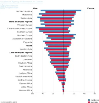
Figure 3.1. Lung cancer incidence and mortality rates by geographical populations and gender (2012 data). ... 34
Figure 3.2. Mutation rates as retrieved by cBioPortal for ADC patients... 38
Figure 3.3. Mutation rates as retrieved by cBioPortal for SCC patients ... 39
Figure 3.4. Top mutated candidate genes in ADC subtype and mutation functional predictions by Polyphen ... 40
Figure 3.5. Top mutated candidate genes in SCC subtype and mutation functional predictions by Polyphen ... 41
Figure 3.6. Top germline mutated candidate genes in ADC subtype and mutation functional predictions by Polyphen... 43
Figure 3.6. Top germline mutated candidate genes in ADC subtype and mutation functional predictions by Polyphen... 43
Figure 3.7. Top germline mutated candidate genes in SCC subtype and mutation functional predictions by Polyphen... 43
Figure 3.8. Expression change of candidate genes in normal and tumor tissue, with normalization by a logarithmic scale of fold change ... 47
Figure 3.9. Schematic representation of SERPINB35’UTR variants location ... 49
Figure 3.10. SERPINB3 protein structure with detected variants positions in reactive center loop highlighted ... 49
Figure A1. Somatic mutations rates of candidate genes in ADC patients with PolyPhen predictions .. 67
Figure A2. Somatic mutations rates of candidate genes in SCC patients with PolyPhen predictions ... 68
Figure A3. Germline mutations rates of candidate genes in ADC patients with PolyPhen predictions . 69 Figure A4. Germline mutations rates of candidate genes in SCC patients with PolyPhen predictions . 70 Figure A5. Somatic mutations rates of candidate genes in ADC patients with SIFT predictions ... 71
Figure A6. Somatic mutations rates of candidate genes in SCC patients with SIFT predictions ... 72
Figure A7. Germline mutations rates of candidate genes in ADC patients with SIFT predictions ... 73
Figure A8. Germline mutations rates of candidate genes in SCC patients with SIFT predictions ... 74
xiii
INDEX OF TABLES
Table 1.1. Summary of the human collagen classes... 5
Table 1.2. Different MMPs clades and their corresponding ECM substrates ... 8
Table 1.3. Clade B SERRPINs with known roles in lung function ... 12
Table 2.1. Proteolysis related candidate genes selected for this study ... 24
Table 2.2. Primers used for the amplification of SERPINB3/B4genes... 29
Table 2.3. Semi-nested PCR primers used for SERPINB3/B4 amplification... 30
Table 2.4. Primers used for SERPINB3/B4 and CTSGsequencing . ... 31
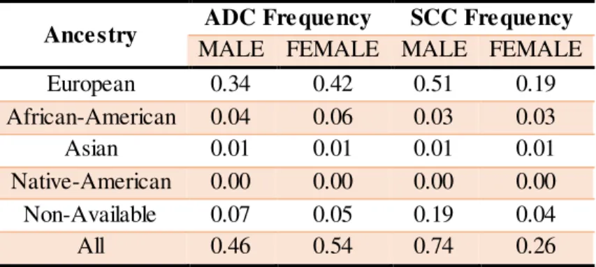
Table 3.1. Distribution of lung cancer subtypes ADC and SCC per patient population ancestry ... 34
Table 3.2. Distribution of ADC and SCC cases per patient gender and ancestry... 35
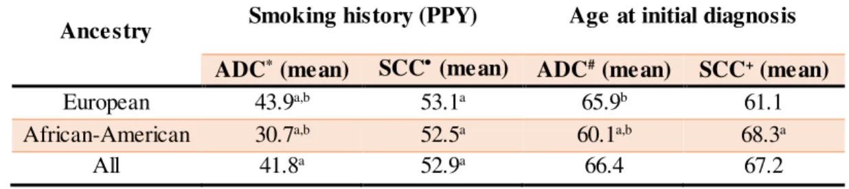
Table 3.3. Smoking history (PPY) and age of onset of ADC and SCC cases in each ancestry ... 36
Table 3.4. Tumor distribution per lung anatomic site for ADC and SCC cases... 37
Table 3.5. COPD Stages of ADC and SCC cases, according to GOLD guidelines... 37
Table 3.6. Significant tendency to co-occurrence of candidate genes in ADC cases ... 41
Table 3.7. Significant tendency to co-occurrence of candidate genes in SCC cases ... 42
Table 3.8. Variants identified in our cohort of Portuguese COPD and LC patients ... 50
Table 3.9. Statistical tests for low frequency variants found in SERPINB3 and SERPINB4 genes... 51
Table T1. PCR conditions for SERPINB3/B4 amplification ... 79
Table T2. Semi-nested PCR conditions for SERPINB3/B4 amplification ... 79
xv
ABBREVIATIONS
AATD Alpha-1 Antitrypsin Deficiency
ADAMs a Disintegrin and Metalloprotease
ADAMTSs a Disintegrin and Metalloprotease with Thrombospondin motifs
ADC Adenocarcinoma
BALF Bronchoalveolar lavage fluid
CNV Copy number variant
COPD Chronic Obstructive Pulmonary Disease
CTSG Cathepsin G
CTSs Cathepsin
DNA Deoxyribonucleic Acid
ECM Extracellular Matrix
EGF Epidermal Growth Factor
ELANE Neutrophil Elastase
ExAc The Exossome Aggregation Consortium
FEV1 Forced Expiratory Volume
FVC Forced Vital Capacity
GAG Glycosaminoglycan
GOLD Global Initiative for Chronic Obstructive Lung Disease
GWAS Genome-wide Association Study
HGF Hepatocyte Growth Factor
IBS Iberian Population in Spain – 1000 Genomes
LC Lung Cancer
MAF Minor Allele Frequency
MMPs Metalloprotease
NSCLC Non-Small Cell Lung Cancer
PCR Polymerase Chain Reaction
PG Proteoglycans
PPY Packs per Year
xvi
RCL Reactive Center Loop
RNA Ribonucleic Acid
ROS Reactive Oxygen Species
SCC Squamous Cell Carcinoma
SCLC Small Cell Lung Cancer
SERPINA1 Alpha-1 Protease Inhibitor
SERPINB3 Squamous Cell Carcinoma Antigen 1
SERPINB4 Squamous Cell Carcinoma Antigen 2
SERPINs Serine Protease Inhibitor
SNV Single-nucleotide Variant
TCGA The Cancer Genome Atlas
TGFα Transforming growth factor alpha
TGFβ Transforming growth factor beta
TIMPs Tissue Inhibitor of Metalloprotease
UTR Untranslated Region
1
1.
Introduction
1.1.
State of the art of Human Genetics and Human Disease
Studies
Understanding the molecular mechanisms of human disease and its genetic basis has been one of the main goals of the scientific community. In a global perspective, Mendelian disorders tend to be less prevalent in worldwide populations, more geographically confined and associated to single genes, where deleterious mutations arise in germline cells and are passed throughout generations rarely reaching polymorphic frequencies (MAF: minor allele frequency >1%). In contrast, complex (or multifactorial) disorders are in general the endpoint result of a combination of both genetic and environmental risk factors, and are often dispersed among diverse ethnic groups. Even though, the full extent of genetic variability associated to these common pathologies is not entirely acknowledged, this is more likely to be connected to germline mutations with a wide spectrum of frequencies (MAF<1% and MAF>1%) and variable contributions to disease susceptibility (small and large effect sizes) (Robinson et al. 2014; Mitchell 2012; Manolio et al. 2010).
Human cancers are by nature multifactorial disorders, however, another layer of complexity is added to these diseases, since a plethora of de novo mutations can originate in tumors (somatic
mutations). These are usually classified into driver and passenger mutations, according to their
outcomes in cancer progression. While driver mutations are accepted to confer a selective advantage,
having critical roles in tumor growth, often reaching higher prevalence within tumor; passengers
mutations are believed to behave neutrally not contributing to tumor clonal expansion (Merid et al. 2014; Stratton et al. 2009).
2 mainly protein-coding regions at high depth for thousands of subjects screened in the scope of different disease-specific and population genetic studies (Lek et al. 2016; Auton et al. 2015). In addition , “The Cancer Genome Atlas” (TCGA) database, resulting also from a collaborative project between the National Cancer Institute (NCI) and National Human Genome Research Institute (NHGRI) in the USA, is supporting the publication of genetic, clinical and other relevant data for 33 tumor types, comprising more than 11.000 patients (Auton et al. 2015; Chang et al. 2013).
Overall, these resources have already contributed to thousands of studies by consortiums themselves and by independent researchers, enabling a comprehensive analysis of the prevalence of many mutations associated to Mendelian and complex diseases. Nevertheless, the architecture of the complex diseases is not yet fully resolved and most variants identified so far seem to explain only a small fraction of heritability, even when using very large cohorts of patients and controls. Several hypotheses have been raised to explain this missing heritability in complex diseases, including the low power to detect gene-gene interactions; the inadequate accounting of shared environments between individuals, and the presence of a large number of variants with small impact in the disease onset, but also the occurrence of rare variants with stronger effects in subject health status (Manolio et al. 2010).
In this scope, this work is focused in the study of two multifactorial disorders affecting the respiratory system, Chronic Obstructive Pulmonary Disease (COPD) and lung cancer (LC), which are prime examples for the interaction between environmental and genetic factors, in disease susceptibility and pathogenesis (gene-by-environment theory). Here, we propose to address this issue by centering our variant screening in proteolysis related genes (proteases, their inhibitors and substrates) and their potential repercussions in the lung extracellular matrix (ECM). We will take advantage of published datasets and our own sample collection.
1.2.
The extracellular Matrix (ECM) in the healthy lung
3 the ECM assemblage can have a large impact in tissue patterning during organogenesis and angiogenesis (Kniazeva & Putnam 2009; Mammoto et al. 2009) and, on the other hand, ECM proteins can influence cell behavior through signaling, binding to growth factors, mediating cell-adhesion and transducing signals into cells (Hynes 2013). One of the key features of the ECM is its ability to respond to diverse physiological and stress stimuli in a biological process often referred as ECM remodeling, in which damaged and proteolytic cleaved proteins are replaced by new ones (Swinehart & Badylak 2017; Bachman et al. 2015).
In the lung, the ECM is mainly responsible for the supportive scaffold of the alveolar wall, branching morphogenesis, and tissue repair after injury (Watson et al. 2016). Moreover, in the respiratory system, the ECM is also specialized for gas changes, the primary function of the lung, while also providing structural support to prevent airway collapse (Balestrini et al. 2016; Parameswaran et al. 2006). For this reason, one of the most important ECM components in the lung are elastin fibers, which confer stretch and recoil properties to pulmonary tissues; and collagen fibers, responsible for parenchyma support and basement membrane barrier functions (Dunsmore et al. 1996). Other key elements of the lungs are fibronectin and laminin fibrils, fundamental for cell-adhesion to the basement membrane and cell survival (Mouw et al. 2015) and glycosaminoglycans (GAG) that together with proteoglycans (PG) control cellular and macromolecule movements, water retention, ion content, and growth factor levels (Papakonstantinou & Karakiulakis 2009).
1.2.1.
The ECM structural macromolecules: Elastin and fibrillar
collagens
Elastin and fibrillar collagens are the main structural components of ECM.
In the lungs, in particular, the elastin provides natural stretching and contractile functions needed to respiratory cycles (Pelosi et al. 2007). This macromolecule, composed mainly by tropoelastin monomers (approximately 60-70 kDa), is encoded by ELN. As a biopolymer, elastin
4 2000). Importantly, during adulthood, such elastogenesis processes are known to drop significantly leading to damage in elastic fibers to be irreversible and remain unrepaired (Humphrey et al. 2015).
5
Table 1.1. Summary of the human collagen classes.
Class Collagen Type Collagen-protein encoding genes
Fibrillar
I COL1A1, COL1A2
II COL2A1
III COL3A1
V COL5A1, COL5A2, COL5A3
XI COL11A1, COL11A2, COL11A3
XXIV COL24A1
XXVII COL27A1
Fibril-associated collagens with
interrupted triple helices (FACIT)
IX COL9A1, COL9A2, COL9A3
XII COL12A1
XIV COL14A1
XVI COL16A1
XIX COL19A1
XX COL20A1
XXI COL21A1
XXII COL22A1
Basement membrane IV COL4A1, COL4A2, COL4A3, COL4A4,
COL4A5, COL4A6
Long chain VII COL7A1
Filamentous VI COL6A1
S hort chain
VIII COL8A1
X COL10A1
Multiplexins
XV COL15A1
XVIII COL18A1
Transmembrane domain (MACIT)
XIII COL13A1
XVII COL17A1
6 In an histological overview of lung tissues, a high percentage of collagens is observed in large bronchi, small airways and large blood vessels like the pulmonary artery; whereas, elastin can be more usually detected in lung parenchyma, bronchi and blood vessels, as well (Balestrini et al. 2016; Townsley 2012; Parameswaran et al. 2006; Pierce & Hocott 1959).
1.2.2.
The ECM multiadhesive macromolecules: fibronectin and
laminins
Fibronectin and laminins are multidomain glycoproteins, whose major functions are to promote the adhesion between ECM structural components, across the later and soluble molecules in the extracellular space, as well as between cells and ECM. In other words, these multiadhesive molecules are capable of binding to other ECM proteins, cell surface receptors and growth factors through specific motifs found in their protein structure (Mouw et al. 2015).
Fibronectin is one of the most important ECM glycoproteins, which is involved in the interstitial organization of ECM and facilitates cell attachment, migration and differentiation (Schwarzbauer & Desimone 2011; Smith et al. 2007). Cellular fibronectin, the isoform most commonly found in the ECM, is secreted mostly by fibroblasts and organized in dimers of 250 kDa. In addition, cellular fibronectin is characterized by the presence of a 70 kDa N-terminal domain, responsible for fibril assembly and binding to the cell surface; and by V region, a key domain for cell motility and matrix assembly that also contains a α4β1 integrin binding site (To & Midwood 2011; Mao & Schwarzbauer 2005; Pankov & Kenneth 2002).
Laminins are ECM glycoproteins found essentially in basal membranes, intervening in the ECM-cell interactions, through the binding of cell surface receptors to ECM components (Lu et al. 2011; Rozario & Desimone 2011). Laminins are heterotrimeric proteins with up to 16 distinct isoforms, composed by five α-, four β- and three γ-chains subunits. Whereas α-chains are encoded by
LAMA1/2/3 genes, β-chains and γ-chains are expressed through LAMB1/2/3 genes LAMC1/2/3,
7
1.2.3.
The role of proteolysis in the ECM remodeling
Multiple proteases and their inhibitors are essential in ECM remodeling to replace damaged macromolecules (protease substrates) and to maintain a fine balance between protein degradation and turnover. Proteases with key activities in ECM remodeling include different classes of metalloproteases, namely matrix metalloproteases (MMPs), desintegrin and metalloprotease domain containing proteins (ADAMs), and desintegrin and metalloprotease with thrombospondin motifs (ADAMTSs); several cysteine and serine proteases: such as neutrophil elastase (ELANE), cathepsin G (CTSG) and proteinase 3 (PRTN3). Whereas tissue inhibitor of metalloproteases (TIMPs) are able to control the activity of MMPs and ADAMs; the family of serine protease inhibitors (SERPINs) efficiently regulates diverse serine and cysteine proteases.
1.2.3.1. The Matrix metalloprotease (MMP) family
The MMP family comprises 23 members that are generally secreted as inactive zymogens and later activated by other MMPs or serine proteases, such as plasmin or neutrophil elastase (ELANE). Briefly, MMPs can be regulated at four different levels: by transcriptional and post-transcriptional regulation of gene expression; in the tissue milieu by proteolytic activation (removal of a propeptide segment); and by specific protease inhibitors (Löffek et al. 2011). During the normal remodeling (wound and healing) process, several MMPs are produced by neutrophils, macrophages, and wounded cells to degrade damaged ECM macromolecules – MMPs substrates.
8
Table 1.2. Different MMPs clades and their corresponding ECM substrates [Adapted from (Cathcart et
al. 2015)].
Clade MMP ECM S ubstrates
Collagenases
M M P-1 Collagens (type I, II, III, VII, VIII, X, XI), fibronectin, laminin, vitronectin, entactin, gelatin, tenascin, aggrecan, and others
M M P-8 Collagens (type I, II, III), aggrecan
M M P-13 Collagens (type I, II, III, IV, VI, IX, X, XIV), fibronectin, gelatin, aggrecan, and others
M M P-18 Collagen type I (rat)
Gelatinases
M M P-2 Collagens (I, II, III, IV, V, VII, X, XI), elastin, fibronectin, gelatin, laminin, vitronectin, tenascin, and others
M M P-9 Collagens (IV, V, XI, XIV), elastin, laminin, vitronectin, and others
S tromelysins
M M P-3 Collagens (III, IV, V, VII, IX, X, XI), elastin, fibronectin, laminin, vitronectin, tenascin, and others
M M P-10 Collagens (III, IV, V), elastin, fibronectin, aggrecan, and others
M M P-11 Collagen type IV, fibronectin, laminin, gelatin
Matrilysins
M M P-7 Collagens (I, IV), elastin, fibronectin, vitronectin, laminin, gelatin, entacin, and others
M M P-26 Gelatin, fibronectin, vitronectin
Membrane-type MMPs
M M P-14 Collagens (I, II, III), gelatin, fibronectin, tenascin, laminin, and others
M M P-15 Fibronectin, tenascin, entacin, laminin, aggrecan, perlecan
M M P-16 Collagen type III, gelatin, fibronectin, vitronectin, laminin
M M P-24 Fibronectin, gelatin, chondroitin and dermatane sulphate proteoglycans
M M P-17 Gelatin
M M P-25 Collagen type IV, gelatin, fibronectin, chondroitin and dermatane sulphate proteoglycans
Others
M M P-12 Collagens (I, IV, V), elastin, gelatin, fibronectin, laminin, vitronectin, entactin, and others
M M P-19 Collagen type IV, gelatin, laminin, entactin, and others
M M P-20 Amelogenin, aggrecan
M M P-21 Gelatin
M M P-23 Gelatin
M M P-27 Gelatin
9
1.2.3.2. The families of Desintegrin and metalloproteases (ADAM and
ADAMTS)
The ADAM family comprises a total of 21 related molecules, in which only 13 show proteolytic activity, these comprise ADAM8-10, 12, 15, 17, 19-21, 28, 30 and 33 (Duffy et al. 2011). Structurally, ADAMs are organized in eight major units: the pre- and propeptide regions; and six other domains with functions as metalloprotease, disintegrin, cysteine-rich, Epidermal Growth Factor (EGF)-like, transmembrane, and cytoplasmic proteins (Giebeler & Zigrino 2016; Duffy et al. 2011). On the other hand, the ADAMTS family includes 19 members with some common features to ADAMs family, aside from the addition of innumerous thrombospondin motifs in the C-terminal domain, and the lack EGF-like, transmembrane and cytoplasmic domains (Kelwick et al. 2015; Paulissen et al. 2009). Based on their domain organization and known functions, ADAMTSs can be divided in 8 subgroups, being the most important the aggrecanase and proteoglycanase group (ADAMTS1, 4, 5, 8, 9, 15, 20), which cleave hyaluronic-binding chondroitin sulfate proteoglycan extracellular proteins. On the other hand, the group of pro-collagen N-peptidases (ADAMTS2, 3, 14) processes pro-collagen molecules (Kelwick et al. 2015). Similarly to MMPs the superfamily of ADAMs and ADAMTS also contain a catalytic domain bound to a zinc-ion (Kelwick et al. 2015; Tallant et al. 2010; Edwards et al. 2009).
1.2.3.3. Cathepsins and other serine proteases
10 of collagen type IV and fibronectin proteolysis. Conversely, CTSK cleaves fibrillary collagens - types I and II (Fonovic & Turk 2014; Kasabova et al. 2011; Wolters & Chapman 2000). CTSC found in alveolar tissues is reported to activate other enzymes such as elastase and CTSG. (Kasabova et al. 2011).
Neutrophil elastase (ELANE) and protease 3 (PTRN3) are two other serine proteases with 29 and 33 kDa, respectively, that are normally stored in the azurophilic granules of polymorphonuclear neutrophils. These proteases are secreted upon neutrophil activation at inflammatory sites, by cleavage of the N-terminal peptide and removal of an aminoterminal dipeptide by CTSC (Korkmaz et al. 2010). Both serine proteases are capable of degrading various ECM structural molecules, including elastin, type IV collagen, and fibronectin. If dysregulated these molecules may compromise integrity of bronchial and alveolar walls. Importantly, ELANE and PTRN3 may also cleave inflammatory mediators, cell receptors and lung surfactant molecules with potential impact in ECM remodeling (Sinden & Stockley 2013; Lucas et al. 2011; Korkmaz et al. 2010).
1.2.3.4. Protease Inhibitors
Tissue metalloproteases inhibitors (TIMPs) are a small family of homologous proteins, mainly synthesized by connective tissue cells and leukocytes that control the activity MMPs, ADAMs and ADAMTSs by means of forming noncovalent complexes with their targeted proteases. Briefly, these proteins comprise two domains (N- and C-terminals) stabilized by three disulfide bonds, in which the N-terminal is the active domain containing two zinc ions, with one folding within itself to bind and inhibit metalloproteases (Mocchegiani et al. 2011; Rocco et al. 2001). Although all four TIMP may work as metalloprotease inhibitors, these have different regulatory efficiencies according to their best affinity to each protease. For example, whereas TIMP2 has a greater affinity to MMP-2, TIMP3 is a stronger inhibitor of MMP-9. Moreover, TIMP1 is capable of controlling the activity of most MMPs, except for some membrane type members (MMP-14-16, -19, -24). In this family, TIMP3 displays the most wide inhibitory range, being able to efficiently regulate a vast number of ADAMs (ADAM10, 12, 17, 28, 33) and ADAMTSs (ADAMTS1, 2, 4, 5) (Arpino et al. 2015; Brew & Nagase 2011). TIMP3 also differs from the other family members in its tissue placement, while it is attached to the ECM, the remaining TIMPs are present as soluble inhibitors (Reunanen & Kähäri 2013).
11 conditioning metalloproteases activity, impairs TGFβ release, regulating that deposition. Furthermore, TIMPs can also control ECM turnover, through the regulation of inflammatory pathways, avoiding the cleavage of cell-surface cytokines and cytokine receptors by metalloproteases (Arpino et al. 2015).
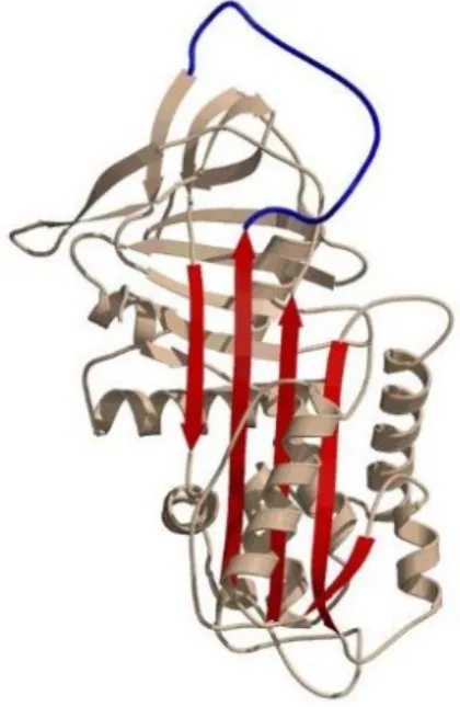
The serine proteases inhibitors (SERPINs) superfamily comprises at least 36 functional members, subdivided into distinct clades (A to I) according to similarities in protein sequence, gene organization and chromosomal location. All SERPINs share a highly conserved three-dimensional structure, characterized by a prototypical molecular arrangement in three -sheets, nine -helixes and an exposed reactive center loop (RCL; Fig. 1.1). This domain contains a pseudo-substrate (P1-P1’)
that once cleaved and covalently bound to target proteases, inhibits their activity in an irreversible fashion. Although SERPINs regulate mostly serine proteases, some are able to control the activity of cysteine proteases and importantly, SERPINs display different affinities toward different proteases, neutralizing not only specific enzymes but also wide classes of proteases (Seixas 2015; Gooptu & Lomas 2009). Alpha-1-antitrypin (SERPINA1), the major protease inhibitor in the serum, represents a critical regulator of ECM degradation in the lower respiratory tract by controlling the enzymatic activity of ELANE, but also CTSG and PRTN3. However, SERPINA1 is also a potential target for MMPs, which are capable of cleaving the RCL, rendering this molecule inactive (Fortelny et al. 2014).
Figure 1.1. Conserved SERPIN three-dimensional structure.
12 Clade B SERPINs are also particularly relevant in the control of lung ECM degradation by preventing cell death (apoptosis and necrosis) and by avoiding promiscuous proteolysis associated to the release of diverse proteases found in the lysosome and cytolytic granules (Table 1.3) (Sun et al. 2016; Houghton 2015; Moroy et al. 2012; Askew & Silverman 2008). SERPINE1 is another example of a protease inhibitor, which functions in lung ECM (alveolar space) by preventing fibrin deposition, an important event in fibrosis and acute lung injury (Askew & Silverman 2008).
Table 1.3. Clade B SERRPINs with known roles in lung function [Adapted from (Askew & Silverman
2008)].
S ERPIN Targets Function
S ERPINB1
Neutrophil elastase (ELANE) Cathepsin G (CTSG) Proteinase-3 (PRTN3)
Protection from elastase activity
S ERPINB2 Urokinase-type plasminogen activator (PLAU)
Tissue-type plasminogen activator (PLAT) Protection against cell death
S ERPINB3 Cathepsins K, L, S, V (CTSK, L, S, V)
Protection from cytosolic lysosomal peptidases;
Inhibition of cell death
S ERPINB4 Cathepsin G (CTSG)
Mast cell proteinase (MCP) Protection against cell death
S ERPINB6 Cathepsin G (CTSG) Protection from granule peptidases
S ERPINB9 Granzyme B (GZMB) Protect cytosolic lymphocytes;
Protection against cell death
S ERPINB10 Trypsin (PRSS1)
Thrombin (F2) Protection against cell death
S ERPINB12 Trypsin (PRSS1)
S ERPINB13 Cathepsins K and L (CTSK, L) Protection against cell death
1.3.
Complex Lung Diseases
13 examples of complex diseases that are linked to dysregulated ECM remodeling. Some of the phenotypes associated to these diseases are the pulmonary emphysema (in COPD) and the pulmonary fibrosis (in LC), both correlated with a dysregulation of ECM remodeling (Bidan et al. 2015; Cox & Erler 2011).
1.3.1.
Chronic Obstructive Pulmonary Disease
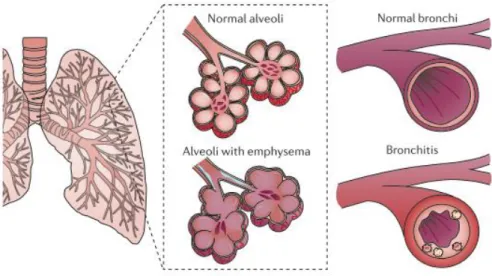
Chronic Obstructive Pulmonary Disease (COPD) is the most common smoking-related disease in Western countries, which is predicted to represent the third cause of death by the end of 2020 (Vestbo et al. 2013). This illness is defined by the presence of progressive airflow obstruction that is not fully reversible and its major clinical manifestations are emphysema and chronic bronchitis (Fig.1.2) (Pauwels et al. 2012). Whereas the former COPD phenotype is characterized by an enlargement and destruction of alveoli, resulting in lower pulmonary oxygenation. The latter one is associated with inflamed and thickened bronchial walls, together with a luminal obstruction by mucus and inflammatory cells, normally causing breathing difficulties and persistent cough (Fischer et al. 2011).
14
Spirometry is the “gold standard” for measuring the extent of airflow limitation. Forced expiratory volume in one second (FEV1), and forced vital capacity (FVC) are two lung function parameters assessed by spirometry that are fundamental to the evaluation of the degree of airflow limitation and its progress overtime. According to the guidelines of the Global Initiative on Chronic Obstructive Pulmonary Disease (GOLD), a health entity working with World Health Organization (WHO) in this field, a COPD diagnosis is confirmed if post-bronchodilator FEV1/FVC ratio is below 70%. In addition, GOLD also recognizes four COPD stages based in their severity, starting from Mild (GOLD 1; FEV1 ≥80%), passing through Moderate (GOLD 2; FEV1 50-79%) and Severe (GOLD 3; FEV1 30-49%), until reaching a Very Severe stage (GOLD 4; FEV1 <30%) (GOLD Guidelines 2017). Although a FEV1/FVC cutoff of <70% is widely used in COPD diagnosis it may be less accurate in elderly, and in adults under 45 years old, resulting in the first case in a overestimation, and in the second in a reduction in the numbers of affected subjects, particularly for milder phenotypes. This phenomenon has been attributed to regular alterations in lung function associated to ageing (Ito & Barnes 2009; Roberts et al. 2006).
Figure 1.2. COPD Phenotypes. Emphysema and Chronic Bronchitis are the most
15
1.3.2.
Lung Cancer
Lung cancer is the most lethal cancer worldwide, and its incidence has increased significantly during the XX century, mainly due to a global raise in smoking exposure. Interestingly, recent studies are pointing to a reduction in the number of affected males, whereas female rates are being maintained constant (Ridge et al. 2013). Despite cigarette smoking has been considered the main environmental risk factor for LC, as in COPD, others inhaled smokes and particles (organic and inorganic) are recognized to play a role in the disease pathogenesis.
Two major types of LC can be identified: the small-cell lung cancer (SCLC) type, a rarer form of the disease observed in 15-20% of patients, and the non-small-cell lung cancer (NSCLC) reported in 80-85% of cases. NSCLC is itself further divided into three major histologic subtypes: squamous-cell carcinoma (SCC) and adenocarcinoma (ADC); the two most frequent subtypes of LC, and large-cell lung cancer (Bracci et al. 2012; Herbst et al. 2008). Notably, ADC usually has a slower growth than other LC types and it is more prevalent in females than males. Moreover, whereas ADC is more frequently detected in peripheral lung parenchyma, SCC, is more usually related to bronchial epithelial lesions and more commonly found in the central region of the lung. Also, both types are highly associated with smoking, although in non-smoking patients, ADC is the most common LC type (The American Cancer Society 2016; Chang et al. 2015).
16
1.4.
Mechanistic links between COP D and LC
Over the latest years, it has been hypothesized that COPD and LC share a common mechanism in their pathogenesis (Houghton 2013; Vermaelen & Brusselle 2013; Young et al. 2011). First, COPD patients present a higher risk for LC (2-5 time higher). Second, not only, LC is a common complication in COPD, as COPD itself is also a prevalent co-morbidity in LC cases (Durham & Adcock 2015; Young et al. 2015). More precisely, in COPD, the emphysema phenotype has been pointed out as the stronger marker for LC risk. Typically, NSCLC (ADC and SCC), the most frequent cancer type among COPD, reaches about 80 to 85% of the cases, with a higher incidence of ADC, in comparison to SCC (Gabrielson 2006; Papi et al. 2004). Third, COPD and LC were found to overlap in several genetic susceptibility factors (see section 1.4.1. below). However, COPD has been reported to display stronger familial aggregation than LC, while COPD estimated heritability ranges from 40-75%, in LC it only reaches 15-25% (Young et al. 2012). Fourth, COPD and LC have as a major
etiological factor cigarette smoking, as well as other inhaled elements, which may be correlated with
increased fields of injury, chronic inflammation, enhanced oxidative stress (see section 1.4.2. bellow), and consequently with altered ECM remodeling (see section 1.4.3. below) (Houghton 2013).
1.4.1.
Genetic Susceptibility factors
The recent efforts made by independent genome wide association studies (GWAS) to underpin common variants increasing COPD and LC susceptibility, uncovered in several instances the same candidate risk genes. For example, these included acetylcholinergic nicotinic receptors, subunits α3 and α5 (CHRNA3 and CHRNA5, respectively) (15q25), hedgehog interacting protein
(HHIP)(4q31), family with sequence similarity 13 member A (FAM13A) (4q24), and iron responsive
element binding protein 2 (IREB2)(15q25) (Khiroya & Turner 2015; Yang et al. 2013; Young et al.
2011).
Notably, CHRNA3/A5 association to COPD and LC has been replicated several times in
17 transformation (Dang et al. 2016; Singh et al. 2011). Conversely, HHIP, which regulates the activity
of Sonic Hedgehog (SHH), has been reported to have a critical role in the signaling pathways for both bronchial embryogenesis and lung development. Also, in COPD and LC, it has been proposed to participate in the cycles of injury and repair induced by environmental risk factors, in which effects in epithelial mesenchymal transition (EMT) can further contribute to LC pathogenesis, by allowing cells to increase their motility (Kugler et al. 2015). Regarding IREB2, a gene located in the same
chromosomic region of CHRNA3/A5 (15q25), its association to COPD and LC has been hypothesized
to be connect to iron regulatory pathways and IREB2 role in iron homeostasis (Ziółkowska-Suchanek
et al. 2015; Alder et al. 2011). The function of FAM13A, not completely understood, it is thought to
be correlated to signal transduction, due to described effects in tumor cell migration and possible impact in cancer growth (Eisenhut et al. 2017; Ziółkowska-Suchanek et al. 2015; Cho et al. 2010).
Still, to date, SERPINA1 deficiency (AATD) remains one of the few proven genetic causes for emphysema, mainly due to a pathogenic variant (rs28929474; p.Glu342Lys) leading to the unopposed activity of ELANE, cleavage of elastin fibers and ECM degradation. However, AATD only accounts for a small proportion of COPD cases (1-3%) and in LC it is not yet clear if it has indeed a role in tumorigenesis. In most GWAS for COPD and LC, no strong association of SERPINA1
to disease susceptibility was detected, although it was described a moderate association of rs28929474 variant with severe airflow limitation in COPD (Jackson et al. 2016; Enewold et al. 2012; Denden et al. 2010).
Sequence variation of MMPs has been extensively investigated in the context of COPD and LC by independent candidate gene approaches. Most interesting associations to lung disease include
MMP12 variants rs652438 and rs2276109 that were associated to emphysema, and severe stages of
COPD (GOLD III-IV) and in MMP2, rs243865 variant, which has been linked to a decay in survival
time of NCSLC patients (González-Arriaga et al. 2012; Haq et al. 2010).
1.4.2.
Oxidative stress, cell injury and inflammation
18 into lung microenvironment. In COPD, these radicals, may additionally cause the inactivation of important proteases inhibitors, resulting in an exacerbation of neutrophil elastase activity, loss of lung elasticity, apoptosis and emphysema (John et al. 2017; Domej & Oettl 2014). On the other hand, in LC individuals, ROS are often implicated in protein degradation and DNA methylation, later contributing to cancer development through the activation of anti-apoptotic molecules that increase cell division and proliferation, and facilitate tumor metastasis (Liou & Storz 2010).
Another outcome of the chronic inflammation in both COPD and LC microenvironments is the perpetuation of tissue injury (repeated cycles of tissue injury and repair) (Vakkila & Lotze 2004). Briefly, lung injuries are initiated by exogenous factors, like cigarette smoking. Then, injured cells start releasing diverse repair-linked mediators such as different families of epidermal and fibroblast growth factors (e.g. TGFα, HGF), chemokines, interleukins and prostaglandins. Later, these molecules are expected to impact ECM remodeling, by processes such as mitosis, migration and repair stimulation, involving collagen, laminin, fibronectin and matrix-metalloproteases, such as MMP-1 and -9 (Crosby & Waters 2010). Conversely, several repair-linked mediators also trigger the recruitment of macrophages and neutrophils to sites of injury that secret diverse proteases capable of degrading ECM elastin and collagens. Importantly, fragments derived from ECM proteolysis can also act as repair-linked mediators, directly or indirectly, supporting further chronic inflammation (Bonnans et al. 2014; Shifren & Mecham 2006). Moreover, in both diseases, the persistence of cell injury is believed to change cell death from apoptosis to necrosis. Contrary to apoptosis, a programmed cell death in which the cells usually shrink and maintain integrity of their membrane, in necrosis, cells lose that integrity and leak their internal contents to extracellular space, promoting an inflammatory response (Rock & Kono 2008).
19
1.4.3.
ECM remodeling and proteolysis in lung disorders
In lungs injured by chronic inflammation is common to observe a disequilibrium between collagen expression, deposition and turnover, with an overall augmented content of collagens in comparison to elastin. In mild and moderate COPD cases, collagen deposition is thought to contribute to the thickening of bronchial walls associated to the development of chronic bronchitis (Eurlings et al. 2014; Annoni et al. 2012; Harju et al. 2010; Kranenburg et al. 2006). In a LC situation, the overexpression and accumulation of collagens type I and III is regarded as an important factor for tumor stroma stiffening and cancer microenvironment (Burgstaller et al. 2017; Vicary et al. 2017; Burgess et al. 2016).
Proteolytic imbalance has been proposed as one of the major causes for COPD as it is occurs in pulmonary emphysema associated to AATD. Still, other studies have implicated proteolytic imbalance in the pathogenesis of COPD and LC in connection mainly to MMPs activities. For example, MMP-1 that was associated with both diseases through different genetic studies, was suggested to promote the metastasis formation in LC, through interaction with STAT3 (Schütz et al. 2015) and described to cause airways enlargement and emphysema, when overexpressed in lungs, with cigarette smoke and other noxious gases exposure, and inflammation as key elements (Churg et
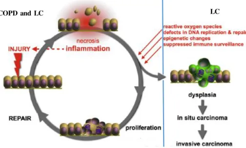
Figure 1.3. Repetitive cycles of tissue injury and repair. These cycles contribute to a state
of chronic inflammation, a feature found in both COPD and LC, which consequently may lead to malignant degeneration. Eventually, inflammatory mediators resulting from this cycles can induce genetic aberrations, perpetuated by the accumulation of cells that escape the apoptotic process, resulting in dysplasia, followed by carcinoma in -situ, and finally an invasive carcinoma state, developing LC [Adapted from (Vermaelen & Brusselle 2013)].
20 al. 2012; Mocchegiani et al. 2011; Greenlee et al. 2007). Furthermore, MMP-12, which is considered an important factor for COPD severity, was found to be highly expressed in patients alveolar macrophages and reported to induce emphysema driven by cigarette smoking (Churg et al. 2012; Soto-quiros et al. 2009). Noticeably, increased levels of MMP-12 were also associated with TIMP1 hydrolysis, SERPINA1 inactivation and increased ELANE activity (Houghton 2015; Lucas et al. 2011).
In LC conditions, a large diversity of MMPs are known to facilitate tumor growth and invasiveness through their impact in the degradation of ECM barriers and as promotors of angiogenesis. Furthermore, MMPs are also known to favor metastization, given that MMPs are critical molecules in ECM cell detachment, thus allowing cancer cells to enter in circulation and to reach distant tissues (Reunanen & Kähäri 2013). In overview, significant changes in the ECM composition associated to its proteolytic degradation and/or wound-healing (fibrosis) can be correlated with the outcomes of COPD and LC.
1.5.
P atient Tailored Therapeutics
To date, patient tailored therapeutics are already available for NSCLC. In particular, for subjects carrying specific somatic mutations in genes such as the Epidermal Growth Factor Receptor (EGFR), Kirsten Rat Sarcoma Viral Oncogen Homolog (KRAS), or Anaplastic Lymphoma Kinase
(ALK) genes (Ridge et al. 2013). Even thought, some of these therapies for ADC and SCC were
already proven to be very successful in some cases, these cannot be administrated widely as most patients lack corresponding molecular targets not showing any response to treatment. Therefore, most patients continue to be medicated with standard drugs with lower response rates and frequent side-effects (Cortinovis et al. 2016; Lazarus & Ost 2013).
In COPD, in spite of a large spectrum of therapeutics available very few take into account patient molecular profile. The only exception is SERPINA1 augmentation therapy in pulmonary emphysema associated to AATD. In near future, possibly some drugs targeting β2-adrenegic receptor (ADRB2) mutations, may promote muscle relaxation and dilation of airways (Nielsen et al. 2017;
Wewers & Crystal 2013).
MMPs and several serine proteases have been investigated as possible therapeutic targets in lung diseases, but so far, there are no perspective of these studies being translated soon into clinical practice (Moroy et al. 2012). Interestingly, somatic mutations in two inhibitor genes, SERPINB3 and
21 were associated with a better outcome in melanoma treatment with anti-CTLA4 antibody (ipilimumab) (Riaz et al. 2016). This drug, ipilimumab, is currently undergoing phase III trials for LC treatment (Bustamante Alvarez et al. 2015), positioning SERPINB3 and SERPINB4 as promising
targets for personalized medicine in LC.
AIMS
In this study, we explore the impact of proteolysis related genes, including proteases, their inhibitors and lung ECM substrates in two correlated lung disorders: COPD and LC that share common and distinctive features of ECM degradation and remodeling. To achieve this main goal, we used different methodological approaches to address the following specific objectives:
1. Survey of The Cancer Genome Atlas (TCGA) database for available clinical and epidemiological data of two LC subtypes (ADC and SCC);
2. Evaluation of TCGA database regarding the mutational (somatic and germline) landscape of 73 proteolysis candidate genes with function in lung ECM;
3. Analysis of the sequence variability of selected genes (SERPINB3/B4 and CTSG) in a small
23
2.
Materials and methods
2.1.
Bioinformatics analysis
2.1.1.
TCGA data
–
Lung Cancer
Clinical and epidemiological information, as well as sequence variation (somatic and germline mutations) and expression data for 522 ADC and 504 SCC patients were retrieved from The Cancer Genome Atlas (TCGA) (https://cancergenome.nih.gov/). For LC samples, TCGA provides several important clinical and epidemiological variables such as tumor histology, anatomic site (upper, middle, lower, right and left lung), age at initial diagnosis, pulmonary function tests (FEV1 and FVC), smoking history (packs per year - PPY), population of origin (United States, Europe, Vietnam, Australia, and Canada), ethnicity (white, black, Hispanic and non-Hispanic) and gender. However, a considerable number of samples was found to lack information for some variables like smoking history and pulmonary function tests (FEV1 and FVC), which in the latter case impaired the identification of COPD burden among ADC and SCC.
TCGA sequence variation data results from whole-exome or whole-genome sequencing (WES and WGS) of both tumor and non-tumor tissues (lung healthy section or blood). In this situation, reads assembling and variant calling were performed through a direct comparison of tumor and non-tumor results using MuTect2 software. Briefly, this software combines several Bayesian methods in the filtering of low-quality sequence data; variant detection in tumor samples; removal of false positives; and in the discrimination of somatic and germline variants (Cibulskis et al, 2013). In
the analysis of matched tumor/normal samples, somatic mutations are only found in tumors, while germline mutations can be present in both samples or if found in tumor their status could not be evaluated. Expression data provided by TCGA, is derived from RNA sequencing.
2.1.2.
Clinical and epidemiological data analysis
24 comparisons within and between LC subtypes, the variables ethnicity and population of origin combined as European ancestry, African-Americans and Asians, age at diagnosis (mean values), gender, tumor anatomic site, and smoking history (mean PPY values). The Mann-Whitney implemented through MedCalc software (version 17.6) was used to appraise statistical significance of all set of comparisons done.
2.1.3.
Candidate genes selection
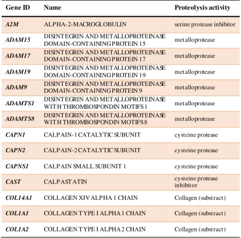
For this study, we selected 73 proteolysis related candidate genes (table 2.1) based in three main criteria: 1) evidence for the occurrence in lung tissues based either in gene expression data or proteomic screenings of biological samples withdrawn from lungs, such as bronchoalveolar lavage fluid (BALF) or sputum (SP) (Ohlmeier et al. 2012; Casado et al. 2007; Plymoth et al. 2006); 2) known function in ECM remodeling (ECM organization – degradation of the ECM) as annotated in the Reactome pathway database (http://reactome.org/); and genes found to be associated to either COPD and/or LC according to the GWAS catalogue (http://www.ebi.ac.uk/gwas/).
Table 2.1. Proteolysis related candidate genes selected for this study.
Gene ID Name Proteolysis activity
A2M ALPHA-2-MACROGLOBULIN serine protease inhibitor
ADAM15 DISINT EGRIN AND METALLOPROTEINASE
DOMAIN-CONTAINING PROTEIN 15 metalloprotease ADAM17 DISINT EGRIN AND METALLOPROTEINASE DOMAIN-CONTAINING PROTEIN 17 metalloprotease
ADAM19 DISINT EGRIN AND METALLOPROTEINASE
DOMAIN-CONTAINING PROTEIN 19 metalloprotease ADAM9 DISINT EGRIN AND METALLOPROTEINASE DOMAIN-CONTAINING PROTEIN 9 metalloprotease
ADAMTS1 DISINT EGRIN AND METALLOPROTEINASE
WIT H THROMBOSPONDIN MOTIFS 1 metalloprotease ADAMTS8 DISINT EGRIN AND METALLOPROTEINASE WIT H THROMBOSPONDIN MOTIFS 8 metalloprotease
CAPN1 CALPAIN-1 CATALYTIC SUBUNIT cysteine protease
CAPN2 CALPAIN-2 CATALYTIC SUBUNIT cysteine protease
CAPNS1 CALPAIN SMALL SUBUNIT 1 cysteine protease
CAST CALPAST ATIN cysteine protease inhibitor
COL14A1 COLLAGEN XIV ALPHA 1 CHAIN Collagen (substract)
COL1A1 COLLAGEN T YPE I ALPHA 1 CHAIN Collagen (substract)
25
COL3A1 COLLAGEN T YPE III ALPHA 1 CHAIN Collagen (substract)
COL4A1 COLLAGEN T YPE IV ALPHA 1 CHAIN Collagen (substract)
COL4A2 COLLAGEN T YPE IV ALPHA 2 CHAIN Collagen (substract)
COL4A3 COLLAGEN T YPE IV ALPHA 3 CHAIN Collagen (substract)
COL5A2 COLLAGEN T YPE V ALPHA 2 CHAIN Collagen (substract)
COL6A1 COLLAGEN T YPE VI ALPHA 1 CHAIN Collagen (substract)
COL6A2 COLLAGEN T YPE VI ALPHA 2 CHAIN Collagen (substract)
COL6A3 COLLAGEN T YPE VI ALPHA 3 CHAIN Collagen (substract)
COL8A1 COLLAGEN T YPE VIII ALPHA 1 CHAIN Collagen (substract)
CST3 CYST AT IN-C cysteine protease inhibitor
CST6 CYST AT IN-M cysteine protease inhibitor
CTSB CAT HEPSIN B cysteine protease
CTSD CAT HEPSIN D aspartic protease
CTSG CAT HEPSIN G serine protease
CTSK CAT HEPSIN K cysteine protease
CTSL1 CAT HEPSIN L1 cysteine protease
CTSS CAT HEPSIN S cysteine protease
ELANE NEUT ROPHIL ELAST ASE serine protease
ELN ELAST IN elastin (substract)
EMILIN2 EMILIN-2 elastin (substract)
FN1 FIBRONECT IN fibronectin (substrate)
FURIN FURIN serine protease
KLK1 KALLIKREIN-1 serine protease
KLK4 KALLIKREIN-4 serine protease
KLKB1 PLASMA KALLIKREIN serine protease
LAMA3 LAMININ SUBUNIT ALPHA 3 Laminin (substrate)
LAMA5 LAMININ SUBUNIT ALPHA 5 Laminin (substrate)
LAMB1 LAMININ SUBUNIT BETA 1 Laminin (substrate)
LAMB3 LAMININ SUBUNIT BETA 3 Laminin (substrate)
LAMC1 LAMININ SUBUNIT GAMMA 1 Laminin (substrate)
26
MMP1 INT ERST ITIAL COLLAGENASE metalloprotease
MMP10 ST ROMELYSIN-2 metalloprotease
MMP12 MAT RIX METALLOPROTEINASE-12 metalloprotease
MMP13 COLLAGENASE 3 metalloprotease
MMP14 MAT RIX METALLOPROTEINASE-14 metalloprotease
MMP15 MAT RIX METALLOPROTEINASE-15 metalloprotease
MMP2 72 KDA T YPE IV COLLAGENASE metalloprotease
MMP3 ST ROMELYSIN-1 metalloprotease
MMP8 NEUT ROPHIL COLLAGENASE metalloprotease
MMP9 MAT RIX METALLOPROTEINASE-9 metalloprotease
PI3 ELAFIN serine protease inhibitor
PLG PLASMINOGEN serine protease
PRTN3 MYELOBLAST IN serine protease
SERPINA1 ALPHA-1-ANTITRYPSIN serine protease inhibitor
SERPINA3 ALPHA-1-ANTICHYMOTRYPSIN serine protease inhibitor
SERPINB1 LEUKOCYT E ELAST ASE INHIBITOR serine protease inhibitor
SERPINB3 SQUAMOUS CELL CARCINOMA ANTIGEN-1 serine protease inhibitor
SERPINB4 SQUAMOUS CELL CARCINOMA
ANTIGEN-2 serine protease inhibitor SERPINB6 PLASMINOGEN ACTIVATOR INHIBITOR 2 serine protease inhibitor
SERPINC1 ANT ITHROMBIN-III serine protease inhibitor
SERPINE1 PLASMINOGEN ACTIVATOR INHIBITOR 1 serine protease inhibitor
SERPINE2 GLIA-DERIVED NEXIN serine protease inhibitor
SERPING1 PLASMA PROTEASE C1 INHIBITOR serine protease inhibitor
SLPI ANT ILEUKOPROTEINASE serine protease inhibitor
THSD4 T HROMBOSPONDIN TYPE-1 DOAMIN-CONT AINING PROTEIN 4 metalloendopeptidase
TIMP1 MET ALLOPROTEINASE INHIBITOR 1 metalloprotease inhibitor
TIMP2 MET ALLOPROTEINASE INHIBITOR 2 metalloprotease inhibitor
27
2.1.4.
Variants expression and impact analysis
In a first step, we examined the data available at the cBioPortal (http://www.cbioportal.org/), a comprehensive web tool that enables the analysis of large-scale cancer genomics datasets, including TCGA. This tool was used to get a glimpse of somatic mutation rates per each candidate gene.
In a more detailed analysis, VCF files containing all SNVs and INDELs (somatic and germline) identified by TCGA consortium were downloaded from the database. Then, these files were filtered for candidate genes genomic regions using Tabix software (version 0.2.6). Variants were
compiled with VCFTools software version 4.0 (http://vcftools.sourceforge.net/), to remove those
mutations with lesser quality (<20 reads).
Filtered somatic and germline variants for the 73 proteolysis candidate genes were next submitted to wANNOVAR software (http://wannovar.wglab.org/) analysis. This web tool has the advantage of compiling the results for a wide number of algorithms, predicting sequence variants functional consequences (SIFT, PolyPhen, CADD) together with variant frequencies for different human populations sequenced by large consortium like 1000 Genomes and ExAc. Here, we choose to consider only variant prediction effects of PolyPhen, SIFT and CADD algorithms. Whereas PolyPhen variant predictions are mainly based in protein sequence and structure, SIFT takes into account levels of evolutionary conservation, and CADD incorporates different metrics regarding functional data (not only protein structure) and conservation, prioritizing deleterious and pathogenic variants across wide range of functional categories (Eilbeck et al. 2017; Richards et al. 2015). Since
CADD generates quantitative values, we used a cutoff of ≥14.5 in scaled CADD score, to denote
most likely deleterious variants.
Candidate gene expression levels were obtained through Firebrowse (http://firebrowse.org/) engine, a TCGA online tool offering a direct comparison of expression differences between tumor and non-tumor samples.
2.2.
Screening of P ortuguese COP D and LC cases
Taking into account the results of our bioinformatics analysis we chose a few candidate genes for laboratory evaluation of sequencing variation in COPD and LC cases. Precisely, the selected genes for follow-up studies in our cases were SERPINB3 and SERPINB4 homologs and CTSG, which is
28
2.2.1.
Samples
Our sample collection included genomic DNA for COPD cases (N=43) sent to our laboratory for the AATD diagnosis and broncho-alveolar lavage fluid (BALF) of LC patients (N=45) gather in the scope of collaborative projects with clinicians from Hospital São João and CEDOC researchers.
Our LC cases included 18 ADC and 2 SCC, for the remaining cases it was not possible to obtain NSCLC subtyping or were classified as belonging to other LC. However, not every sample was completely sequenced.
2.2.2.
DNA Extraction
All samples derived from AATD diagnosis were previously extracted from blood using standard salting out methods or using Generation Capture Column kit (QIAGEN).
For BALF samples, DNA extraction was previously done using QIAamp mini kit (QIAGEN),
according to manufacture instructions.
2.2.3.
Polymerase Chain Reaction (PCR) amplification and
sequencing
For COPD samples, which were found to have higher DNA concentrations, SERPINB3 and SERPINB4 genes were amplified in five different PCR reactions fragments using the primer pairs
listed in Table 2.2. As high homologous genes, similar experimental schemes were used for
SERPINB3 and SERPINB4 amplification, as schematized in Figure 2.1.
Briefly, fragment A spanning over 2.5 kb contained exons 1 to 3; fragments B and E ranged about 1.5 kb each and included exons 4-5 and exon 8, respectively, and finally, fragments C and D with 400 bp each, covered the remaining exon 6 and 7. All reactions were performed with the following reagents: 1x KAPA Taq ReadyMix or 1x MyTaq Mix and 0.5-1µM concentrations for each
29
Table 2.2. Primers used for the amplification of SERPINB3/B4genes.
Gene(s) Fragments Primers
SERPINB3/B4
A
FW (B3): 5’- TGCTAAATGGAA GGA CCACCAA -3’ FW (B4): 5’- TGCTAAACAGAAGGA CCATTGA -3’ RV: 5’- CACTCTGTATGTCTCAATCT –3’
B
FW: 5’- ACAGACTTAGCATGGGTTTA -3’
RV (B3): 5’- TGTGATAATCCCTGCAGAACTTGT -3’ RV (B4): 5’- TGTGATAATCCCTGCAGAACACAT –3’
C
FW: 5’- TGGTCAGTGAGTCTAACAAT -3’ RV (B3): 5’- TCATTAACTATGCCTTCAGTT -3’ RV (B4): 5’- CAGAAATGTTTAACATTCCA -3’
D
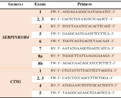
FW (B3): 5’- AATTTAAACATTTCTGATGGAATG -3’ FW (B4): 5’- TAATATGTTAATACATGGAATGT -3’ RV: 5’- AATATGAAGGTGAGTCATCA -3’
SERPINB3 E FW: 5’- TGACACATGTAGTAGGCTGT -3’ RV: 5’- CTTTCCCTTTCCAGAGAGAAAATG -3’
SERPINB4 E FW: 5’- TGACACATGTAGTAGGCTGT -3’ RV: 5’- TGCCCTTTCCAGAGAGAAAACAG -3’
For CTSG amplification a single PCR reaction was carried out to cover all five exons in a ~3
kb fragment. The sequences of the primers used are: Fw: 5’- TGAAACCTTTCATGGTAGCA -3’ and Rv: 5’- GATCTTAGACTTCTTAGCCTCT -3’. PCR reaction mix consisted of 1x KAPA Taq ReadyMix or MyTaq Mix, 0.75 µM concentrations for each primer, and 15-150 ng of genomic DNA.
30 In lung cancer samples, due to lower DNA concentrations from BALFs, SERPINB3 and SERPINB4 were amplified using semi-nested PCR reactions for fragments A, B and E. After a first
round of PCR reactions using primer pairs listed in Table 2.2, LC samples were submitted to second round PCR using the primers listed in Table 2.3. Similarly to PCR reactions used in first round amplification, semi-nested mixtures were done using the following reagents: 1x KAPA Taq ReadyMix
or MyTaq Mix, 0.5-1µM concentration for each primer, and 2 µL of the first PCR product diluted
1:50. PCR conditions of semi-nested reaction are shown in Annex Table T2.
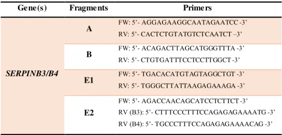
Table 2.3. Semi-nested PCR primers used for SERPINB3/B4 amplification.
Gene(s) Fragments Primers
SERPINB3/B4
A FW: 5’- AGGAGAAGGCAATAGAATCC -3’ RV: 5’- CACTCTGTATGTCTCAATCT –3’
B FW: 5’- ACAGACTTAGCATGGGTTTA -3’ RV: 5’- CTGTGATTTCCTCCTTGGCT -3’
E1 FW: 5’- TGACACATGTAGTAGGCTGT -3’ RV: 5’- TGGGCTTATTAAGAGAAAGA -3’
E2
FW: 5’- AGACCAACAGCATCCTCTTCT -3’
RV (B3): 5’- CTTTCCCTTTCCAGAGAGAAAATG -3’ RV (B4): 5’- TGCCCTTTCCAGAGAGAAAACAG -3’ Figure 2.1. Schematic representation of SERPINB3/SERPINB4 amplification. Location of
SERPINB3 (B3) and SERPINB4 (B4) genes in chromosome 18 is shown on top (marked as red). The common gene structure is shown below with the amplicons represented as grey arrows.
![Table 1.3. Clade B SERRPINs with known roles in lung function [Adapted from (Askew & Silverman 2008)].](https://thumb-eu.123doks.com/thumbv2/123dok_br/16672898.742794/29.918.128.785.354.810/table-clade-serrpins-known-function-adapted-askew-silverman.webp)