Dissertation of master degree in Bioengineering
Antioxidant delivery systems for
cosmetic application
Microencapsulation of rosmarinic acid by spray drying and
single emulsion solvent evaporation
Specialization in Biological Engineering
June, 2016
Developed within the discipline of Dissertation
Conducted at Laboratory for Process Engineering, Environment, Biotechnology and Energy
Department of Chemical Engineering, Faculty of Engineering, University of Porto
Supervisor: Dr. Lúcia Santos
i
Acknowledgements
I would like to formally express my gratitude to the following institutions and persons.
I would like to thank my supervisor Dr. Lúcia Santos, first for proposing such an interesting theme and for all the help, support, patience and availability during the semester. I am also grateful to Dr. Berta Estevinho for all the help, guidance and optimism.
The Laboratory for Process Engineering, Environment, Biotechnology and Energy (LEPABE) and the Department of Chemical Engineering for providing the facilities, equipment and materials employed in this work.
This work was financially supported by: Project POCI-01-0145-FEDER-006939 (Laboratory for Process Engineering, Environment, Biotechnology and Energy – LEPABE funded by FEDER funds through COMPETE2020 - Programa Operacional Competitividade e Internacionalização (POCI) – and by national funds through FCT - Fundaç ão para a Ciência e a Tecnologia.
I also would like to express my gratitude to the whole 201 Lab group for receiving me so well and for the good work atmosphere. Specially, I want to thank Eng. Isabel Carvalho for all the dedication, encouragement and support in the laboratory.
To my co-oriented friends, I would like to thank for the friendship and patiecet. A special thanks to Joana Aguiar and Marta Xavier for all the sharing and mutual support.
Finally, I would like to say thank you to my parents, my brother, my close friends and family for always being there and believing in me.
iii
Abstract
Antioxidants are molecules capable of oxidizing themselves instead or before other molecules. For cosmetic application, antioxidants may be used to retard the skin ageing process caused by oxidative stress. In addition, this ingredient may also be used as preservative avoiding the rancidification of the product or protecting other sensitive ingredients from oxidation.
The aim of this work was to perform the microencapsulation of rosmarinic acid (RA) in carboxymethyl cellulose (CMC) and ethyl cellulose (EC) polymers, using as encapsulation methods spray drying and o/w solvent evaporation, respectively. After encapsulation the microparticles were characterized in terms of shape and size distribution, using scanning electron microscopy and coulter counter-LS 230 particle size analyzer, respectively. Controlled release studies were performed in water and octanol, during 24 hours to simulate cosmetic vehicles. The antioxidant capacity of the encapsulated rosmarinic acid was evaluated using ABTS radical scavenging assay.
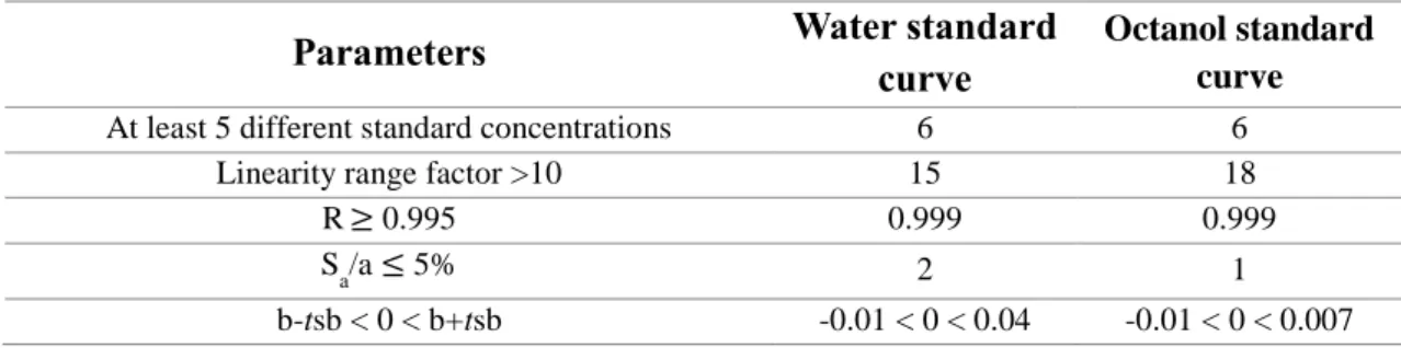
UV-Vis spectrophotometry analytical method was used to quantify the amount of rosmarinic acid released from microparticles. All the measurements were performed at 324 nm. The limit of quantification (LOQ) in water and octanol was 1.66 and 0.700 mg/L of rosmarinic acid, respectively while, the limit of detection (LOD) in water and octanol was 0.497 and 0.210 mg/L of rosmarinic acid, respectively. The analytical method was validated for both mediums with correlation coefficients greater than 0.995. The intermediate precision and repeatability only evidenced coefficients of variation (%CV) greater than 5% for the lowest RA concentration (3 mg/L). Accuracy studies showed recovery values (%R) greater than 97% for all the concentrations except for 9 mg/L of RA in water (94.10%). Therefore, no significant variations in the measurements were observed which demonstrates that the analytical method reproduced reliable results.
Regarding particles size, considering the number distribution, CMC and CMC-RA particles had a mean size of 0.156 and 0.358 µm, respectively while, taking into account the volume distribution, CMC and CMC-RA microparticles had mean size of 13.49 and 10.90 µm, respectively. These results suggest a size heterogeneity of the powder or agglomeration. CMC-RA microparticles prepared by spray drying presented a smooth surface and a spherical regular shape. EC-RA microparticles prepared by o/w solvent evaporation presented an irregular shape as well as some roughness on the surface.
O/W solvent evaporation method presented high product yield (95.4%) but low encapsulation efficiency (6.87%). On the other hand, spray drying microparticles showed higher encapsulation efficiency (90.8%) but lower product yield (38.4%).
The controlled release studies in water revealed that CMC-RA microparticles had a faster release (17.5 mg RA/g microparticles at 2 h) than EC-RA microparticles (2.41 mg RA/g microparticles at 2 h). In addition, EC-RA microparticles only released around 20% of their drug content in water. The release of EC-RA microparticles in octanol (58% at 2 h) was faster than in water (13%).
iv
Finally, the antioxidant activity of encapsulated rosmarinic acid in CMC (208 µM TE) and EC (197 µM TE) was slightly higher than the respective free rosmarinic acid solution.
These preliminary studies of rosmarinic acid controlled release, using CMC and EC as wall materials, suggest that they can be used for its microencapsulation since they retarded the release of the antioxidant and they did not reduce its antioxidant activity.
Key words: rosmarinic acid, antioxidants, cosmetics, delivery systems, microencapsulation, spray drying,
v
Resumo
Os antioxidantes são moléculas capazes de se oxidarem a elas mesmas prevenindo a oxidação de outras. Na cosmética, os antioxidantes poderão ser usados para retardar o envelhecimento na pele causado pelo stress oxidativo. Para além disso, este ingrediente poderá também ser utilizado como conservante combatendo a possível oxidação de compostos lipídicos presentes no produto ou outros ingredientes sensíveis à oxidação.
O trabalho realizado consistiu na microencapsulação de ácido rosmarínico (RA) em polímeros de carboximetil celulose (CMC) e etil celulose (EC), utilizando as técnicas de secagem por atomização e evaporação do solvente de emulsões óleo em água, respetivamente. Após a microencapsulação, as micropartículas formadas foram caracterizadas relativamente à sua distribuição em tamanho e forma, usando um analisador de tamanho de partículas coulter counter-LS 230 e microscopia eletrónica de varrimento (SEM), respetivamente. Os estudos de libertação controlada do RA encapsulado foram realizados em água e octanol, durante 24 horas de forma a simular as condições de formulações cosméticas simples. A capacidade antioxidante do ácido rosmarínico foi também avaliada recorrendo ao ensaio de eliminação de radicais de ABTS.
A espectrofotometria ultravioleta-visível foi utilizada como método analítico para quantificar o RA libertado das micropartículas. Todas as medições foram efetuadas com o comprimento de onda de 324 nm. O método analítico foi validado para ambos os meios de libertação com coeficientes de correlação superiores a 0,995. Os limites de quantificação (LOQ) em água e octanol obtidos foram 1,66 e 0,700 mg/L de RA, respetivamente. Os limites de deteção em água (LOD) e octanol foram 0,497 e 0,210 mg/L de RA, respetivamente. A precisão intermédia e repetibilidade apenas apresentaram valores de coeficientes de variação (CV%) superiores a 5% para a concentração mais baixa (3 mg/L). A exatidão apresentou valores de recuperação (R%) acima dos 97% exceto para a concentração de 9 mg/L de RA em água (94,10%). Assim, não foram observadas variações significativas nas medições o que demonstra a fiabilidade dos resultados obtidos usando este método.
Relativamente à distribuição de tamanhos, considerando uma distribuição em número, as partículas de CMC sem antioxidante e de CMC com RA revelaram um diâmetro médio de 0,156 e 0,358 µm, respetivamente. Considerando uma distribuição em volume, as micropartículas vazias de CMC e contendo RA encapsulado apresentaram um diâmetro médio 13,49 e 10,90 µm, respetivamente. Estes resultados sugerem uma heterogeneidade do tamanho das partículas do pó. Pela analise de SEM verificou-se que as micropartículas de CMC com RA apresentaram uma geometria esférica e uma superfície lisa. Por outro lado, as micropartículas de EC com RA demonstraram uma forma irregular e uma superfície mais rugosa.
A técnica de evaporação do solvente de emulsões óleo em água apresentou elevados rendimentos (95,4%), mas eficiências de encapsulação baixas (6,87%). Por outro lado, a secagem por atomização demonstrou altos valores de eficiência de encapsulação (90,8%), mas valores relativamente baixos de rendimento (38,4%).
vi
Os ensaios de libertação controlada em água revelaram que as micropartículas de CMC com RA conduziram a uma libertação mais rápida do antioxidante (17,5 mg RA/g micropartículas em 2 h) que as micropartículas de EC (2,41 mg RA/g micropartículas em 2 h). Para além disso, as micropartículas de EC apenas libertaram aproximadamente 20% do seu conteúdo em RA no fim das 24 horas de ensaio. A libertação do antioxidante das micropartículas de EC em octanol (58% em 2 h) foi mais rápida que na água (13%).
Por último, a atividade antioxidante do RA encapsulado em CMC (208 µM TE) e em EC (197 µM TE) foi ligeiramente superior a uma solução contendo RA que não sofreu o processo de encapsulação. Assim, estes estudos preliminares de libertação controlada de RA, utilizando CMC e EC como agentes encapsulantes, sugerem que a microencapsulação poderá ser uma boa solução para retardar a libertação do antioxidante sem comprometer o ser desempenho.
Palavras chave: ácido rosmarínico, antioxidantes, cosmética, microencapsulação, secagem por
vii
Contents
Acknowledgements ... i Abstract ... iii Resumo ... v Contents ... vii Figure list ... ix Table list ... xi Glossary ... xiiiObjectives and thesis organization ... xv
Background motivation ... 1
I. Introduction ... 3
I.1. Antioxidants ... 3
I.1.1. Rosmarinic Acid ... 6
I.2. Delivery systems... 8
I.2.1. Nanoparticles and Microparticles ... 9
I.3. Transdermal barrier ... 10
I.4. Incorporation of delivery systems in cosmetic formulations ... 13
I.5. Microencapsulation ... 15
I.5.1. Microencapsulation methods ... 15
I.5.2. Encapsulating agent ... 18
I.5.2.1. Carboxymethyl cellulose and ethyl cellulose ... 19
I.5.3. Microparticles controlled release ... 20
II. State of the art ... 23
II.1. Delivery system ingredients ... 24
II.2. Microparticles and preparation methods - Spray drying and solvent evaporation ... 25
II.3. Controlled release studies ... 26
II.4. Skin permeation and penetration ... 28
II.5. Incorporation of delivery systems in formulations ... 29
III. Equipment and reagents... 33
III.1. Reagents ... 33
III.2. Equipment ... 33
IV. Methods ... 35
IV.1. Analytical method for rosmarinic acid quantification ... 35
IV.1.1. Standard solutions preparation ... 35
IV.1.2. Standard curves validation... 35
viii
IV.2.1. Spray drying ... 37
IV.2.2. Solvent evaporation - O/W emulsion ... 37
IV.3. Characterization of the microparticles ... 38
IV.4. Controlled release studies ... 38
IV.5. Antioxidant activity assessment ... 38
V. Results and discussion ... 41
V.1. Analytical methods validation ... 41
V.2. Microparticles characterization ... 43
V.2.1. Product yield, encapsulation efficiency, drug loading ... 43
V.2.2. Particles morphology ... 44
V.2.3. Size distribution analyses ... 45
V.3. Controlled release studies ... 47
V.4. Antioxidant activity ... 50
VI. Conclusion... 53
Future work and limitations ... 55
References ... 57
Appendix 1 ... 71
Appendix 2 ... 73
Appendix 3 ... 83
ix
Figure list
Figure 1 - Possible skin oxidative stress effects [11]. ... 3
Figure 2 – Example of autoxidation mechanism [16] ... 4
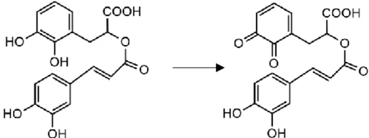
Figure 3 - Example of rosmarinic acid oxidation to the respective quinone ... 7
Figure 4 - Schematic representation of nano/microcapsules and nano/microsphere ... 9
Figure 5 - Schematic representation of skin structure [192], [193]. ... 10
Figure 6 - Possible mechanisms of action of vesicles as skin drug delivery systems [96] ... 13
Figure 7 - Schematic representation of an example of preparation of a cosmetic cream formulation [99], [194] ... 14
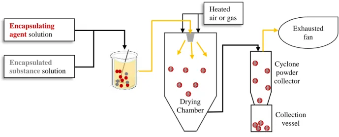
Figure 8 - Schematic representation of a spray dryer process ... 17
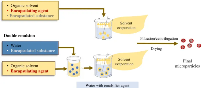
Figure 9 - Schematic representation of solvent evaporation method by single and double emulsion ... 18
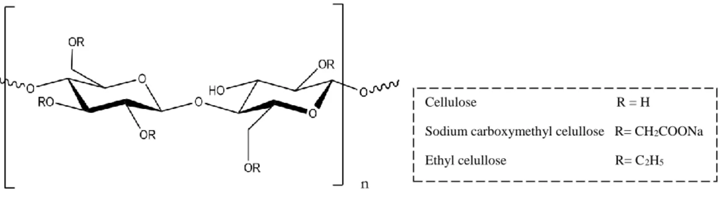
Figure 10 - Chemical structure of cellulose, sodium CMC and EC polymers ... 19
Figure 11 - Release mechanisms: (A) diffusion through water-filled pores, (B) diffusion through the polymer, (C) osmotic pumping and (D) erosion [130] ... 20
Figure 12 - Release profiles consisting of different phases for PLGA capsules [130]. ... 21
Figure 13 - Distribution of each type of delivery systems (last 10 years) from Table B and Table C from appendix 2 ... 23
Figure 14 - Distribution of type of skin tests performed ... 28
Figure 15 – Skin deposition within the skin layer ... 28
Figure 16 – Product yield of EC-RA and CMC-RA microparticles ... 43
Figure 17 – Encapsulation efficiency of EC-RA and CMC-RA microparticles ... 43
Figure 18 - SEM image of CMC-RA microparticles prepared by spray drying ... 44
Figure 19 - SEM image of EC-RA microparticles prepared by o/w solvent evaporation ... 45
Figure 20 - Number (A) and Volume (B) distribution of CMC microparticles prepared by spray drying ... 46
Figure 21 - Number (A) and Volume (B) distribution of CMC loaded RA microparticles prepared by spray drying ... 46
Figure 22 - Release profile of CMC-RA microparticles, prepared by spray drying, in water ... 49
Figure 23 - CMC-RA microparticles after 48h of release in octanol ... 49
Figure 24 - Release profile of CMC-RA and EC-RA microparticles, prepared by spray drying and O/W solvent evaporation, respectively, in water ... 49
Figure 25 - Release profile of EC-RA microparticles, prepared by O/W solvent evaporation in water and octanol ... 49
xi
Table list
Table 1 - Examples of the mostly used natural antioxidants in cosmetic formulation ... 5
Table 2 - Physical and Chemical properties of rosmarinic acid [45]... 6
Table 3 - Example of delivery systems used in cosmetics and their materials ... 8
Table 4 - Expression to calculate encapsulating efficiency, drug loading and product yield for spray drying and solvent evaporation methods ... 16
Table 5 - Spray drying operating conditions ... 37
Table 6 - Linearity conditions for the validation of the UV-Vis-spectrophotometry standard curves ... 41
Table 7 - Limit of quantification and limit of detection of the UV-Vis-spectrophotometry water and octanol standard curves ... 42
Table 8- Intermediate precision, repeatability, accuracy for rosmarinic acid quantification method in water and octanol ... 42
Table 9 - Experimental and theoretical drug loading of CMC-RA and EC-RA microparticles ... 43
Table 10 - Number and Volume average size for CMC and CMC-RA microparticles ... 46
xiii
Glossary
% CV Coefficient of variation %R Recovery percentage ~y Absorbance average 4CL Hydroxycinnamate coenzyme A A Regression slopeABTS 2,2'-azino-bis(3-ethylbenzothiazoline-6-sulphonic acid) B Intercept of the regression
C4H Cinnamic acid 4 hydroxylase CAGR Compound annual growth rate CMC Carboxymethyl cellulose
CMC-RA Carboxymethyl cellulose rosmarinic acid loaded microparticles
DL Drug loading
DMF Dimethylformamide
DMSO Dimethyl sulfoxide
EC Ethyl cellulose
EC-RA Ethyl cellulose rosmarinic acid loaded microparticles EE Encapsulation efficiency
HPLC High-performance liquid chromatography HPPR 4 hydroxyphenylpyruvate reductase Kow Coefficient of partition octanol- water LMWA Low molecular weight antioxidants
LOD Limit of Detection
LOQ Limit of quantification NLC Nanostructured lipid carriers PAL Phenylalanine ammonia- lyase PBS Phosphate-buffered saline PLGA Poly(lactic-co-glycolic acid) PLLA Poly(l-lactic acid)
PVA Polyvinyl alcohol
R Coefficient of correlation
RA Rosmarinic acid
ROS Reactive oxygen species Sa Slop standard deviation Sb Intercept standard deviation
xiv
SC Stratum corneum
SEM Scanning electron microscopy SLN Solid lipid nanoparticles Sy Absorbance standard deviation T Confidence interval of 95% TAT Tyrosine aminotransferase
xv
Objectives and thesis organization
The aim of the experimental work was to perform the microencapsulation of rosmarinic acid (RA) in carboxymethyl cellulose (CMC) and ethyl cellulose (EC) polymers, using as encapsulation methods spray drying and o/w solvent evaporation, respectively. After encapsulation the microparticles were characterized in terms of shape and size distribution, using scanning electron microscopy and coulter counter-LS 230 particle size analyzer, respectively. Controlled release studies were performed in water and octanol, during 24 hours to simulate cosmetic vehicles. The antioxidant capacity of the encapsulated rosmarinic acid was evaluated using ABTS radical scavenging assay.
This master thesis is organized in six chapters: introduction, state of the art, equipment and reagents, methods, results and discussion, conclusion. Other sections such as background motivation and future work and limitations are also presented. Chapter I, introduction, presents the basic explanation of the importance of antioxidants focusing in rosmarinic acid. In addition, it also describes some of the concepts of the transdermal barrier and the basic fundamentals of microencapsulation. Some of the challenges behind the development of a delivery system are also mention during the whole chapter. Chapter II, state of the art, compiles and describes some of the information presented in appendix 2 providing the reader with the description of some research articles using delivery systems for cosmetic application. Chapter III, equipment and reagents, presents the equipment and reagents used in the experimental work. Chapter IV, methods, describes the methods used to perform the work as well as the conditions that allowed the validation of the analytical method. Chapter V, results and discussion, presents the main results regarding the analytical method validation, characterization of the microparticles, controlled release studies and antioxidant activity assessments. Finally, chapter VI, conclusion, presents the main conclusions of this work. All the references used in this thesis may be consulted in references section as well as some additional information in appendix 1, 2, 3 and 4.
1
Background motivation
Cosmetics and personal care products are market sectors that represent billions of dollars worldwide. United States of America is one of the main countries in this industry with a revenue stream around 60.58 billion U.S dollars and employing about 62,450 people, by 2015 [1]. On the other side, in Europe, Germany was the greatest country in 2014, with a sales volume of approximately 13 billion euros, followed by France (10.6 billion euros) and United Kingdom (10.4 billion euros) [2]. Among the different cosmetic categories, skincare products are clearly dominant with a market share of 35.3% [3].
Despite the expected expansion of cosmetics industry, with an annual growth rate of 3.6% in 2014, a constant innovation is crucial to catch the attention of new consumers and keep the current consumers loyal to specific brands [1]. In order to do so, new ingredients with different and promising functions have been studied, in combination with recent technologies and formulations to extend the product shelf-life and improve its performance.
Antioxidants are extensively used and advertised on cosmetic products that claim, most of the times, anti-ageing properties (Appendix 1). This ingredient can be either synthetic or natural, easily found in a different number of plants, fruits, grains [4]. Although synthetic antioxidants represent around 60% of the total consumption of antioxidants in several industries, the demand for natural compounds is increasing [5]. Besides its health benefits, antioxidants may be used as product preservatives as well. However, these molecules are unstable and may be sensitive to light, pH, temperature, oxygen which can lead to degradation and loss of effectiveness of the product. For topical application, they may also have some difficulties crossing the transdermal barrier due to their physical chemical properties and the heterogeneous skin constitution [6].
Delivery systems are innovative technologies used to ensure the stabilization, protection and targeted release of an active ingredient. Some of the most common delivery systems used in cosmetics and personal care products comprise liposomes, microparticles, emulsions and it is expected that their global market will reach 543,373.2 thousand US dollars in 2020 [7]. Microencapsulation is mostly used to protect and stabilize the active agent by surrounding it with shell material. Several methods and materials may be used to entrap the active ingredient leading to the formation of different types of particles. The materials and operational conditions must be chosen and optimized according to the final application of the microparticle.
There are several companies that produce microparticles to incorporate in cosmetic formulations [7]. Besides that, a great number of published articles and patents were also found, suggesting a crescent and common interest of this technology by scientific community and industries [8]–[10].
3
I. Introduction
I.1.
Antioxidants
Antioxidants are molecules capable of oxidizing themselves instead or before other molecules. They are compounds or systems that can interact with free radicals and terminate a chain reaction before vital molecules are damaged [4]. The use of antioxidants is reported in food, cosmetics, beverages, pharmaceuticals and even in animal feed industry. They may be used as supplements and active ingredients, with health benefits, or as preservatives [5].
Reactive oxygen species (ROS) are one of the major causes of oxidative stress which enhances the skin ageing process [11]. Intrinsic ageing is associated with natural process of ageing while extrinsic ageing is related to external factors (e.g. air pollution, UV radiation, pathogenic microorganisms) that affect the ageing process. Photo ageing is probably the main reason of ROS production and acceleration of skin ageing process. Several potential targets can be found to interact with ROS in the skin (e.g. lipids, DNA, proteins) [12]. Figure 1 presents some of the potential oxidative stress effects in the skin.
Antioxidant molecules can be enzymes or low molecular weight antioxidants (LMWA) that can act by donating an electron to reactive species interrupting the radical chain reaction, by preventing the reactive oxidants formation, by acting as metal chelators, oxidative enzyme inhibitors or antioxidant enzyme cofactor’s [13], [14]. LMVA include a great number of substances and extracts that can be obtained from a variety of plants, grains, fruits and used in cosmetic industry. Table 1 presents some examples of natural antioxidants as well as their natural sources. Rosmarinic acid was the antioxidant studied in this work.
Antioxidants can also be used as preservatives avoiding the rancidity of lipid ingredients. In fact, lipid oxidation (Figure 2) is present in chemical products but also in the human body. In this way, antioxidants may have multi functions when present in a product. In the initiation phase of lipid oxidation, the number of radicals is expanded. During the propagation phase, molecular oxygen and fatty acid radicals react leading to the
Figure 1 - Possible skin oxidative stress effects [11].
Skin oxidative stress effects
Induce inflammation process Induce skin barrier disruption Induce skin roughness
Stimulate sebaceous glands excretion (due to the excessive production of oxidized lipids)
Affect melanocytes activity (pigmented and depigmented skin areas) Degradation of extracellular matrix
4
formation of hydroperoxide products. Hydroperoxide are unstable and can degrade to produce radicals that will accelerate the propagation reaction. The termination phase is dominated by reactions between radicals [15]. Antioxidants can prevent lipid oxidation by reacting with lipid and peroxy radicals, converting them to more stable and non-radical products. In addition, antioxidants are also able to reduce hydroperoxides to hydroxy compounds, deplete molecular oxygen, deactivate singlet oxygen, remove prooxidative metal ions, replenish hydrogen to other antioxidants and absorb UV light [14], [16].
Some studies report that neurodegenerative diseases like Alzheimer or Parkinson can be caused by oxidation of polyunsaturated lipids of cell membranes by free radicals. In this way, antioxidants have been studied as possible therapy for this kind of diseases [17], [18]. According to literature, antioxidants can also be involved in cancer treatments since the production of reactive oxygen species is altered during tumorigenesis [19]. Other mechanisms and implications in cancer disease are also reported in literature [18], [20], [21]. In addition, antioxidants play a role in diabetes disease, cardiovascular and hepatic diseases. Besides, they have anti-inflammatory, immuno-stimulant and anti-microbial properties as well [18], [22–26].
Nevertheless, antioxidants have some limitations that narrow their inclusion on all type of products mentioned above. According to literature, the stability of antioxidants is influenced by light, pH, temperature and oxygen [27–29]. Additionally, they can react with other matrix compounds, degrade and lose their activity [30]. Some antioxidants are used in the form of extracts which can give some taste or smell to the product as well [31], [32]. One of the main challenges for antioxidants, mainly for topical application, remains in the ability of crossing the dermal barrier. This difficulty is explained by the distinct characteristics that are associated to the different skin layers [33], [34].
Figure 2 – Example of autoxidation mechanism [16] RH- fatty acid R• - fatty acid radical ROO• - peroxy radicals
5
Table 1 - Examples of the mostly used natural antioxidants in cosmetic formulation
Classification
Antioxidant
Source
Chemical Structure
K
owReference
Vitamins
Vitamin C Apple, bayberry, broccoli, citrus peel,
garlic, peppermint, spearmint 1.850 [4], [35–37]
Vitamin E Olives and olive oil, palm oil, pumpkin
seeds, sunflower seeds and sunflower oil 12.180 [4], [36], [38]
Polyphenols
Quercetin Black pepper, onions, curly kale, leeks,
broccoli, blueberry, red wine and tea 1.5 [6], [39–42]
Resveratrol
Red wine, grape berry skins and seeds, peanuts, dried roots of plant Polygonum
cuspidatum
3.10 [43], [44]
Rosmarinic acid
Oregano, rosemary, marjoram, clary sage,
thyme, basil 1.82 [45]
Carotenoids
Lycopene Apricots, grapefruit, guava, watermelon,
papaya and carrots 16.6 [4], [46–48]
Lutein Spinach, leaf lettuce, peas, oranges, kale,
6
I.1.1. Rosmarinic Acid
Rosmarinic acid (RA) is a polyphenol found in a wide variety of plants from the Lamiaceae family: oregano (Origanum vulgare L.) rosemary (Rosmarinus officinalis L.) , marjoram ( Origanum majorana L.), clary sage (Salvia sclarea L.) , thyme (Thymus vulgaris L.), basil (Ocimum basilicum L.) [45]. This compound was first isolated in 1958 by two Italian chemistries, Scarpati and Oriente [53]. Table 2 presents some chemical and physical properties of rosmarinic acid.
Table 2 - Physical and Chemical properties of rosmarinic acid [45]
Chemical structure IUPAC name (2R)-3-(3,4-dihydroxyphenyl)-2-[(E)-3-(3,4-dihydroxyphenyl)prop-2-enoyl]oxypropanoic acid Molecular Formula C18H16O8 Melting point (ºC) 171-175 Vapor pressure at 25 ºC (mmHg) 1.1x10⁻¹³ log Kow 1.82
Solubility (mg/mL) DMSO: 25 DMF: 25 Ethanol: 25
PBS:15 Water:1.0
Rosmarinic Acid is an ester of caffeic acid and d 3,4-dihydroxyphenyllactic acid. The biosynthesis of this antioxidant is initiated by two parallel pathways - phenylpropanoid pathway and tyrosine derived pathway. The first via comprises three steps, using phenylalanine ammonia-lyase (PAL), cinnamic acid 4-hydroxylase (C4H) and hydroxycinnamate coenzyme A ligase (4CL). The second via involves two successive steps using tyrosine aminotransferase (TAT) and 4-hydroxyphenylpyruvate reductase (HPPR). Subsequently, the two resulting products (4-coumaroyl-CoA and 4-hydroxyphenyllactic acid) act as substrates for rosmarinic acid precursor (4-coumaroyl-4'-hydroxyphenyllactic acid) and then to the final conversion to rosmarinic acid [54]. The chemical synthesis of this antioxidant was first achieved in 1991 [53].
According to literature, there are several benefits related to rosmarinic acid besides its strong antioxidant activity: anti-inflammatory, anti-mutagenic, anti-bacterial, anti-viral, anti-cholinesterase and anti-tumor effects [53], [55]. Its antioxidant activity is higher than vitamin E and it also acts as scavenger of free radical [56]. During oxidation reaction, rosmarinic acid molecules may be transformed to quinones (Figure 3) [57].
7
Studies show that rosmarinic acid can be involved in therapies related to ocular disease, since data suggests rosmarinic acid could be a potent inhibitor of retinal neovascularization and may be used in the vasoproliferative retinopathies treatments [30]. Furthermore, rosmarinic acid have proven neuroprotective action in animal models of neurodegenerative diseases such as Alzheimer and Parkinson. Literature also shows the effectiveness of this antioxidant against memory deficits induced by permanent focal cerebral ischemia in mice [58]. Rosmarinic acid can also play a role in epilepsy disease, liver/brain damage following ischemia and reperfusion, rheumatoid and arthritis disease [59–61]. Several studies report its involvement in cancer treatments such as colon cancer (tested in mice) or leukemia (tested in vitro) [62], [63]. The use of rosmarinic acid as preservative can also be beneficial for cosmetic or food products. Thymus nummularius methanol extract containing rosmarinic acid revealed antimicrobial activity against pathogenic microorganisms such E. coli, P. aeruginosa, S. aureus, S. pyogenes and C. albicans [64]. For dermatologic application, published articles evidenced the role of rosmarinic acid in melanin production, which can be used as a therapeutic agent for skin pigmentation disorders, and it can have a role in protection against photo carcinogenesis [65–67]. Atopic dermatitis treatment was also evaluated in mice using rosmarinic acid and it presented positive results [60]. The photoprotective effect of rosmarinic acid in UVA induced changes in human keratinocytes was also verified. Rosmarinic acid decreased the production of ROS, DNA damage and increased the keratinocytes cell viability [68]. Therefore, this antioxidant is a substance of interest for cosmetic industry.
However, it has some particularities that limit its use in cosmetics formulations (emulsions and creams): low water solubility, discoloration and instability [56]. The low Kow (1.82) makes it difficult to cross the transdermal barrier in order to reach the deeper layers of the skin and once there, reduce the amount of ROS [34].
The use of delivery systems can overcome these limitations. Delivery systems may not only guarantee the protection and stabilization of rosmarinic acid during storage and manufacturing process, but also they may improve its physical chemical properties to facilitate the transport across biological barriers. The choice of the carrier material is crucial since it has to be biocompatible and it has to be able to solve all the limitation mentioned above.
8
I.2.
Delivery systems
Nowadays, consumers are not only focused in cosmetic products that only promote their beauty. They are seeking for products claiming health benefits, the use of natural ingredients combined with recent technologies that can improve the cosmetic performance. Drug delivery systems are engineered technologies used to carry an active ingredient promoting a controlled and targeted delivery. Human skin acts as a barrier against the permeation of exogenous molecules. Drug delivery systems are able to enhance the permeation of the active ingredient through the skin layers, controlling its concentration in the formulation and on the skin. For cosmetics purpose, it is a major concern to keep the active ingredient in the superficial skin layers and avoid the systemic absorption [69]. Burst and sustained release are two major features that may be associated to cosmetic delivery systems. Burst release is important to improve the penetration of active molecule, while sustained release becomes important when the active ingredient is irritating at high concentrations, or to supply the skin for long periods of time [70]. The protection of the active ingredient is another advantage of using delivery systems. The shelf life demanded for a cosmetic product is usually no less than 2 years. Therefore, sensitive ingredients to external conditions like light, oxygen, heat must be protected to ensure the stability of the product. In addition, drug delivery systems can also prevent the reaction between the encapsulated ingredient and other molecules in the product matrix [71]. Several types of delivery systems can be found in cosmetic products (Table 3). They may be divided into vesicular systems (liposomes, niosomes, transfersomes, emulsions (microemulsions and nanoemulsions), particulate systems (micro particles, nanoparticles) [69].
Table 3 - Example of delivery systems used in cosmetics and their materials
Delivery system Material Ref.
Liposomes Lipids: Soya phosphatidylcholine, dipalmityl phosphatidylcholine, distearyl
phosphatidylcholine [72]
Niosomes
Nonionic surfactant: Polyoxyethylene alcohol, polyoxyethylene glycol alkyl
ethers, alkyl ethoxylate, alkyl phenol ethoxylate, fatty acid alkanolamides, propylene oxide-modified polymethylsiloxane
[73]
Transfersomes
Lipids: Soya phosphatidylcholine, egg phosphatidylcholine, dipalmityl
phosphatidylcholine, distearyl phosphatidylcholine
Edge activator: Sodium cholate, sodium deoxy cholate, Tween 80, Span 80
[74] Solid lipid Nanoparticles (SLN) & Nanostructured lipid carriers (NLC)
Solid lipids: Tristearin, stearic acid, cetyl palmitate, cholesterol, Precirol®
ATO 5, Compritol® 888 ATO, Dynasan®, 116, Dynasan® 118, Softisan® 154, Cutina® CP, Imwitor® 900 P, Geleol®, Gelot® 64, Emulcire® 61
Liquid lipids: Medium chain triglycerides, paraffin oil, 2-octyl dodecanol, oleic
acid, squalene, isopropyl myristate, vitamin E, Miglyol® 812, Transcutol® HP, Labrafil Lipofile® WL 1349, Labrafac® PG, Lauroglycol® FCC, Capryol® 90
9
I.2.1. Nanoparticles and Microparticles
Polymeric microparticles were the main focus of this work. Nano/microparticles are used to entrap active ingredients (core material) inside or scattered in a surrounding material (shell material) [77]. The exact definition of nanoparticles and microparticles is not consensual among authors. Some researchers consider that nanoparticles size should range between 1 and 100 nm, while others claim that nanoparticles must range between 1-1000 nm [78], [79]. Nano/microparticle is a general term comprising both nano/microspheres and nano/microcapsules. Nano/microspheres consist of a homogenously dispersion of the active ingredient in the polymeric matrix whereas nano/microcapsules are reservoirs where distinct domains of core and wall material are present (Figure 4) [80]. One of the main purposes of this procedure is the creation of a physical barrier that avoids the contact of the active agent with the external matrix protecting sensitive substances from moisture, pH, light, oxygen and other molecules present in the matrix [81]. This barrier is also responsible for a controlled release of the active ingredient which can be regulated by heat, mechanical action, pH, biodegradation, diffusion and dissolution [82]. Nano/microparticles are often used to enhance the permeation of the active ingredient through the skin without the penetration of the particle. Nanoparticles, in particular, can be applied as preservatives and antibacterial agents in cosmetics [83].
The preparation of nanoparticles and microparticles may be performed by the same methods (i.e. evaporation or extraction of the solvent, interfacial polymerization, spray drying) with controlled operational conditions in order to obtain the desired product [84].
Delivery system Material Ref.
Polymeric Nanoparticles and
microparticles
Natural: Collagen, albumin, whey protein, casein, gelatin, chitosan, agarose,
alginate, carrageenan, dextran, pectin, cyclodextrins, hyaluronic acid
Synthetic: Poly(lactic acid), poly(glycolicacid), poly(lactic-co-glycolic acid),
poly(ε-caprolactone), poly(alkylene succinates), poly(hydroxyl butyrate),
[76] Table 3 - Example of delivery systems used in cosmetics and their materials
Matrix Mononuclear Polynuclear
Nano/microcapsules Nano/microsphere
10
A wide variety of encapsulating agents can be used, extending from synthetic to natural. Its choice should be made according to the particle application, the selected core material, the physical and chemical stability, the required particle size, the release mechanism and manufacturing costs [82], [83]. Occasionally, a combination of wall materials is used to achieve the desired properties [84].
Nanoparticles have a higher tendency to aggregate due to their high surface area and the type of interaction that they can establish with each other [85]. Additionally, particles size also interferes with the release of the active ingredient. Active ingredient entrapped in smaller particles have greater access to the external phase which can lead to a higher release by diffusion, a faster penetration of water into the particle and a lower drug loading. The adsorption of molecules on the surface also occurs during particle formation and it is more accentuated the smaller the particle is. On the other hand, smaller particles may have a better binding for a unit of particle mass than larger ones which could be useful to adhere on the skin [86]. Transdermal barrier and the interactions of the different delivery systems with it is another critical aspect that must be considered.
I.3.
Transdermal barrier
Human skin can be divided in three distinct layers: epidermis, dermis and subcutaneous layer (Figure 5). Epidermis is the outer layer (thickness: 50-150 µm) that has a protective role since it is in contact with the environment. It comprises several layers from the stratum corneum (upper layer) to the basal cell layer that are constantly being regenerated. The second layer of human skin is dermis and it composed of glycosaminoglycans, blood vessels, nerves, sweat glands, hair follicles and sebaceous glands. Elastin and collagen are here synthetized and they are responsible for the tensile strength, resilience and elasticity of the skin, respectively. The inner layer is hypodermis or subcutaneous layer which is composed of adipose tissue and it is responsible for the thermal control [34], [87], [88].
11
Stratum corneum is composed of enucleated and completely differentiated keratinocytes cells, called corneocytes, mostly filled with keratin. Each corneocyte cell is surrounded by a lipid matrix and contains intact or degraded corneodesmosomes that are responsible for keeping cells together. According to literature, stratum corneum is composed of 15 layers of corneocytes with a diameter of 40 µm and thickness of 0.3 to 0.8 µm. Under air-dried conditions the gap between cells is 75 nm. Looking from the top to the inside, it is possible to observe that corneocytes have a hexagonal shape and are grouped in clusters up to 12 units [89]. Pathways between the clusters offer less resistance delimiting the intercorneocyte penetration pathway. Transcellular penetration pathways are not significant for transdermal passage [90]. Stratum corneum hydration is an important factor to consider in drug delivery and it may be regulated by the proteolysis of the corneocyte content. The degradation products consist in a mixtures of osmotically active amino acids that capture water and act as moisturizing factors.
The lipid matrix is composed of different types of lipids, i.e. cholesterol and its derivatives, ceramides, free fatty acids and triglycerides, that are responsible for its space organization. However, the basis of the lipid matrix structure consists in lamellar repeating hydrophilic and lipophilic bilayers where approximately 80% of the lipid are non-polar, with hydroxyl groups, able to settle hydrogen bond interactions with adjacent lipids or water [89]. As we go deeper in the skin layers the content in water increases: the lipophilic stratum corneum contains around 13% of water, the viable epidermis about 50% of water and the dermis around 70% of water [34]. In stratum corneum, hydrophilic regions are created mainly in the lateral cell junctions due to the non-planarity of the corneocytes outer membrane. Less ordered lipids and flexible hydrophobic chains are also found in interlamellar and linker regions. Such characteristics are extremely important for transepidermal diffusion of lipophilic and amphiphilic molecules since they guarantee the necessary space for migration. Hydrophilic molecules diffuse preferably “laterally” through the water filled inter lamellar spaces or the free spaces between the lamella and the corneocyte outer membrane [90]. The chemical composition of stratum corneum is also responsible for a pH gradient ranging from 4.5 and 5.5, in mammalian stratum corneum surfaces, to neutral values in stratum granulosum interface.
Therefore, the passage through the stratum corneum is limited to low molecular weight molecules (<500 Da), preferably uncharged and with Kow values between 1 and 3. Regarding the use of delivery systems, they have to be smaller than 5–7 nm to potentially diffuse throughout the fluid lipid bilayers or smaller than 36 nm to eventually go through the aqueous pores [34], [89].
The penetration of active substances and nanoparticles is also possible through the pilosebaceous units and it has been demonstrating an increased interest. Pilosebaceous units are composed of the hair follicle, the adjoining arrector pili muscle and the associated sebaceous glands [91]. Their function is to synthetize and excrete a mixture of squalene, waxes, cholesterol derivatives, triglycerides fatty acids and cell debris, called sebum [34]. Sebum excretion acts as barrier that prevents the passage of molecules and particles to the inner layers. However, literature describes this non polar lipid mixture as a possible responsible for the uptake of some lipophilic molecules. Therefore, penetration agents may be able to disperse themselves in sebum [92].
12
Corneocytes barrier in the lower hair follicle infundibulum seems to be crumbly and smaller which makes this area more susceptible to penetration. Hair follicle infundibulum can also act as reservoir for nano and micro sized particles. It has been suggested that larger particles release high concentrations of the active ingredients to the follicle, while smaller particles (less than 40 nm) can pass through the disrupted skin barrier [93]. The hair follicle can be considered as an invagination of the epidermis extended deep to the dermis layer thus providing a greater improvement of penetration surface area. Cosmetic relevant drug targeting, through pilosebaceous units, can be directed to the sebaceous gland, viable skin epidermis, follicular papilla and hair matrix cells. The sebaceous gland area is associated to skin problems such as acne and it was recently found the existence of melanocytes stem cells. Follicular papilla and hair matrix are believed to be involved in hair growth and pigmentation. Regarding hair grow cycle, it can influence the penetration of active ingredients [92].
In order to improve skin delivery, penetration enhancers may be used. These substances may be used to increase the diffusion coefficient of the penetration molecule through the stratum corneum, to increase the effective concentration of the drug in the vehicle, to improve the partitioning between the formulation and stratum corneum. Examples of classic penetration enhancers consists in water, ethanol, dimethyl sulphoxide, laurocapram, oleic acid, surfactants. Ethanol is a commonly used solvent in cosmetic formulations. The mechanisms by which ethanol acts as penetration enhancer are different: as a solvent it can improve the solubility of an active ingredient in the vehicle; it can permeate and change the solubility properties of the stratum corneum; its rapid permeation or evaporation may alter the thermodynamic properties of the drug within the formulation; when used in high concentrations, it can extract some lipid fraction from the stratum corneum [94]. Recent studies suggest the use of peptides as penetration enhancers [95]. They may be considered as safer than traditional penetration enhancers since the last ones can cause skin irritation, cytotoxicity or alter irreversibly the skin barrier.
Vesicular delivery systems are frequently reported in cosmetic formulations. Their dimensions can range between the nano and the micro scale. Roughly speaking, they can be divided in deformable systems and rigid ones. Lipid vesicular systems can deliver the active ingredient just for releasing it from the vesicle, can enhance the transdermal passage due to interactions with stratum corneum, can fuse and exchange lipid material with stratum corneum, transferring the active ingredient to the skin (Figure 6) [96]. Intact vesicular skin penetration is associated to deformable systems since they can squeeze through the interclusters and intercorneocytes pathways using hydrating gradient as driving force [97], [98]. Deformable vesicles are often smaller than rigid ones which also justifies why they are frequently found deeper in the skin. The vesicular chemical composition is the key factor to establish the predominant mechanism by which a molecule is delivered through the skin.
Regarding rigid particulate systems (SLN, NLC, micro/nano particles), its penetration mostly depends on the particle size being its composition and charge less significant. SLN and NLC are claimed to form an occlusive film on skin surface that enhances the active ingredient penetration [89].
13
I.4.
Incorporation of delivery systems in cosmetic formulations
Generally cosmetic cream formulations are manufactured by preparing separately two distinct phases with the respective soluble ingredients. The homogenization step must be equipped with a vacuum line or a de-airing step should be added [99]. Thermal or volatile excipients must be added after the mixture is cooled down to a suitable temperature. The adjustment of pH can be performed in the end of the formulation or by adding the neutralizing agent generally to the aqueous phase. Some excipients can be sensitive to pH modifications therefore, a careful choice of the cosmetics ingredients should be made to ensure stability [100], [101]. Figure 7 represents a schematic example of the preparation of cream formulation.
A deliver system can be formulated in a liquid state or as powder. Regardless of the physical state, it has to be incorporated in a vehicle (i.e. cream, lotion) to be applied on the skin. After incorporation, it is necessary to guarantee a uniform product, sensorially attractive for the consumer and with a long term stability. Therefore, experimental formulations must be tested regarding its spreadability, rheological properties, color changes, pH changes, storage temperatures. Taking into account rheological properties, for instance, cosmetic formulations may be characterized by a non-Newtonian behavior since it is desired to decrease the viscosity of semi solid formulation when it is spread on the skin (i.e. an external force is applied). Furthermore, Newtonian systems such as liquids or emulsions may not form occlusion films, since they spread rapidly, affecting the effectiveness of the formulation [102]. A good stability after the rheological tests is also important since it indicates that the product will remain stable even while it is rubbed on skin. Additionally, in vivo tests should also be taken in order to evaluate the efficacy of the product or adverse irritation reaction that it may cause on the skin [103].
A- Fusion and lipid material exchange with stratum corneum B- Enhancement of the transdermal passage
C- Free releasing from the vesicle
D- Intact vesicular skin penetration (only flexible vesicles) A B C D Vesicle Active ingredient E p id er m is & Der m is Stra tu m co rn eu m
14
The characterization of the delivery system can help to predict the behavior when it is incorporated in the cosmetic formulation. After prepared, delivery systems can be characterized concerning the size and morphology (scanning electron microscopy), mechanical properties (atomic force microscopy), thermal analysis (differential scanning calorimetry), zeta potential. Thermal analysis can be useful to determine the delivery system integrity after incorporation [104]. The size and zeta potential are two measurements that could help to prevent agglomeration since nano-sized particles and zeta potential values close to zero tend to aggregate [85]. After entrapment of an active ingredient into a delivery system (e.g. antioxidant), it is also advisable to evaluate its antioxidant activity since the process of encapsulation may affect its antioxidant properties. The protection of the antioxidant to light, oxygen or any other external factor should also be assessed for short or long periods of time.
The incorporation of a delivery system into a cosmetic formulation does not follow a single method or rule. Delivery systems can be incorporated in the final product, in the aqueous phase or in the oily phase. In addition, they may be incorporated in gels, dissolved in solutions or slurries that are further mixed. Considering SLN and NLC, the incorporation in the final product implies a reduction in the water content to incorporate an aqueous solution containing the lipid particles. Usually the cream formulations are produced, cooled and the particle lipid concentrate is added, stirring gently. This incorporation may lead to a viscosity increase and so, sometimes, it is necessary to reduce the lipid content of the initial formulation. Another method to produce SLN and NLC cosmetic formulations is to initially replace some water content by the lipid aqueous phase. To perform the last method, it is fundamental to assure the physical and chemical stability of the lipid particles during processing [105]. Another concern is the amount of active ingredient present in the final cosmetic formulation. Usually during the process, the formulations are diluted 10 to 20 times. In the final formulation, typically the particle concentration is between 2 - 4% while the active ingredient concentration should be 0.05 - 0.10% [106]. Therefore, the drug loading of the particles as well as the final proper concentrations of particles
Figure 7 - Schematic representation of an example of preparation of a cosmetic cream formulation [99], [194]
O i l y P h a s e H e a t a n d s t i r i n g H o mo g e n i z a t i o n C o o l i n g A q u e o u s P h a s e H e a t a n d s t i r i n g A d d i t i o n o f s e n s i t i v e i n g r e d i e n t s Fragances Antioxidants Preservative ss Humectants Thickener agents p H a d j u s t me n t F i l t r a t i o n Emmolients Emulsifiers S t o r a g e
15
and active ingredient must be considered and combined. Polymeric microparticles and nanoparticles may be added to any phase of the cosmetic formulation, according to its affinity to form a dispersion and avoid agglomeration. They can be added as a powder, a slurry or a wet cake. Powder microparticles can be directly incorporated or firstly dispersed in a solvent. Mechanical stirring and temperature are important factors to take into account since they can influence the integrity of the particle and lead to the release of the active encapsulated ingredient during manufacturing [107]. Microparticles will be further discussed to better understand some of the fundamentals of this type of delivery system since it is the main objective of this work.
I.5.
Microencapsulation
Although several delivery systems may present micro scale dimensions, the concept of microencapsulation is usually intensively associated to the formation of polymeric microparticles. Microencapsulation techniques have been used worldwide and they have numerous applications in food, pharmaceutical, household products, cosmetics, agrochemicals etc. Some examples of the utilization of microparticles include carbonless paper, ‘‘scratch and sniff’’ fragrance sampling, ‘‘intelligent’’ textiles, controlled release of drugs and cosmetic active agents [82].
Microencapsulation has many advantages comparing to non-encapsulated substances since it allows the protection and stabilization of the core material and its controlled, timed and targeted release [108]. Product appearance and flow properties may also be improved, enhancing its handling, usage and storage. Undesirable organoleptic properties can be masked and the evaporation of volatile ingredients can be controlled using microencapsulation. This technique can also be used to reduce the amount of ingredients in formulation being a cost saving alternative [109].
Cosmetic industry represented 8% of microencapsulation market in 2013 [82]. Several companies are involved in the production of microparticles for a wide range of applications: Ronald T. Dodge Company, Lipo Technologies, Evonik Industries AG, GAT Microencapsulation GmbH. The market is expected to growth at a compound annual growth rate (CAGR) of 9.7% from 2014 to 2020 [110]. Microencapsulation is a suitable technique to solve some of the cosmetic ingredients (e.g. antioxidants) limitations since it protects sensitive substances prolonging the shelf life and stability of the product. It can also control the release of the active ingredient improving its penetration across the skin.
I.5.1. Microencapsulation methods
There are several methods for microencapsulation however its choice should be well considered and analyzed. The microencapsulation method should be selected based on the intended particle size, the biodegradability and biocompatibility of the particles, the physicochemical properties of the core and shell material, the final application of the particles, the proposed mechanism for core material release, the production scale and the processing cost. Several variables can also influence the formation of the microparticles.
16
Therefore, an optimization of the microencapsulation process should be performed in order to select the proper materials and conditions to get the desired outcome.
Generally, microencapsulation techniques can be divided into two major categories: chemical and physical methods; the latter one can be subdivided into physicochemical and physico-mechanical techniques. Chemical methods comprise interfacial, emulsion and suspension polymerization. Physicochemical methods are for instance, solvent evaporation /extraction, coacervation, sol-gel encapsulation, ionotropic gelation. Spray drying, spray chilling, fluid bed coating may be considered physical methods [111–113].
After microencapsulation, microparticles should be characterized in order to evaluate the encapsulation method. Some of the basic process aspects are the encapsulation efficiency, drug loading capacity and the product yield. Encapsulation efficiency (EE) refers to the amount of active ingredient associated with the particles comparing to the feed solution. Drug loading (DL) refers to the amount of the active agent present in the microparticles. Yield product is the ratio between the output mass obtained and the initial solid content of the feed solution [114]. Since the ratio between the core material and the encapsulating agent can be considered constant during spray drying, EE should be calculated indirectly. Free RA is determined by measure the amount of rosmarinic acid right after dispersion of the microparticles in the solvent. However, for solvent evaporation techniques the same consideration cannot be taken since some antioxidant can escape to the aqueous phase. Therefore, it is only possible to calculate EE directly. Table 4 presents a sum up of the last information.
Table 4 - Expression to calculate encapsulating efficiency, drug loading and product yield for spray drying and solvent evaporation methods
I.5.1.1. Spray drying and solvent evaporation
Spray drying is probably the mostly used microencapsulation technique (Figure 8). A solution, a suspension or an emulsion containing the core material and the shell material is homogenized and then fed to a spray drying equipment. Then, the process can be divided in three steps: (1) atomization of the liquid solution (2) the contact of the fine droplets with a hot gas stream to evaporate the solvent (3) the separation and collection of the powder. Commercially available atomization systems consist of pressure nozzles, centrifugal atomizer, kinetic energy nozzle, and sonic energy atomizer. The spray drying operation mode can be in counter
Spray drying Solvent evaporation
Encapsulation Efficiency (%)
Initial RA weight − Free RA weight × 100 Initial RA weight
Entrapped RA weight × 100 Initial RA weight
Drug loading (%) RA weight in microparticles × 100
Microparticles weight
Product yield (%) Output mass × 100
17
current, co-current or a combination of both. The use of co-current operation mode is suitable for heat sensitive products since the solvent evaporation is fast with a very short residence time. The counter current mode is suitable for non-heat sensitive products and has a more efficient heat utilization. The combination of both operation mode is usually associated to smaller spray dryers. To achieve good encapsulation efficiency operational parameters like feed temperature, air inlet temperature and air outlet temperature should be optimized. The air inlet temperature is usually from 150 to 220 °C and air outlet temperature is from 50 to 80 °C. The drying times are in range of 5–100 s and in well design systems 15–30 s is a fair time [115]. Spray drying is a relatively simple process, with an operating low cost, easy to scale up and it can operate in continuous. However, some of the main drawback of this method are the high cost of the equipment, the low overall thermal efficiency and the possibility of loss of low-boiling point substances. The final product may not have a uniform size and may need further processing [82], [113].
Solvent evaporation is a simple method frequently used since it allows the encapsulation of hydrophobic and hydrophilic substances. In this method, the polymer is dissolved in a water immiscible solvent and the encapsulated substance is dispersed or dissolved in the mixture (Figure 9). The resulting solution or dispersion is then slowly added to a water phase, frequently in a presence of an emulsifier agent, under stirring, to form an emulsion. To form the solid microparticle the solvent must now diffuse into the aqueous phase and evaporate. The microparticles can be further washed and collected by filtration, centrifugation and then, dried. Several factors affect the formation of microparticles which should be thus optimized. The surfactant ratio, the rate of solvent evaporation, the polymer molecular weight, the agitation rate, the organic phase volume are some examples of parameters than can interfere with the microparticle formation [116]. Considering the solvent evaporation method there are two specific techniques that are widely employed: single o/w emulsion and double w/o/w emulsion (Figure 9). In the first an oily phase containing the encapsulating agent and the core substance is added to the aqueous phase. In double emulsion, a first w/o emulsion is prepared and then it
Figure 8 - Schematic representation of a spray dryer process Heated air or gas Encapsulating agent solution Encapsulated substance solution Drying Chamber Cyclone powder collector Exhausted fan Collection vessel
18
is added to the aqueous phase to form a w/o/w emulsion. Double emulsion technique was developed to entrap hydrophilic molecules since they have tendency to escape to the aqueous phase when the single emulsion technique is used. Solvent evaporation method allows the incorporation of thermosensitive drugs since it does not require high temperatures. Some of the main drawback of this method is the use of high amounts of solvents, the low drug encapsulating efficiency and the hard scale up [112], [117].
I.5.2. Encapsulating agent
The encapsulating agent selection is a very important factor to consider during microencapsulation process since it affects its efficiency and the microparticles stability. However, most of the times the selection is made by trial-error procedures due to the unpredictable behavior of the formed complex [115].
The selection of the wall material should ponder the physicochemical properties of both core (e.g. porosity, solubility) and shell material (e.g. viscosity, mechanical properties). Furthermore, the wall material should not be soluble or react with the core. Besides the particle size, the final application of the microparticle is an important criterion to select the wall material. Different applications require different degrees of protection of the core and different release mechanisms of the active agent [118]. Sometimes one shell material does not have all the required characteristics so a combination of encapsulation agents may be used.
Wall materials can be classified as natural, semi-synthetics and synthetic polymers. Natural wall materials can be divided into carbohydrates (e.g. starch, maltodextrin, cyclodextrin), gums (e.g. arabic gum, acacia gum, alginate, carrageenan), proteins (e.g., gelatin, casein, soy protein). Synthetic shell material can be classified in polyesters (poly (lactic-co-glycolic acid), poly(ε-caprolactone), poly (alkylene succinates)), acrylic polymers (poly (methyl methacrylate), poly hydro(ethylmethacrylate), polymethacrylates),
Single emulsion Double emulsion Filtration/centrifugation Drying Final microparticles • Organic solvent • Encapsulating agent •Encapsulated substance Solvent evaporation • Water • Encapsulated substance • Organic solvent • Encapsulating agent Solvent evaporation
Water with emulsifier agent
19
phosphorous-based polymers and polyamides [76]. In a less extent, silicates and clays can also be used as encapsulating agents [82].
In order to use in cosmetic applications, wall materials should be biocompatible and once in contact with human body, they must not induce any adverse reaction or its degradation products must not cause any negative effect on health. The carrier must also have a proper resistance to protect and deliver the core material [118], [119].
I.5.2.1. Carboxymethyl cellulose and ethyl cellulose
The use of carbohydrates as wall materials is very attractive since they are usually cheap materials, biocompatible, biodegradable, non-toxic and can be used with different encapsulating methods and core materials [120]. Cellulose (Figure 10) is natural water insoluble biopolymer composed of β-linked glucopyranose residues and may have several types of substitution that modify the physical chemical properties of the polymer.
Carboxymethyl cellulose (CMC) is a water soluble anionic cellulose derivative with carboxymethyl substitution groups. It can be obtained by the etherification of the polymer with chloroacetic acid. Typically, the commercial form of carboxymethyl cellulose is as white sodium salt and this derivative finds application in a wide range of fields such as food, cosmetic, pharmaceutical, textiles, detergents. Carboxymethyl cellulose is already widely used in cosmetic and personal products as emulsifier, stabilizer, film-former and thickener agent [121]. It is not soluble in organic solvents but it can be dissolved in mixtures of water and water-miscible solvents (e.g. ethanol, methanol). The dissolution rate is proportional to the degree of substitution but it decreases with the molecular weight (i.e. viscosity) [122]. Carboxymethyl cellulose hydrogel swelling behavior is pH and ionic strength dependent due to the presence of electrostatic charges in the polymer network originated from sodium ion and the carboxymethyl group [123]. Films formed using carboxymethyl cellulose have a moderate strength in aqueous solutions although such property is also dependent on the degree of substitution and molecular weight [124], [125]. Carboxymethyl cellulose is frequently used with cross linking agents to improve its performance [123].
Cellulose R = H
Sodium carboxymethyl celullose R= CH2COONa
Ethyl celullose R= C2H5
n
20
Ethyl cellulose (EC) is another derivative of cellulose where some of the hydroxyl groups of cellulose monomer were replaced by ethyl ether groups. It may result from the etherification with ethyl chloride [121]. Its appearance ranges between a white and a light tan powder or granular substance. It is only soluble in organic solvents such as alcohols, ketones, ether and esters which makes its use attractive as encapsulation agent for both water and organic solvents. Although its water insolubility, ethyl cellulose can take up water due to the hydrogen bond potential of the oxygen ethyl group atom with water. In addition, it is biocompatible, odorless, tasteless, non-toxic and non-irritant although it is not biodegradable. Ethyl cellulose is also stable under light, oxygen, heat and it is able to absorb pressure being thus resistant to mechanical stress [126]. Several options of ethyl cellulose are commercially available depending on the degree of substitution and molecular weights (i.e. viscosity). Ethyl cellulose is also used in cosmetic industry as base of cosmetic pastes and to form gels of liquid oils [127]. In fact, ethyl cellulose has been studied to replace petrolatum as cosmetic ingredient [128].
I.5.3. Microparticles controlled release
The term controlled release can be defined as the rate that certain molecules become available under particular conditions or stimulus (e.g. pH, temperature, moisture, enzymes, etc.) [129]. The mechanism by which the core material is released from the microparticles is called release mechanism and it depends on external factors (pH, moisture, enzymes, temperature, mechanical strengths) as well as interactions between the core and shell materials. There are several release mechanisms by which core material can be released: hydrolysis, diffusion through water-filled pores, osmotic pumping, erosion, water absorption/swelling etc. However, they can be summarized in four main processes that are presented in Figure 11.
Diffusion processes and osmotic pumping involve the transport of the core material whereas erosion is caused by polymer disintegration. Assuming water as the fluid where the microparticle is placed, diffusion through water pores is the mostly common mechanism involved in core material release. The fluid penetrates the shell and then dissolves the core that is released through the pores. The release depends on 1) the rate that the fluid penetrates the shell; 2) the rate that the core dissolves in the fluid; 3) the rate that the dissolved core leaks out through pores. In addition, dissolution of the polymer in water may occur at the same time the core material diffuses. Diffusion through the polymer can occur when the core material is small and has a suitable hydrophobicity to diffuse through the polymer. Osmosis can be defined as the transport of core material through water filled pore by a force such osmotic pressure (convection).
Figure 11 - Release mechanisms: (A) diffusion through water-filled pores, (B) diffusion through the polymer, (C) osmotic pumping and (D) erosion [130]
![Figure 1 - Possible skin oxidative stress effects [11] . Skin oxidative](https://thumb-eu.123doks.com/thumbv2/123dok_br/19204581.955339/21.892.115.768.525.769/figure-possible-skin-oxidative-stress-effects-skin-oxidative.webp)
![Figure 5 - Schematic representation of skin structure [192], [193].](https://thumb-eu.123doks.com/thumbv2/123dok_br/19204581.955339/28.892.130.767.799.1075/figure-schematic-representation-skin-structure.webp)
![Figure 6 - Possible mechanisms of action of vesicles as skin drug delivery systems [96]](https://thumb-eu.123doks.com/thumbv2/123dok_br/19204581.955339/31.892.142.753.116.388/figure-possible-mechanisms-action-vesicles-skin-delivery-systems.webp)
![Figure 7 - Schematic representation of an example of preparation of a cosmetic cream formulation [99], [194]](https://thumb-eu.123doks.com/thumbv2/123dok_br/19204581.955339/32.892.97.805.123.407/figure-schematic-representation-example-preparation-cosmetic-cream-formulation.webp)
![Figure 12 - Release profiles consisting of different phases for PLGA capsules [130].](https://thumb-eu.123doks.com/thumbv2/123dok_br/19204581.955339/39.892.101.774.739.998/figure-release-profiles-consisting-different-phases-plga-capsules.webp)