Mjhg
Universidade Nova de Lisboa
Instituto de Higiene e Medicina Tropical
Characterization of T follicular helper (Tfh) cells and B cell
isotype switching induced by type 1 and type 2 adjuvants
Patrícia Isabel Figueiredo Campos
DISSERTAÇÃO PARA A OBTENÇÃO DE GRAU DE MESTRE EM CIÊNCIAS BIOMÉDICAS, ESPECIALIDADE EM BIOLOGIA MOLECULAR EM SAÚDE TROPICAL E
INTERNACIONAL
Universidade Nova de Lisboa
Instituto de Higiene e Medicina Tropical
Characterization of T follicular helper (Tfh) cells
and B cell isotype switching induced by type 1 and
type 2 adjuvants
Autor: Patrícia Isabel Figueiredo Campos
Orientador: Sílvia Almeida, Laboratório LGraca, [email protected] Unidade de Regulação de Linfócitos
Coorientador: Luís Graça, Laboratório LGraca, [email protected] Unidade de Regulação de Linfócitos
Dissertação apresentada para cumprimento dos requisitos necessários à obtenção do grau de Mestre em Ciências Biomédicas, Regulamento n.º 128/2012
Este trabalho é dedicado aos meus pais, que sempre acreditaram nos meus sonhos e me apoiaram em todas as decisões que tomei. Especialmente ao meu padrinho, Ivan Figueiredo, que sei
Agradecimentos
Durante este ano sinto que cresci e aumentei o meu conhecimento, não só a nível profissional mas também pessoal. Todo o trabalho resultante nesta tese contou com o apoio de diversas pessoas às quais tenho de agradecer.
Tive o prazer de trabalhar em estreita colaboração com a Doutora Sílvia Almeida, uma excelente investigadora e amiga que brilhantemente me ensinou, orientou, inspirou e encorajou ao longo deste tempo. Por tudo o que fez por mim, muito obrigada, Sílvia. Agradeço ao Professor Doutor Luís Graça pela oportunidade de realizar a minha tese de mestrado no seu laboratório e dar os primeiros passos no mundo da imunologia.
Obrigada à Raquel Oliveira, Yaquelin Ortiz, Raquel Filipa e Andreia Carneiro pelo companheirismo, apoio e partilha de experiência e conhecimento, contribuindo para um ótimo ambiente de trabalho.
Aos restantes membros do grupo, obrigada pelo entusiasmo e momentos de discussão no lab meeting, que permitiram com que este trabalho progredisse.
Em particular, um obrigada especial à Raquel Rodrigues pela sua amizade e por compartilharmos todos os bons e maus momentos. Obrigada à Cátia Patrício pela sua dedicação e por me ter acompanhado ao longo destes dois anos de mestrado.
Finalmente, gostaria de agradecer à minha família e ao João Renato por todo o apoio e amor incondicional. Em especial, à minha Mãe por partilhar tão intensamente todas as etapas comigo. Obrigada pela paciência, amizade, ajuda e compreensão.
ix
Resumo
A principal função dos linfócitos T CD4+ é fornecer apoio a outras células no sentido de
gerar uma resposta imunitária eficiente. As interações entre as células T e B são essenciais para a produção de respostas humorais, sendo que foi recentemente demonstrado que as células T foliculares auxiliares (Tfh) desempenham um papel crucial neste processo. Caracteristicamente, expressam o fator de transcrição Bcl-6, o recetor de quimiocinas CXCR5 e o marcador de superfície PD-1. A expressão destes marcadores é única e fundamental para que estas células possam aceder ao folículo de células B, onde orientam as reações no centro germinativo (GC), levando à consequente mudança de isótipo, maturação da afinidade, produção de anticorpos de alta afinidade e células B de memória. Neste projeto, foram testadas duas hipóteses opostas no sentido de caracterizar fenotipicamente as células Tfh. Propomos investigar se estas são especializadas no fornecimento de auxílio do tipo Th1 ou Th2, que designamos de células hipotéticas "Tfh1" e "Tfh2" (Hipótese 1) ou se são uma subpopulação genérica que responde igualmente na presença de diferentes antigénios, células Tfh (Hipótese 2).
Deste modo, murganhos C57BL/6J e Balb/c foram imunizados na almofada plantar da pata traseira, utilizando proteína Ovalbumin (OVA) combinada com diferentes tipos de adjuvantes: CpG ODNs isoladamente e em combinação com TiterMax® Gold (TMX), Sigma Adjuvant System (SAS) e Montanide ISA 720 VG, testados como adjuvantes tipo 1, e por sua vez Incomplete Freund’s Adjuvant (IFA) e Alum experimentados como adjuvantes do tipo 2. A técnica de ELISA permitiu determinar no soro dos murganhos o tipo de resposta gerada, através da medição de imunoglobulinas específicas para OVA (IgG2a para Th1, IgG1 e IgE total para Th2). CpG ODNs e IFA foram considerados como os adjuvantes mais apropriados para induzir respostas Th1 e Th2, respetivamente. Células T que reconhecem especificamente OVA foram colhidas de murganhos OT-II Rag-/- e DO11.10 Rag-/- e transferidas para murganhos congénicos. De seguida,
procedeu-se à imunização tal como descrito acima. Os nódulos linfáticos drenantes foram recolhidos no pico da reação do centro germinativo (11 dias após imunização), assim como as células Tfh específicas para OVA (CD4+CD44+ CXCR5+PD-1+ Thy1.2+Vβ5+Vα2+/DO11.10+) e as células T auxiliares ativadas específicas para OVA
(CD4+CD44+CXCR5-PD-1- Thy1.2+Vβ5+Vα2+/DO11.10+).
A caracterização molecular destas populações de células T está a ser analisada através da sequenciação dos seus transcritos pela técnica de RNA-sequencing. Além disso, a expressão de marcadores de Th1 e Th2 em células Tfh foi analisada através de citometria de fluxo e Reação em Cadeia da Polimerase quantitativa por Transcrição Reversa (RT-qPCR). Neste estudo, foi demonstrado que as células Tfh co-expressam Bcl-6 e T-bet e também produzem IFN-γ, quando sensibilizadas com OVA-CpG ODNs, características concordantes com os marcadores fenotípicos de uma célula Tfh e célula Th1. A expressão de Gata-3 (marcador Th2) só foi detetada sob estimulação IFA-OVA, embora em níveis mais baixos do que as determinadas para T-bet.
Palavras-chave: Células Th1 e Th2, Adjuvantes, células Tfh, hipotéticas "Tfh1" e "Tfh2", RNA-Sequencing.
xi
Abstract
The major function of CD4+ T cells is to provide help to other lymphocytes to mount an
efficient immune response. T and B cell interactions are essential for humoral responses and it was recently shown that T follicular helper (Tfh) cells play a crucial role in this process. They characteristically express the transcription factor Bcl-6, chemokine receptor CXCR5 and PD-1. These markers are unique as their expression is pivotal to acquire access to the B cell follicle and drive germinal centre (GC) reactions, leading to isotype switching, affinity maturation, and production of high affinity antibodies and memory B cells.
In this project, two competing hypothesis investigating the phenotype of Tfh cells were tested. We propose to dissect whether Tfh cells are specialized in providing Th1 or Th2 help, which we call putative “Tfh1” and “Tfh2” cells (hypothesis 1), or if they are a more generic Th subset that responds equally in the presence of different antigens, which we designate as Tfh cells (hypothesis 2).
Therefore, we immunized C57BL/6J and Balb/c mice in the footpad using Ovalbumin (OVA) protein combined with different adjuvant types: CpG ODNs only and combined with TiterMax® Gold (TMX), Sigma Adjuvant System (SAS) and Montanide ISA 720 VG, as type 1 adjuvant, and Incomplete Freund’s Adjuvant (IFA) and Alum as type 2 adjuvants. Using ELISA assays to determine the type of response generated by measuring serum immunoglobulins of distinct clones (specific IgG2a for Th1 and OVA-specific IgG1 and total IgE for Th2), we considered CpG ODNs and IFA as the most appropriate adjuvants to induce Th1 and Th2 responses, respectively.
OVA-specific cells were transferred from OT-II Rag-/- and DO11.10 Rag-/- mice into congenic mice subsequent to immunization as described above. Draining LNs were collected at the peak of the GC reaction (day 11 post-immunization) and OVA-specific Tfh cells (CD4+ CD44+ CXCR5+PD-1+ Thy1.2+Vβ5+Vα2+/DO11.10+) and OVA-specific
activated-Th cells (CD4+ CD44+ CXCR5-PD-1- Thy1.2+Vβ5+Vα2+/DO11.10+) were
sorted.
The molecular signature of these T cell populations is being analysed via RNA-Sequencing. Moreover, the expression of Th1 and Th2 markers on Tfh cells was investigated via flow cytometry and Reverse Transcription quantitative Polymerase Chain Reaction (RT-qPCR). In this study, it could be shown that Tfh cells of mice immunized with OVA-CpG ODNs co-expressed Bcl-6 and T-bet and also produced IFN-γ, both concordant features with the phenotypic markers of a Tfh cell and of a Th1 cell. As for the expression of Gata-3, it has only been detected in mice under IFA-OVA stimulation, even though at levels lower than the ones determined for T-bet.
Keywords: Th1 and Th2 cells, Adjuvants, Tfh cells, putative “Tfh1” and “Tfh2”, RNA-Sequencing
xiii
Table of Contents
Agradecimentos ... vii Resumo ... ix Abstract ... xi Abbreviations list ... xv 1. Introduction ...1 1. Lymphoid organs ...41.1 Secondary lymphoid organs ...4
1.2 Germinal centre ...5
2. T helper cells (CD4+ cells) ...7
2.1 CD4 T cell differentiation ...7
3. T follicular helper (Tfh) cells ... 11
3.1 Phenotypic features of Tfh cells ... 12
3.2 Bcl-6 as Master Regulator of Tfh cells differentiation ... 13
3.3 Tfh cell differentiation ... 14
3.3.1 Stage 1: Generation of pre-Tfh ………...15
3.3.2 Stage 2: Guiding pre-Tfh into GC …...……….…15
3.3.3 Stage 3: Tfh differentiation in GCs ………...……..….15
3.3.4 Stage 4: Tfh cells can exit GCs ...……….…16
3.4 The transcriptional signature of Tfh cells ... 16
4. Adjuvants ... 17
2. Objectives ... 19
3. Materials and methods ... 23
1. Experimental mice ... 25
2. Ethics statement ... 25
3. Immunoglobulin quantification using Enzyme-Linked Immunosorbent Assay (ELISA) ... 25
4. Adoptive cell transfers and mice immunization ... 26
5. Flow cytometry analysis ... 28
5.1 OVA-specific Tfh cell sorting ... 28
xiv
6.1 Generation of mouse bone marrow-derived Dendritic Cells ... 29
6.2 Priming of naïve T cells ... 29
6.3 Cell surface and intracellular staining ... 31
7. Real-time quantitative-PCR (RT-qPCR) ... 31
7.1 RNA extraction from T cells ... 31
7.2 cDNA synthesis from total RNA ... 32
7.3 Quantitative Real time PCR ... 32
4. Results ... 35
1. CpG ODNs was the best adjuvant to induce Th1 responses, whereas Incomplete Freund’s Adjuvant (IFA) was better at inducing Th2 responses ... 37
2. Phenotypic characterization and recovery of Tfh cells ... 41
2.1 Tfh cells were phenotypically characterized as CD4+ CXCR5+PD-1+ while GC B cells were identified as CD19+GL-7+FAS+ ... 42
2.2 Tfh cells express high levels of the activation marker CD44, while expressing no CD25 ... 46
2.3 IFA induces a better recruitment of CD4+ T cells into GCs than does CpG ODNs ... 48
3. Molecular characterization of Tfh cells: Expression of transcription factors and cytokine production ... 51
3.1 in vitro polarized Th1 cells expressed T-bet and produced IFN-γ, while induced Th2 cells expressed Gata-3 and IL-13 ... 51
3.2 Bcl-6, T-bet, Gata-3 and IFN-γ expression were detected in Tfh cells ex vivo . 53 5. Discussion and Conclusions ... 63
Concluding remarks and future perspectives ... 68
6. References... 71
7. Attachment ... 81
1. Adoptive cell transfer from OT-II Rag-/- mice into B6 Thy1.1 recipient mice was not successful ... 83
xv
Abbreviations list
ACK Ammonium-Chloride-Potassium APC Antigen-presenting cell
Bcl-6 B-cell lymphoma 6 BCR B cell receptor BFA BrefeldinA
Blimp-1 B lymphocyte-induced maturation protein-1 BM Bone marrow
BM-DC Bone marrow-derived dendritic cells CCR CC chemokine receptor
CD40L CD40 ligand
cDNA complementary Deoxyribonucleic Acid cTfh circulating T follicular helper cell CXCR CXC chemokine receptor
DC Dendritic cell DZ Dark zone
ELISA Enzyme-linked immunosorbent assay FCS Foetal calf serum
FDC Follicular dendritic cell GC Germinal Centre
GM-CSF Granulocyte Macrophage Colony-stimulating factor HRP Horseradish Peroxidase
ICOS Inducible co-stimulator IFA Incomplete Freund’s adjuvant IFN Interferon
Ig Immunoglobulin
IGC Instituto Gulbenkian Ciência IL Interleukin
IMM Instituto de Medicina Molecular i.v Intravenous
xvi
LPS Lipopolysaccharides LZ Light zone
MALT Mucosal associated lymphoid tissues MHC Major Histocompatibility complex MPLA Monophosphoril lipid A
NK Natural killer cell NKT Natural killer T cell OVA Ovalbumin
PBS Phosphate buffered saline PD-1 Programed dead-1
PI3K Phosphoinositide-3-kinase PMA Phorbol-12-myristate-13-acetate PRRs Pattern recognition receptors RAG Recombination activating gene RNA Ribonucleic acid
RNA-seq Ribonucleic acid sequencing RT Room temperature
RT-qPCR Reverse transcription quantitative polymerase chain reaction SAP SLAM-associated protein
SAS Sigma Adjuvant System s.c Subcutaneous
SHM Somatic hypermutation SPF Specific pathogen free
STAT Signal transducer and activator of transcription TCR T cell receptor
Tfh T follicular helper cell TGF Transforming growth factor Th T helper
TLR Toll-like receptor TMX TiterMax® Gold TNF Tumour necrosis factor Tregs Regulatory T cell
1
3
The immune system is a remarkably versatile defence system that specifically recognizes and eliminates a variety of antigens (1) and is comprised of two different components, the innate and adaptive immune system.
Innate immunity provides the first line of defence against infection and includes physical and chemical defensive barriers as well as cellular barriers, namely phagocytes (macrophages and dendritic cells), granulocytes (neutrophils, eosinophils, basophils or mast cells) and natural killer (NK) cells (1). These cells express on their surface pattern recognition receptors (PRRs), that recognize conserved pathogen-associated molecular patterns (PAMPs) (2). Phagocytic cells internalize antigens through the endocytic pathway and process them into peptides in association with major histocompatibility complex (MHC) molecules. MHC-class II are expressed mostly by antigen-presenting cells (APC), manily macrophages, dendritic cells (DCs), and B cells. These cells are professional in antigen processing and presentation, both critical for T cell activation (1,3). Furthermore, innate activated cells produce a myriad of inflammatory cytokines and chemokines pivotal for the polarization of the appropriate subsequent immune response, directly impacting on effector function of T cells (1,2).
Adaptive immunity relies on lymphocytes to ensure specific long-lasting immunity, thereby protecting the host from subsequent exposure to the same antigen. An effective adaptive immune response is orchestrated by two major lymphocyte populations—B cells and T cells (1). Naïve B cells develop in the bone marrow (BM) and exit to circulate in blood and lymph, towards secondary lymphoid organs, where immune reactions take place. B cells can recognize soluble antigens through the B-cell receptor (BCR) with a single antigenic specificity (1,3). T cells develop and mature in the thymus and exit to the periphery as CD4+ T helper (Th) or CD8+ cytotoxic T (Tc) cells, the two main effector T cell populations of the αβ lineage. Unlike B cells, the T-cell receptor (TCR) can only recognize antigens that are presented by MHC molecules, with CD4+ T cells and CD8+ T cells recognizing antigen bound to class II MHC molecules or to class I MHC molecules, respectively (1–3).
4
1. Lymphoid organs
Lymphoid organs can be largely divided into primary and secondary lymphoid organs (1–3). Primary lymphoid organs, the thymus for T cells and the BM for B cells, are the place of lymphoid development. On the other hand, the morphologically and functionally distinct secondary lymphoid organs, constitute the sites where specific adaptive immune responses take place, through the presentation of a given antigen by APCs, resulting in the recruitment, activation and maturation of responding effector T cells and B cells.
1.1 Secondary lymphoid organs
The secondary (or peripheral) lymphoid organs are the spleen, lymph nodes and various mucosal associated lymphoid tissues (MALT), which trap antigen and provide places for interaction between mature naïve lymphocytes and its cognate antigen (4).
Lymph nodes are extremely organized structures, interconnected by a system of lymphatic vessels, which drain lymph, an extracellular fluid continuously produced through filtration of the blood and tissues. Morphologically, they can be divided into three roughly concentric regions, each supporting a distinct microenvironment: the cortex, the paracortex and the medulla (1). The cortex is arranged in primary follicles that contain mostly B cells, macrophages, and follicular dendritic cells (FDCs). The primary follicles enlarge, upon antigenic challenge, into secondary follicles, each containing a germinal centre. The paracortex, located between the cortex and the medulla, is populated largely by T cells and also interdigitating dendritic cells. These cells express high levels of MHC class II which are necessary for presenting the antigen, trapped in tissues and then drained, to T helper cells. The medulla is the innermost layer of a lymph node, formed mainly by plasma cells actively secreting antibody (1–4).
5
1.2 Germinal centre
Germinal centres (GC) are dynamic microenvironments of secondary lymphoid organs that provide a unique niche for B-cell affinity maturation (5). GCs develop only in response to an antigen and are initially engaged by migration of antigen-activated B cells that expand at around 6 days after a primary immunization (6). In GCs two distinct areas were identified, the dark zone (DZ) containing B cells and the light (LZ) zone that contains a rich network of FDCs and T follicular helper cells (Tfh) (7) (Figure 1.1). Affinity maturation takes place in B cells that actively proliferate in the DZ of GCs. These cells express high levels of the chemokine receptor CXCR4 that allows their maintenance in the DZ, in response to chemokine CXCL12 produced by stromal cells (8). The DZ B cells, known as centroblasts, then reduce expression of surface immunoglobulin (Ig) and undergo somatic hypermutation (SHM) of heavy- and light-chain variable region genes (5,9). Somatic hypermutation is induced by the enzyme cytidine deaminase (AID) and occurs randomly. Consequently, cells expressing receptors (BCR) of unchanged or lower affinity will be generated, as well as cells with receptors of higher affinity for a particular antigen (5) (Figure 1.1).
These cells also suffer class switch recombination, a biological mechanism that allows the change of the constant region of the antibody-heavy chain and, consequently, the alteration of the antibody class produced by an activated B cell (10). Naïve B cells express the IgM isotype and begin to express both IgM and IgD when they reach maturity. If antigen-activated B cells encounter specific signalling molecules via their CD40 and cytokine receptors, they undergo antibody class switching to produce IgG, IgA or IgE antibodies, allowing interaction with different effector molecules that have defined roles in the immune system (10).
Then, surviving DZ B cells will then upregulate the chemokine receptor CXCR5 and migrate toward the LZ. There, FDCs produce CXCL13, the ligand for CXCR5 and the major chemoattractant in the LZ (11). At this stage B cells, called centrocytes, stop dividing, increase the expression of surface Ig and are selected by the ability to recognize and bind antigen displayed on the surface of FDCs (6). Furthermore, the centrocyte must also receive survival signals from interaction with cognate Tfh cells (9) (Figure 1.1).
6
B cells that successfully interact with FDC and Tfh cells can follow three potential fates. First, GC B cells can upregulate CXCR4 to undergo recycling and re-entry into the DZ, for further proliferation and SHM. Second, GC B cells can also differentiate into memory B cells that stop dividing and enter the G0 phase of the cell cycle, during a primary response. Finally, cells can exit the GC as a plasma cell. As a consequence of SHM and class switching into the DZ, plasma cells produce higher antigen-affinity and express additional antibody isotypes, including IgG, IgA, and IgE (12). Additionally, differentiation of antibody-secreting plasma cells involves changes in gene expression that allows the gain of protein production and secretion functions. Such modifications are mediated by the master transcription factor B lymphocyte-induced maturation protein-1 (Blimp-1) (13).
Figure 1.1- Compartments and dynamic of the GC. Centroblasts undergo somatic hypermutation and clonal expansion into the DZ. During proliferation, the process of SHM introduces changes into the V(D)J region of the rearranged immunoglobulin variable region (IgV) genes. Dark zone B cells then move to the LZ, where the modified BCR is selected for improved binding to the immunizing antigen. Tfh and FDCs participates actively in this process (7).
7
GC B cells can be identified by their expression of high levels of death receptor Fas and
n-glycolylneuraminic acid (the ligand of antibody GL-7), loss of surface IgD and
anti-apoptotic molecule Bcl-2 (6). The transcriptional factor Bcl-6 is selectively upregulated during the GC stage as the critical regulator in the generation of the GC B cell phenotype.
2. T helper cells (CD4
+cells)
CD4+ T helper (Th) cells play a critical role in providing help to other lymphocytes and
innate immune cells to mount an efficient immune response. CD4+ T cells have the
capacity to secrete appropriate cytokines and express a variety of essential co-stimulatory molecules, important for their effector functions (14). Th cells carry out multiple tasks, ranging from activation and recruitment of other cells, including neutrophils and monocytes, B-lymphocytes, cytotoxic T cells, as well as non-immune cells. They also play critical role in the suppression of immune reaction (14).
2.1 CD4 T cell differentiation
The initial step of naïve CD4+ T cells differentiation is the antigenic stimulation through interaction of TCR with antigen-MHC II complex, presented by APCs. It consequently induces a network of signalling pathways which lead to cell proliferation and differentiation into specific effector cells. Lineage-specific differentiation depends on the cytokine milieu of the microenvironment as well as on the concentration of antigens, type of APCs, and co-stimulatory molecules (15). Co-stimulatory signals enhance TCR signal, and the main co-stimulatory receptor is CD28 expressed in all naïve T cells. The CD28 ligands, CD80 and CD86, are upregulated upon activation of DCs. The initial source of cytokines are from the APCs as well as other members of the innate immune system. Subsequently, CD4+ T cell differentiation into various effector subtypes (16) is
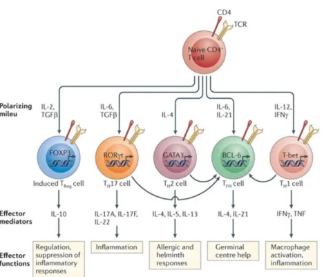
coordinated by genetic programs and epigenetic modifications, triggered by appropriate transcription factors to direct expression of distinct soluble mediators and surface molecules that support interactions with other immune cells. Besides the classical Th1 and Th2, other subsets have been identified, including Th17, regulatory T cell (Treg) and Tfh cells, each with a characteristic profile and function (17).
8
Th1 cells
Interleukin 12 (IL-12) and interferon γ (IFN-γ) are both critical cytokines for initiating the signalling cascade to develop Th1 cells (16). IL-12 is secreted largely by APCs, after their activation through the PRRs (15), and, in turn, IL-12 induces NK cells to produce IFN-γ.
T-bet is the principal master regulator that, in coordination with other transcription factors, induces full differentiation of Th1 cells. T-bet enhances the production of IFN-γ which actively stimulates the signal transducer and activator of transcription 1 (STAT-1). T-bet expression was found to be strongly dependent on STAT-1. T-bet further induces IFN-γ and TNF-α production by the differentiating cells, thereby amplifying T-bet expression and upregulating the expression of IL-12 receptors (IL-12R). Then, cells can be selected by the abundant IL-12 from the APCs, ensuring selective expansion of the differentiating Th1 cells (15).
IFN-γ stimulates the microbicidal activity of macrophages, up-regulates the level of class II MHC and increases the production of more IL-12 (18). Furthermore, IFN-γ produced by Th1 cells also induces antibody-class switching to some IgG classes, with production of IgG2a (in mice). The IgG2a isotype has been shown to be particularly potent in host defence against viral infections due to its ability in binding to Fc receptors expressed on phagocytes, activating the complement system and inducing Ab-dependent cell mediated cytotoxicity (19).
Importantly, T-bet suppresses the development of Th2 cells by inhibiting the crucial IL-4 gene and impairing the function of the Th2 master regulator Gata- 3 (20). Th17 lineage is also inhibited in consequence of the interaction between T-bet and Rorc promoter, which encodes RORγt, the principal transcription factor of Th17 cells.
The Th1 subset is responsible for many cell-mediated functions (e.g. promoting differentiation of CD8+ cytotoxic T cells, that perform an essential role in inhibiting replication of intracellular pathogens such as viruses) and for the production of opsonizing IgG antibodies (i.e. antibodies that bind to the high-affinity Fc receptors of phagocytes and interact with the complement system). This subset is also associated with the promotion of excessive inflammation and tissue injury (1,2,18).
9
Th2 cells
IL-4 and IL-2 are the critical cytokines for Th2 differentiation. IL-4 upregulates the expression of the major transcription factor involved in Th2 lineage, the master regulator Gata-3, which consequently enhances the production of IL-4, IL-5, and IL- 13 (1,2). STAT-5 is readily activated by IL-2 and has an important role in Th2 lineage commitment. For full differentiation of Th2 cells, the coordinated activity of STAT-5 and Gata-3 is required, since they bind to different sites of the IL-4 locus.
IL-4 promotes a pattern of class switching that produces neutralizing IgG1 (in mice) and IgE by activating transcription factors such as STAT-6, and also inhibits IgG2a and IgG2c isotypes in mice (16). IL-13 can also contribute to IgE class switch that plays a role in eosinophil-mediated defence, because eosinophils express Fc receptors specific for IgE and some IgG antibodies. IgE is essential against helminth infections and is also the principal mediator of allergic reactions (21).
Moreover, IL-4 and IL-13 induce alternative macrophage activation and induce secretion of growth factors that stimulate fibroblast proliferation and collagen synthesis, which contributes to tissue repair and fibrosis. IL-13 stimulates mucus production by airway epithelial cells, an important component of allergic reactions. IL-5 is important for eosinophil activation, growth and differentiation, and serves as the principal association between T cell activation and eosinophilic inflammation (1,2).
Gata-3 also inhibits Th1 differentiation, presumably through interaction with T-bet, downregulating the expression of STAT-4 and the signalling chain of the IL-12R (15). Th2 cells are involved in allergic diseases, promoting airway inflammation, eosinophilia, asthma and exacerbation of helminth infections (22,23).
Th17 cells
IL-6, IL-21, IL-23, and TGF-β are the major signalling cytokines involved in Th17 cells differentiation, and retinoic acid receptor-related orphan receptor gamma-T (RORγt) is the master regulator (15). IL-6, IL-21 and IL-23 activate STAT-3 signalling that plays an important role in RORγt expression and Th17 differentiation process.
10
These cells appear to be abundant in mucosal tissues, particularly of the gastrointestinal tract, suggesting that Th17 cells may be specialists in combating intestinal infections and in the development of intestinal inflammation. Most of inflammatory actions of these cells are mediated by IL-17, which stimulates the production of chemokines and other cytokines that recruit neutrophils to the sites of infection. IL-17 also stimulates the production of antimicrobial substances (1,2). The main effector function of Th17 cells is to control bacterial and fungal infections (24) through neutrophilic inflammation. Th17 cells have been associated to the pathogenesis of inflammatory diseases, e.g. psoriasis, rheumatoid arthritis and multiple sclerosis (25,24).
Treg cells
Regulatory T cells (Tregs) express Foxp3 and secrete anti-inflammatory cytokines, such as TGF-β and IL-10. Tregs manifest their function through numerous mechanisms that include the secretion of immunosuppressive soluble factors such as IL-9, IL-10 and TGF-β, cell contact mediated regulation via the high affinity TCR and other costimulatory molecules (26,27).
Naturally occurring Foxp3+CD4+CD25+ Treg cells develop in the thymus and display a diverse TCR repertoire that is specific for self-antigens (28)However, Treg cells can also be induced from effector T cells during inflammatory processes in peripheral tissues (29). Regulatory T cells (Tregs) are critical to maintain immune homeostasis, limiting the magnitude of immune response against pathogens and control inflammatory and autoimmune reactions (26,27,30).
Tfh cells
T follicular helper cells (Tfh) express Bcl-6 as the major transcription factor and produce IL-21. These cells are specialized in providing support to B cells during the GC reaction, essential for the generation of high-affinity memory B cells and antibody-producing plasma cells (31). Tfh cells will be discussed in detail in the following section.
11
Figure 1.2- Major pathways of naïve CD4+ T cell differentiation into effector cells. Upon encountering antigens presented by the professional APCs, naïve CD4+ T cells differentiate into Th1, Th2, or Th17 effector cells. Cytokines present in the environment during differentiation play the major role in determining the phenotype that the CD4+ T cell will acquire (32).
3. T follicular helper (Tfh) cells
In the early 2000s, a new population of CD4+ T cells was reported in human tonsils and also in lymphoid organs of immunized mice (33). The original defining feature of this designated Tfh cell was the expression of the chemokine receptor CXCR5, which was found to be indispensable for the co-localization of cognate T and B cells, which can interact during GC reactions (34). However, the original description of Tfh as CXCR5+ cells was insufficient to establish it as a distinct lineage of CD4 T cells. In 2009, B-cell lymphoma-6 (Bcl-6) was described by three independent groups as the master transcription regulator of Tfh cells (35–37). Tfh cells are pivotal for germinal centre formation, providing help and support to B cells while undergoing affinity maturation and class switch recombination, culminating in the secretion of high affinity antibodies, plasma cells and memory B cells.
12
3.1 Phenotypic features of Tfh cells
Tfh cells express a range of cell surface molecules that are essential for their identification and allow their functions in their interactions with B cells. As referred above, Tfh cells express high levels of CXCR5 that, together with down-regulation of CCR7, facilitates their migration into B cells follicles, in response to CXCL13 (11). This co-localization is critical for T-B interactions and important for proper generation of functional Tfh cells (38).
Typically, Tfh cells are also identified by the co-expression of other surface markers, most commonly programmed death-1 (PD-1) and inducible co-stimulator (ICOS). Both these molecules are members of the CD28 family and are up-regulated on T cells following activation. PD-1 is a negative regulator that provides an inhibitory signal to GC B cells, that express PD-1 ligand 1 or 2, preventing excess of Tfh cells proliferation in GC (31). PD-1 and PD-1 ligand deficient mice displayed higher frequencies of Tfh cells after protein immunization (39), alongside worsened B cell responses due to increased GC B cell apoptosis (39).
ICOS is a co-stimulatory molecule expressed on activated T cells, whereas its ligand ICOSL exists on B cells, macrophages and other APCs. Several studies have demonstrated that ICOS signalling seems to be dependent on its ability to activate phosphoinositide-3-kinase (PI3K) signalling (40). ICOS is able to up-regulate Tfh cell-associated genes such as c-Maf, IL-4 and IL-21. This plays an important role in Tfh cell generation, GC formation and antibody production (41).
Additional surface molecules are importantly expressed by Tfh cells. CD40 ligand (CD40L) provides survival signals to B cells through CD40 molecule and induces B cell differentiation and class-switching. The signalling adaptor SLAM-associated protein (SAP) plays an indispensable role for stable T and B interactions required for Tfh cell differentiation.
IL-21 is produced by activated CD4+ T cells and NKT cells and are highly expressed by Tfh and GC Tfh cells (42). This cytokine plays a major role in Tfh cell survival and GC B cell proliferation, survival and differentiation (42). IL-21 is the most potent inducer of plasma cell differentiation in both mice and humans, a process that is STAT-3 dependent.
13
Additionally, IL-21 can induce expression of Blimp-1 and Bcl-6 transcription factors on B cells in a mutually exclusive manner (43).
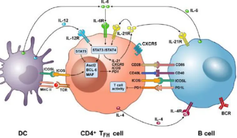
Figure 1.3- Cellular and molecular interactions between DCs, Tfh and B cells in GC. Tfh cells express a range of surface molecules, ICOS, CD28, CD40L and PD-1, important for interaction with DC in the T-cell zones and B cells in GC of lymph nodes (44).
3.2 Bcl-6 as Master Regulator of Tfh cells differentiation
Like other Th lineages, Tfh cells are associated with expression of a canonical transcription factor. Bcl-6 was identified as a master regulator transcription factor in Tfh cell differentiation. T cells deficient in Bcl-6 are unable to differentiate into Tfh cells or to sustain GC reactions, while Bcl-6 overexpression facilitates the expressions of Tfh-associated molecules such as CXCR5 and PD-1 (45).
Bcl-6 appears to participate in control of at least four major categories of genes: genes involved in cell migration, repression of alternative fates, Tfh differentiation and Tfh cytokines production (37). Bcl-6 controls GC B cell differentiation by regulating cell cycle genes and DNA damage response genes, supressing a host of signalling pathways, including BCR signalling (36,46,47). Tfh cells express high levels of Bcl-6 while non-Tfh cells (i.e Th1, Th2, Th17) express Blimp-1. Blimp-1 is a reciprocal antagonist of Bcl-6 and can inhibit Tfh cell development (35). Importantly, Bcl-Bcl-6 can antagonize transcription factors important for Th1 (T-bet), Th2 (Gata-3) and Th17 (RORγt) differentiation. However, this inhibition can be, and is most frequently, incomplete.
14
3.3 Tfh cell differentiation
In 2011 Shane Crotty proposed a canonical Tfh cell differentiation model, i.e. a multistage and multifactorial process (31) involving several events, molecules and two different cells as APCs. Nowadays, although much more is understood about the multiple stages and signals involved in the process, critical knowledge is still lacking because it has not been possible to reproducibly mimic Tfh cell differentiation in vitro. Tfh cell differentiation comprises four different stages, with characteristic molecules, signals and APCs playing essential roles for a successful process culminating in the development of a fully differentiated Tfh cell.
Figure 1.4- Canonical model of Tfh cell differentiation. Multiple signals and steps for the generation of Tfh cells. (a) Within the T cell zone, naïve CD4+ T cells
encounter DCs, activating T cells that move towards B cell follicles. (b) At the T-B border, activated T cells become pre-Tfh cells interacting with cognate activated B cells. (c) In the germinal centre, pre-Tfh cells become Tfh cells and provide help for B cell differentiation into plasma cells and memory B cells. Tfh cell differentiation involves a series of co-stimulatory molecules and cytokines, which are important for their function in providing help to B cells (45).
15
3.3.1 Stage 1: Generation of pre-Tfh
After an infection or immunization, naïve CD4+ T cells in the T cell zone interact with antigen presenting-DCs. Naïve CD4+ T cells acquire features of pre-Tfh or early Tfh cell and undergo a cell fate decision within the first few rounds of cell division (38). The increased expression of Bcl-6 and downregulation of antagonist Blimp-1 is taking place during this early stage, within 3 days after immunization or infection. This program is influenced by IL-6 and IL-21 (in mice) or IL-12 (in humans) and co-stimulation through ICOS and CD28 (36). ICOS plays a role in both Tfh cell differentiation and migration, and there is data supporting a synergistic role of ICOS and IL-6 (40) (Figure 1.4). The transcription factor Bcl-6 is crucial for maintenance of the Tfh program, including the expression of CXCR5 and the downregulation of CCR7. Thus, the early Tfh cell will migrate to the border of the B cell follicle and undergo further Tfh cell differentiation (48). Thus, the interplay between IL-6, ICOS, IL-21, and TCR signaling orchestrates early induction of mouse Tfh cell differentiation during DC priming via control of CXCR5, Bcl6, and other targets (49) (Figure 1.4).
3.3.2 Stage 2: Guiding pre-Tfh into GC
The second stage of Tfh cell differentiation occurs in the T-B border, where pre-Tfh cells contact with cognate activated B cells, which serve both as APCs and as a source of ICOSL (48,49). The stable T-B conjugates so formed move into the GC, while low affinity T cells that fail to interact with their cognate B cell, do not gain access to the GC and, expectedly, accumulate in the T-B border (31,45) (Figure 1.4).
3.3.3 Stage 3: Tfh differentiation in GCs
The third stage occurs in the GC, where pre-Tfh cells finally differentiate into Tfh cells which help GC B cells during affinity maturation, class switch recombination and differentiation into memory B cells or plasma cells (50). Tfh cells express high levels of CXCR5, PD-1, Bcl6 and ICOS (31,37) (Figure 1.4). Moreover, SAP expression is also essential for Tfh cell development, GC development, and the generation of the majority of memory B cells and memory plasma cells (31,51).
16
In the absence of SAP, Tfh cells display defective adhesion to GC B cells and fail to be retained in GCs. In addition, IL-21 produced by Tfh cells prompts their own differentiation, while IL-6 produced by B cells is important for Tfh cells maintenance (49) (Figure 1.4).
3.3.4 Stage 4: Tfh cells can exit GCs
After Tfh cells differentiation, they can exit the GC in several ways. Tfh cells can transit to neighboring follicles and enter a different GC (52), or temporarily reside in an adjacent B cell follicle before re-entering the same GC. Alternatively, Tfh cells can downregulate Bcl-6 and develop into a memory Tfh cell (53). Tfh memory cells are characterized by intermediate expression of CXCR5 and CCR7. Despite the fact that Bcl-6 expression is not detected in memory Tfh cells, c-Maf and other transcription factors are maintained at low or intermediate levels. Furthermore, ICOS, IL-21 and PD-1 are absent in antigen-specific memory Tfh cells in mice. One study has demonstrated that antigen-antigen-specific memory cells expressing CXCR5, provided accelerated B cell responses and antibody class switching. The authors suggested that CXCR5 expression promotes their rapid migration to the GC (53).
3.4 The transcriptional signature of Tfh cells
The transcriptional profiling of Tfh cells has revealed a distinct repertoire of expressed genes that distinguish them from Th1, Th2, or Th17 cells (54). However, it is not yet defined whether Tfh cells can be themselves sub-divided into distinct populations, which can provide specific help to B cells, depending on antigen type.
Recently, few studies have reported that Tfh cells in GCs have the capacity to produce either IFN-γ or IL-4, depending on how they were primed (31,35,55). Nevertheless, this fact does not distinguish a scenario where a cell of the Tfh lineage subsequently acquires the capacity to produce IL-4 or IFN-γ, from another where Th2 or Th1 cells could acquire the capacity to act as Tfh cells (35).
17
This was interpreted as evidence that Tfh cells can produce cytokines associated with canonical helper T effector subsets depending on environmental conditions. According to these, further studies are lacking to clarify this question.
4. Adjuvants
Immunization with purified protein antigens typically results in the induction of a modest antibody response, with little or no T cell involvement. In order to overcome this, adjuvants, i.e. compounds with the capacity to enhance the immune response against co-inoculated antigens, have been used to boost immune responses to an antigen of interest. They can sustain and improve the immunogenicity of antigens, effectively modulating appropriate immune responses, thereby reducing the amount of inoculated antigen and/or the number of immunizations. Some of the features involved in adjuvant selection are the particular antigen, the formulation and safety aspects, the animal species, the route of administration and, importantly, the type of immune response, or more appropriately, the phenotype of the effector T and B cells so generated. In a nutshell, adjuvants should promote an appropriate immune response, stimulating both cellular and humoral immunity.
Adjuvants can be classified according to their component sources, physiochemical properties or mechanisms of action (56). Adjuvants usually fall in two major functional groups, based on being dependent (Type 1) or not (Type 2) on Toll-like-receptors (TLRs) signalling (Figure 1.5).
Type 1 adjuvants are recognised by TLR and directly act on the immune system to increase responses to an antigen, while type 2 adjuvants, present antigens in an optimal manner, including controlled release and depot delivery systems, thereby enhancing the specific immune response to the immunizing antigen. (56) (Figure 1.5).
18
Figure 1.5- Putative mechanisms of action of adjuvants. Many adjuvants can act as ligands for PRRs that activate an innate immune response. Receptor signalling can then activate transcription factors that induce the production of cytokines and chemokines that help direct a particular immune response, such as a Th1 or Th2 type response. Some adjuvants also influence presentation of antigen by MHC (57).
19
21
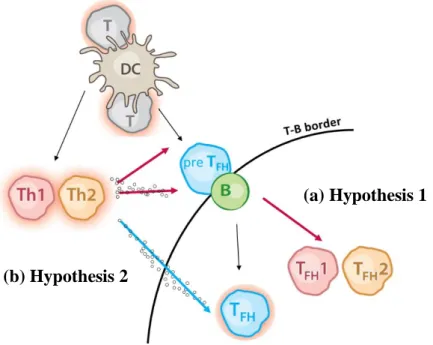
Tfh cell differentiation remains controversial because it is still unknown whether Tfh cells can be subdivided in different subsets what we call putative “Tfh1” or “Tfh2” (Hypothesis 1), or if they are a more “general” subset of T helper cells, Tfh (Hypothesis 2) such as Th1, Th2 or Th17. Our proposal addressed these two competing hypotheses as shown on figure 2.1.
The first hypothesis (a) postulates the existence of Tfh subsets specialized in providing Th1 or Th2-type “help”, a subset we named here putative “Tfh1” or “Tfh2”. The acquisition of such specialized Tfh function may be a consequence of early interaction with DCs or following interaction with B cells in the T-B border, and it may be influenced by cytokines produced by Th1, Th2 or Th17 cells.
The alternative model (b) defends that “help” provided by Tfh cells in the course of a Th1 or Th2-associated immune response is the responsibility of a non-specialized and generic cell population, Tfh cells. Thus, isotype-switch is influenced by contact with generic Tfh cells and cytokines produced in the vicinity by extra-follicular Th1 or Th2 cells.
Figure 2.1- Two alternative models for Tfh cells contribution in GC reaction. (a) Extra-follicular environment can lead to acquisition of type-1 or type-2 characteristics by Tfh cells, originating specialized “Tfh1” or “Tfh2” cells that then drive GC formation. (b) As an alternative, Tfh cell contribute as non-specialized Tfh cells together with cytokines produced by extra-follicular Th1 or Th2 cells.
(a) Hypothesis 1
22
In this work, we aimed to characterize the phenotype of putative specialized “Tfh1” and “Tfh2” subsets, or exclude their existence, in two different genetic backgrounds, Balb/c mice or C57BL/6J mice (which will be generically designed as B6 mice). We will dissect and assign, should it exist, a molecular signature to the at presumed Tfh1 and Tfh2 cells, through RNA-sequencing analysis of purified antigen-specific cells.
To achieve this goal, the work has been organized in two tasks:
i. Identification of ideal immunizing conditions to drive Th1 and Th2 humoral responses.
Primary, mice will be subcutaneously immunized in the footpad with OVA (protein) with either a type 1 or a type 2 adjuvant to generate Th1 or Th2 immunity, respectively. We will collect serum at day 14 in order to determine the type of response generated, by measuring serum OVA-specific (IgG2a for Th1, IgG1 for Th2) and total IgE (Th2) by ELISA. We will also collect draining LNs at the peak of GC reaction (Day 11) for further evaluation of the presence of effector Th and Tfh cells, by flow cytometry.
ii. Purification of antigen-specific populations of Th1, Th2 humoral responses. Then, using established immunizing conditions, CD4+ T cells will be isolated from
OVA-specific TCR-transgenic OT-II or DO11.10 mice and adoptively transferred intravenously into congenic B6 or Balb/c hosts, respectively. Mice will be subsequently immunized as described with OVA in conjugation with defined adjuvants.
Draining LNs will be collected and Tfh cells (CD4+CXCR5+PD-1+) and extra-follicular
cells (CD4+CXCR5-PD-1-) will be identified by flow cytometry. From each condition we
will sort antigen-specific Tfh cells and antigen-specific extra-follicular cells (Th1 or Th2). These T cell populations are being further analyzed by RNA-Sequencing, an objective outside the scope of this thesis.
23
25
1. Experimental mice
Balb/c, C57BL/6 (B6), B6 Thy1.1, B6 Thy1.1/Thy1.2, DO11.10 Rag-/-, OT-II Rag+/+ and OT-II Rag-/- mice were bred and maintained in specific pathogen free (SPF) conditions at the Instituto Gulbenkian de Ciência (IGC) and Instituto de Medicina Molecular (IMM). All mice were used between 6–10 weeks of age.
2. Ethics statement
Procedures were conducted in accordance with the Portuguese and European laws (Portaria 1005/92 and Directive 2010/63/EU) and following the FELASA recommendations. Procedures were conducted in accordance with guidelines from the Animal User and Institutional Ethical Committees.
3. Immunoglobulin quantification using Enzyme-Linked
Immunosorbent Assay (ELISA)
Immunoglobulin concentration in the serum was determined using ELISA assay. Serum was obtained through centrifugation (1000g, 10min, RT) from whole blood collected by cardiac puncture on the day indicated. For measurement of IgG OVA-specific antibody levels, plates were coated with 50μl/well of OVA solution (20µg/ml, Sigma Grade V, Cat. No. 5503). For detection of total IgE, the plates were coated with capture purified anti-mouse IgE monoclonal antibody (diluted 1:250, eBioscience, Cat. No. 88-50460). All reagents were diluted in ELISA coat (Table 3.1), overnight at 4C.
Serum was serially diluted and tested as presented in table 3.2. The standard curve was obtained using a mouse anti-Ovalbumin IgG1 monoclonal antibody (AbCam, Cat. No. 17293), mouse anti-Ovalbumin IgG2a monoclonal antibody (Chondrex, Cat. No. 7095) and recombinant mouse IgE (eBioscience, Cat. No. 88-50460), starting the curve at 1µg/ml for IgG1 and IgG2a and 100ng/ml for IgE. Standards and samples were incubated on ELISA buffer (50µl/well) for a minimum of 1h30min.
26
As detection reagents, anti-mouse-IgG1-Horseradish Peroxidase (HRP) or goat-mouse-IgG2a-HRP (diluted 1:2000, SouthernBiotech) and biotin-conjugated anti-mouse IgE (diluted 1:250, eBioscience) were incubated for 45min (50µl/well, RT). Additionally, to detect IgE, plates were incubated with Streptavidin-HRP (diluted 1:400, eBioscience) for 30min (50µl/well, RT). The TMB substrate was added and the reaction was stopped with H2SO4 (1M, 25µl/well) and the colorimetric product was evaluated
through spectrophotometric absorbance at 450nm.
Table 3.1- Solutions and substrate solution used in ELISA assay.
Solution Composition
ELISA coat Sodium carbonate, 50mM, pH 9.5
ELISA wash Phosphate buffered saline (PBS) 1X + 0.1% (v/v) Tween-20
ELISA buffer PBS 1X + 0.1% (v/v) Tween-20 + 1% (w/v) Bovine Serum Albumin- BSA (Sigma A3912)
TMB substrate TMB solution, equal parts of substrate reagent A and B, BD OptEIA
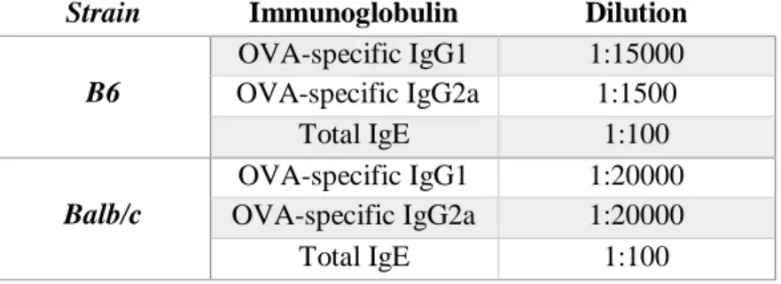
Table 3.2- Serum dilutions used for the detection of IgG2a, IgG1 and IgE concentrations in B6 and Balb/c mice.
Strain Immunoglobulin Dilution
B6 OVA-specific IgG1 1:15000 OVA-specific IgG2a 1:1500 Total IgE 1:100 Balb/c OVA-specific IgG1 1:20000 OVA-specific IgG2a 1:20000 Total IgE 1:100
4. Adoptive cell transfers and mice immunization
OVA-specific cells were collected from spleen and LNs of OT-II Rag-/- or DO11.10 Rag-/- mice and transferred into B6 Thy1.1, B6 Thy1.1/Thy1.2 or Balb/c recipient mice, respectively (in 100µl saline/mouse, intra venous (i.v) retro orbital injection (Day -1). Mice immunized with type 1 adjuvants received 8-10x106 cells/mouse, while mice immunized with type 2 adjuvants received 2-5x10⁶ cells/mouse.
27
The following day (Day 0) mice were immunized in the hind footpads with 80µg/50µl/footpad of OVA protein(Sigma-Aldrich, Cat. No. 031M7037V), conjugated with the indicated type 1 or type 2 adjuvant (Table 3.3).
Emulsions of OVA protein conjugated with TiterMax® Gold (TMX), Sigma Adjuvant System (SAS), Montanide ISA 720 VG or Incomplete Freund’s Adjuvant (IFA) were prepared mixing equal volumes of the aqueous OVA solution and of the chosen adjuvant (1:1 volume) in glass syringe with Luer Lock Tips, to allow a tight fit and the formation of a perfect emulsion. After mixing, the syringes were rested in the fridge to guarantee that the emulsions formed were stable and ready to be administered to the mice, following transfer to 1 ml plastic hypodermal syringes. The Aluminium hydroxide gel (Alum) has a white gelatinous precipitate appearance and is supplied at a 2% (v/v) concentration, with an aluminium content varying from 9.0–11.0mg/ml. Adsorption of the OVA to the Alum was performed by mixing the OVA in aqueous solution to an equal volume of Alum and slowly stirring for 30min at 4ºC. Preparation of CpG ODNs was achieved by mixing the OVA and CpG ODNs aqueous solution by vortexing for a few minutes.
Table 3.3- Adjuvants tested to induce Th1 or Th2 responses. TMX, SAS, CpG ODNs and Montanide ISA 720 VG were used as type 1 adjuvants, while Alum and IFA were tested as type 2 adjuvants.
Adjuvant Vendor (Cat. No.) Formulation CpG ODNs /footpad OVA antigen /footpad Type 1 TMX Sigma-Aldrich T2684 Water-in-oil emulsion (1:1) 30 μg 80 μg SAS Sigma-Aldrich S6322-1VL Oil-in-water-emulsion (1:1) 30 μg 80 μg CpG ODNs Invivogen vac-1826-1 Aqueous (saline) solution 30 μg 80 μg Montanide ISA 720 VG SEPPIC Water-in-oil emulsion (1:1) 30 μg 80 μg Type 2 Alum Serva 12261.01 Water-in-oil emulsion (1:1) --- 80 μg IFA Sigma-Aldrich F5506-10X10ML Water-in-oil emulsion (1:1) --- 80 μg
28
5. Flow cytometry analysis
Eleven days after immunization (Day 11), draining LNs (popliteal and inguinal LNs) from B6 and Balb/c mice, were harvested and mashed through a 70μm mesh into PBS 1X, 2% (v/v) of Foetal calf serum (FCS). For identification of OVA-specific Tfh cells and GC B cells, 1x10⁶ cells were initially incubated with purified anti-CD16/32 to block unspecific binding of antibodies to Fc receptors, and then labelled with anti-CXCR5-biotin (15min at 37ºC and 15min at RT).
After washing (300g, 5min, 4ºC), cells were labelled with anti-DO11.10 (Balb/c mice), anti-Vβ5 and anti-Vα2 (B6 mice), anti-Fas, anti-PD-1, streptavidin, anti-GL-7, anti-CD25,
anti-CD44, anti-CD19, anti-CD4, anti-Thy1.2, anti-Thy1.1 and anti-CD3 (30min, 4ºC) (Table 7.1 of attachments). Whenever possible, exclusion of dead cells was performed using a Viability dye (Life Technologies, Cat. No. L34957). Stainings were performed in PBS 1X, 2% (v/v) FCS. All samples were analysed using a LSR Fortessa (BD Biosciences) and the results processed with the FlowJo software version 8.7.1 (Tree Star Inc).
5.1 OVA-specific Tfh cell sorting
For sorting of antigen specific-Tfh and antigen specific-Th cells, CD4+ T cells from each mice were processed individually and first enriched using 10µl/107cells of CD4 MicroBeads (L3T4) (Miltenyi Biotec-Citomed, Cat. No.130-049-201). Then, the positively selected cells were subsequently isolated using MACS Separation Columns as described by the manufacturer (Miltenyi Biotec-Citomed, Cat. No.130-042-401). OVA-specific Tfh cells (CD4+CD44+ CXCR5+PD-1+ Thy1.2+ DO11.10+/ Vβ5+Vα2+)and
OVA-specific activated-Th cells (CD4+CD44+ CXCR5-PD-1- Thy1.2+DO11.10+/
Vβ5+Vα2+) were collected to lysis buffer (3,5µl of 0,2% (v/v) Triton X-100, 2U/µl RNAse
29
6. Th1 and Th2 polarization in vitro
6.1 Generation of mouse bone marrow-derived Dendritic Cells
Bone marrow-derived dendritic cells (BM-DC) were generated from B6 mice. The femurs were separated from muscle tissue, briefly cleaned in 70% (v/v) ethanol and washed with complete RPMI (RPMI 1X supplemented with 10% (v/v) heat inactivated FCS, 10mM HEPES, 50µM 2-mercaptoethanol, 100U/ml penicillin/ 100μg/mL Streptomycin, 2mM glutamine and 100nM sodium pyruvate- cRPMI). The ends of the bones were cut and a 27-gauge needle fitted into a syringe was used to flush the BM out with cRPMI. Following homogenization, erythrocytes were lysed by incubation (5min, RT) in Ammonium-Chloride-Potassium (ACK) lysing buffer (8,024mg/l of NH4Cl, 1,001mg/l
KHCO3 and 3.722 mg/l EDTA.Na2·2H2O). Cells were washed in PBS 1X, 2% (v/v) FCS
and cultured, in sterile plate without tissue culture treatment, at 1x10⁶ cells/ml in cRPMI
media supplemented with 20ng/ml recombinant murine Granulocyte macrophage colony-stimulating factor (GM-CSF) (PeproTech, Cat. No.315-03). The medium was changed every 3 days (Day 3 and Day 6). DCs were harvested on day 7 and co-cultured with naïve CD4+ T cells, as indicated below.
6.2 Priming of naïve T cells
Naïve CD4+ T cells were purified and enriched from spleen, mesenteric and inguinal LNs of OT-II Rag-/- mice. Red blood cells were eliminated using ACK lysis method and CD4+ T cells positively selected using CD4 (L3T4) MicroBeads, as described above.
Naïve T cells (3x10⁴ cells) were cultured at a ratio of 2:1 with allogeneic DCs in 96-well
flat-bottom plates (Costar, Corning, NY, USA). T-DC co-culture was maintained for 4 days at 37ºC and 5% CO2 in the conditions presented in table 3.4. All cells were cultured
with DCs, OVA antigen and IL-2 as a basal stimulation. For Th1 polarization, anti-IL-4 and IL-12 were added, whereas anti-IFN-γ and IL-4 were used for Th2 polarization. As a negative control (non-polarized cells), naïve CD4+ T cells were cultured just with
DCs and OVA. As a positive control (plate-bound), naïve CD4+ T cells received
30
Table 3.4- Culture conditions for differentiation of naïve CD4+ T cells.
Condition Reagent Stock Work
concentration
Vendor Cat. No.
Basal stimulation
Ovalbumin 500 µM/ml 10 µM /ml Sigma, Grade
5503
Mouse IL-2 Recombinant Protein
100 µl/ml 5 ng/ml eBioscience 14-8021-64 Th1
condition
Anti-mouse IL-4 Purified BVD6-24G2
0,5 mg/ml 10 µg/ml PeproTech 214-14 Murine IL-12 100 µl/ml 10 ng/ml PeproTech
210-12-B Th2
condition
Anti-mouse IFN-γ Purified R4-6A2
1 mg/ml 10 ng/ml LabClinics 16-7312-85 Recombinant Murine IL-4 100 µl/ml 10 ng/ml LabClinics 14-7042-85 Plate bound
Anti-Mouse CD3e Functional Grade Purified
1 mg/ml 3 µg/ml eBioscience 16-0031-86 Anti-Mouse CD28 Purified 500 µl/ml 2 µl/ml eBioscience 14-0281-86
The primed Th1 and Th2 cells were stimulated with phorbol-12-myristate-13-acetate (PMA), Ionomycin, BrefeldinA (BFA) and Golgi stop in cRPMI (4h, 37ºC and 5% CO2).
Table 3.5- Stimulation conditions for promotion of cytokine production.
Reagent Stock concentration Work concentration
PMA 2,5 mg/ml 50 ng/ml
Ionomycin 2,5 mg/ml 500 ng/ml
BFA 2,5 mg/ml 5 μg/ml
31
6.3 Cell surface and intracellular staining
Cultured cells were incubated with purified anti-CD16/32 (15min, 4ºC) and then surface stained with anti-CD25, anti-CD4 and Livedead (30min, 4ºC). Cells were washed with PBS 1X, 2% (v/v) FCS, BFA and Golgi stop and fixed with Direct Fixation (eBioscience, Cat. No.88-8823-88) or 1:3- Fix/Perm concentrate: Fix/Perm diluent (eBioscience, Cat. No.00-5521-00) for cells that will be stained for cytokines or transcription factors, respectively (30min, on ice).
Then, after centrifugation (300g, 3min, 4ºC), cells were permeabilized with Perm Buffer (eBioscience, Cat. No.00-5521-00) and stained with anti-IFN-γ, anti-IL-13, anti-IL-4, anti-IL-21, anti-Gata-3, anti-T-bet and anti-Bcl-6 diluted in Perm Buffer. Cells were washed (PBS 1X, 2% (v/v) FCS) and data collected using the LSR Fortessa flow cytometer (BD Biosciences) and analysed using FlowJo software version 8.7.1 (Tree Star Inc).
7. Real-time quantitative-PCR (RT-qPCR)
Sorted cells (5x10⁴ cells), namely Tfh cells (CD4+CD44+ CXCR5+PD-1+), activated-Th
cells (CD4+CD44+CXCR5-PD-1-), non-activated Th cells (CD4+CD44- CXCR5-PD-1-),
B cells (CD19+ FAS-GL-7-) and GC B cells (CD19+ FAS+GL-7+) were collected from
naïve mice and from immunized mice with type 1 or type 2 adjuvants.
7.1 RNA extraction from T cells
Cells were lysed in 350μl of lysis buffer (buffer RLT, RN Easy MiniKit, Qiagen) with 10µl/ml β-mercaptoethanol and homogenized by vortexing. For RNA extraction, the Quick-StartProtocol provided by RNeasy® Mini Kit (QUIAGEN) was followed.
32
7.2 cDNA synthesis from total RNA
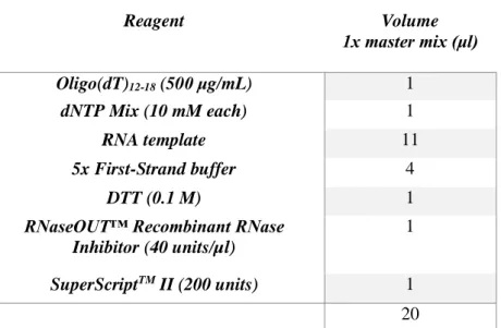
cDNA was synthesized from total RNA (11μl) by reverse transcription using Oligo(dT) and SuperScript II Reverse Transcriptase following the protocol available from Invitrogen and partially available on table 3.6. cDNA synthesis was performed using a PCR thermal cycler: 65ºC 5min, quick chill on ice, 42ºC 2min, 42ºC 50min and 70ºC 15min.
Table 3.6- 1x SuperScript II reverse transcriptase master mix.
Reagent Volume 1x master mix (μl) Oligo(dT)12-18 (500 μg/mL) 1 dNTP Mix (10 mM each) 1 RNA template 11 5x First-Strand buffer 4 DTT (0.1 M) 1
RNaseOUT™ Recombinant RNase Inhibitor (40 units/µl)
1
SuperScriptTM II (200 units) 1
20
7.3 Quantitative Real time PCR
Quantitative Real time polymerase chain reaction (qPCR) was performed using primers, specific for CXCR5, Bcl-6, Gata-3, T-bet, IL-4 and IFN-γ genes (Table 3.8), using Power SYBR® Green PCR Master Mix (Life Technologies) to detect the amplified PCR product from synthetized cDNA. Primers amplifying a segment of the housekeeping gene encoding β-2-microglobulin, since it is constitutively expressed by most cells, and in relation to which relative quantification will be performed. Reaction was processed in the RT-PCR Viia 7 System machine, following programed PCR thermal cycler: 50ºC 2min, 95°C 10min, 95ºC 15sec, 60 °C 1min (40-45 cycles); Melt curve, 95ºC 15sec, 65°C 1min, 95ºC 15sec. Relative expression was calculated using the 2-ΔCt method.
33
Table 3.7- RT-qPCR master mix reaction.
Reagent Work concentration Volume
1x master mix (μl) Template 1:5 dilution 2 SYBR® Green (1x) 1x 4 Primer F (10 nM) 100 nM 0,08 Primer R (10 nM) 100 nM 0,08 H2O --- 1,84
Table 3.8- Primers used in analysis of Tfh, activated-Th and non-activated-Th cells, B cells and GC B cells.
Primers (Target gene)
Sequence Orientation
CXCR5 GAG CTG CAG CTA TGA ACT AC Forward
AGG AGG AAG ATG AGG CTG TA Reverse
Bcl-6 AGT GAG AGT CAC TCA CCA CT Forward
GCA GTC ACA TTC GTT GCA GA Reverse
Gata-3 CAC TAC CTT TGC AAT GCC TG Forward
AGC TTG TAG TAC AGC CCA CA Reverse
T-bet GGA AGC TAA AGC TCA CCA AC Forward
AGC TGA GTG ATC TCT GCG TT Reverse
IL-4 CAT ATC CAC GGA TGC GAC AA Forward
TCT TCA AGC ATG GAG TTT TC Reverse
IFN-γ ACT GGC AAA AGG ATG GTG AC Forward
GTG GGT TGT TGA CCT CAA AC Reverse
β-2- CTG CAG AGT TAA GCA TGC CAG TAT Forward
35
37
1. CpG ODNs was the best adjuvant to induce Th1 responses,
whereas Incomplete Freund’s Adjuvant (IFA) was better at
inducing Th2 responses
With the purpose of dissecting the phenotype of antigen-specific Tfh cells that are generated after immunization with OVA protein in conjunction with either type 1 or type 2 adjuvants, it was essential to optimize the ideal immunizing conditions.
In these experiments, 6-8 weeks old mice were immunized in the footpad with each of the adjuvants described and as indicated in scheme 4.1. Serum was collected on day 14 in order to analyse the humoral response induced by each adjuvant using ELISA assay.
Scheme 4.1- Experimental design of the immunization assay of B6 and Balb/c mice. (a) On day 0, mice were immunized with 80μg of OVA along with the indicated type 1 or type 2 adjuvants (listed in (b)). On day 14, serum was recovered from peripheral blood and used to determine antibody titres by ELISA.
Adjuvant CpG ODNs /footpad OVA antigen /footpad Type 1 TMX 30 μg 80 μg SAS 30 μg 80 μg CpG ODNs 30 μg 80 μg Montanide ISA 720 VG 30 μg 80 μg Type 2 Alum --- 80 μg IFA --- 80 μg (a) (b) Adjuvant CpG ODNs /footpad OVA antigen /footpad Type 1 TMX 30 μg 80 μg SAS 30 μg 80 μg CpG ODNs 30 μg 80 μg Montanide ISA 720 VG 30 μg 80 μg Type 2 Alum --- 80 μg IFA --- 80 μg (b)
38
To investigate Th1 responses, several type 1 adjuvants were tested in Balb/c and B6mice. More specifically, CpG ODNs only or in combination with TMX and SAS were tested in both strains, while Montanide ISA 720 VG was tested only in B6 mice.
Synthetic oligodeoxynucleotides containing unmethylated CpG motifs (CpG ODNs) are normally present in bacterial DNA at a 20-fold greater frequency compared to mammalian DNA. CpG ODNs are recognized by TLR-9, which induces the activation of DCs and Th1 cells, that, in turn, secrete typical pro-inflammatory cytokines, such as TNF-α, IFN-γ, IFN-α and IL-12 (58). In this project a mouse specific type B CpG ODNs was used as an efficient inducer of Th1 immunity, with predominant production of IgG2a. TMX is a stable water-in-oil emulsion that, upon subcutaneous administration, forms a depot effect with slow release of the antigen, stimulating cellular and humoral immune responses. It is characterized by the production of high antibody titres, mainly IgG2a, even without boost immunization (59).
SAS is a stable oil-in-water emulsion which contains Monophosphoryl lipid A (MPLA), an immunostimulating TLR-4 agonist formed by detoxified lipopolysaccharide (LPS) from Salmonella minnesota R595. MPLA is considerably less toxic than LPS, whilst maintaining immunostimulatory activity, inducing a strong Th1 response (60).
Montanide ISA 720 VG, tested only in B6 mice, is also a water-in-oil emulsion that promotes the formation of a depot at the site of injection (61). It has been shown that it induces both Th1 and Th2 responses, as antigen-specific IgG1 and IgG2c titres were observed in response to vaccine consisting of Schistosoma mansoni cathepsin B formulated in Montanide ISA 720 VG (62).
As for type 2 adjuvants, IFA and Alum were tested in both strains and in Balb/c mice only, respectively. IFA induces a predominantly Th2-biased response through the formation of a depot at the injection site, enabling the slow release of antigen and the stimulation of high and long-lasting antibody producing plasma cells (63). This adjuvant efficiently induced the production of high titres of IgG1 and also of IgE.